
Influence of Cavity Size on the Survival of Single Surface Atraumatic Restorative Treatment Using Glass Ionomer Cement with or without Chlorhexidine Diacetate—A Randomized Trial

Abstract
:1. Introduction
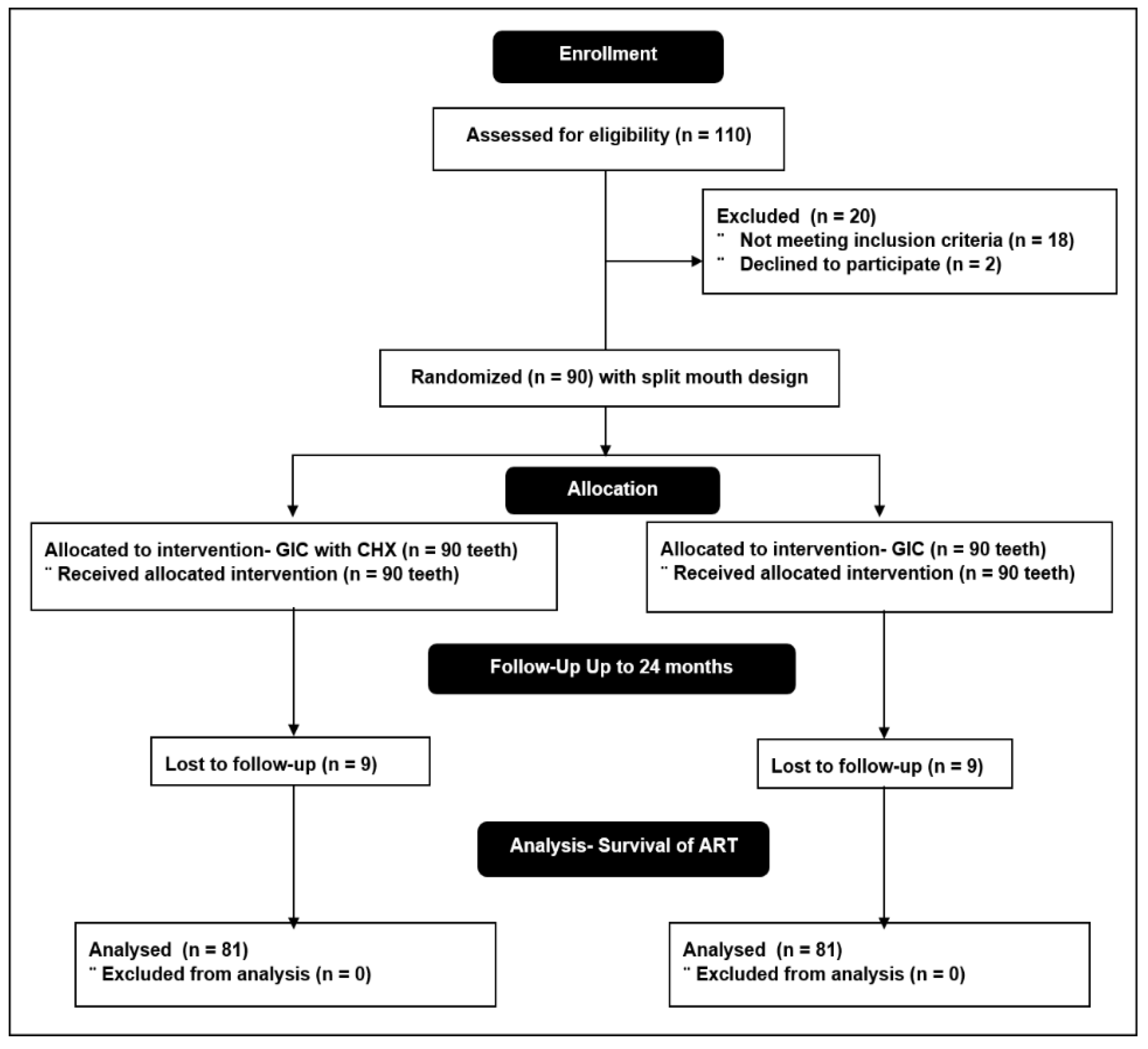
2. Materials and Methods
3. Results
4. Discussion
5. Conclusions
Author Contributions
Funding
Institutional Review Board Statement
Informed Consent Statement
Data Availability Statement
Acknowledgments
Conflicts of Interest
References
- Frencken, J.E.; Leal, S.; Navarro, M.F.L. Twenty-five-year atraumatic restorative treatment (ART) approach: A comprehensive overview. Clin. Oral Investig. 2012, 16, 1337–1346. [Google Scholar] [CrossRef] [Green Version]
- Saber, A.M.; El-Housseiny, A.A.; AlAmoudi, N.M. Atraumatic Restorative Treatment and Interim Therapeutic Restoration: A Review of the Literature. Dent. J. 2019, 7, 28. [Google Scholar] [CrossRef] [Green Version]
- Nkwocha, F.G.; A Akinyamoju, G.; O Ogbode, S.; Lawal, F.B. Management of Dental Caries with Atraumatic Restorative Treatment Under Field Condition in Primary Schools in Oyo State, Nigeria. Ann. Ib. Postgrad. Med. 2019, 17, 75–80. [Google Scholar]
- Frencken, J.E. Atraumatic restorative treatment and minimal intervention dentistry. Br. Dent. J. 2017, 223, 183–189. [Google Scholar] [CrossRef] [PubMed]
- Menezes-Silva, R.; Velasco, S.R.M.; Bresciani, E.; Bastos, R.D.S.; Navarro, M.F.L. A prospective and randomized clinical trial evaluating the effectiveness of ART restorations with high-viscosity glass-ionomer cement versus conventional restora-tions with resin composite in Class II cavities of permanent teeth: Two-year follow-up. J. Appl. Oral. Sci. 2021, 29, e20200609. [Google Scholar] [CrossRef] [PubMed]
- Konde, S.; Raj, S.; Jaiswal, D. Clinical evaluation of a new art material: Nanoparticulated resin-modified glass ionomer ce-ment. J. Int. Soc. Prev. Community Dent. 2012, 2, 42–47. [Google Scholar] [CrossRef] [Green Version]
- Yip, H.-K.; Smales, R.J.; Ngo, H.C.; Tay, F.R.; Chu, F.C. Selection of restorative materials for the atraumatic restorative treatment (ART) approach: A review. Spéc. Care Dent. 2001, 21, 216–221. [Google Scholar] [CrossRef]
- American Academy of Pediatric Dentistry. Pediatric restorative dentistry. In the Reference Manual of Pediatric Dentistry; American Academy of Pediatric Dentistry: Chicago, IL, USA, 2020; pp. 371–383. [Google Scholar]
- Sidhu, S.K.; Nicholson, J.W. A Review of Glass-Ionomer Cements for Clinical Dentistry. J. Funct. Biomater. 2016, 7, 16. [Google Scholar] [CrossRef]
- Elkady, D.M.; Khater, A.G.A.; Schwendicke, F. Chlorhexidine to improve the survival of ART restorations: A systematic re-view and meta-analysis. J. Dent. 2020, 103, 103491. [Google Scholar] [CrossRef]
- Mishra, A.; Pandey, R.K.; Manickam, N. Antibacterial effect and physical properties of chitosan and chlorhexi-dine-cetrimide-modified glass ionomer cements. J. Indian Soc. Pedod. Prev. Dent. 2017, 35, 28–33. [Google Scholar] [PubMed]
- Neelima, B.; Reddy, J.S.; Singh, P.T.; Suhasini, K.; Hemachandrika, I.; Hasanuddin, S. Comparative evaluation of antimi-crobial efficacy of glass ionomer cement added with propolis, chitosan, and chlorhexidine against Streptococcus mu-tans and Lactobacillus acidophilus: An in vitro study. J. Indian Soc. Pedod. Prev. Dent. 2020, 38, 367–373. [Google Scholar]
- Joshi, J.S.; Roshan, N.M.; Sakeenabi, B.; Poornima, P.; Nagaveni, N.B.; Subbareddy, V.V. Inhibition of Residual Cariogenic Bacteria in Atraumatic Restorative Treatment by Chlorhexidine: Disinfection or Incorporation. Int. J. Clin. Pediatr. Dent. 2017, 39, 308–312. [Google Scholar]
- Marti, L.M.; Da Mata, M.; Ferraz-Santos, B.; Azevedo, E.R.; Giro, E.M.A.; Zuanon, A.C.C. Addition of Chlorhexidine Gluconate to a Glass Ionomer Cement: A Study on Mechanical, Physical and Antibacterial Properties. Braz. Dent. J. 2014, 25, 33–37. [Google Scholar] [CrossRef] [Green Version]
- Duque, C.; Aida, K.L.; Pereira, J.A.; Teixeira, G.S.; Caldo-Teixeira, A.S.; Perrone, L.R.; Caiaffa, K.S.; Negrini, T.C.; Castilho, A.R.F.; Costa, C.A.S. In vitro and in vivo evaluations of glass-ionomer cement containing chlorhexidine for Atraumatic Re-storative Treatment. J. Appl. Oral. Sci. 2017, 25, 541–550. [Google Scholar] [CrossRef]
- Jiang, M.; Fan, Y.; Li, K.Y.; Lo, E.C.M.; Chu, C.H.; Wong, M.C.M. Factors affecting success rate of atraumatic restorative treatment (ART) restorations in children: A systematic review and meta-analysis. J. Dent. 2020, 104, 103526. [Google Scholar] [CrossRef]
- Kemoli, A.M.; van Amerongen, W.E. Influence of the cavity-size on the survival rate of proximal ART restorations in primary molars. Int. J. Paediatr. Dent. 2009, 19, 423–430. [Google Scholar] [CrossRef] [PubMed]
- Mobarak, E.H.; Shabayek, M.M.; El-Deeb, H.A.; Mulder, J.; Hassan, F.M.; Van der Sanden, W.J.M.; Frencken Jo, E. Survival of occlusal ART restorations using high-viscosity glass-ionomer with and without chlorhexidine: A 2-year split-mouth quad-ruple-blind randomized controlled clinical trial. J. Adv. Res. 2019, 17, 117–123. [Google Scholar] [CrossRef] [PubMed]
- Kemoli, A.M.; Van Amerongen, W.E.; Opinya, G.N. Influence of different isolation methods on the survival of proximal ART restorations in primary molars after two years. Eur. Arch. Paediatr. Dent. 2010, 11, 136–139. [Google Scholar] [CrossRef] [PubMed]
- Kemoli, A.M.; Van Amerongen, W.E.; Opinya, G.N. Influence of the experience of operator and assistant on the survival rate of proximal ART restorations: Two-year results. Eur. Arch. Paediatr. Dent. 2009, 10, 227–232. [Google Scholar] [CrossRef]
- Roshan, N.M.; Sakeenabi, B. Survival of occlusal ART restorations in primary molars placed in school environment and hospital dental setup- one year follow-up study. Med. Oral Patol. Oral Cir. Bucal. 2011, 16, e973-7. [Google Scholar]
- Frencken, J.E.; Liang, S.; Zhang, Q. Survival estimates of atraumatic restorative treatment versus traditional restorative treatment: A systematic review with meta-analyses. Br. Dent. J. 2021, 1–11. [Google Scholar] [CrossRef]
- De Amorim, R.G.; Frencken, J.E.; Raggio, D.P.; Chen, X.; Hu, X.; Leal, S.C. Survival percentages of atraumatic restorative treatment (ART) restorations and sealants in posterior teeth: An updated systematic review and meta-analyses. Clin. Oral Investig. 2018, 22, 2703–2725. [Google Scholar] [CrossRef]
- Lo, E.; Holmgren, C. Provision of Atraumatic Restorative Treatment (ART) restorations to Chinese pre-school children—A 30-month evaluation. Int. J. Paediatr. Dent. 2001, 11, 3–10. [Google Scholar] [CrossRef] [PubMed]
- Rahimtoola, S.; van Amerongen, W.E. Comparison of two tooth-saving preparation techniques for one surface cavities. J. Dent. Child 2002, 69, 16–26. [Google Scholar]
- Honkala, E.; Behbehani, J.; Ibricevic, H.; Kerosuo, E.; Al-Jame, G. Comparable survival rate of class I ART in primary dentition (GIC and Amalgam) in Beirut after 2 years of follow-up. Int. J. Paediatr. Dent. 2003, 13, 172–179. [Google Scholar] [CrossRef] [PubMed]
- van Gemert-Schriks, M.C.M.; van Amerongen, W.E.; Cate, J.M.T.; Aartman, I.H.A. Three-year survival of single- and two-surface ART restorations in a high-caries child population. Clin. Oral Investig. 2007, 11, 337–343. [Google Scholar] [CrossRef] [Green Version]
- Taifour, D.; Frencken, J.E.; Beiruti, N.; van‘t Hof, M.A.; Truin, G.J. Effectiveness of glass-ionomer (ART) and amalgam res-torations in the deciduous dentition: Results after 3 years. Caries Res. 2002, 36, 437–444. [Google Scholar] [CrossRef]
- Arrow, P.; Klobas, E. Minimal intervention dentistry for early childhood caries and child dental anxiety: A randomized con-trolled trial. Aust. Dent. J. 2017, 62, 200–207. [Google Scholar] [CrossRef] [Green Version]
- Farag, A.; Van Der Sanden, W.J.M.; Abdelwahab, H.; Frencken, J.E. Survival of ART restorations assessed using selected FDI and modified ART restoration criteria. Clin. Oral Investig. 2010, 15, 409–415. [Google Scholar] [CrossRef] [Green Version]


| Inclusion criteria |
|
| Exclusion criteria |
|
| Code | Criteria |
|---|---|
| 0 | The restoration is present and in good condition |
| 1 | The restoration is present, with a slight marginal defect; no repair is needed |
| 2 | The restoration is present, with slight wear; no repair is needed |
| 3 | The restoration is present, with marginal defect >0.5 mm; repair is needed |
| 4 | The restoration is present, with wear >0.5 mm; repair is needed |
| 5 | The restoration is not present, it is partly or completely lost |
| 6 | The restoration is not present, it is replaced by another restoration |
| 7 | The tooth is missing, exfoliated, or extracted |
| 8 | Restoration not assessed; child not present |
| Cavity Size Categories | |||
|---|---|---|---|
| Cavity Sizes | Depth n (Mean) | Mesio-Distal n (Mean) | Bucco-Lingual n (Mean) |
| <2 mm | 68 (1.6 mm) | 38 (1.5 mm) | 51 (1.4 mm) |
| 2.1–3 mm | 97 (2.3 mm) | 65 (2.6 mm) | 68 (2.4 mm) |
| 3.1–4 mm | 15 (3.2 mm) | 44 (3.3 mm) | 37 (3.3 mm) |
| >4 mm | 0 | 33 (4.2 mm) | 24 (4.2 mm) |
| Total | 180 | 180 | 180 |
| Restoration Status | 24 Months | ||||
|---|---|---|---|---|---|
| GIC | CHX-GIC | Kruskal–Wallis | |||
| 81 | % | 81 | % | p | |
| 51 | 63.0 | 46 | 56.8 | 0.07 |
| 8 | 9.9 | 13 | 16.0 | 0.09 |
| 9 | 11.1 | 8 | 9.9 | 0.09 |
| 5 | 6.2 | 6 | 7.4 | 0.11 |
| 4 | 4.9 | 4 | 4.9 | 0.12 |
| 3 | 3.7 | 3 | 3.7 | 0.11 |
| 1 | 1.2 | 1 | 1.2 | NA |
| Success | 68 | 83.9 | 67 | 82.7 | 0.12 |
| Failure | 13 | 16.0 | 14 | 17.3 | 0.11 |
| Overall success | 135 (83.3) | ||||
| Drop-out | 9 | ||||
| Cavity Size | GIC | CHX GIC | Chi-Square, p-Value |
|---|---|---|---|
| Success | Success | ||
| Cavity Depth success | |||
| a. <2 mm (n = 46) | 24 (52.2) | 22 (47.8) | 0.08 |
| b. 2.1–3 mm (n = 79) | 40 (50.6) | 39 (49.4) | 0.09 |
| c. 3.1–4 mm (n = 10) | 4 (40) | 6 (60) | 0.03 |
| Mesio-distal width, success | |||
| <2 mm (n = 21) | 10 (47.6) | 11 (52.4) | 0.07 |
| 2.1–3 mm (n = 55) | 27 (49.1) | 28 (50.9) | 0.09 |
| 3.1–4 mm (n = 35) | 17 (48.6) | 18 (51.4) | 0.08 |
| >4 mm (n = 24) | 14 (58.3) | 10 (41.7) | 0.06 |
| Bucco-lingual width, success | |||
| <2 mm (n = 39) | 18 (46.2) | 21 (53.8) | 0.06 |
| 2.1–3 mm (n = 62) | 33 (53.2) | 29 (46.8) | 0.07 |
| 3.1–4 mm (n = 20) | 9 (45) | 11 (55) | 0.06 |
| >4 mm (n = 14) | 8 (57.1) | 6 (42.9) | 0.06 |
| Cavity Volume Category | Cavity at Baseline (n = 180). n (Mean Volume) | Survival of ART Restorations at the 24-Month Assessment | |||||
|---|---|---|---|---|---|---|---|
| Overall Survival of ART Restorations (n = 162) | Conventional GIC (n = 81) | CHX–Modified GIC (n = 81) | |||||
| Success (n = 135) n (%) | Failure (n = 27) n (%) | Success (n = 68) n (%) | Failure (n = 13) n (%) | Success (n = 67) n (%) | Failure (n = 14) n (%) | ||
| a. 0–9.9 mm3 | 23 (7.9) | 16 (11.8) | 5 (18.5) | 9 (13.2) | 3 (23.1) | 7 (10.4) | 2 (14.3) |
| b. 10–19.9 mm3 | 59 (18.4) | 55 (40.7) | 2 (7.4) | 25 (36.8) | 0 | 30 (44.8) | 2 (14.3) |
| c. 20–29.9 mm3 | 39 (26.2) | 33 (24.4) | 1 (3.7) | 19 (27.9) | 0 | 14 (20.9) | 1 (7.1) |
| d. 30–39.9 mm3 | 27 (37.3) | 14 (10.4) | 8 (29.6) | 8 (11.8) | 4 (30.8) | 6 (8.9) | 4 (28.6) |
| e. 40–49.9 mm3 | 21 (43.4) | 12 (8.9) | 7 (25.9) | 5 (7.3) | 3 (23.1) | 7 (10.4) | 4 (28.6) |
| f. >50 mm3 | 11 (61.4) | 5 (3.7) | 4 (14.8) | 2 (2.9) | 3 (23.1) | 3 (4.5) | 1 (7.1) |
| Kruskal–Wallis H test, p value | 0.03 | 0.08 | 0.03 | 0.07 | 0.03 | 0.09 | |
| Man-Whitney U test | b > a, c, d, e, f | NA | b > a, c, d, e, f | NA | b > a, c, d, e, f | NA | |
| Time Interval (Months) | GIC | CHX–GIC | ||||||||
|---|---|---|---|---|---|---|---|---|---|---|
| n e | n f | n c | Survival % | SE | n e | n f | n c | Survival % | SE | |
| 0–6 | 90 | 3 | 3 | 96.6 | 1.7 | 90 | 4 | 3 | 95.4 | 1.9 |
| 6–12 | 86 | 8 | 4 | 90.7 | 2.1 | 86 | 9 | 4 | 89.5 | 2.3 |
| 12–18 | 83 | 12 | 7 | 85.5 | 3.1 | 83 | 14 | 7 | 83.1 | 3.6 |
| 18–24 | 81 | 13 | 9 | 83.9 | 3.8 | 81 | 14 | 9 | 82.7 | 3.9 |
Publisher’s Note: MDPI stays neutral with regard to jurisdictional claims in published maps and institutional affiliations. |
© 2021 by the authors. Licensee MDPI, Basel, Switzerland. This article is an open access article distributed under the terms and conditions of the Creative Commons Attribution (CC BY) license (https://creativecommons.org/licenses/by/4.0/).
Share and Cite
Mohamed, R.N.; Basha, S.; S. Joshi, J.; Parameshwarappa, P. Influence of Cavity Size on the Survival of Single Surface Atraumatic Restorative Treatment Using Glass Ionomer Cement with or without Chlorhexidine Diacetate—A Randomized Trial. Appl. Sci. 2021, 11, 10438. https://doi.org/10.3390/app112110438
Mohamed RN, Basha S, S. Joshi J, Parameshwarappa P. Influence of Cavity Size on the Survival of Single Surface Atraumatic Restorative Treatment Using Glass Ionomer Cement with or without Chlorhexidine Diacetate—A Randomized Trial. Applied Sciences. 2021; 11(21):10438. https://doi.org/10.3390/app112110438
Chicago/Turabian StyleMohamed, Roshan Noor, Sakeenabi Basha, Jooie S. Joshi, and Poornima Parameshwarappa. 2021. "Influence of Cavity Size on the Survival of Single Surface Atraumatic Restorative Treatment Using Glass Ionomer Cement with or without Chlorhexidine Diacetate—A Randomized Trial" Applied Sciences 11, no. 21: 10438. https://doi.org/10.3390/app112110438
APA StyleMohamed, R. N., Basha, S., S. Joshi, J., & Parameshwarappa, P. (2021). Influence of Cavity Size on the Survival of Single Surface Atraumatic Restorative Treatment Using Glass Ionomer Cement with or without Chlorhexidine Diacetate—A Randomized Trial. Applied Sciences, 11(21), 10438. https://doi.org/10.3390/app112110438