Article

A Short S-Equol Exposure Has a Long-Term Inhibitory Effect on Adipogenesis in Mouse 3T3-L1 Cells

by
Gilberto Mandujano-Lázaro
1,
Carlos Galaviz-Hernández
2,
César A. Reyes-López
3,
Julio C. Almanza-Pérez
4,
Abraham Giacoman-Martínez
4,5,
César López-Camarillo
6,
Fengyang Huang
7 and
Laurence A. Marchat
1,*
1
Laboratorio 2 de Biomedicina Molecular, ENMH, Instituto Politécnico Nacional, Ciudad de México 07320, Mexico
2
Academia de Genómica, CIIDIR-Durango, Instituto Politécnico Nacional, Durango 34220, Mexico
3
Laboratorio de Bioquímica Estructural, ENMH, Instituto Politécnico Nacional, Ciudad de México 07320, Mexico
4
Laboratorio de Farmacología, Departamento de Ciencias de la Salud, División de Ciencias Biológicas y de la Salud, Universidad Autónoma Metropolitana-Iztapalapa, Ciudad de México 09340, Mexico
5
Departamento de Farmacobiología, Centro de Investigación y de Estudios Avanzados del IPN, Sede Sur, Ciudad de México 14330, Mexico
6
Posgrado en Ciencias Genómicas, Universidad Autónoma de la Ciudad de México, Ciudad de México 03100, Mexico
7
Laboratorio de Farmacología y Toxicología, Hospital Infantil de México Federico Gómez, Ciudad de México 06720, Mexico
*
Author to whom correspondence should be addressed.
Appl. Sci. 2021, 11(20), 9657; https://doi.org/10.3390/app11209657
Submission received: 18 August 2021 / Revised: 28 September 2021 / Accepted: 11 October 2021 / Published: 16 October 2021
(This article belongs to the Special Issue Drug Development for Cancer, Diabetes and Obesity)

Abstract

:
In the search for new drugs against obesity, the chronic disease that threatens human health worldwide, several works have focused on the study of estrogen homologs because of the role of estrogen receptors (ERs) in adipocyte growth. The isoflavone equol, an ERβ agonist, has shown beneficial metabolic effects in in vivo and in vitro assays; however, additional studies are required to better characterize its potential for body weight control. Here, we showed that the treatment of 3T3-L1 cells with 10 μM of S-equol for the first three days of the adipocyte differentiation protocol was able to prevent cells becoming semi-rounded and having a lipid droplet formation until the seventh day of culture; moreover, lipid accumulation was reduced by about 50%. Congruently, S-equol induced a reduction in mRNA expression of the adipogenic markers C/EBPα and PPARγ, and adipokines secretion, mainly Adiponectin, Leptin, Resistin, and MCP-1, while the release of PAI-1 was augmented. Moreover, it also reduced the expression of ERα and attenuated the subexpression of ERβ associated with adipogenesis. Altogether, our data suggested that S-equol binding to ERβ affects the transcriptional program that regulates adipogenesis and alters adipocyte functions. Future efforts will focus on studying the impact of S-equol on ER signaling pathways.

1. Introduction

Obesity and overweight have become one of the main health problems worldwide because they increase the risk of suffering from diabetes, hypertension, cancer, and other diseases associated with the metabolic syndrome [1]. Obesity is associated with an abnormal expansion of adipose tissue and a deregulation of the adipogenesis process [2], which affects the synthesis of adipokines, such as Leptin, Adiponectin, Resistin, Plasminogen activator inhibitor (PAI-1), Monocyte chemoattractant protein 1 (MCP-1), Interleukin-6 (IL-6), and Tumor necrosis factor alpha (TNFα), with autocrine and paracrine functions in inflammation, homeostasis regulation, and adipogenesis, among other processes [3,4]. Obesity is mainly exacerbated by the excessive intake of highly caloric food and the lack of physical activity [5], but estrogen reduction is also an important cause of augmented adiposity, mainly in postmenopausal women [6]. Estrogen effects depend on the binding to estrogen receptors (ERs) that act as transcription factors to regulate different processes. Notably, experiments in 3T3-L1 preadipocytes and adipose tissue from obese mice showed that ERα regulates insulin sensitivity and glucose tolerance, while ERβ modulates the expression of genes related to adipogenesis by inhibiting the function of the pro-adipogenic marker PPARγ [7]. Although estradiol therapy seems to be promising for obesity control, adverse effects usually related to the overexpression of ERα in female reproductive organs and breasts, and its implication in the induction of proliferation, strongly limit its therapeutic use [8,9]. Consequently, works have focused on ERβ-selective ligands that trigger body weight and fat mass reduction, as well as the subexpression of PPARγ without producing secondary effects [10,11].
In the search for new drugs against obesity, plant and food components have attracted increased attention worldwide. Notably, abundant epidemiological evidence suggests that the intake of soy-containing foods has many health benefits [12,13]. Daidzein is a soy isoflavone that is hydrolyzed by specific gut bacteria to form S-equol, which is a structural homolog of 17-β-estradiol. S-equol has a higher binding affinity to ERβ (Ki = 0.73 ± 0.2 nmol/L) than ERα (Ki = 6.41 ± 1 nmol/L), which makes it an interesting molecule for obesity control through ER pathway modulation. The presence of a chiral carbon atom at position C-3 of the furan ring allows the formation of a synthetic enantiomer form called R-equol, with a weaker affinity for both ERs [14,15,16]. A decade ago, Jackson et al. published an interesting report showing that ≥80% of Asian people are equol producers versus ≤25% in Western countries (mainly United States and Europe), which is consistent with the highest consumption of soybean and the lowest rate of overweight/obesity in Eastern regions [17]. Congruently, some studies have reported the beneficial effect of equol for the control of overweight and obesity. An epidemiological study conducted on 54 Japanese patients with obesity supplemented with 10 mg of S-equol per day for 12 weeks evidenced reductions in the levels of glycosylated hemoglobin (HbAlc), low-density lipoprotein (LDL-C), vascular Cardio-ankle index (CAVI), and Leptin [18]. On the other hand, treatment of obese diabetic C57BL/6J mice with 0.05% equol in diet for three weeks induced reductions in serum glucose, cholesterol, and triglyceride levels, as well as in weight gain [19]. Additionally, ovariectomized Long-Evans rats treated with a subcutaneous injection of 5 mg of S-equol/kg/day showed a decrease in body weight gain [20], and ovariectomized Sprague-Dawley rats treated with 6.87 mg of equol/day for six weeks presented a reduced weight gain and abdominal fat accumulation, as well as a diminution in plasma Leptin, cholesterol, and triglyceride levels [21]. In contrast, a recent study performed in C57/BL6 mice on a high-fat diet (HFD) revealed that S-equol supplementation did not affect body weight and food intake, but exacerbated several aspects of HFD-induced metabolic disease, such as hyperglycemia and energy expenditure reduction; moreover, it also evidenced opposite effects on serum insulin and Leptin levels depending on sex [22].
To our knowledge, there are few reports about the effect of S-equol on adipogenesis in vitro. In 2010, Cho et al. evidenced that the racemic mixture of (R,S)-equol at 0.1 to 20 μmol/L significantly increased adipocyte differentiation and insulin-stimulated glucose uptake in C3H10T1/2 pluripotent stem cells. Moreover, (R,S)-equol at 10 and 20 μmol/L significantly increased PPARγ transcriptional activity in 3T3-L1 preadipocytes [23]. Later, Nishide et al. confirmed the stimulating effect of low concentrations of (R,S)-equol on adipogenesis. Importantly, they demonstrated that 100 μM of (R,S)-equol inhibits adipocyte differentiation of the 3T3-L1 mouse cell line, observing a 40% decrease in lipid accumulation, as well as a reduced expression of the pro-adipogenic factors PPARγ, C/EBPα, and FAS, suggesting a bi-phasic effect of (R,S)-equol on adipogenesis in vitro [24].
The conflicting results about the beneficial metabolic effects of equol in in vivo and in vitro assays clearly indicate that additional studies are required to better understand the potential of S-equol for obesity and obesity-related diseases control. An additional problem is that some studies have not clarified the use of S-equol or (R,S)-equol, being a possible cause of variability in the results. Here, we aimed to characterize how the ERβ agonist S-equol affects adipocyte differentiation and function.

2. Materials and Methods

2.1. Cell Culture

Mouse 3T3-L1 fibroblasts (ATCC-CL-173) were grown at 37 °C and 5% CO2 in growth medium (GM) containing Dulbecco’s modified Eagle’s medium (DMEM) (ATCC-30-2002) supplemented with newborn calf serum 10% v/v (Gibco 16010-159), 2 mM of L-Glutamine (Gibco 25030), 1X nonessential amino acids (Sigma M7145), 1 mM of sodium pyruvate (Gibco 11360), and penicillin-streptomycin 1% v/v (10,000 unit/mL; Gibco 30-2300).

2.2. MTT Assays

3T3-L1 fibroblasts were seeded at a density of 5 × 10³ cells/well in 96-well plates and cultivated in GM with increasing amounts of S-equol (Sigma SML2147) (1, 3, 10, 30, 100, and 300 µM), for 24 and 48 h at 37 °C and 5% CO2. In another assay, confluent 3T3-L1 cells were cultured in differentiation medium I (DM-I) containing DMEM/high glucose supplemented with 10% v/v fetal bovine serum (Gibco 16000-044), 2 mM of L-Glutamine, 1X nonessential amino acids, 1 mM of sodium pyruvate, penicillin-streptomycin 1% v/v (10,000 unit/mL; Gibco 30-2300), 0.5 mM of isobutylmethylxanthine (IBMX) (Sigma I5879), 0.25 µM of dexamethasone (DEX) (Sigma D4902), and 0.2 UI/mL of insulin (Novolin), with S-equol (0 to 300 µM), for 3 days at 37 °C and 5% CO2. Then, the medium was removed, and cells were incubated with MTT reagent (0.10 mg/mL) for 2 h (Sigma M2128). The formazan crystals produced were dissolved with acidic isopropanol and the absorbance was measured at 570 nm, in an Epoch microplate spectrophotometer (Biotek, Winooski, VT, USA). Cells without S-equol, with 2% DMSO, were used as controls. Data were expressed as mean ± standard deviation (SD) from three independent experiments performed in triplicate. Data corresponding to cells without treatment were taken as 100%.

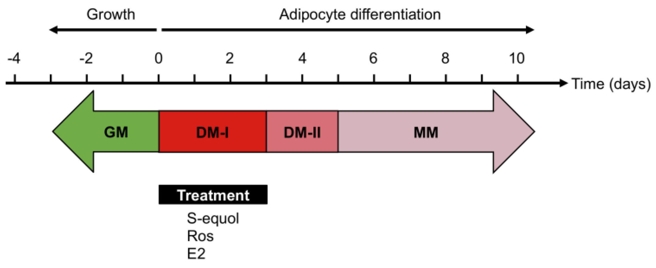
2.3. Adipocyte Differentiation

The adipocyte differentiation was performed according to the protocol reported by Zebisch et al. (2012) [25] with some modifications (Figure 1). 3T3-L1 cells were seeded at a density of 6 × 10⁴ cells/well in six-well plates in GM at 37 °C and 5% CO2 for 48 h until cells reached 80% confluence (day 0). Then, GM was replaced by DM-I and cells were incubated at 37 °C and 5% CO2 for three days in order to induce adipocyte differentiation. Then, DM-I was replaced with differentiation medium II (DM-II) containing (DMEM)/high glucose supplemented with 10% v/v fetal bovine serum, 2 mM of L-Glutamine, nonessential amino acids 1X, 1 mM of sodium pyruvate, penicillin-streptomycin 1% v/v (10,000 unit/mL; Gibco 30-2300), and 0.1 UI/mL of insulin (Novolin); cells were incubated at 37 °C and 5% CO2 for two days in order to favor and promote the maturation and differentiation of adipocytes. Finally, cells were kept in maintenance medium (MM) containing (DMEM)/high glucose supplemented with fetal bovine serum 10% v/v, 2 mM of L-Glutamine, 0.1 mM of nonessential amino acids, 1 mM of sodium pyruvate, and penicillin-streptomycin 1% v/v (10,000 unit/mL; Gibco 30-2300), at 37 °C and 5% CO2; the medium was changed every two days until observing morphological changes associated with adipocyte differentiation by microscopy.
To determine the effect of S-equol on adipocyte differentiation, cells were induced to differentiation using DM-I in the presence of different concentrations of S-equol (1, 3, and 10 µM). Cells incubated in DM-I with 10 µM of estradiol (E2) (Sigma E2758) or 2 µM of rosiglitazone (Ros) (Avandia gsk) were used as controls of inhibition and stimulation of adipocyte differentiation, respectively (Figure 1).

2.4. Oil Red O Staining

3T3-L1 adipocytes were fixed with 3.7% formaldehyde for 30 min, washed three times with PBS 1X, and stained with Oil Red O (ORO; Sigma O 0625)-60% isopropanol/PBS solution (1.2 mg of ORO/mL isopropanol-PBS 1X solution) for 10 min. Then, cells were washed with 70% ethanol/PBS 1X to remove excess dye, and stained lipid droplets were documented by optical microscopy at 40×. Finally, ORO dye droplets were recovered with isopropanol and quantified by spectrophotometry at 510 nm. Data were expressed as the mean ± SD from three independent experiments performed in triplicate and normalized against cells without treatment.

2.5. Real-Time qRT-PCR (Real-Time Quantitative Reverse Transcription-PCR)

Total RNA was extracted from 3T3-L1 adipocytes using TRIzol reagent (Invitrogen) according to the manufacturer’s protocol. Integrity was determined by electrophoresis on 1% agarose gel of GelRed (Biotium)-stained RNA and by observation on a BioDoc-It Imaging System (UVP). Then, 1 µg of RNA was treated with 1 µL of DNaseI (1000 U) (Thermo Scientific, Waltham, MA, USA) at 37 °C for 10 min and reverse-transcribed to cDNA (in a final reaction of 20 μL) using 1 µL of random primers (50 µM) (Invitrogen N8080127) and 1 µL of M-MLV Reverse transcriptase [200 U] (Invitrogen 28025013) following the manufacturer’s recommendations (25 °C for 10 min, 37 °C for 50 min, and 75 °C for 15 min for inactivation) in a GeneQ Thermal Cycler (BIOER). Finally, qPCR assays were performed using SYBR green SensiFASTTM Hi-ROX (meridian BIOSCIENCE, BIO-92005). The oligonucleotides used were: PPARγ Fw, 5′-TTTTCAAGGGTGCCAGTTTC-3′/Rv 5′-AATCCTTGGCCCTCTGAGAT-3′ (Tm = 60 °C); C/EBPα Fw, 5′-TTGAAGCACAATCGATCCATCC-3′/Rv 5′-GCACACTGCCATTGCACAAG-3′ (Tm = 60 °C); β-Actin Fw, 5′-CCACAGCTGAGAGGGAAATC-3′/Rv 5′-AAGGAAGGCTGGAAAAGAGC-3′ (Tm = 60 °C), previously described [24]; ERα Fw, 5′-AATGCAAGAACGTTGTGCCC-3′/Rv 5′-TCAGATCGTGTTGGGGAAGC-3′ (Tm = 61 °C); ERβ Fw, 5′-ATCTCCGGAAGAGCAGAGGT-3′/Rv 5′-TGTGTCACTGTGTCGATGGG-3′ (Tm = 61 °C), designed using the Oligoperfect software. All reactions were performed in a StepOne Real-Time PCR system (Applied Biosystems) and the cycling conditions were: 95 °C for 2 min, 40 cycles of 95 °C for 5 s, and Tm °C for 30 s. Experiments were carried out in triplicate and the relative expression of mRNA was determined by the 2-ΔΔCt method. Data corresponding to PPARγ, C/EBPα, Erα, and ERβ were normalized against the constitutive control β-Actin, and values were compared to control differentiated cells without S-equol.

2.6. Determination of Adipokine Secretion

The culture supernatant of 3T3-L1 adipocytes was collected on days 7 and 9, and the release of Adiponectin, Leptin, Resistin, PAI-1, MCP-1, IL-6, and TNFα adipokines was analyzed by the Milliplex® MAP mouse adipocyte magnetic bead 96-Well Plate Assay (Millipore, MADCYMAG-72K). Fluorescence values were detected in a MAGPIX® System (Luminex Technology) and adipokine concentrations were calculated through the standard curve for each adipokine according to the manufacturer’s protocol.

2.7. Statistical Analyses

Significant differences between mean values were determined by Student’s t-test for comparisons between two groups or ANOVA with post hoc tests for multiple comparisons using the GraphPad Prism software; p values < 0.05 were considered statistically significant.

3. Results

3.1. S-Equol at 1, 3, and 10 µM Does Not Affect 3T3-L1 Cell Viability

To evaluate the effect of S-equol on cell viability, 3T3-L1 fibroblasts cultured in GM were treated with different concentrations of S-equol (1, 3, 10, 30, 100, and 300 µM) for 24 and 48 h and MTT assays were performed to determine the concentrations that allow at least 85% cell viability. Results showed that treatment with 1, 3, and 10 µM of S-equol did not significantly modify cell viability at 24 h and 48 h, while higher concentrations of 30, 100, and 300 µM of S-equol induced a significant decrease of about 25% in cell viability. Notably, cell viability was only 66.7% and 37.8% when 3T3-L1 fibroblasts were treated with 100 µM of S-equol for 24 h and 48 h, respectively (Figure 2A,B). Subsequently, we wanted to determine the effect of S-equol on 3T3-L1 preadipocyte viability. For this, 3T3-L1 cells were induced to differentiation by DM-I with or without S-equol for three days, and cell viability was determined by MTT assay. As shown in Figure 2C, cell viability remained above 85% in the presence of low concentrations of S-equol (1 to 30 µM), while it was reduced by at least 90% when higher concentrations were used.

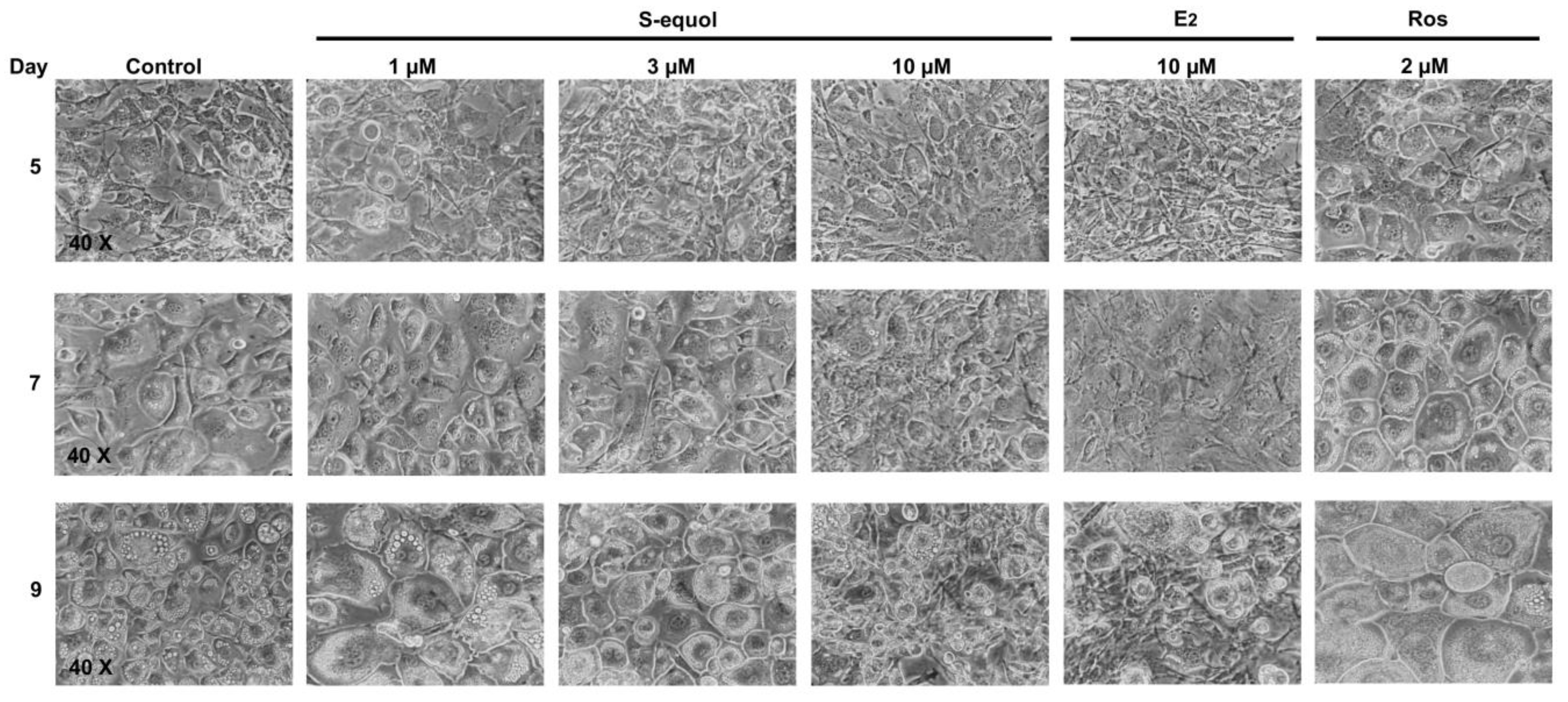
3.2. S-Equol Inhibits Adipocyte Differentiation of Cells 3T3-L1

To examine the effect of S-equol on adipocyte differentiation, confluent 3T3-L1 fibroblasts were induced to differentiation in DM-I containing 1, 3, and 10 µM of S-equol for three days and subsequently kept in S-equol free DM-II and MM, as described above. As expected, the size of control cells without S-equol progressively increased from day 5 of adipocyte differentiation; the shape became semi-rounded and intracellular lipid droplets were formed; notably, these morphological changes that are associated with an initial stage of adipocyte differentiation were more visible in the following days. These changes were even more pronounced in cells treated with 2 µM of rosiglitazone used as a positive control; on day 9, the cell monolayer appeared similar to mature adipose tissue (Figure 3). Treatment with 1 and 3 µM of S-equol did not significantly affect cell differentiation in comparison with control cells, as cells exhibited a similar increase in size, the same morphological changes as the formation of lipid droplets, notably from day 5. Interestingly, cells treated with 10 µM of S-equol did not present the changes in lipid droplet formation associated with adipocyte differentiation on day 5; and this inhibitory effect remained until the seventh day of culture. On day 9, cells seemed to recover from the 10 µM S-equol effects, their size slightly increased, and lipid droplets were formed (Figure 3). Similar observations were made in cells treated with 10 µM of estradiol used as an inhibitor of adipocyte differentiation.

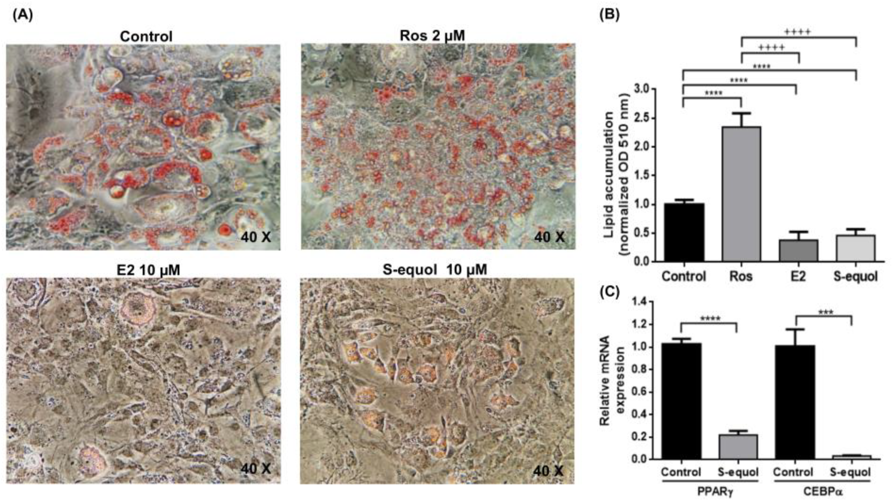
3.3. S-Equol Inhibits Lipid Accumulation

One of the first functions of adipocytes is lipid accumulation for energy storage. Therefore, we examined lipid accumulation through ORO staining on day 7 in order to better characterize the effect of 10 µM of S-equol on adipocytes. As expected, cells treated with 2 µM of rosiglitazone had a greater number of ORO-stained lipid droplets when compared to control cells without any treatment. In contrast, lipid staining was lower in cells treated with 10 µM of estradiol. Interestingly, a reduction in lipid droplet staining was also observed in cells treated with 10 µM of S-equol (Figure 4A). Quantification of ORO dye droplets confirmed that cells treated with 2 µM of rosiglitazone accumulated about 2.35-fold more lipids than control cells, whereas lipid accumulation was reduced by about 60% in cells treated with 10 µM of estradiol. Remarkably, a similar reduction of about 50% was also observed in cells treated with 10 µM of S-equol (Figure 4B).

3.4. S-Equol Affects the Expression of Pro-Adipogenic Markers

As C/EBPα and PPARγ are two master pro-adipogenic transcription factors, we analyzed their mRNA expression by real-time qRT-PCR in 3T3-L1 cells exposed to S-equol (10 μM) during the first three days of the adipocyte differentiation process. As shown in Figure 4C, treatment with S-equol significantly decreased the expression of PPARγ and C/EBPα by 78% and 97%, respectively, when compared to control cells on day 7 of adipocyte differentiation, which is consistent with the reduced adipogenesis.

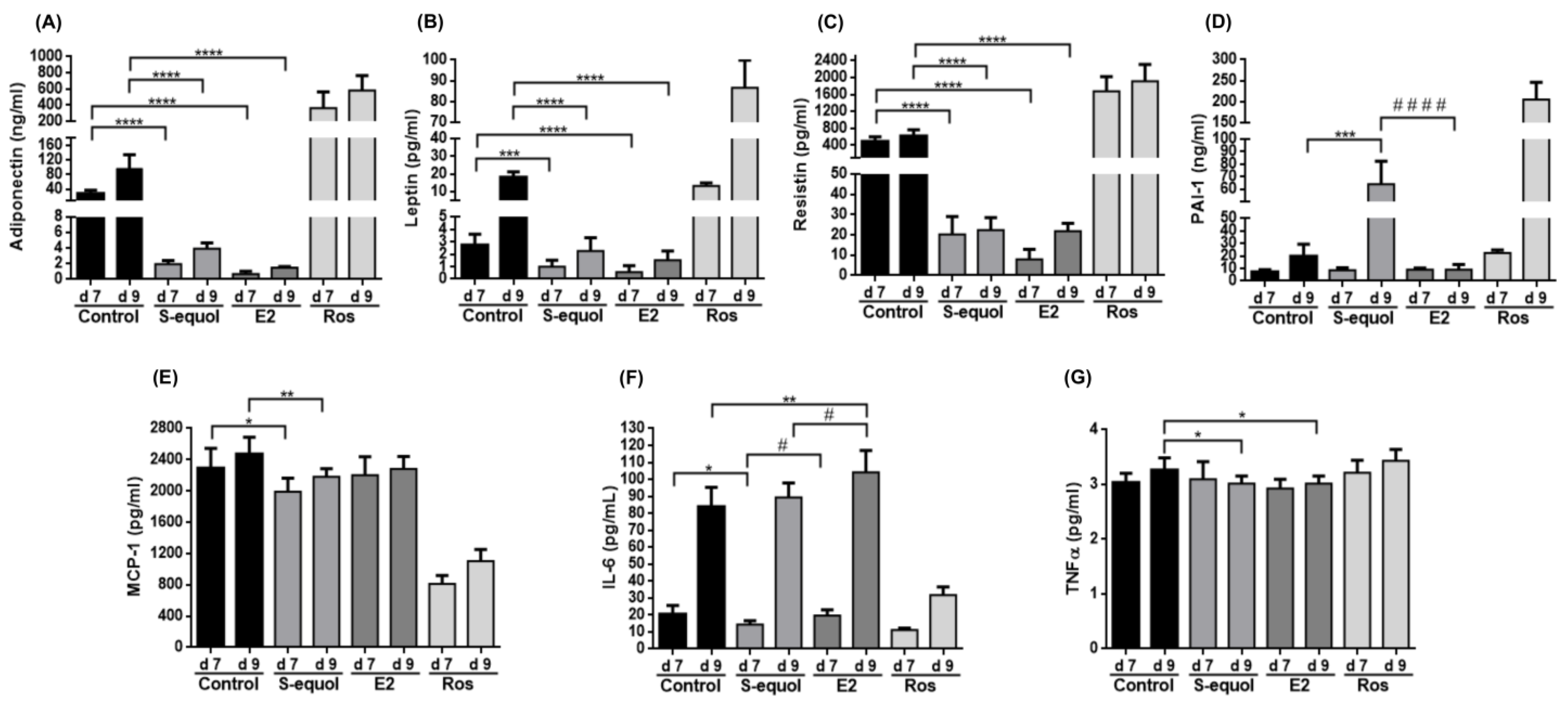
3.5. S-Equol Reduces Adipokine Secretion

In addition to energy storage through accumulation of fatty acids, adipocytes have an endocrine function in regulating homeostasis, inflammatory processes, and adipogenesis, among other events [3,4]; moreover, the production of adipokines is positively correlated with adipocyte size and adipocyte differentiation, mainly in the middle and late stages of adipogenesis [26,27]. Therefore, we evaluated how S-equol affects the secretion of adipokines in 3T3-L1 adipocytes (Figure 5). As expected, control cells released Adiponectin, Leptin, Resistin, PAI-1, MCP-1, IL-6, and TNFα on day 7, with an increase on day 9, mainly in the cases of Adiponectin (3.4-fold), Leptin (6.7-fold), PAI-1 (2.74-fold), and IL-6 (4.09-fold). In cells treated with rosiglitazone, the secretion of Adiponectin, Leptin, Resistin, and PAI-1 was clearly exacerbated; it was reduced in the cases of MCP-1 and IL-6, while the release of TNFα remained unchanged. Treatment with S-equol and estradiol significantly reduced the secretion of Adiponectin, Leptin, Resistin, and TNFα compared to control cells. Interestingly, the release of MCP-1 was slightly reduced in S-equol-treated cells, the secretion of IL-6 was reduced by S-equol on day 7, while it was increased by estradiol on day 9. Finally, there were no significant differences in PAI-1 levels between the control, S-equol, and estradiol-treated cells on day 7; however, the PA1-1 release was about 3.25-fold increased in cells exposed to S-equol on day 9, while it remained low in the case of estradiol. Altogether, these data suggest that both ER ligands have different effects on 3T3-L1 adipocytes.

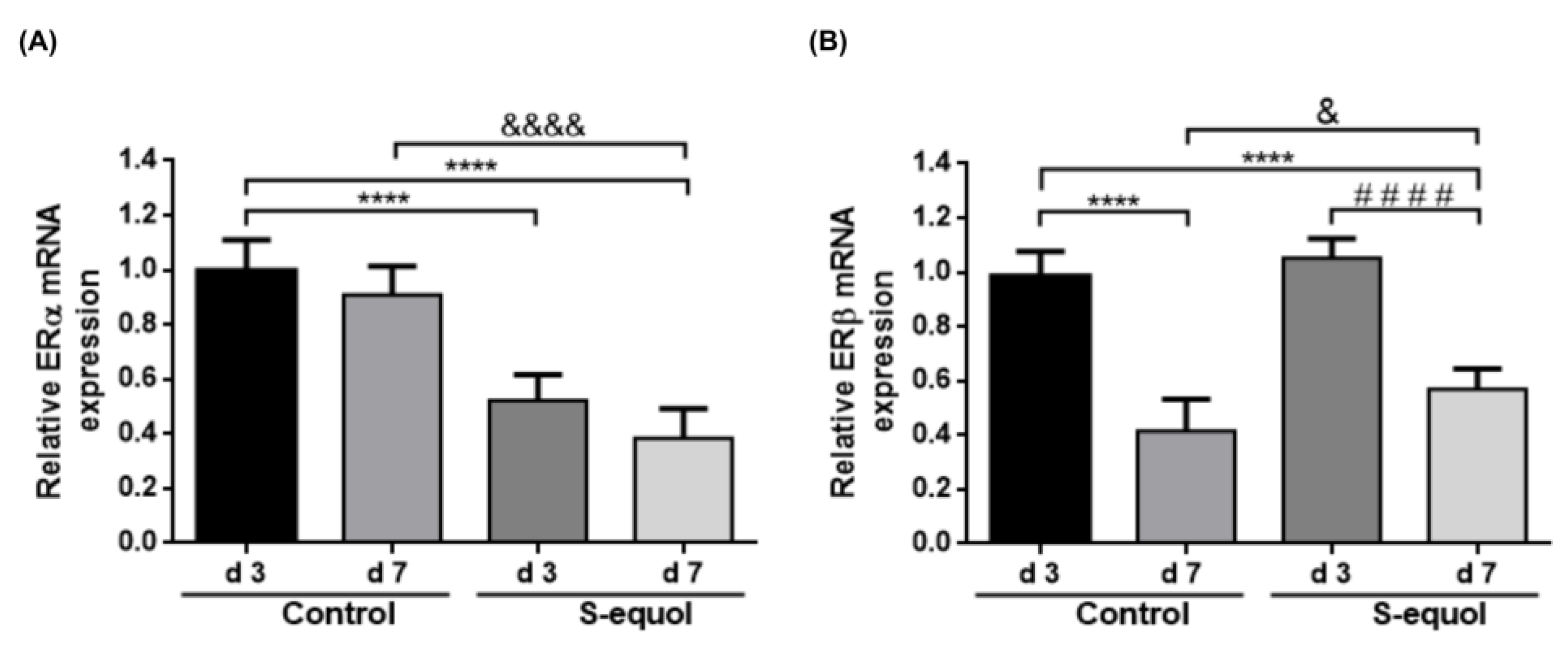
3.6. S-Equol Reduces ERα Expression and Attenuates the Subexpression of ERβ

The results described above showed that the ERβ agonist S-equol affects adipocyte differentiation and functions. It has been reported that the expression of ERα is greater and constant during earlier stages of adipogenesis compared to ERβ, whose expression decreases during adipocyte differentiation, suggesting an association between the expression of ERs and adipogenesis progression [28,29,30]. Therefore, we analyzed the effect of S-equol on both ERs by real-time qRT-PCR assays (Figure 6). As previously reported, ERα mRNA levels remained constant during the differentiation process in control cells; in contrast, they were reduced by about 48% and 62% in cells treated with S-equol on days 3 and 7, respectively, with respect to control cells (Figure 6A). Regarding ERβ, its expression was decreased by about 59% on day 7 in control cells; treatment with S-equol did not affect ERβ mRNA levels on day 3, but it only produced a 43% reduction on day 7 (Figure 6B). These data indicated that S-equol exposure has an effect on ER expression in 3T3-L1 cells.

4. Discussion

The supplementation with dietary isoflavones has been shown to have positive effects on body weight reduction. In this context, there are contradictory reports about the effect of equol, an analog of estradiol, on adipocyte differentiation, mainly because of the use of S-equol or the racemic mixture of (R,S)-equol, which creates some uncertainty about the effectiveness of equol as an adipogenesis inhibitor. To our knowledge, this is the first work that describes the effect of S-equol on 3T3-L1 adipocyte differentiation in vitro.
Low concentrations of S-equol (1 to 10 µM) did not significantly affect the viability of 3T3-L1 cells, while higher concentrations led to cell death. Interestingly, S-equol (1–30 µM) did not affect the viability of differentiating cells either. Considering that a racemic mixture is a 50:50 mix of two enantiomers, this observation roughly corresponds to the previous report of Nishide et al., 2013, which described that the cell viability of differentiating 3T3-L1 cells remained almost constant in the presence of 0.1 to 100 µM of (R,S)-equol [24]. As expected for an estradiol analog and an ERβ ligand, S-equol at 10 µM inhibited adipocyte differentiation, and this anti-adipogenic effect was associated with a reduced expression of the pro-adipogenic factors C/EBPα and PPARγ; moreover, there was a reduction in lipid accumulation and changes in adipokine release, mainly Adiponectin, Leptin, Resistin, MCP-1, and PAI-1, indicating that S-equol also affected the adipocyte role in energy storage and endocrine function.
Adipokines have important roles for body weight control and related processes. Adiponectin is involved in the modulation of insulin sensitivity/resistance [31,32]. Leptin is considered the master regulator of homeostasis by modulating food intake and expenditure energy through the hypothalamic pathway [33]. Resistin participates in insulin resistance, glucose intolerance, and the development of inflammatory processes [34,35]. PAI-1 presents a dual role in the context of obesity, modulating various metabolic and physiologic processes mainly associated with insulin resistance [36]. MCP-1 is involved in insulin resistance and macrophage infiltration in adipose tissue, contributing to the development of the cellular obesogenic microenvironment [37,38]. The effect of soy isoflavones, including equol, on the synthesis and secretion of adipokines has been previously described in 3T3-L1 cells and animal models. For example, Niwa et al. reported a dose-dependent reduction in Leptin secretion in differentiated 3T3-L1 adipocytes treated with 5 to 40 µM of S-equol for two days [39]. Regarding animal models, Wagner et al. demonstrated that a supplementation with soy isoflavones reduced plasma Adiponectin levels and increased the insulin response in obese monkeys [40]. Nagarajan et al. showed that a diet with isoflavones, including (R,S)-equol, inhibits MCP-1 expression and release in mice ApoE-/- mice, contributing to atherosclerotic lesions improvement [41].
Importantly, several reports have shown that adipokines can also regulate adipocyte differentiation. Notably, the overexpression of Adiponectin promotes proliferation, lipid accumulation, and adipocyte differentiation in 3T3-L1 cells through the increased expression of pro-adipogenic markers PPARγ, C/EBPα, and ADD1/SREBP1 [42]. Similar results have been reported in differentiated 3T3-L1 cells stimulated with LPS [43]. High Leptin levels were associated with an increase in fat tissue and positively correlated with weight gain, cardiovascular risk, and triglyceride and cholesterol levels [44,45]. Treatment with 4 and 40 nM of Leptin stimulated adipocyte differentiation, increased lipid accumulation, and enhanced the expression of PPARγ, PLIN1, SREBP1C, as well as TNFα and Adiponectin, in differentiating 3T3-L1 cells [46]. Resistin knockdown produced an anti-adipogenic effect, reducing the formation of lipid droplets and accumulation of intracellular triglycerides in differentiating 3T3-L1 cells, independently of PPARγ and C/EBPα, through the reduction in lipogenic genes such as ChREBP, FASN, and SCD1 [47]. The overexpression of PAI-1 in 3T3-L1 inhibited adipocyte differentiation through the reduction in PPARγ and CEBPα expression, while PAI-1 inhibition stimulated adipogenesis [48]. Therefore, it is possible that the lower release of Adiponectin, Leptin, and Resistin, and the higher levels of PAI-1, following exposure to S-equol and ERβ activation, could also contribute to reduce adipogenesis in 3T3-L1 cells.
Interestingly, S-equol seems to produce the same anti-adipogenic effects than estradiol [49,50,51], confirming that S-equol is a genuine estrogen analog. However, there were several differences in adipokines synthesis. Notably, the increase in PAI-1 release on day 9 was only observed in S-equol-treated cells, indicating that distinct molecular mechanisms may be activated by both ERβ agonists. It has been previously reported that a high concentration of (R,S)-equol (100 μM of equol) is required to significantly repress proliferation, adipogenesis, and expression of PPARγ, C/EBPα, FAS, and CD36, while a lower concentration (10 μM) tends to promote adipocyte differentiation by activating the expression of genes related to adipocyte differentiation in MC3T3-L1 [24]. Conversely, low concentrations of equol (0–20 µmol/L) increased adipogenesis and PPARγ transcriptional activity in mesenchymal stem cell 10T1/2 [23]. Both enantiomeric forms of equol have different binding affinities for ER, with S-equol being the best ligand for ERβ. The fact that the effective concentration of S-equol to inhibit adipogenesis was about 10-fold lower than the concentration of the racemic mixture highlights the relevance of ERβ for adipocyte formation. Congruently, Naaz et al. showed that the effects of ERβ on body weight and adipocyte size reduction are independent of ERα using ovariectomized mice lacking ERα [52]. In the absence of ERα, ERβ also has a protective role in reducing inflammation and extracellular matrix markers, to modulate the expansion of adipose tissue [53]. These data confirm the relevance of specific ERβ ligands, such as S-equol, for adipogenesis control. In this context, several studies have shown that ERβ agonists reduce body weight and WAT through the reduction in PPARγ activity [11,54]. Zhang et al. showed that the activation of ERβ by the DPN agonist repressed adipogenesis and downregulated PPARγ, PGC-1α, and UCP-1 expression in adipose-derived stem cells (ASCs) of brown adipose tissue extracted from male C57BL/6 mice; in contrast, the use of PPT, an ERα agonist, was associated with proliferation and migration processes [28]. Moreover, ERβ activation by DPN negatively correlated with Leptin expression in differentiating 3T3-L1 cells [55]. In an Achilles tendon injury, ERβ deletion stimulated adipocyte accumulation through activation of the PPARγ signaling pathways; moreover, the activation of ERβ by a specific agonist inhibited the adipogenic differentiation of Tendon-derived stem cells (TDSCs) [56]. Therefore, we hypothesize that the binding of S-equol to ERβ during the early step of adipogenesis induction promotes the activation of the receptor, which results in reductions in PPARγ and C/EBPα, and therefore an inhibition of adipogenesis and adipocyte functions. A remarkable point of our work is that 3T3- L1 cells were treated with S-equol for only three days, during the induction step, and the inhibitory effect was maintained until the seventh day of adipocyte differentiation. These results indicate that treatment with a low concentration of S-equol in the early step of adipogenesis produces changes in the expression of specific genes that alter the molecular program of the differentiation and maintenance steps, and therefore the adipogenesis process and adipocyte functions. They also suggest that efficient body weight control should not require a daily S-equol administration, which would reduce the cost of the treatment for people with obesity.
An interesting finding of our work is the effect of S-equol on the expression of ERα and ERβ as changes in ERα and ERβ expression have been associated with the modulation of adipogenesis. ERβ expression decreases during adipocyte differentiation in vitro [30]. ERβ activation is related to adipogenesis inhibition, while ERα activation promotes adipogenesis, proliferation, and migration [28]. Moreover, ERβ modulates the transcriptional activity of ERα by reducing its expression [57,58,59]. Therefore, by attenuating the downregulation of ERβ on day 7, S-equol could help to maintain the anti-adipogenic effect due to ERβ activation. Moreover, by reducing ERα expression on days 3 and 7, S-equol could also prevent the negative side-effects associated with ERα activation.
Altogether, our results confirmed the potential of ERβ ligands, particularly the soy-derived isoflavonoid S-equol, as anti-obesity molecules. Based on our results and data previously described for estradiol, we proposed a hypothetical model to explain the anti-adipogenic effects produced by a short exposure to S-equol (Figure 7). It has been reported that ER interacts with transcription factors C/EBPβ and NF-kB and inhibits their binding on the IL-6 gene promoter [60]. During adipogenesis, C/EBPβ induces the expression of PPARγ, which, in turn, promotes C/EBPα expression [61,62] that activates the transcription of Resistin, Leptin, and Adiponectin genes [63,64,65,66]. On the other hand, NF-kB is associated with the expression of MCP-1 [37,67]. The activation of membrane ER by estradiol stimulates the activation of intracellular pathways that induce ER binding to the PPARγ promoter in the nucleus, reducing its expression and inhibiting adipogenesis in differentiating 3T3-L1 and ADSC cells [68]. Therefore, we hypothesize that S-equol binds to ERβ in the cytoplasm and is translocated to the nucleus to interact with C/EBPβ and NF-kB, which prevents the transcription of PPARγ and subsequently C/EBPα, reducing Leptin (LEP), Resistin (RETN), Adiponectin (ADIPO), and MCP-1 production. These effects could also be due to ERβ activation at the plasma membrane by S-equol followed by a signaling cascade in the cytoplasm that leads to transcriptional inactivation of the PPARγ gene. On the other hand, it has been shown that PA1-1 promoter activity depends on the ERβ/ERα ratio, and an increase in ERβ modulates the induction of the PAI-1 promoter [69]. The presence of Estrogen Response Elements (ERE) in the promoter region of human ERβ indicates a possible mechanism of autoregulation that leads to the modulation of ERα transcription [58,59,70]. A search using the Software LASAGNA-Search 2.0 [71] indicated the presence of ERE in mouse ERβ and PAI-1 (data not shown). Then, it is also possible that ERβ activated by S-equol in the cytoplasm could be translocated to the nucleus to bind ERE in promoter regions, modulating the transcription of ERβ and PAI-1. As a result, ERβ activation by S-equol though these different pathways diminishes pro-adipogenic marker expression, adipokine levels, lipid accumulation, and, therefore, adipocyte hypertrophy, contributing to the mitigation of the expansion of adipose tissue and preventing complications derived from an excessive accumulation of body fat.

5. Conclusions

The development of new strategies to combat obesity and overweight is a worldwide priority to control comorbidities that threaten human health. In this context, various works have demonstrated the potential of ERβ-selective ligands. In the present study, we demonstrated that a single and short exposure to the estrogenic analog, S-equol, produced an anti-adipogenic effect that was maintained for seven days, showing a reduction in lipid accumulation, pro-adipogenic markers expression, and adipokines release, as well as an attenuation of ERβ downregulation, which probably helps to maintain the long-term anti-adipogenic effect. Additionally, the reduction in ERα expression could avoid the side-effects associated with its activation by estrogen treatments. Altogether, our findings confirmed the potential of ERβ ligands, particularly the soy-derived isoflavonoid S-equol, as anti-obesity molecules and allowed us to propose a hypothetical model to explain the anti-adipogenic effects produced by a short exposure to S-equol. However, further experiments are required to confirm these assumptions and elucidate the exact molecular mechanisms by which S-equol inhibits the adipogenesis process.

Author Contributions

G.M.-L., Conceptualization, Methodology, Investigation, Writing—Original Draft; C.G.-H., Conceptualization, Formal Analysis, Writing—Review and Editing; J.C.A.-P., Methodology, Investigation, Writing—Review and Editing; A.G.-M., Validation, Formal Analysis, Writing—Review and Editing; C.A.R.-L. and C.L.-C., Methodology, Investigation, Writing—Review and Editing; F.H., Methodology, Visualization, Writing—Review and Editing; L.A.M., Conceptualization, Supervision, Funding Acquisition, Writing—Original Draft. All authors provided critical feedback and gave their approval of the final version of the manuscript.

Funding

This research was funded by SIP/IPN (20194920 and SIP20201142) (Mexico). LAM is supported by COFAA-IPN and EDI-IPN (Mexico). GML is a CONACyT fellowship (632860) (Mexico).

Acknowledgments

We thank Rodolfo Gamaliel Avila-Bonilla for his technical support in real-time qRT-PCR assays.

Conflicts of Interest

The authors declare no conflict of interest.

References

  1. Engin, A. The Definition and Prevalence of Obesity and Metabolic Syndrome. Adv. Exp. Med. Biol. 2017, 906, 1–17. [Google Scholar] [CrossRef]
  2. Kim, S.M.; Lun, M.; Wang, M.; Senyo, S.; Guillermier, C.; Patwari, P.; Steinhauser, M.L. Loss of White Adipose Hyperplastic Potential Is Associated with Enhanced Susceptibility to Insulin Resistance. Cell Metab. 2014, 20, 1049–1058. [Google Scholar] [CrossRef] [PubMed] [Green Version]
  3. Coelho, M.; Oliveira, T.; Fernandes, R. Biochemistry of adipose tissue: An endocrine organ. Arch. Med. Sci. 2013, 9, 191–200. [Google Scholar] [CrossRef] [Green Version]
  4. Abranches, M.V.; de Oliveira, F.C.E.; da Conceição, L.L.; Peluzio, M.D.C.G. Obesity and diabetes: The link between adipose tissue dysfunction and glucose homeostasis. Nutr. Res. Rev. 2015, 28, 121–132. [Google Scholar] [CrossRef] [Green Version]
  5. Jeffery, E.; Church, C.D.; Holtrup, B.; Colman, L.; Rodeheffer, M.S. Rapid depot-specific activation of adipocyte precursor cells at the onset of obesity. Nat. Cell Biol. 2015, 17, 376–385. [Google Scholar] [CrossRef]
  6. Leeners, B.; Geary, N.; Tobler, P.; Asarian, L. Ovarian hormones and obesity. Hum. Reprod. Updat. 2017, 23, 300–321. [Google Scholar] [CrossRef]
  7. Foryst-Ludwig, A.; Clemenz, M.; Hohmann, S.; Hartge, M.; Sprang, C.; Frost, N.; Krikov, M.; Bhanot, S.; Barros, R.; Morani, A.; et al. Metabolic Actions of Estrogen Receptor Beta (ERβ) are Mediated by a Negative Cross-Talk with PPARγ. PLoS Genet. 2008, 4, e1000108. [Google Scholar] [CrossRef]
  8. Santen, R.J.; Allred, D.C.; Ardoin, S.P.; Archer, D.F.; Boyd, N.; Braunstein, G.D.; Burger, H.G.; Colditz, G.A.; Davis, S.R.; Gambacciani, M.; et al. Postmenopausal Hormone Therapy: An Endocrine Society Scientific Statement. J. Clin. Endocrinol. Metab. 2010, 95, s1–s66. [Google Scholar] [CrossRef] [Green Version]
  9. Hua, H.; Zhang, H.; Kong, Q.; Jiang, Y. Mechanisms for estrogen receptor expression in human cancer. Exp. Hematol. Oncol. 2018, 7, 24. [Google Scholar] [CrossRef] [PubMed] [Green Version]
  10. Savva, C.; Korach-André, M. Estrogen Receptor beta (ERβ) Regulation of Lipid Homeostasis—Does Sex Matter? Metabolites 2020, 10, 116. [Google Scholar] [CrossRef] [PubMed] [Green Version]
  11. Yepuru, M.; Eswaraka, J.; Kearbey, J.D.; Barrett, C.M.; Raghow, S.; Veverka, K.A.; Miller, D.D.; Dalton, J.T.; Narayanan, R. Estrogen Receptor-β-selective Ligands Alleviate High-fat Diet- and Ovariectomy-induced Obesity in Mice. J. Biol. Chem. 2010, 285, 31292–31303. [Google Scholar] [CrossRef] [PubMed] [Green Version]
  12. Křížová, L.; Dadáková, K.; Kasparovska, J.; Kašparovský, T. Isoflavones. Molecules 2019, 24, 1076. [Google Scholar] [CrossRef] [Green Version]
  13. Setchell, K.D. The history and basic science development of soy isoflavones. Menopause 2017, 24, 1338–1350. [Google Scholar] [CrossRef]
  14. Setchell, K.D.R.; Clerici, C.; Lephart, E.D.; Cole, S.J.; Heenan, C.; Castellani, D.; Wolfe, B.E.; Nechemias-Zimmer, L.; Brown, N.M.; Lund, T.D.; et al. S-Equol, a potent ligand for estrogen receptor β, is the exclusive enantiomeric form of the soy isoflavone metabolite produced by human intestinal bacterial flora. Am. J. Clin. Nutr. 2005, 81, 1072–1079. [Google Scholar] [CrossRef] [PubMed] [Green Version]
  15. Setchell, K.D.R.; Clerici, C. Equol: History, Chemistry, and Formation. J. Nutr. 2010, 140, 1355S–1362S. [Google Scholar] [CrossRef] [Green Version]
  16. Setchell, K.D.; Borriello, S.P.; Hulme, P.; Kirk, D.N.; Axelson, M. Nonsteroidal estrogens of dietary origin: Possible roles in hormone-dependent disease. Am. J. Clin. Nutr. 1984, 40, 569–578. [Google Scholar] [CrossRef] [PubMed]
  17. Jackson, R.L.; Greiwe, J.S.; Schwen, R.J. Emerging evidence of the health benefits of S-equol, an estrogen receptor β agonist. Nutr. Rev. 2011, 69, 432–448. [Google Scholar] [CrossRef]
  18. Usui, T.; Tochiya, M.; Sasaki, Y.; Muranaka, K.; Yamakage, H.; Himeno, A.; Shimatsu, A.; Inaguma, A.; Ueno, T.; Uchiyama, S.; et al. Effects of naturalS-equol supplements on overweight or obesity and metabolic syndrome in the Japanese, based on sex and equol status. Clin. Endocrinol. 2012, 78, 365–372. [Google Scholar] [CrossRef]
  19. Cheong, S.H.; Furuhashi, K.; Ito, K.; Nagaoka, M.; Yonezawa, T.; Miura, Y.; Yagasaki, K. Antihyperglycemic effect of equol, a daidzein derivative, in cultured L6 myocytes andob/obmice. Mol. Nutr. Food Res. 2014, 58, 267–277. [Google Scholar] [CrossRef]
  20. Blake, C.; Fabick, K.M.; Setchell, K.D.; Lund, T.D.; Lephart, E.D. Neuromodulation by soy diets or equol: Anti-depressive & anti-obesity-like influences, age- & hormone-dependent effects. BMC Neurosci. 2011, 12, 28. [Google Scholar] [CrossRef] [Green Version]
  21. Rachoń, D.; Vortherms, T.; Seidlovä-Wuttke, D.; Wuttke, W. Effects of dietary equol on body weight gain, intra-abdominal fat accumulation, plasma lipids, and glucose tolerance in ovariectomized Sprague-Dawley rats. Menopause 2007, 14, 925–932. [Google Scholar] [CrossRef]
  22. Bax, E.N.; Cochran, K.E.; Mao, J.; Wiedmeyer, C.E.; Rosenfeld, C.S. Opposing effects of S-equol supplementation on metabolic and behavioral parameters in mice fed a high-fat diet. Nutr. Res. 2018, 64, 39–48. [Google Scholar] [CrossRef] [PubMed]
  23. Cho, K.W.; Lee, O.-H.; Banz, W.J.; Moustaid-Moussa, N.; Shay, N.F.; Kim, Y.-C. Daidzein and the daidzein metabolite, equol, enhance adipocyte differentiation and PPARγ transcriptional activity. J. Nutr. Biochem. 2010, 21, 841–847. [Google Scholar] [CrossRef] [PubMed]
  24. Nishide, Y.; Tousen, Y.; Inada, M.; Miyaura, C.; Ishimi, Y. Bi-Phasic Effect of Equol on Adipocyte Differentiation of MC3T3-L1 Cells. Biosci. Biotechnol. Biochem. 2013, 77, 201–204. [Google Scholar] [CrossRef] [Green Version]
  25. Zebisch, K.; Voigt, V.; Wabitsch, M.; Brandsch, M. Protocol for effective differentiation of 3T3-L1 cells to adipocytes. Anal. Biochem. 2012, 425, 88–90. [Google Scholar] [CrossRef] [PubMed]
  26. Skurk, T.; Alberti-Huber, C.; Herder, C.; Hauner, H. Relationship between Adipocyte Size and Adipokine Expression and Secretion. J. Clin. Endocrinol. Metab. 2007, 92, 1023–1033. [Google Scholar] [CrossRef]
  27. Ojima, K.; Oe, M.; Nakajima, I.; Muroya, S.; Nishimura, T. Dynamics of protein secretion during adipocyte differentiation. FEBS Open Bio. 2016, 6, 816–826. [Google Scholar] [CrossRef]
  28. Zhang, W.; Schmull, S.; Du, M.; Liu, J.; Lu, Z.; Zhu, H.; Xue, S.; Lian, F. Estrogen Receptor α and β in Mouse: Adipose-Derived Stem Cell Proliferation, Migration, and Brown Adipogenesis In Vitro. Cell. Physiol. Biochem. 2016, 38, 2285–2299. [Google Scholar] [CrossRef] [PubMed]
  29. Dieudonné, M.N.; Leneveu, M.C.; Giudicelli, Y.; Pecquery, R. Evidence for functional estrogen receptors α and β in human adipose cells: Regional specificities and regulation by estrogens. Am. J. Physiol. Cell Physiol. 2004, 286, C655–C661. [Google Scholar] [CrossRef]
  30. Pedersen, S.B.; Bruun, J.; Hube, F.; Kristensen, K.; Hauner, H.; Richelsen, B. Demonstration of estrogen receptor subtypes α and β in human adipose tissue: Influences of adipose cell differentiation and fat depot localization. Mol. Cell. Endocrinol. 2001, 182, 27–37. [Google Scholar] [CrossRef]
  31. Kawano, J.; Arora, R. The Role of Adiponectin in Obesity, Diabetes, and Cardiovascular Disease. J. CardioMetabolic Syndr. 2009, 4, 44–49. [Google Scholar] [CrossRef] [PubMed]
  32. Chandran, M.; Phillips, S.A.; Ciaraldi, T.; Henry, R.R. Adiponectin: More Than Just Another Fat Cell Hormone? Diabetes Care 2003, 26, 2442–2450. [Google Scholar] [CrossRef] [Green Version]
  33. Friedman, J.M. Leptin, leptin receptors, and the control of body weight. Nutr. Rev. 1998, 56, S38–S46. [Google Scholar] [CrossRef] [PubMed]
  34. Steppan, C.M.; Lazar, M.A. Resistin and obesity-associated insulin resistance. Trends Endocrinol. Metab. 2002, 13, 18–23. [Google Scholar] [CrossRef]
  35. Filkova, M.; Haluzik, M.; Gay, S.; Senolt, L. The role of resistin as a regulator of inflammation: Implications for various human pathologies. Clin. Immunol. 2009, 133, 157–170. [Google Scholar] [CrossRef] [Green Version]
  36. Kaji, H. Adipose Tissue-Derived Plasminogen Activator Inhibitor-1 Function and Regulation. Compr. Physiol. 2016, 6, 1873–1896. [Google Scholar] [CrossRef]
  37. Panee, J. Monocyte Chemoattractant Protein 1 (MCP-1) in obesity and diabetes. Cytokine 2012, 60, 1–12. [Google Scholar] [CrossRef] [Green Version]
  38. Kanda, H.; Tateya, S.; Tamori, Y.; Kotani, K.; Hiasa, K.-I.; Kitazawa, R.; Kitazawa, S.; Miyachi, H.; Maeda, S.; Egashira, K.; et al. MCP-1 contributes to macrophage infiltration into adipose tissue, insulin resistance, and hepatic steatosis in obesity. J. Clin. Investig. 2006, 116, 1494–1505. [Google Scholar] [CrossRef]
  39. Niwa, T.; Yokoyama, S.-I.; Ito, T.; Osawa, T. Reduction of leptin secretion by soy isoflavonoids in murine adipocytes in vitro. Phytochem. Lett. 2010, 3, 122–125. [Google Scholar] [CrossRef]
  40. Wagner, J.D.; Zhang, L.; Shadoan, M.K.; Kavanagh, K.; Chen, H.; Tresnasari, K.; Kaplan, J.R.; Adams, M.R. Effects of soy protein and isoflavones on insulin resistance and adiponectin in male monkeys. Metabolism 2008, 57, S24–S31. [Google Scholar] [CrossRef] [PubMed] [Green Version]
  41. Nagarajan, S.; Burris, R.L.; Stewart, B.W.; Wilkerson, J.E.; Badger, T.M. Dietary Soy Protein Isolate Ameliorates Atherosclerotic Lesions in Apolipoprotein E-Deficient Mice Potentially by Inhibiting Monocyte Chemoattractant Protein-1 Expression. J. Nutr. 2008, 138, 332–337. [Google Scholar] [CrossRef] [Green Version]
  42. Fu, Y.; Luo, N.; Klein, R.L.; Garvey, W.T. Adiponectin promotes adipocyte differentiation, insulin sensitivity, and lipid accumulation. J. Lipid Res. 2005, 46, 1369–1379. [Google Scholar] [CrossRef] [PubMed] [Green Version]
  43. Yang, W.; Yang, C.; Luo, J.; Wei, Y.; Wang, W.; Zhong, Y. Adiponectin promotes preadipocyte differentiation via the PPARγ pathway. Mol. Med. Rep. 2017, 17, 428–435. [Google Scholar] [CrossRef] [PubMed] [Green Version]
  44. Ekmen, N.; Helvaci, A.; Gunaldi, M.; Sasani, H.; Yildirmak, S.T. Leptin as an important link between obesity and cardiovascular risk factors in men with acute myocardial infarction. Indian Hear. J. 2016, 68, 132–137. [Google Scholar] [CrossRef] [Green Version]
  45. Liuzzi, A.; Savia, G.; Tagliaferri, M.; Lucantoni, R.; Berselli, M.; Petroni, M.; De Medici, C.; Viberti, G. Serum leptin concentration in moderate and severe obesity: Relationship with clinical, anthropometric and metabolic factors. Int. J. Obes. 1999, 23, 1066–1073. [Google Scholar] [CrossRef] [Green Version]
  46. Palhinha, L.; Liechocki, S.; Hottz, E.D.; Pereira, J.A.D.S.; De Almeida, C.J.; Vieira, P.; Bozza, P.T.; Maya-Monteiro, C.M. Leptin Induces Proadipogenic and Proinflammatory Signaling in Adipocytes. Front. Endocrinol. 2019, 10, 841. [Google Scholar] [CrossRef] [PubMed] [Green Version]
  47. Ikeda, Y.; Tsuchiya, H.; Hama, S.; Kajimoto, K.; Kogure, K. Resistin affects lipid metabolism during adipocyte maturation of 3T3-L1 cells. FEBS J. 2013, 280, 5884–5895. [Google Scholar] [CrossRef]
  48. Liang, X.; Kanjanabuch, T.; Mao, S.-L.; Hao, C.-M.; Tang, Y.-W.; Declerck, P.; Hasty, A.; Wasserman, D.H.; Fogo, A.B.; Ma, L.-J. Plasminogen activator inhibitor-1 modulates adipocyte differentiation. Am. J. Physiol. Endocrinol. Metab. 2006, 290, E103–E113. [Google Scholar] [CrossRef] [PubMed] [Green Version]
  49. Yoon, M.; Jeong, S. Repression of PPARγ Activity on Adipogenesis by 17β-estradiol in Differentiated 3T3-L1 Cell. J. Exp. Bio-Med. Sci. 2009, 15, 179–185. [Google Scholar]
  50. Jeong, S.; Yoon, M. 17β-Estradiol inhibition of PPARγ-induced adipogenesis and adipocyte-specific gene expression. Acta Pharmacol. Sin. 2011, 32, 230–238. [Google Scholar] [CrossRef] [Green Version]
  51. Yoon, M.; Jeong, S. 17β-estradiol Prevents the Expression of CEBPα-mediated Adipocyte Marker Genes in Female Ovariecto-mized C57BL/6 Mice. J. Exp. Biomed. Sci. 2008, 14, 131–137. [Google Scholar]
  52. Naaz, A.; Zakroczymski, M.; Heine, P.; Taylor, J.; Saunders, P.; Lubahn, D.; Cooke, P.S. Effect of Ovariectomy on Adipose Tissue of Mice in the Absence of Estrogen Receptor Alpha (ERα): A Potential Role for Estrogen Receptor Beta (ERβ). Horm. Metab. Res. 2002, 34, 758–763. [Google Scholar] [CrossRef] [PubMed]
  53. Davis, K.E.; Neinast, M.D.; Sun, K.; Skiles, W.M.; Bills, J.D.; Zehr, J.A.; Zeve, D.; Hahner, L.D.; Cox, D.W.; Gent, L.M.; et al. The sexually dimorphic role of adipose and adipocyte estrogen receptors in modulating adipose tissue expansion, inflammation, and fibrosis. Mol. Metab. 2013, 2, 227–242. [Google Scholar] [CrossRef] [PubMed]
  54. Ponnusamy, S.; Tran, Q.T.; Harvey, I.; Smallwood, H.S.; Thiyagarajan, T.; Banerjee, S.; Johnson, D.L.; Dalton, J.T.; Sullivan, R.D.; Miller, D.D.; et al. Pharmacologic activation of estrogen receptor α increases mitochondrial function, energy expenditure, and brown adipose tissue. FASEB J. 2017, 31, 266–281. [Google Scholar] [CrossRef] [PubMed] [Green Version]
  55. Yi, K.W.; Shin, J.-H.; Seo, H.S.; Lee, J.K.; Oh, M.-J.; Kim, T.; Saw, H.S.; Kim, S.-H.; Hur, J.-Y. Role of Estrogen Receptor-α and -β in Regulating Leptin Expression in 3T3-L1 Adipocytes. Obesity 2008, 16, 2393–2399. [Google Scholar] [CrossRef] [PubMed]
  56. Bian, X.; Liu, T.; Zhou, M.; He, G.; Ma, Y.; Shi, Y.; Wang, Y.; Tang, H.; Kang, X.; Yang, M.; et al. Absence of estrogen receptor beta leads to abnormal adipogenesis during early tendon healing by an up-regulation of PPARγ signalling. J. Cell. Mol. Med. 2019, 23, 7406–7416. [Google Scholar] [CrossRef] [Green Version]
  57. Lindberg, M.K.; Movérare, S.; Skrtic, S.; Gao, H.; Dahlman-Wright, K.; Gustafsson, J.-A.; Ohlsson, C. Estrogen Receptor (ER)-β Reduces ERα-Regulated Gene Transcription, Supporting a “Ying Yang” Relationship between ERα and ERβ in Mice. Mol. Endocrinol. 2003, 17, 203–208. [Google Scholar] [CrossRef] [PubMed]
  58. Hall, J.M.; McDonnell, D.P. The Estrogen Receptor β-Isoform (ERβ) of the Human Estrogen Receptor Modulates ERα Transcriptional Activity and Is a Key Regulator of the Cellular Response to Estrogens and Antiestrogens1. Endocrinology 1999, 140, 5566–5578. [Google Scholar] [CrossRef]
  59. Matthews, J.; Wihlén, B.; Tujague, M.; Wan, J.; Ström, A.; Gustafsson, J.-A. Estrogen Receptor (ER) β Modulates ERα-Mediated Transcriptional Activation by Altering the Recruitment of c-Fos and c-Jun to Estrogen-Responsive Promoters. Mol. Endocrinol. 2006, 20, 534–543. [Google Scholar] [CrossRef] [Green Version]
  60. Stein, B.; Yang, M.X. Repression of the interleukin-6 promoter by estrogen receptor is mediated by NF-kappa B and C/EBP beta. Mol. Cell. Biol. 1995, 15, 4971–4979. [Google Scholar] [CrossRef] [Green Version]
  61. Wu, Z.; Xie, Y.; Bucher, N.L.; Farmer, S. Conditional ectopic expression of C/EBP beta in NIH-3T3 cells induces PPAR gamma and stimulates adipogenesis. Genes Dev. 1995, 9, 2350–2363. [Google Scholar] [CrossRef] [Green Version]
  62. Zuo, Y.; Qiang, L.; Farmer, S. Activation of CCAAT/Enhancer-binding Protein (C/EBP) α Expression by C/EBPβ during Adipogenesis Requires a Peroxisome Proliferator-activated Receptor-γ-associated Repression of HDAC1 at the C/ebpα Gene Promoter. J. Biol. Chem. 2006, 281, 7960–7967. [Google Scholar] [CrossRef] [Green Version]
  63. Hartman, H.B.; Hu, X.; Tyler, K.X.; Dalal, C.K.; Lazar, M.A. Mechanisms Regulating Adipocyte Expression of Resistin. J. Biol. Chem. 2002, 277, 19754–19761. [Google Scholar] [CrossRef] [Green Version]
  64. Mason, M.M.; He, Y.; Chen, H.; Quon, M.J.; Reitman, M. Regulation of Leptin Promoter Function by Sp1, C/EBP, and a Novel Factor 1. Endocrinology 1998, 139, 1013–1022. [Google Scholar] [CrossRef] [PubMed]
  65. Qiao, L.; MacLean, P.; Schaack, J.; Orlicky, D.J.; Darimont, C.; Pagliassotti, M.; Friedman, J.E.; Shao, J. C/EBP Regulates Human Adiponectin Gene Transcription Through an Intronic Enhancer. Diabetes 2005, 54, 1744–1754. [Google Scholar] [CrossRef] [Green Version]
  66. Fox, K.E.; Fankell, D.M.; Erickson, P.F.; Majka, S.M.; Crossno, J.T., Jr.; Klemm, D.J. Depletion of cAMP-response Element-binding Protein/ATF1 Inhibits Adipogenic Conversion of 3T3-L1 Cells Ectopically Expressing CCAAT/Enhancer-binding Protein (C/EBP) α, C/EBP β, or PPARγ2. J. Biol. Chem. 2006, 281, 40341–40353. [Google Scholar] [CrossRef] [PubMed] [Green Version]
  67. Tsuchiya, K.; Yoshimoto, T.; Hirono, Y.; Tateno, T.; Sugiyama, T.; Hirata, Y. Angiotensin II induces monocyte chemoattractant protein-1 expression via a nuclear factor-κB-dependent pathway in rat preadipocytes. Am. J. Physiol. Endocrinol. Metab. 2006, 291, E771–E778. [Google Scholar] [CrossRef] [PubMed] [Green Version]
  68. Ahluwalia, A.; Hoa, N.; Ge, L.; Blumberg, B.; Levin, E.R. Mechanisms by Which Membrane and Nuclear ER Alpha Inhibit Adipogenesis in Cells Isolated from Female Mice. Endocrinology 2020, 161, bqaa175. [Google Scholar] [CrossRef]
  69. Smith, L.H.; Coats, S.R.; Qin, H.; Petrie, M.S.; Covington, J.W.; Su, M.; Eren, M.; Vaughan, D.E. Differential and Opposing Regulation of PAI-1 Promoter Activity by Estrogen Receptor α and Estrogen Receptor β in Endothelial Cells. Circ. Res. 2004, 95, 269–275. [Google Scholar] [CrossRef] [PubMed]
  70. Li, L.-C.; Yeh, C.-C.; Nojima, D.; Dahiya, R. Cloning and Characterization of Human Estrogen Receptor β Promoter. Biochem. Biophys. Res. Commun. 2000, 275, 682–689. [Google Scholar] [CrossRef]
  71. Lee, C.; Huang, C.-H. LASAGNA-Search: An integrated web tool for transcription factor binding site search and visualization. BioTechniques 2013, 54, 141–153. [Google Scholar] [CrossRef] [PubMed]
Figure 1. Flowchart of the differentiation protocol of 3T3-L1 cells. Fibroblasts were cultivated in growth medium (GM) until 80% confluence (day 0). Then, adipogenesis was induced by incubation in differentiation medium I and II (DM-I and DM-II) for three and two days, respectively. Finally, differentiating cells were kept in maintenance medium (MM). Cells were treated with S-equol in DM-I for three days. Rosiglitazone (Ros) and estradiol (E2) were used as stimulation and inhibition controls, respectively.
Applsci 11 09657 g001
Figure 2. Effect of S-equol on 3T3-L1 cell viability. 3T3-L1 fibroblasts were treated with S-equol for 24 (A) and 48 h (B), while differentiating 3T3-L1 were treated with S-equol for three days (C), and cell viability was determined by MTT assay. Data were obtained from three independent experiments in triplicate and expressed as mean ± SD. Statistical analysis was performed by one-way ANOVA with the Bonferroni post hoc test. **** p < 0.0001, ** p < 0.01, * p < 0.05 vs. control. Dotted line, 85% cell viability.
Applsci 11 09657 g002
Figure 3. Effect of S-equol on 3T3-L1 adipocyte differentiation. 3T3-L1 fibroblasts treated with S-equol for 72 h were induced to differentiation and morphological changes were documented on days 5, 7, and 9. Control, cells without treatment; E2, estradiol; Ros, rosiglitazone.
Applsci 11 09657 g003
Figure 4. Effect of S-equol on lipid accumulation in differentiated 3T3-L1. 3T3-L1 fibroblasts treated with S-equol (10 μM) for 72 h and induced to differentiation for seven days were stained with Oil Red O (A) and lipid accumulation was quantified as absorbance at 510 nm (B). (C) Expression of PPARγ and C/EBPα genes by real-time qRT-PCR in differentiating 3T3-L1 cells untreated (Control) and treated with S-equol, 10 μM (S-equol). Rosiglitazone (Ros) and estradiol (E2) were used as positive and negative controls, respectively. Data were obtained from three experiments in triplicate and expressed as mean ± SD. All values were normalized with respect to β-Actin and the change in expression between treatments was calculated and compared with control cells without treatment. Statistical analysis was performed by one-way ANOVA with Tukey’s multiple comparison post hoc test for lipid accumulation (B) and Student’s t test for pro-adipogenic marker expression (C) using the GraphPad Prism software. **** p < 0.0001, *** p < 0.001 vs. control; ++++ p < 0.0001 vs. Ros.
Applsci 11 09657 g004
Figure 5. Effect of S-equol on adipokine secretion in adipocyte differentiation. 3T3-L1 fibroblasts treated with S-equol (10 μM) for 72 h were induced to differentiation, and the secretion of Adiponectin (A), Leptin (B), Resistin (C), PAI-1 (D), MCP-1 (E), IL-6 (F), and TNFα (G) was measured in the maintenance medium collected on days 7 and 9 of adipogenesis. Untreated cells, and cells treated with rosiglitazone (Ros) and estradiol (E2) were used as controls. Data were obtained from three independent experiments in triplicate and expressed as mean ± SD. Statistical analysis was performed by one-way ANOVA with Tukey’s multiple comparison post hoc test using the GraphPad Prism software. **** p < 0.0001; *** p < 0.001; ** p < 0.01; * p < 0.05 vs. control; #### p < 0.0001; # p < 0.05 S-equol vs. E2.
Applsci 11 09657 g005
Figure 6. Effect of S-equol on the expression of estrogen receptors α (A) and β (B). 3T3-L1 fibroblasts treated with S-equol (10 μM) for 72 h were induced to differentiation, and mRNA levels of ERα (A) and ERβ (B) were determined by real-time qRT-PCR on days 3 and 7. Untreated cells were used as control. Data were obtained from three experiments in triplicate and expressed as mean ± SD. All values were normalized with respect to β-Actin, and the change in expression was calculated and compared with control cells without treatment on day 3. Statistical analysis was performed by one-way ANOVA with Tukey’s multiple comparison post hoc test using the GraphPad Prism software. **** p < 0.0001 vs. control on day 3; &&&& p < 0.0001; & p < 0.05 vs. control on day 7; #### p < 0.0001 S-equol on day 3 vs. S-equol on day 7.
Applsci 11 09657 g006
Figure 7. Hypothetical model of the anti-adipogenic effects mediated by S-equol in 3T3-L1 preadipocytes.
Applsci 11 09657 g007
Publisher’s Note: MDPI stays neutral with regard to jurisdictional claims in published maps and institutional affiliations.

