Review

In Vivo Efficacy of Contact Lens Drug-Delivery Systems in Glaucoma Management. A Systematic Review

Ophthalmology, Department of Surgical, Medical, Molecular Pathology and of the Critical Area, University of Pisa, 56126 Pisa, Italy
*
Authors to whom correspondence should be addressed.
Appl. Sci. 2021, 11(2), 724; https://doi.org/10.3390/app11020724
Submission received: 12 December 2020 / Revised: 6 January 2021 / Accepted: 12 January 2021 / Published: 13 January 2021
(This article belongs to the Special Issue Glaucoma: Innovative Drug Delivery Systems for Its Treatment)

Abstract

:

Featured Application

A systematic review of in vivo studies available on the contact lens as a drug-delivery system in glaucoma therapy, also focusing on technical limitations and future perspectives.

Abstract

Adherence is crucial in medical glaucoma therapy, although half of the patients skip eyedrops. In recent years alternative drug-delivery systems have been developed. One of the most promising seems the contact lens (CL). This systematic review aims to present the in vivo efficacy of different CL drug-delivery systems. A total of 126 studies were identified following a literature search adhering to the preferred reporting items for systematic reviews and meta-analyses (PRISMA) guidelines. After full-text evaluation, 19 studies about CL drug-delivery systems were included. To date, the following drug-delivery systems have been investigated in vivo: drug-soaked CL, CL with physical barriers (vitamin E), molecularly imprinted CL, CL with implants, and nanoparticle-loaded CL. Nanoparticle-loaded CL and CL with implants seem the most promising drug-delivery systems, although initial burst drug release and patient acceptance may limit their widespread use in current practice. Clinical trials are warranted to understand the role of CL as a drug-delivery system in improving glaucomatous patient care.

1. Introduction

Glaucoma is a leading cause of blindness worldwide. Its prevalence ranges between 2.65 and 3.54% of the global population and around 111.8 million people are estimated to be affected in 2040, mainly due to life expectancy increase [1]. Open-angle glaucoma (OAG) is the most frequent type of glaucoma, with a prevalence of 3.05% compared to 0.50% of angle-closure glaucoma (ACG) [1]. OAG causes bilateral blindness in 5% and monolateral blindness in 27% of affected patients within 20 years of the diagnosis [2].
Glaucoma treatment aims to maintain the patient’s visual function and related quality of life [3]. Multiple randomised controlled studies show with no doubt that lowering the intraocular pressure (IOP) reduces glaucoma progression [4]. This target can be reached surgically or medically. Initial therapy may be laser trabeculoplasty or topical medications, although the latter remains the most widely prescribed [5]. Unfortunately, glaucoma is almost asymptomatic in the beginning, and treatment adherence is the main drawback in patient management, as with other chronic diseases. Even when patients are aware of being monitored, they forget to instil the hypotensive medication in many cases [6,7,8].
Multiple factors may jeopardise adherence, such as multiple topical treatments, medication side effects, poor understanding of treatment aims, poor instillation techniques (including physical barriers, e.g., arthritis and tremor), and cost [9,10,11,12,13].
Furthermore, multiple fixed-combination eyedrops are available on the market, allowing patients to take two hypotensive drugs with a single eye drop. All fixed combinations include a beta-blocker, except one which contains brimonidine and brinzolamide [5].
Another step to improve patient cooperation has been the introduction of preservative-free eyedrops [14]. In particular, preservatives induce toxic effects such as the detergent effect on tear film (altering the external lipid layer), the direct toxic effect on corneal and conjunctival epithelia, and the immune-allergic effect [15,16]. Nevertheless, recent studies showed that preservative-free multidose droppers required an increased squeezing force compared to multidose standard droppers, being less user-friendly for older patients or those with comorbidities [17]. Even if single vial droppers are not related to handling difficulties, it remains an expensive technology that also causes an environmental burden [14,15].
Despite the improvements mentioned above, low adherence is one of the principal causes of medical treatment failure in glaucoma [6,7,8].
Moreover, eyedrops medications are characterised by a pulsatile drug-delivery, with a concentration peak followed by a tough until the next drop instillation [18]. Consequently, drug effect may not be constant over time, and IOP may change during the day [18].
Furthermore, eyedrops have low bioavailability because only 1–7% of the drug is absorbed by the eye [18]. In particular, the lacrimal drainage consequent to blinking removes about 80% of the instilled drop [19]. The remaining dose penetrates through the cornea with a diffusion mechanism, primarily driven by the dose concentration [20]. Therefore, to improve penetration, it is necessary to increase the retention time or the corneal permeability [19].
A sustained drug-delivery system could overcome all the issues mentioned—adherence, pulsatile therapeutic effect, and low retention time. Therefore, over the last decade, the pharmaceutical industry has developed alternative drug-delivery systems [19,21]. These solutions can be classified as intraocular (e.g., implants, nanoparticles, and inserts) or topical (e.g., conjunctival inserts, contact lens (CL), gels, nanoparticles, mucoadhesive polymers, ointments, solutions, and suspensions) [19]. The ideal sustained drug-delivery system releases the drug regularly without requiring invasive surgery or injection to be placed [18].
There are already 140 million people worldwide wearing a CL, with a forecasted increase in the next future [22]. Therefore, the CL as a drug-delivery system does not affect the routine lifestyle of these patients. Moreover, the CL could allow an excellent sustained drug delivery because once placed, they could release the drug for several days. This systematic review aims to analyse and discuss the in vivo efficacy of the CL as an alternative drug-delivery system to eyedrops to manage glaucoma disease.

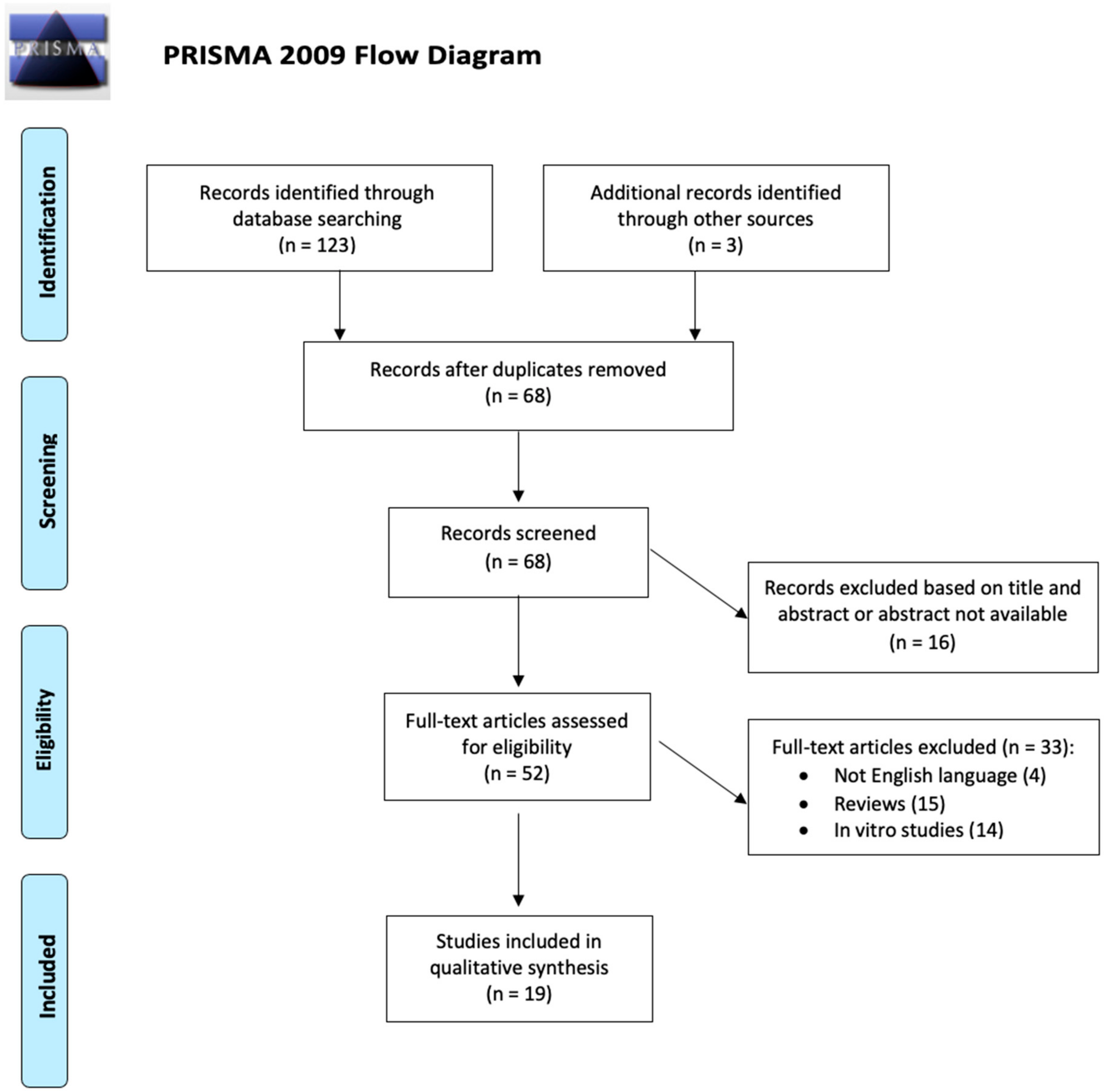
2. Materials and Methods

The PubMed database was consulted on 1 November 2020. The search terms used were “contact lens AND drug delivery AND glaucoma” and “contact lenses AND drug delivery AND glaucoma”. The search workflow was designated in adherence to the preferred reporting items for systematic reviews and meta-analyses (PRISMA) statement (Figure 1) [23]. The process applied for this review consisted of a systematic search of all available articles regarding the CL drug-delivery system in glaucoma medical treatment.
All manuscripts were enlisted, and two independent researchers (F.S. and M.M.) screened each article’s title and abstract identifying any relevant articles. All in vivo studies available in the literature reporting original data on CL drug-delivery systems in glaucoma treatment were initially included without restriction for study design, sample size, or intervention performed. Exclusion criteria were review studies, articles written in languages other than English, and in vitro studies. Moreover, the reference lists of identified articles were checked manually to detect any potential studies.
The same reviewers selected the captured studies according to inclusion and exclusion criteria, examining the articles’ full text. A senior author arbitrated when necessary (M.F.). For unpublished data, no effort was made to contact the corresponding authors.
Two blinded researchers (F.S. and M.M.) assessed the strength of evidence of clinical studies (four) according to the Oxford Centre for Evidence-Based Medicine (OCEM) 2011 guidelines and the Scottish Intercollegiate Guideline Network (SIGN) assessment system for individual studies as implemented for Preferred Practice Patterns by the American Academy of Ophthalmology [24,25]. Then, their quality of evidence was evaluated based on the Grading of Recommendations Assessment, Development and Evaluation (GRADE) system [26]. The included clinical studies were listed in the Supplementary Materials (Table S1).
Preclinical studies (animal studies) (15) were evaluated according to animal research: reporting of in vivo experiments (ARRIVE) guidelines, focusing on the “Material and Methods” section to assess the quality of evidence [27,28]. A score based on three levels for the evaluation of the categories of the guidelines was established, as follows: 0, not mentioned (total absence of any information); 1, unclear/not complete (items not mentioned entirely in the category assess); and 2, adequate/clear (complete information for all items corresponding to the category evaluated) [29]. Finally, the quality of evidence based on the Grading of Recommendations Assessment, Development and Evaluation (GRADE) system for animal studies was also assessed [30]. The included preclinical studies were listed in the Supplementary Materials (Table S2).

3. Results

The search strategy returned 126 candidate articles. Sixty-eight abstracts were screened after eliminating duplicated records, but only 52 of these met the inclusion/exclusion criteria for full-text review. Thirty-three articles were excluded: four not written in English, 15 review papers, and 14 in vitro studies. After full-text evaluation, 19 studies about CL drug-delivery systems were included. The included studies were listed in the Supplementary Materials [31,32,33,34,35,36,37,38,39,40,41,42,43,44,45,46,47,48,49].
No data synthesis was possible for the heterogeneity of available data and the design of the available studies. The current systematic review reports a qualitative analysis, detailed issue-by-issue narratively. Finally, technical limitations and future perspectives of CL drug-delivery systems were presented.

3.1. The Drug-Soaked CL

The drug-soaked CL was the first method to be developed as a CL drug-delivery system, as described by Witcherle and colleagues in 1960 [50]. The CL was soaked in a solution containing the drug to uptake it. Once applied, the CL released the drug over 60–90 min, as demonstrated by Schultz and colleagues’ in vitro study (Figure 2) [31].
North successfully treated two patients affected by acute primary ACG with a CL soaked in pilocarpine 4% [32]. In both cases, IOP decreased in two hours (from 70 mmHg to 12 mmHg and from 72 mmHg to 36 mmHg) and symptoms resolved. However, details on the soaking method were not reported.
Assef et al. compared the pilocarpine penetration into monkeys’ aqueous humour after installing topical eyedrops or applying a soaked CL [33]. A CL contained a drug dose of 400 μg when soaked in 1% of pilocarpine solution for 2 min. Conversely, a pilocarpine eyedrop (0.1 mL) at 1% contained 1000 μg of the drug, at 4%, 4000 μg, and 8%, 8000 μg. The soaked CL delivered the same pilocarpine amount in the aqueous as the 4% eyedrops. Moreover, the CL achieved the highest aqueous drug concentration after two hours, even when compared to the instilled pilocarpine eyedrop at 8%. Finally, miosis in the eye treated with the soaked CL was more pronounced than in eyes treated with eyedrops even 24 h later. These authors concluded that the soaked CL enhanced the drug’s penetration and prolonged the drug’s effect [33].
Podos et al. treated 10 patients affected by ocular hypertension [34]. In particular, they soaked a CL in 0.5% pilocarpine for two minutes and obtained an IOP-lowering for the 23 h of wearing (from 28 mmHg to 24 mmHg, 14.7%). Moreover, 24 h after treatment, an IOP reduction of 20% occurred after 30 min of wearing a soaked CL in 1% of pilocarpine and after 60 min with a CL soaked in 0.5% of pilocarpine [34].
Hillman et al. treated 25 patients with acute primary ACG with a CL soaked in 1% of pilocarpine and 500 mg of intramuscular acetazolamide [35]. Results were retrospectively compared with 25 patients treated with topical pilocarpine 4% eyedrops instilled every minute for 5 min, then every 5 min for half an hour, and finally every 15 min for 90 min and 500 mg of intramuscular acetazolamide. The CL released 400 μg of pilocarpine, although the eyedrops instillation delivered about 40,000 μg. All patients well-tolerated lens wearing for two hours, and no one developed corneal abrasion. The CL drug delivery showed an increased efficacy compared to the eyedrop treatment, with a mean IOP-lowering of 54.8% compared to 49.7%, respectively [35]. In the following years, other studies confirmed the CL’s therapeutic role in ACG and OAG when soaked with pilocarpine [36,51].
The soaked CL was a straightforward drug-delivery system, although it had several limitations. The CL drug’s affinity was a double-edged sword because when elevated, the CL poorly released the drug. Instead, if the affinity was low, the drug release immediately peaked after lens application. Moreover, to be available on the market, the soaked CL should be sterilised, altering the drug efficacy or release.
Recently Schultz et al. evaluated the IOP-lowering effect of a CL soaked with timolol or brimonidine [31]. Three patients affected by OAG were enrolled. After three weeks of washout, patients wore, for 30 min a day for 21 days, a CL soaked with the same drug that they were already taking before the washout (one with brimonidine and two with timolol). The CL were loaded with 0.65 mg/mL timolol maleate, but no details were provided about the brimonidine tartrate-loaded CL. All patients showed IOP values similar to the ones with eyedrops, for all the periods. However, the CL released a drug quantity ten times inferior to the eyedrops [31].
Latanoprost drug delivery with a soaked CL has also been recently investigated, although only an in vitro study has been conducted [52].

3.2. Vitamin-E-Loaded CL

To extend the drug release time and prolong the therapeutic effect, a physical barrier may be added to the CL matrix, without altering CL transparency and oxygen permeation [53]. To pursue this solution, the CL can be soaked in a vitamin E ethanol before soaking in the drug solution or a vitamin E ethanol can be added to the drug solution, followed by ethanol removal with water (Figure 3) [53].
Peng and colleagues tested a vitamin-E loaded-CL in dogs affected by primary OAG [37,38]. In a first study, a vitamin-E-loaded CL was compared to a CL without barrier and to timolol eyedrops. Both CL types were soaked in two timolol solution, one at 2.65 mg/mL and one at 8 mg/mL, to allow a drug release capacity of 67 μg and 200 μg, respectively. The drug release capacity of 0.5% timolol eyedrop was estimated at 150 μg. Five treatment groups were designed, and the contralateral eyes constituted the control group. IOP-lowering was not statistically different among treated groups; however, eyedrops treatment showed a significantly higher bilateral IOP reduction than CL groups. This effect seemed related to the increased systemic uptake, supporting that the CL drug-delivery system reduced it. Also, no statistical difference in IOP-lowering was reported between the CL with vitamin E or without. However, in this study, the CL was replaced every day. Nevertheless, in 24 h, the vitamin E CL released only 80% of their drug capacity. Finally, dogs with the vitamin-E-loaded CL did not report any systemic or local toxicity [37].
In the following study with identical CL applied on glaucomatous dogs, Peng et al. replaced the CL without barrier every 24 h or every four days, and the vitamin-E-loaded CL every four days [38]. The vitamin E CL showed similar efficacy to eyedrops and the CL without barrier replaced daily, without signs of toxicity or hypoxia. Conversely, the CL without barrier replaced every four days failed to lower the IOP significantly [38].
The vitamin-E-loaded CL was also soaked with two drugs (e.g., timolol and dorzolamide) [39]. In particular, the CL without barrier and the vitamin E CL were able to release 217.8 μg and 676.7 μg of dorzolamide and 60.6 μg and 191.4 μg of timolol, respectively. The co-loading extended drug delivery time compared to the single-drug-loaded lens. This effect could be related to hydrogen bonding between drug molecules. The vitamin E co-loaded CL was able to extend drug delivery up to 48 h in vitro. Moreover, the IOP-lowering continued for a week after CL removal maybe thanks to drug accumulation inside corneal epithelial cells in beagle dogs. It opened the possibility of wearing a CL for a day to obtain a therapeutic effect lasting a week. The vitamin E CL also achieved a superior IOP-lowering to that of eyedrops (4.35 ± 0.18 mmHg versus 3.79 ± 0.28 mmHg), with an 11-fold lower drug dose. No ocular or systemic toxicity was reported [39].
Vitamin A has recently been used as a physical barrier to extend drug delivery time, although only in vitro study was available [54].

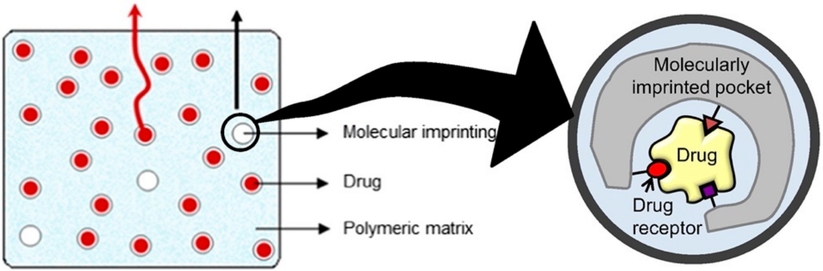
3.3. Molecularly Imprinted CL

Another type of CL drug-delivery system was molecular imprinting [55]. During CL polymerisation, a template drug and functional monomers created multiple macromolecular memory sites, similar to drug receptors [56]. The CL was then washed and reloaded with the active drug, which was entrapped in the CL memory site (Figure 4) [56]. This technique increased the CL drug loading capacity [56]. It has been applied to different ophthalmic drugs—hyaluronic acid, prednisolone, diclofenac, ketotifen fumarate, norfloxacin, and acyclovir [57,58,59,60,61,62]. However, to date, only timolol has been investigated in vivo among hypotensive drugs.
Hiratani et al. compared a molecularly imprinted CL to a timolol-soaked CL and timolol eyedrops [40]. In particular, timolol concentration and bioavailability in rabbits’ tears were evaluated. The molecularly imprinted CL (30 μg of drug dose) delivered timolol at a measurable concentration for 180 min, longer than the soaked CL (21 μg of drug dose) (90 min) and eyedrops (34 and 125 μg of drug dose) (60 min). Furthermore, molecularly imprinted CL bioavailability was 3.3- and 8.7-fold superior to the soaked CL and eyedrops, respectively [40].
However, further studies are warranted to evaluate the molecularly imprinted CL as an extended drug-delivery system.

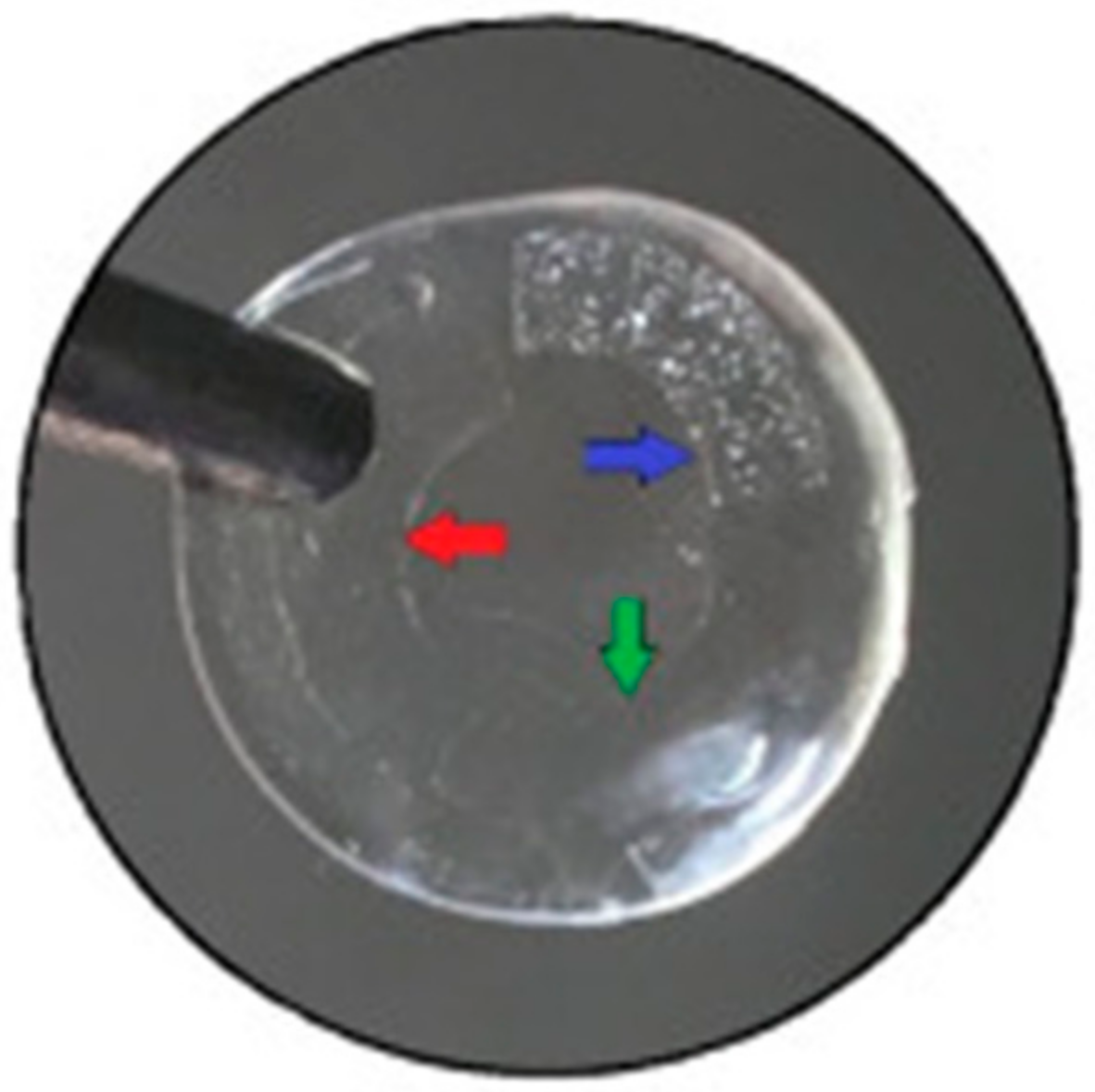
3.4. CL with Implant

Desai et al. proposed to include drug implants in the outer periphery of a silicone CL [41,42]. This technique prevented the limiting of vision through the CL centre. Each implant was loaded with bimatoprost, timolol, or hyaluronic acid. Hyaluronic acid improved comfort during the extended CL wear (Figure 5).
In the first study on rabbit tears, the CL with timolol and hyaluronic acid implants was compared to the timolol-soaked CL and timolol eyedrops [41]. Eyedrops reduced IOP by 2.24 mmHg on average, returning to baseline after 12 h. The soaked CL and the CL with implants lowered IOP by 2.95 mmHg and 3.19 mmHg on average, and IOP recovered after 96 h and 144 h, respectively. Both the soaked CL and the CL with implant showed an initial burst drug release. It was interesting to note that the CL with implant obtained this achievement with a drug dose of 154 μg, although a single eyedrop contained 250 μg [41].
In the following study, a CL with bimatoprost–timolol–hyaluronic acid implants was compared with soaked a bimatoprost–timolol CL and bimatoprost-timolol eyedrops in glaucomatous rabbits [42]. Similar results to the previous study were obtained, bearing in mind that prostaglandin does not affect rabbits IOP. Eyedrops reduced IOP by 3.10 mmHg at maximum, returning to baseline after 12 h. The soaked CL and the CL with implants lowered IOP at maximum by 4.33 mmHg and 5.00 mmHg and kept IOP below baseline values for 96 h and 120 h, respectively. The CL with implants showed a sustained drug release in tears (17.26 h) compared to the soaked CL (6.61 h) and eyedrops (11.47 min). Interestingly, the soaked CL and the CL with the implants showed a reduced initial burst of 26.18- and 77.11-fold, respectively, compared to the eyedrops. No signs of ocular irritation were reported [42].

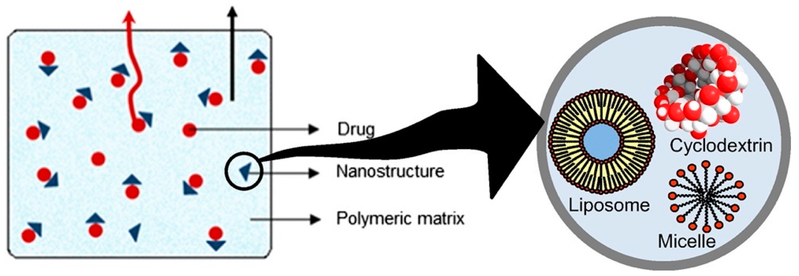
3.5. Nanoparticle-Loaded CL

Another CL drug-delivery technique was including drugs into nanoparticles that were incorporated inside the CL. Nanoparticles are defined as all particles having dimensions around or 10–1000 nm, such as liposomes or micelles, which acted as a carrier for the drug, although in some cases the drug itself was the nanoparticle [63]. The nanoparticles aimed to allow precise drug delivery with toxicity reduction and increased biocompatibility [63].
Nanoparticles allowed a high drug-loading capacity compared to a soaked CL. Furthermore, both hydrophilic and hydrophobic drugs could be loaded in the CL [63]. However, excessive nanoparticle loading could alter CL transparency [63]. Different nanoparticles were available—nanosuspension, nanodiamonds, nanocrystals, liposomes, niosomes, dendrimers, micelles, and cyclodextrins (Figure 6).
Jung et al. incorporated nanoparticles of propoxylated glyceryl triacylate (PGT) containing timolol in a CL [43]. The CL was soaked in a solution of nanoparticles in ethanol. Then, the CL was applied continuously to dogs affected by OAG. The IOP lowered significantly for five days, although the therapeutic effect was detectable after day 2 of application. No adverse effects were reported. Interestingly, the authors compared a freshly soaked lens with a lens soaked and packaged for 15 days. The latter group showed a significantly reduced drug release rate, maybe related to the diffusion of the timolol–PGT particles into the packaging medium [43].
Ciolino et al. developed a film of latanoprost and poly(lactic-co-glycolic) acid (PLGA) nanoparticle encapsulated in methafilcon by ultraviolet light polymerisation [44]. Then, the methafilcon block was lathed into the CL. This CL was continuously applied to glaucomatous rabbits for one month. Latanoprost was released for 28 days at a steady-state aqueous concentration significantly higher than the mean aqueous concentration with latanoprost eyedrops (p < 0.05). However, on day one, the CL showed a drug concentration burst release higher than the one following latanoprost eyedrops instillation at 0.005% (854–1473 ng/mL versus 54 ng/mL). To reduce it, the latanoprost-eluting CL was soaked in phosphate-buffered saline for 1 or 3 days before the application. Both types of preconditioned CL showed a smaller initial burst (46–74 ng/mL) but a significantly reduced steady-state aqueous latanoprost concentration compared to the non-preconditioned version (5.5–5.7 ng/mL versus 21.0–39.6 ng/mL). It is essential to remark that latanoprost concentration was reported because it does not induce IOP-lowering in rabbits. In 25% of rabbits, the lenses moved slightly inferiorly, away from the corneal centre, and some of them developed limbal corneal neovascularisation (exact percentage not reported) [44].
Ciolino et al. also compared the latanoprost–PLGA CL to latanoprost eyedrops in glaucomatous monkeys [45]. Two versions of latanoprost–PLGA CL were developed, one with a high drug dose and one with a low drug dose. Each animal consecutively received the following treatment: eight days with the low-dose CL, eight days with latanoprost eyedrops, and eight days with the high-dose CL. The researchers observed a three-week washout period between different treatments. Only the high-dose CL showed a significantly greater IOP reduction than eyedrops at day 3 (p = 0.001), day 5 (p = 0.015), and day 8 (p < 0.05). No adverse events were reported [45].
Maulvi and colleagues developed an ethylcellulose-nanoparticle-laden ring loaded with the drug and installed it in a hydrogel CL [46]. This CL loaded with timolol was compared to timolol eyedrops in glaucomatous rabbits. The eyedrop group’s IOP was lowered by 4.4 ± 0.50 mmHg after 2 h and returned after 12 h to baseline values. Conversely, after an initial burst release, the CL lowered the IOP by 6.3 ± 1.92 mmHg 3 h later and kept below baseline values for 192 h (8 days) [46].
Other interesting polymeric nanoparticles were cyclodextrins that increased hydrophobic drugs’ affinity to the CL polymer matrix, prolonging the drug release time [47]. In particular, the interior part of cyclodextrins was hydrophobic and complexed with hydrophobic drugs. Conversely, the exterior part was hydrophilic and easily penetrated the CL polymer matrix. Xu et al. loaded a CL with a pueranin–cyclodextrin complex [47]. Pueranin is a hydrophobic substance with a beta-blocker effect, used in Chinese medicine to treat ocular hypertension and glaucoma. The CL and eyedrops at 1% released a mean pueranin dose of 232.64 μg and the 500 μg, respectively. The CL and eyedrops showed the same bioavailability in the tear film. However, the mean residency time of pueranin in the CL was significantly longer (6.04 times more) in rabbits tear fluid than that of 1% of pueranin eyedrops. Data about IOP-lowering or drug concentration in the rabbit aqueous were not detected [47].
Furthermore, cyclodextrins allowed the incorporation of acetazolamide in the CL, opening the possibility to use this drug topically [64]. However, no in vivo study was available.
Moreover, gold nanoparticles (GNP) have been investigated as CL drug delivery [48]. GNP was added to the timolol soaking solution or was merged into the CL during manufacturing, without altering the CL’s physical properties. Glaucomatous rabbits were treated with timolol eyedrops, or a CL soaked with GNP or CL with GNP merged. All three treatments showed an average IOP-lowering of 2mmHg. In the eyedrops group, the IOP returned to baseline values after 12 h. Conversely, in both CL groups the IOP returned to baseline values after 72 h. No adverse effects were reported [48].
Also, micelles have been loaded in a CL, as reported by Xu et al. [49]. These authors compared the CL with micelles containing timolol or latanoprost to timolol or latanoprost eyedrops in glaucomatous rabbits. The initial burst was reduced with the CL compared to the eyedrops. The CL loaded with micelles extended drug residence time in the tear fluid for more than 120 h with timolol and 96 h with latanoprost. In particular, the mean residence time and bioavailability for both timolol and latanoprost delivered by CL were enhanced, 79.6-fold and 122.2-fold and 2.2-fold and 7.3-fold, respectively, compared to eyedrops. IOP returned to baseline after 168 h of CL application and 72 h with eyedrops (data available only with timolol because prostaglandin did not affect IOP in rabbits). However, the CL became rough with extended wearing, resulting in discomfort, red eyes, or giant papillary conjunctivitis [49].

4. Discussion

Recently, Peral et al. detailed technical aspects of a CL drug-delivery system in glaucoma, highlighting the increased drug bioavailability [21]. Simultaneously, administering a lower drug dose, adverse events’ frequency and severity were reduced [21]. In particular, only 20% of the drug dose was required to produce a similar therapeutic effect with CL drug-delivery systems compared to the dose needed with eyedrops delivery systems [65].
Our review illustrated the in vivo findings of CL drug-delivery system, focusing on IOP-lowering and assessment versus eyedrops. Therefore, these data provided a scaffold to analyse the potential impact of CL drug-delivery systems in managing glaucomatous patients.
During recent years, multiple attempts to improve patient cooperation, such as educational brochures, pamphlets, and fact sheets have been developed [66]. Video and Web-based patient educational tools were designed to encourage patient adherence and stimulate dialogue with the clinician. Also, reminders and recall systems were developed to avoid missed appointments or skipped drugs [66]. Nevertheless, the adherence issue is still unresolved, and there is still much to do.
The development of minimally invasive glaucoma surgery allows surgery to be performed earlier than before [67]. In particular, these techniques are currently adopted in selected glaucoma patients with early or moderate disease, usually associated with phacoemulsification [4,68]. However, patients consider surgery a last-chance treatment and still prefer non-invasive treatment, such as alternative drug-delivery systems [69].
To increase adherence, preservative-free topical drugs have also been developed. Long-term therapy has usually caused topical toxic effects such as discomfort during instillation, foreign body sensation, burning, and ocular inflammation signs [10]. These side effects reduced patients’ cooperation; consequently, the European Medicines Agency (EMA) in 2009 provided crucial recommendations for preservative-free topical drug development [70]. Even with the massive development in preservative-free eyedrop delivery systems in recent years, this technology has remained expensive. It also does not resolve other obstacles limiting patients’ adherence, such as incorrect drop instillation, skipped instillation, and poor doctor–patient communication.
For these reasons, alternative systems for drug delivery in ophthalmology are currently under development, such as nanoparticles, punctal plugs, and CL-eluting glaucoma medications.
A recent survey of 199 glaucomatous patients investigated the acceptance of drug-delivery systems [69]. The CL showed the second-lowest acceptance rate (31%) compared to triple combination eyedrops (85%), eye spray (54%), periocular ring inserts (43%), injectable subconjunctival drug inserts (32%), and injectable anterior chamber implants (30%). Moreover, patients with greater disease severity and prior incisional glaucoma surgery were more likely to pursue alternatives to traditional eyedrops [69].
Another survey of 150 glaucomatous patients compared the drug-eluting CL, ring inserts, punctal plugs, and sub-conjunctival injections as an alternative to eyedrops [71]. In this study, the CL acceptance rate of these drug-delivery systems was higher than ring inserts (36% more) or sub-conjunctival injections (32% more). However, alternative drug-delivery systems would be accepted if surgery was avoided or they were more effective than eyedrops [71].
However, multiple obstacles limit their adoption in current practice.

4.1. Barriers to Widespread Use in Clinical Practice

Today, no CL drug-delivery system is available on the market because different issues prevent its widespread use [50]. In particular, the drug-soaked CL and the CL with vitamin E have limited drug loading capability. The drug-soaked CL also cannot release a drug for an extended period at a constant rate. Moreover, extended CL wear is related to several ocular complications, classifiable into infectious and non-infectious [72].
The infectious side effect related to CL wear is mainly microbial keratitis. The CL wearer has an 80-times increased risk of developing it compared to healthy people, and visual acuity decreases in 10–15% of CL wearers after microbial keratitis resolution [73,74]. Principal pathogens are Pseudomonas ssp., Acanthamoeba, and Candida spp., although the causative organism varies according to the region [72]. Traumatic or incorrect lens insertion or removal may cause a corneal abrasion, that needs strict control to prevent microbial keratitis development [72].
Chronic hypoxia belongs to the non-infectious group and leads to ocular and limbal erythema, microcystic and stromal oedema, vascularisation, epithelial thinning, endothelial polymegethism, and pleomorphism [75,76,77]. However, these adverse events are usually reversible once CL wear is discontinued [78]. Other non-infectious complications related to extended CL wear are a peripheral corneal ulcer, single or multiple corneal infiltrates without anterior chamber reaction, conjunctival hyperaemia, and papillary conjunctivitis [72].
CL wearing is also a risk factor for dry-eye disease [79]. The exact responsible mechanism remains unclear. Maybe the CL blocks meibomian gland ducts with desquamated epithelial cells aggregates, due to mechanical trauma after each blinking [72]. Once the external lipid layer is altered, the tear film became unstable, and the vicious circle of dry eye is set. Other suggested mechanisms are inflammation and chronic hypoxia [80].
Corneal warpage is another crucial complication due to extended CL wear. It is defined as topographic corneal alterations that affect refraction and are distinguished by other ectasic disorders [81,82].
Another significant limitation is that people not accustomed to wearing a CL may hesitate to adopt this therapeutic solution, even if wearing it for a day allows an IOP-lowering lasting a week [39]. Main concerns could be the impact on vision quality in patients without refractive errors, cosmetic appearance, difficulties in day-to-day activities, and inconveniences rendered in daily life.
Likely, CL drug-delivery systems may overcome issues related to prescription refill or white-coat adherence (instilling the therapy just five to seven days before the visit). Nevertheless, some patients may not be able to wear a CL autonomously, as for eyedrops. In particular, 11.3–60.6% of patients cannot instil eyedrops correctly, and 6.8–37.3% of patients miss the eye when instilling the eyedrops [12]. The main risk factors are older age, severe visual field defect, lower educational level, and comorbidities such as arthritis [12]. In these cases, even with a CL drug-delivery system, a caregiver’s presence is still necessary. However, this technology may reduce the burden on relatives or health-care providers because the CL can be changed just every fifteen or thirteen days.
Finally, CL drug-delivery system manufacturing increases the number of synthesis and processing steps or requires long periods until the drug-loaded CL is usable, with a not negligible economic burden [83].

4.2. Future Perspectives

To date, only the nanoparticle-loaded CL or the CL with implants can release drug for weeks. However, the initial burst is an issue that should be overcome. An exception is the CL loaded with micelles, where initial burst release is reduced compared with eyedrops, but with extended wearing, this particular CL cause ocular irritation [49]. Also, in CL with implants, the initial burst is limited by entrapping the drugs in the implant’s matrix structure. However, it is still present [41].
Another possible solution could be loading the nanoparticles in a thin, collagen membrane, embeddable into the CL. A recent article compared in vitro CL with or without collagen membrane, both loaded with nanoparticles containing lidocaine [84]. The CL with this membrane released 23.4% of the drug loading after seven days compared to the CL without, which released 64% of it. The CL with membrane also showed a reduced initial burst release (16.2% of the initial drug loading) compared to the CL without (41.8% of the initial loading). However, in vivo studies are warranted to confirm these results [84].
Furthermore, Lee et al. developed a CL with nanopores (100–250 nm) interconnected via multiple nano-channels to limit the initial burst release [85]. This CL released the drug according to the initial drug load and the loading method. In particular, a deep drug embedding with soaking and centrifuging avoided the initial burst release. It also showed a sustained drug release for 30 days. In vivo studies are recommended to confirm these results.
The same authors loaded this lens with thermosensitive nanogel without altering optic CL physical properties opening the possibility of temperature-triggered drug release [85]. The following study of Lee and co-workers showed this possibility in glaucomatous rabbits [86]. IOP decreased significantly by 33% in the group wearing a CL compared to the untreated control group (p < 0.01).
It is essential to mention two techniques recently developed for a drug-delivery system based on the CL—the supercritical solvent impregnation and ion-exchange reaction [83,87,88]. The first method uses a supercritical solvent as a carrier for the drug that also swells the polymeric matrix, increasing its free volumes and facilitating the drug diffusion [83]. This method allows tuning of the loaded drug, changing the operational settings, such as temperature and pressure. Also, it applies to a commercially available CL without altering its physical properties.
Instead, the ion-exchange reaction is based on adding a cationic functional group inside the CL polymeric matrix [87,88]. Thanks to this method, the CL can store an anionic drug and release it once applied. However, further studies are required to investigate the in vivo behaviour of both techniques.

5. Conclusions

Today, therapy adherence still represents one of the leading causes of treatment failure in glaucoma disease. Alternative drug-delivery systems could play an essential role in the future. The CL seems one of the most promising and could be accepted by the patient to postpone or avoid surgery. The soaking method was the first approach developed; however, it did not allow an extended drug release, and no study has recently investigated it. The addition of a physical barrier such as Vitamin E overcame this issue. However, other approaches have been recently developed, such as molecular imprinting, implants and the nanoparticle-loaded lens. However, CL wear can also cause several side effects. Further preclinical and clinical studies are warranted to understand this alternative drug-delivery system’s role in humans and how it can impact the patient’s adherence to glaucoma treatment.

Supplementary Materials

The following are available online at https://www.mdpi.com/2076-3417/11/2/724/s1, Table S1: Clinical studies included in the systematic review, Table S2: Preclinical studies included in the systematic review.

Author Contributions

Conceptualisation, F.S.; methodology, F.S.; validation, M.F.; formal analysis, C.P.; investigation, M.M.; data curation, M.M.; writing—original draft preparation, F.S.; writing—review and editing, M.F.; visualisation, G.C.; supervision, G.C. and C.P.; project administration, M.F. All authors have read and agreed to the published version of the manuscript.

Funding

This research received no external funding.

Institutional Review Board Statement

Not applicable.

Informed Consent Statement

Not applicable.

Data Availability Statement

Not applicable.

Conflicts of Interest

The authors declare no conflict of interest.

References

  1. Tham, Y.C.; Li, X.; Wong, T.Y.; Quigley, H.A.; Aung, T.; Cheng, C.Y. Global prevalence of glaucoma and projections of glaucoma burden through 2040: A systematic review and meta-analysis. Ophthalmology 2014, 121, 2081–2090. [Google Scholar] [CrossRef]
  2. Hattenhauer, M.G.; Johnson, D.H.; Ing, H.H.; Herman, D.C.; Hodge, D.O.; Yawn, B.P.; Butterfield, L.C.; Gray, D.T. The probability of blindness from open-angle glaucoma. Ophthalmology 1998, 105, 2099–2104. [Google Scholar] [CrossRef]
  3. European Glaucoma Society Terminology and Guidelines for Glaucoma, 4th Edition—Part 1 Supported by the EGS Foundation. Br. J. Ophthalmol. 2017, 101, 1–72. [CrossRef] [Green Version]
  4. European Glaucoma Society Terminology and Guidelines for Glaucoma, 4th Edition—Chapter 3: Treatment principles and options Supported by the EGS Foundation. Part 1 Foreword Introd. Gloss. Chapter 3 Treat. Princ. Options 2017, 101, 130–195. [CrossRef] [Green Version]
  5. Lusthaus, J.; Goldberg, I. Current management of glaucoma. Med. J. Aust. 2019, 210, 180–187. [Google Scholar] [CrossRef] [PubMed]
  6. Okeke, C.O.; Quigley, H.A.; Jampel, H.D.; Ying, G.S.; Plyler, R.J.; Jiang, Y.; Friedman, D.S. Adherence with topical glaucoma medication monitored electronically the Travatan Dosing Aid study. Ophthalmology 2009, 116, 191–199. [Google Scholar] [CrossRef] [PubMed]
  7. Kinast, R.M.; Mansberger, S.L. Glaucoma Adherence-From Theriac to the Future. Am. J. Ophthalmol. 2018, 191, xiii–xv. [Google Scholar] [CrossRef]
  8. Gatwood, J.D.; Johnson, J.; Jerkins, B. Comparisons of Self-reported Glaucoma Medication Adherence with a New Wireless Device: A Pilot Study. J. Glaucoma 2017, 26, 1056–1061. [Google Scholar] [CrossRef]
  9. Tsai, J.C.; McClure, C.A.; Ramos, S.E.; Schlundt, D.G.; Pichert, J.W. Compliance barriers in glaucoma: A systematic classification. J. Glaucoma 2003, 12, 393–398. [Google Scholar] [CrossRef]
  10. Jaenen, N.; Baudouin, C.; Pouliquen, P.; Manni, G.; Figueiredo, A.; Zeyen, T. Ocular symptoms and signs with preserved and preservative-free glaucoma medications. Eur. J. Ophthalmol. 2007, 17, 341–349. [Google Scholar] [CrossRef]
  11. Gilissen, L.; De Decker, L.; Hulshagen, T.; Goossens, A. Allergic contact dermatitis caused by topical ophthalmic medications: Keep an eye on it! Contact Dermat. 2019, 80, 291–297. [Google Scholar] [CrossRef]
  12. Davis, S.A.; Sleath, B.; Carpenter, D.M.; Blalock, S.J.; Muir, K.W.; Budenz, D.L. Drop instillation and glaucoma. Curr. Opin. Ophthalmol. 2018, 29, 171–177. [Google Scholar] [CrossRef]
  13. Schwartz, G.F.; Quigley, H.A. Adherence and persistence with glaucoma therapy. Surv. Ophthalmol. 2008, 53 (Suppl. S1), S57–S68. [Google Scholar] [CrossRef] [PubMed]
  14. Figus, M.; Agnifili, L.; Lanzini, M.; Brescia, L.; Sartini, F.; Mastropasqua, L.; Posarelli, C. Topical preservative-free ophthalmic treatments: An unmet clinical need. Expert Opin. Drug Deliv. 2020. [Google Scholar] [CrossRef]
  15. Kaur, I.P.; Lal, S.; Rana, C.; Kakkar, S.; Singh, H. Ocular preservatives: Associated risks and newer options. Cutan. Ocul. Toxicol. 2009, 28, 93–103. [Google Scholar] [CrossRef] [PubMed]
  16. Burstein, N.L. The effects of topical drugs and preservatives on the tears and corneal epithelium in dry eye. Trans. Ophthalmol. Soc. UK 1985, 104 (Pt 4), 402–409. [Google Scholar]
  17. Marx, D.; Birkhoff, M. New devices for dispensing ophthalmic treatments may be the key to managing the life cycles of established products. Drug Deliv. Technol. 2010, 10, 16–21. [Google Scholar]
  18. Kompella, U.B.; Hartman, R.R.; Patil, M.A. Extraocular, periocular, and intraocular routes for sustained drug delivery for glaucoma. Prog. Retin. Eye Res. 2020, 100901. [Google Scholar] [CrossRef]
  19. Souto, E.B.; Dias-Ferreira, J.; Lopez-Machado, A.; Ettcheto, M.; Cano, A.; Camins Espuny, A.; Espina, M.; Garcia, M.L.; Sanchez-Lopez, E. Advanced Formulation Approaches for Ocular Drug Delivery: State-Of-The-Art and Recent Patents. Pharmaceutics 2019, 11. [Google Scholar] [CrossRef] [Green Version]
  20. Gaudana, R.; Ananthula, H.K.; Parenky, A.; Mitra, A.K. Ocular drug delivery. Aaps. J. 2010, 12, 348–360. [Google Scholar] [CrossRef] [PubMed]
  21. Peral, A.; Martínez Águila, A.; Pastrana, C.; Huete-Toral, F.; Carpena-Torres, C.; Carracedo, G. Contact Lenses as Drug Delivery System for Glaucoma: A Review. Appl. Sci. 2020, 10, 5151. [Google Scholar] [CrossRef]
  22. Barr, J.; Mack, C. Contact Lenses Yesterday, Today, and the Best Yet to Come. Contact Lens Spectr. 2011, 26, 14–15. [Google Scholar]
  23. Moher, D.; Liberati, A.; Tetzlaff, J.; Altman, D.G.; Group, P. Preferred reporting items for systematic reviews and meta-analyses: The PRISMA statement. J. Clin. Epidemiol. 2009, 62, 1006–1012. [Google Scholar] [CrossRef] [PubMed]
  24. Guyatt, G.; Oxman, A.D.; Akl, E.A.; Kunz, R.; Vist, G.; Brozek, J.; Norris, S.; Falck-Ytter, Y.; Glasziou, P.; DeBeer, H.; et al. GRADE guidelines: 1. Introduction-GRADE evidence profiles and summary of findings tables. J. Clin. Epidemiol. 2011, 64, 383–394. [Google Scholar] [CrossRef] [PubMed]
  25. Keaney, M.; Lorimer, A.R. Auditing the implementation of SIGN (Scottish Intercollegiate Guidelines Network) clinical guidelines. Int. J. Health Care Qual. Assur. Inc. Lead. Health Serv. 1999, 12, 314–317. [Google Scholar] [CrossRef] [PubMed]
  26. Howick, J.; Glasziou, P.; Aronson, J.K. Evidence-based mechanistic reasoning. J. R. Soc. Med. 2010, 103, 433–441. [Google Scholar] [CrossRef] [Green Version]
  27. Kilkenny, C.; Browne, W.; Cuthill, I.C.; Emerson, M.; Altman, D.G.; Group, N.C.R.R.G.W. Animal research: Reporting in vivo experiments: The ARRIVE guidelines. Br. J. Pharmacol. 2010, 160, 1577–1579. [Google Scholar] [CrossRef]
  28. Schwarz, F.; Iglhaut, G.; Becker, J. Quality assessment of reporting of animal studies on pathogenesis and treatment of peri-implant mucositis and peri-implantitis. A systematic review using the ARRIVE guidelines. J. Clin. Periodontol. 2012, 39 (Suppl. 12), 63–72. [Google Scholar] [CrossRef]
  29. Alemán-Laporte, J.; Alvarado, G.; Sa Garcia-Gomes, M.; Fonseca Brasil Antiorio, A.T.; Zúñiga-Montero, M.; Cabrera Mori, C.M. Quality of Adherence to the ARRIVE Guidelines in the Material and Methods Section in Studies Where Swine Were Used as Surgical Biomodels: A Systematic Review (2013–2018). Animals (Basel) 2019, 9, 947. [Google Scholar] [CrossRef] [Green Version]
  30. Wei, D.; Tang, K.; Wang, Q.; Estill, J.; Yao, L.; Wang, X.; Chen, Y.; Yang, K. The use of GRADE approach in systematic reviews of animal studies. J. Evid. Based Med. 2016, 9, 98–104. [Google Scholar] [CrossRef]
  31. Schultz, C.L.; Poling, T.R.; Mint, J.O. A medical device/drug delivery system for treatment of glaucoma. Clin. Exp. Optom. 2009, 92, 343–348. [Google Scholar] [CrossRef] [PubMed]
  32. North, D.P. Treatment of acute glaucoma. Can. Med. Assoc. J. 1971, 105, 561. [Google Scholar]
  33. Asseff, C.F.; Weisman, R.L.; Podos, S.M.; Becker, B. Ocular penetration of pilocarpine in primates. Am. J. Ophthalmol. 1973, 75, 212–215. [Google Scholar] [CrossRef]
  34. Podos, S.M.; Becker, B.; Asseff, C.; Hartstein, J. Pilocarpine therapy with soft contact lenses. Am. J. Ophthalmol. 1972, 73, 336–341. [Google Scholar] [CrossRef]
  35. Hillman, J.S. Management of acute glaucoma with pilocarpine-soaked hydrophilic lens. Br. J. Ophthalmol. 1974, 58, 674–679. [Google Scholar] [CrossRef] [PubMed] [Green Version]
  36. Ruben, M.; Watkins, R. Pilocarpine dispensation for the soft hydrophilic contact lens. Br. J. Ophthalmol. 1975, 59, 455–458. [Google Scholar] [CrossRef] [PubMed] [Green Version]
  37. Peng, C.C.; Ben-Shlomo, A.; Mackay, E.O.; Plummer, C.E.; Chauhan, A. Drug delivery by contact lens in spontaneously glaucomatous dogs. Curr. Eye Res. 2012, 37, 204–211. [Google Scholar] [CrossRef]
  38. Peng, C.C.; Burke, M.T.; Carbia, B.E.; Plummer, C.; Chauhan, A. Extended drug delivery by contact lenses for glaucoma therapy. J. Control. Release 2012, 162, 152–158. [Google Scholar] [CrossRef]
  39. Hsu, K.H.; Carbia, B.E.; Plummer, C.; Chauhan, A. Dual drug delivery from vitamin E loaded contact lenses for glaucoma therapy. Eur. J. Pharm. Biopharm. 2015, 94, 312–321. [Google Scholar] [CrossRef] [PubMed]
  40. Hiratani, H.; Fujiwara, A.; Tamiya, Y.; Mizutani, Y.; Alvarez-Lorenzo, C. Ocular release of timolol from molecularly imprinted soft contact lenses. Biomaterials 2005, 26, 1293–1298. [Google Scholar] [CrossRef]
  41. Desai, A.R.; Maulvi, F.A.; Pandya, M.M.; Ranch, K.M.; Vyas, B.A.; Shah, S.A.; Shah, D.O. Co-delivery of timolol and hyaluronic acid from semi-circular ring-implanted contact lenses for the treatment of glaucoma: In vitro and in vivo evaluation. Biomater. Sci. 2018, 6, 1580–1591. [Google Scholar] [CrossRef] [PubMed]
  42. Desai, A.R.; Maulvi, F.A.; Desai, D.M.; Shukla, M.R.; Ranch, K.M.; Vyas, B.A.; Shah, S.A.; Sandeman, S.; Shah, D.O. Multiple drug delivery from the drug-implants-laden silicone contact lens: Addressing the issue of burst drug release. Mater. Sci. Eng. C 2020, 112, 110885. [Google Scholar] [CrossRef]
  43. Jung, H.J.; Abou-Jaoude, M.; Carbia, B.E.; Plummer, C.; Chauhan, A. Glaucoma therapy by extended release of timolol from nanoparticle loaded silicone-hydrogel contact lenses. J. Control Release 2013, 165, 82–89. [Google Scholar] [CrossRef] [PubMed]
  44. Ciolino, J.B.; Stefanescu, C.F.; Ross, A.E.; Salvador-Culla, B.; Cortez, P.; Ford, E.M.; Wymbs, K.A.; Sprague, S.L.; Mascoop, D.R.; Rudina, S.S.; et al. In vivo performance of a drug-eluting contact lens to treat glaucoma for a month. Biomaterials 2014, 35, 432–439. [Google Scholar] [CrossRef] [PubMed] [Green Version]
  45. Ciolino, J.B.; Ross, A.E.; Tulsan, R.; Watts, A.C.; Wang, R.F.; Zurakowski, D.; Serle, J.B.; Kohane, D.S. Latanoprost-Eluting Contact Lenses in Glaucomatous Monkeys. Ophthalmology 2016, 123, 2085–2092. [Google Scholar] [CrossRef]
  46. Maulvi, F.A.; Lakdawala, D.H.; Shaikh, A.A.; Desai, A.R.; Choksi, H.H.; Vaidya, R.J.; Ranch, K.M.; Koli, A.R.; Vyas, B.A.; Shah, D.O. In vitro and in vivo evaluation of novel implantation technology in hydrogel contact lenses for controlled drug delivery. J. Control Release 2016, 226, 47–56. [Google Scholar] [CrossRef]
  47. Xu, J.; Li, X.; Sun, F. Preparation and evaluation of a contact lens vehicle for puerarin delivery. J. Biomater. Sci. Polym. Ed. 2010, 21, 271–288. [Google Scholar] [CrossRef] [PubMed]
  48. Maulvi, F.A.; Patil, R.J.; Desai, A.R.; Shukla, M.R.; Vaidya, R.J.; Ranch, K.M.; Vyas, B.A.; Shah, S.A.; Shah, D.O. Effect of gold nanoparticles on timolol uptake and its release kinetics from contact lenses: In vitro and in vivo evaluation. Acta Biomater. 2019, 86, 350–362. [Google Scholar] [CrossRef]
  49. Xu, J.; Ge, Y.; Bu, R.; Zhang, A.; Feng, S.; Wang, J.; Gou, J.; Yin, T.; He, H.; Zhang, Y.; et al. Co-delivery of latanoprost and timolol from micelles-laden contact lenses for the treatment of glaucoma. J. Control Release 2019, 305, 18–28. [Google Scholar] [CrossRef]
  50. Wichterle, O.; LÍM, D. Hydrophilic Gels for Biological Use. Nature 1960, 185, 117–118. [Google Scholar] [CrossRef]
  51. Marmion, V.J.; Yurdakul, S. Pilocarpine administration by contact lens. Trans Ophthalmol. Soc. UK 1977, 97, 162–163. [Google Scholar] [PubMed]
  52. Horne, R.R.; Rich, J.T.; Bradley, M.W.; Pitt, W.G. Latanoprost uptake and release from commercial contact lenses. J. Biomater. Sci. Polym. Ed. 2020, 31, 1–19. [Google Scholar] [CrossRef]
  53. Carvalho, I.M.; Marques, C.S.; Oliveira, R.S.; Coelho, P.B.; Costa, P.C.; Ferreira, D.C. Sustained drug release by contact lenses for glaucoma treatment-a review. J. Control Release 2015, 202, 76–82. [Google Scholar] [CrossRef] [PubMed]
  54. Lee, D.; Cho, S.; Park, H.S.; Kwon, I. Ocular Drug Delivery through pHEMA-Hydrogel Contact Lenses Co-Loaded with Lipophilic Vitamins. Sci. Rep. 2016, 6, 34194. [Google Scholar] [CrossRef]
  55. Alvarez-Lorenzo, C.; Hiratani, H.; Gomez-Amoza, J.L.; Martinez-Pacheco, R.; Souto, C.; Concheiro, A. Soft contact lenses capable of sustained delivery of timolol. J. Pharm. Sci. 2002, 91, 2182–2192. [Google Scholar] [CrossRef] [PubMed]
  56. White, C.J.; Byrne, M.E. Molecularly imprinted therapeutic contact lenses. Expert Opin. Drug Deliv. 2010, 7, 765–780. [Google Scholar] [CrossRef]
  57. Alvarez-Lorenzo, C.; Yanez, F.; Barreiro-Iglesias, R.; Concheiro, A. Imprinted soft contact lenses as norfloxacin delivery systems. J. Control Release 2006, 113, 236–244. [Google Scholar] [CrossRef]
  58. Vaughan, A.D.; Zhang, J.B.; Byrne, M.E. Enhancing therapeutic loading and delaying transport via molecular imprinting and living/controlled polymerization. AIChE J. 2010, 56, 268–279. [Google Scholar] [CrossRef]
  59. Ali, M.; Byrne, M.E. Controlled release of high molecular weight hyaluronic Acid from molecularly imprinted hydrogel contact lenses. Pharm. Res. 2009, 26, 714–726. [Google Scholar] [CrossRef]
  60. Xu, J.; Li, X.; Sun, F. In vitro and in vivo evaluation of ketotifen fumarate-loaded silicone hydrogel contact lenses for ocular drug delivery. Drug Deliv. 2011, 18, 150–158. [Google Scholar] [CrossRef] [Green Version]
  61. Malaekeh-Nikouei, B.; Ghaeni, F.A.; Motamedshariaty, V.S.; Mohajeri, S.A. Controlled release of prednisolone acetate from molecularly imprinted hydrogel contact lenses. J. Appl. Polym. Sci. 2012, 126, 387–394. [Google Scholar] [CrossRef]
  62. Varela-Garcia, A.; Gomez-Amoza, J.L.; Concheiro, A.; Alvarez-Lorenzo, C. Imprinted Contact Lenses for Ocular Administration of Antiviral Drugs. Polymers (Basel) 2020, 12, 2026. [Google Scholar] [CrossRef] [PubMed]
  63. De Jong, W.H.; Borm, P.J. Drug delivery and nanoparticles:applications and hazards. Int. J. Nanomed. 2008, 3, 133–149. [Google Scholar] [CrossRef] [PubMed] [Green Version]
  64. Dos Santos, J.F.; Couceiro, R.; Concheiro, A.; Torres-Labandeira, J.J.; Alvarez-Lorenzo, C. Poly(hydroxyethyl methacrylate-co-methacrylated-beta-cyclodextrin) hydrogels: Synthesis, cytocompatibility, mechanical properties and drug loading/release properties. Acta Biomater. 2008, 4, 745–755. [Google Scholar] [CrossRef] [PubMed]
  65. Kwon, S.; Kim, S.H.; Khang, D.; Lee, J.Y. Potential Therapeutic Usage of Nanomedicine for Glaucoma Treatment. Int. J. Nanomed. 2020, 15, 5745–5765. [Google Scholar] [CrossRef] [PubMed]
  66. Kowing, D.; Messer, D.; Slagle, S.; Wasik, A.; Group, V.P.S. Programs to optimize adherence in glaucoma. Optometry 2010, 81, 339–350. [Google Scholar] [CrossRef]
  67. Nardi, M.; Casini, G.; Guidi, G.; Figus, M. Emerging surgical therapy in the treatment of glaucoma. Prog. Brain Res. 2015, 221, 341–357. [Google Scholar] [CrossRef]
  68. Fea, A.M.; Bron, A.M.; Economou, M.A.; Laffi, G.; Martini, E.; Figus, M.; Oddone, F. European study of the efficacy of a cross-linked gel stent for the treatment of glaucoma. J. Cataract Refract. Surg. 2020, 46, 441–450. [Google Scholar] [CrossRef]
  69. Wang, B.B.; Lin, M.M.; Nguyen, T.; Turalba, A.V. Patient attitudes toward novel glaucoma drug delivery approaches. Digit. J. Ophthalmol. 2018, 24, 16–23. [Google Scholar] [CrossRef]
  70. Agency, E.M. Emea/622721/2009. Available online: https://www.ema.europa.eu/en/documents/public-statement/emea-public-statement-antimicrobial-preservatives-ophthalmic-preparations-human-use_en.pdf (accessed on 27 May 2020).
  71. Varadaraj, V.; Kahook, M.Y.; Ramulu, P.Y.; Pitha, I.F. Patient Acceptance of Sustained Glaucoma Treatment Strategies. J. Glaucoma 2018, 27, 328–335. [Google Scholar] [CrossRef]
  72. Lim, C.H.L.; Stapleton, F.; Mehta, J.S. Review of Contact Lens-Related Complications. Eye Contact Lens. 2018, 44 (Suppl. 2), S1–S10. [Google Scholar] [CrossRef] [PubMed]
  73. Dart, J.K.; Stapleton, F.; Minassian, D. Contact lenses and other risk factors in microbial keratitis. Lancet 1991, 338, 650–653. [Google Scholar] [CrossRef]
  74. Schein, O.D.; Buehler, P.O.; Stamler, J.F.; Verdier, D.D.; Katz, J. The impact of overnight wear on the risk of contact lens-associated ulcerative keratitis. Arch. Ophthalmol. 1994, 112, 186–190. [Google Scholar] [CrossRef] [PubMed]
  75. Holden, B.A.; Sweeney, D.F.; Swarbrick, H.A.; Vannas, A.; Nilsson, K.T.; Efron, N. The vascular response to long-term extended contact lens wear. Clin. Exp. Optom. 1986, 69, 112–119. [Google Scholar] [CrossRef] [PubMed] [Green Version]
  76. Holden, B.A.; Sweeney, D.F.; Vannas, A.; Nilsson, K.T.; Efron, N. Effects of long-term extended contact lens wear on the human cornea. Investig. Ophthalmol. Vis. Sci. 1985, 26, 1489–1501. [Google Scholar]
  77. Weissman, B.A.; Mondino, B.J. Risk factors for contact lens associated microbial keratitis. Cont. Lens Anterior Eye 2002, 25, 3–9. [Google Scholar] [CrossRef]
  78. Doughty, M.J.; Aakre, B.M.; Ystenaes, A.E.; Svarverud, E. Short-term adaptation of the human corneal endothelium to continuous wear of silicone hydrogel (lotrafilcon A) contact lenses after daily hydrogel lens wear. Optom. Vis. Sci. 2005, 82, 473–480. [Google Scholar] [CrossRef] [Green Version]
  79. Willcox, M.D.P.; Argüeso, P.; Georgiev, G.A.; Holopainen, J.M.; Laurie, G.W.; Millar, T.J.; Papas, E.B.; Rolland, J.P.; Schmidt, T.A.; Stahl, U.; et al. TFOS DEWS II Tear Film Report. Ocul. Surf. 2017, 15, 366–403. [Google Scholar] [CrossRef] [Green Version]
  80. Pisella, P.J.; Malet, F.; Lejeune, S.; Brignole, F.; Debbasch, C.; Bara, J.; Rat, P.; Colin, J.; Baudouin, C. Ocular surface changes induced by contact lens wear. Cornea 2001, 20, 820–825. [Google Scholar] [CrossRef]
  81. Tseng, S.S.; Hsiao, J.C.; Chang, D.C. Mistaken diagnosis of keratoconus because of corneal warpage induced by hydrogel lens wear. Cornea 2007, 26, 1153–1155. [Google Scholar] [CrossRef]
  82. Schornack, M. Hydrogel contact lens-induced corneal warpage. Cont. Lens Anterior Eye 2003, 26, 153–159. [Google Scholar] [CrossRef]
  83. Costa, V.P.; Braga, M.E.M.; Duarte, C.M.M.; Alvarez-Lorenzo, C.; Concheiro, A.; Gil, M.H.; de Sousa, H.C. Anti-glaucoma drug-loaded contact lenses prepared using supercritical solvent impregnation. J. Supercrit. Fluids 2010, 53, 165–173. [Google Scholar] [CrossRef]
  84. Sharma, M.; Bhowmick, R.; Gappa-Fahlenkamp, H. Drug-Loaded Nanoparticles Embedded in a Biomembrane Provide a Dual-Release Mechanism for Drug Delivery to the Eye. J. Ocul. Pharmacol. Ther. 2016, 32, 565–573. [Google Scholar] [CrossRef] [PubMed]
  85. Lee, S.H.; Kim, H.J.; Kim, D.H.; Chang, W.S.; Vales, T.P.; Kim, J.W.; Kim, K.H.; Kim, J.K. Thermo-sensitive nanogel-laden bicontinuous microemulsion drug-eluting contact lenses. J. Biomed. Mater. Res. B Appl. Biomater. 2019, 107, 1159–1169. [Google Scholar] [CrossRef] [PubMed]
  86. Lee, S.H.; Shin, K.S.; Kim, J.W.; Kang, J.Y.; Kim, J.K. Stimulus-Responsive Contact Lens for IOP Measurement or Temperature-Triggered Drug Release. Transl. Vis. Sci. Technol. 2020, 9, 1. [Google Scholar] [CrossRef] [PubMed] [Green Version]
  87. Uchida, R.; Sato, T.; Tanigawa, H.; Uno, K. Azulene incorporation and release by hydrogel containing methacrylamide propyltrimenthylammonium chloride, and its application to soft contact lens. J. Control Release 2003, 92, 259–264. [Google Scholar] [CrossRef]
  88. Sato, T.; Uchida, R.; Tanigawa, H.; Uno, K.; Murakami, A. Application of polymer gels containing side-chain phosphate groups to drug-delivery contact lenses. J. Appl. Polym. Sci. 2005, 98, 731–735. [Google Scholar] [CrossRef]
Figure 1. PRISMA flowchart.
Applsci 11 00724 g001
Figure 2. Drug diffusion through the polymeric matrix of a contact lens (CL).
Applsci 11 00724 g002
Figure 3. Vitamin E effect on drug diffusion through the polymeric matrix of a CL. Reproduced with permission from [53], @Copyright 2015, Elsevier.
Applsci 11 00724 g003
Figure 4. Drug release by molecularly imprinted CL. Reproduced with permission from [53], @Copyright 2015, Elsevier.
Applsci 11 00724 g004
Figure 5. CL with multiple implants in the periphery, highlighted by red, green, and blue arrows. Reproduced with permission from [42], @Copyright 2020, Elsevier.
Applsci 11 00724 g005
Figure 6. Drug release from a CL loaded with nanostructures. Reproduced with permission from [53], @Copyright 2015, Elsevier.
Applsci 11 00724 g006
Publisher’s Note: MDPI stays neutral with regard to jurisdictional claims in published maps and institutional affiliations.

