
A Facile HPLC-UV-Based Method for Determining the Concentration of the Bacterial Universal Signal Autoinducer-2 in Environmental Samples


Abstract
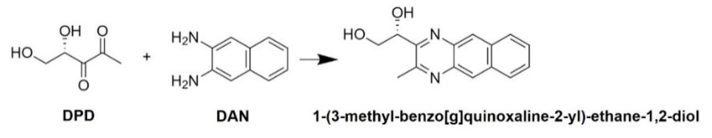
:1. Introduction
2. Materials and Methods
2.1. Chemicals
2.2. Bacterial Strains and Culture Conditions
2.3. Procedures of Sample Preparation and Derivatization for AI-2 Detection
2.4. Chromatographic Procedures
3. Results and Discussion
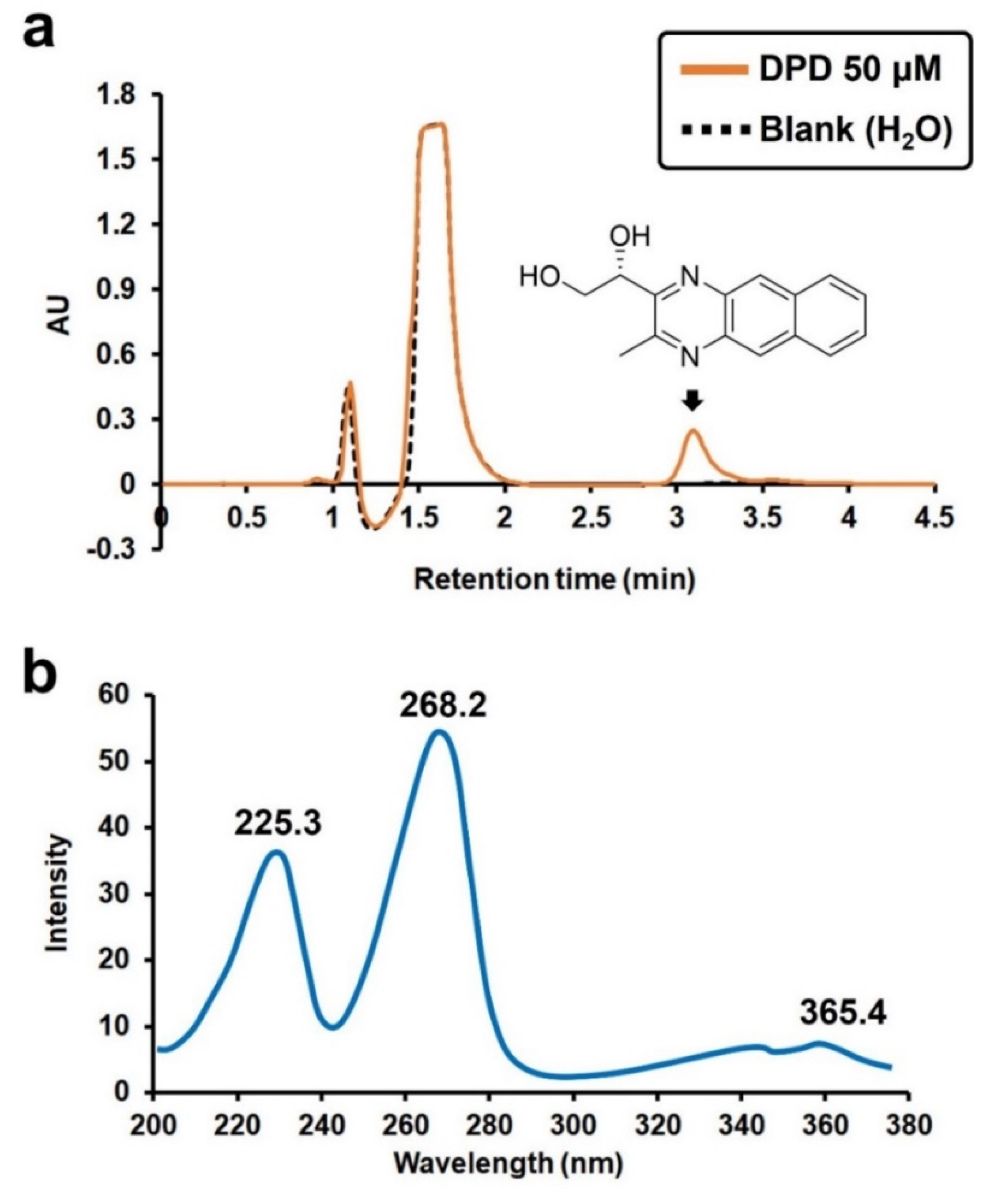
3.1. Optimal UV Wavelength for the Detection of the AI-2 Derivative
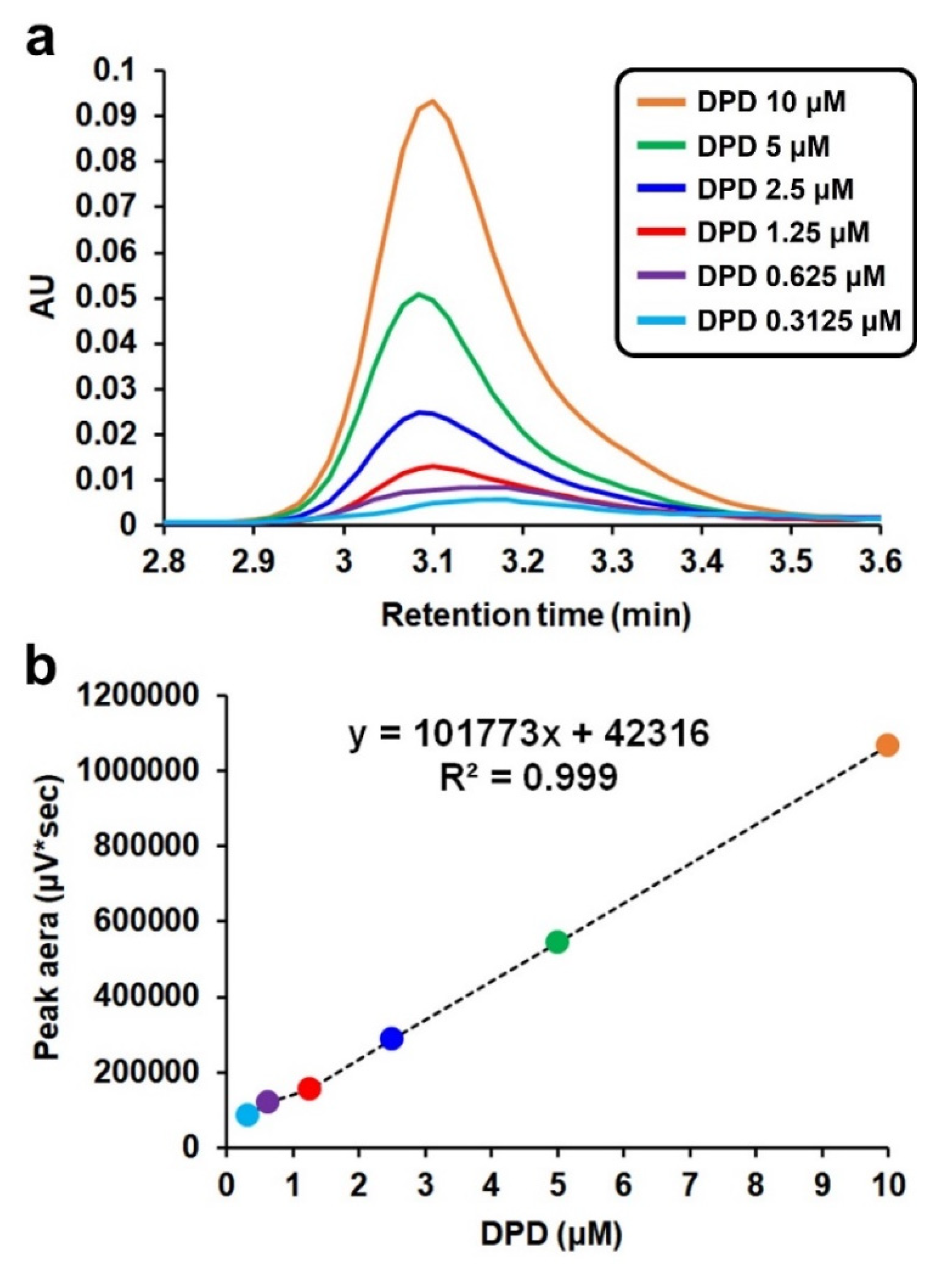
3.2. Validation of the Method for the Quantitative Analysis of DPD Concentration
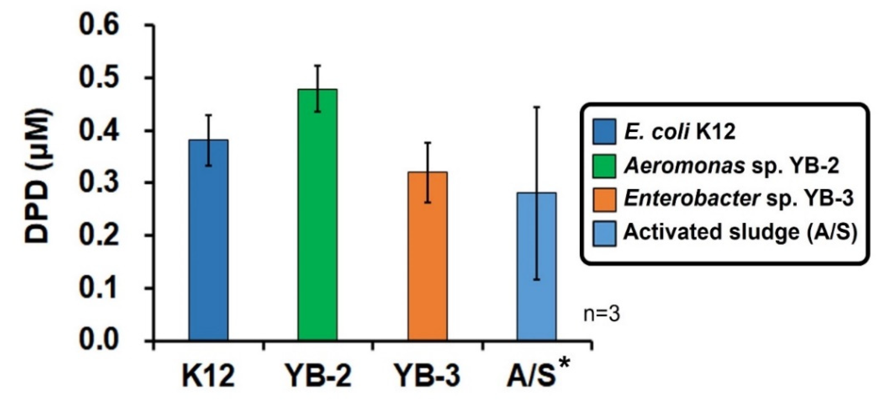
3.3. Measurement of the DPD Concentration in Various Environmental Samples
4. Conclusions
Supplementary Materials
Author Contributions
Funding
Institutional Review Board Statement
Informed Consent Statement
Data Availability Statement
Acknowledgments
Conflicts of Interest
References
- Waters, C.M.; Bassler, B.L. Quorum sensing: Cell-to-cell communication in bacteria. Annu. Rev. Cell Dev. Biol. 2005, 21, 319–346. [Google Scholar] [CrossRef] [Green Version]
- Davies, D.G.; Parsek, M.R.; Pearson, J.P.; Iglewski, B.H.; Costerton, J.W.; Greenberg, E.P. The involvement of cell-to-cell signals in the development of a bacterial biofilm. Science 1998, 280, 295–298. [Google Scholar] [CrossRef] [Green Version]
- Bischofs, I.B.; Hug, J.A.; Liu, A.W.; Wolf, D.M.; Arkin, A.P. Complexity in bacterial cell–cell communication: Quorum signal integration and subpopulation signaling in the Bacillus subtilis phosphorelay. Proc. Natl. Acad. Sci. USA 2009, 106, 6459–6464. [Google Scholar] [CrossRef] [Green Version]
- Pereira, C.S.; Thompson, J.A.; Xavier, K.B. AI-2-mediated signalling in bacteria. FEMS Microbiol. Rev. 2013, 37, 156–181. [Google Scholar] [CrossRef] [PubMed] [Green Version]
- Lee, K.; Kim, Y.W.; Lee, S.; Lee, S.H.; Nahm, C.H.; Kwon, H.; Park, P.K.; Choo, K.H.; Koyuncu, I.; Drews, A. Stopping AI-2 chatter by means of an indigenous bacterium (Acinetobacter sp. DKY-1): A new anti-biofouling strategy in an MBR for wastewater treatment. Environ. Sci. Technol. 2017, 52, 6237–6245. [Google Scholar] [CrossRef] [PubMed]
- Waheed, H.; Xiao, Y.; Hashmi, I.; Zhou, Y. The selective pressure of quorum quenching on microbial communities in membrane bioreactors. Chemosphere 2020, 247, 125953. [Google Scholar] [CrossRef] [PubMed]
- Lee, K.; Yu, H.; Zhang, X.; Choo, K.-H. Quorum sensing and quenching in membrane bioreactors: Opportunities and challenges for biofouling control. Bioresour. Technol. 2018, 270, 656–668. [Google Scholar] [CrossRef] [PubMed]
- Maddela, N.R.; Sheng, B.; Yuan, S.; Zhou, Z.; Villamar-Torres, R.; Meng, F. Roles of quorum sensing in biological wastewater treatment: A critical review. Chemosphere 2019, 221, 616–629. [Google Scholar] [CrossRef]
- Lee, K.; Lee, S.; Lee, S.H.; Kim, S.-R.; Oh, H.-S.; Park, P.-K.; Choo, K.-H.; Kim, Y.-W.; Lee, J.-K.; Lee, C.-H. Fungal quorum quenching: A paradigm shift for energy savings in membrane bioreactor (MBR) for wastewater treatment. Environ. Sci. Technol. 2016, 50, 10914–10922. [Google Scholar] [CrossRef]
- Oh, H.-S.; Lee, C.-H. Origin and evolution of quorum quenching technology for biofouling control in MBRs for wastewater treatment. J. Membr. Sci. 2018, 554, 331–345. [Google Scholar] [CrossRef]
- Fuqua, C.; Winans, S.C. Conserved cis-acting promoter elements are required for density-dependent transcription of Agrobacterium tumefaciens conjugal transfer genes. J. Bacteriol. 1996, 178, 435–440. [Google Scholar] [CrossRef] [PubMed] [Green Version]
- Zhu, J.; Beaber, J.W.; Moré, M.I.; Fuqua, C.; Eberhard, A.; Winans, S.C. Analogs of the autoinducer 3-oxooctanoyl-homoserine lactone strongly inhibit activity of the TraR protein of Agrobacterium tumefaciens. J. Bacteriol. 1998, 180, 5398–5405. [Google Scholar] [CrossRef] [PubMed] [Green Version]
- McClean, K.H.; Winson, M.K.; Fish, L.; Taylor, A.; Chhabra, S.R.; Camara, M.; Daykin, M.; Lamb, J.H.; Swift, S.; Bycroft, B.W. Quorum sensing and Chromobacterium violaceum: Exploitation of violacein production and inhibition for the detection of N-acylhomoserine lactones. Microbiology 1997, 143, 3703–3711. [Google Scholar] [CrossRef] [Green Version]
- Raut, N.; Pasini, P.; Daunert, S. Deciphering bacterial universal language by detecting the quorum sensing signal, autoinducer-2, with a whole-cell sensing system. Anal. Chem. 2013, 85, 9604–9609. [Google Scholar] [CrossRef] [PubMed]
- Teiber, J.F.; Draganov, D.I. High-performance liquid chromatography analysis of N-acyl homoserine lactone hydrolysis by paraoxonases. In Quorum Sensing; Springer: New York, NY, USA, 2011; pp. 291–298. [Google Scholar]
- Kim, S.-R.; Oh, H.-S.; Jo, S.-J.; Yeon, K.-M.; Lee, C.-H.; Lim, D.-J.; Lee, C.-H.; Lee, J.-K. Biofouling control with bead-entrapped quorum quenching bacteria in membrane bioreactors: Physical and biological effects. Environ. Sci. Technol. 2013, 47, 836–842. [Google Scholar] [CrossRef]
- Patel, N.M.; Moore, J.D.; Blackwell, H.E.; Amador-Noguez, D. Identification of unanticipated and novel N-acyl L-homoserine lactones (AHLs) using a sensitive non-targeted LC-MS/MS method. PLoS ONE 2016, 11, e0163469. [Google Scholar] [CrossRef] [PubMed]
- Huang, S.; Zhang, H.; Ng, T.C.A.; Xu, B.; Shi, X.; Ng, H.Y. Analysis of N-Acy-L-homoserine lactones (AHLs) in wastewater treatment systems using SPE-LLE with LC-MS/MS. Water Res. 2020, 177, 115756. [Google Scholar] [CrossRef] [PubMed]
- Sheng, H.; Song, Y.; Bian, Y.; Wu, W.; Xiang, L.; Liu, G.; Jiang, X.; Wang, F. Determination of N-acyl homoserine lactones in soil using accelerated solvent extraction combined with solid-phase extraction and gas chromatography-mass spectrometry. Anal. Methods 2017, 9, 688–696. [Google Scholar] [CrossRef]
- Miller, S.T.; Xavier, K.B.; Campagna, S.R.; Taga, M.E.; Semmelhack, M.F.; Bassler, B.L.; Hughson, F.M. Salmonella typhimurium recognizes a chemically distinct form of the bacterial quorum-sensing signal AI-2. Mol. Cell 2004, 15, 677–687. [Google Scholar] [CrossRef]
- Song, X.-N.; Qiu, H.-B.; Xiao, X.; Cheng, Y.-Y.; Li, W.-W.; Sheng, G.-P.; Li, X.-Y.; Yu, H.-Q. Determination of autoinducer-2 in biological samples by high-performance liquid chromatography with fluorescence detection using pre-column derivatization. J. Chromatogr. A 2014, 1361, 162–168. [Google Scholar] [CrossRef]
- Campagna, S.R.; Gooding, J.R.; May, A.L. Direct quantitation of the quorum sensing signal, autoinducer-2, in clinically relevant samples by liquid chromatography-tandem mass spectrometry. Anal. Chem. 2009, 81, 6374–6381. [Google Scholar] [CrossRef]
- Zhang, X.; Lee, K.; Yu, H.; Mameda, N.; Choo, K.-H. Photolytic quorum quenching: A new anti-biofouling strategy for membrane bioreactors. Chem. Eng. J. 2019, 378, 122235. [Google Scholar] [CrossRef]
- Thiel, V.; Vilchez, R.; Sztajer, H.; Wagner-Döbler, I.; Schulz, S. Identification, Quantification, and Determination of the Absolute Configuration of the Bacterial Quorum-Sensing Signal Autoinducer-2 by Gas Chromatography–Mass Spectrometry. Chem. Biol. Chem. 2009, 10, 479–485. [Google Scholar] [CrossRef]
- Taga, M.E.; Xavier, K.B. Methods for analysis of bacterial Autoinducer-2 production. Curr. Protoc. Microbiol. 2011, 23, 1C.1.1–1C.1.15. [Google Scholar] [CrossRef]
- Vilchez, R.; Lemme, A.; Thiel, V.; Schulz, S.; Sztajer, H.; Wagner-Döbler, I. Analysing traces of autoinducer-2 requires standardization of the Vibrio harveyi bioassay. Anal. Bioanal. Chem. 2007, 387, 489–496. [Google Scholar] [CrossRef] [Green Version]
- Li, J.; Attila, C.; Wang, L.; Wood, T.K.; Valdes, J.J.; Bentley, W.E. Quorum sensing in Escherichia coli is signaled by AI-2/LsrR: Effects on small RNA and biofilm architecture. J. Bacteriol. 2007, 189, 6011–6020. [Google Scholar] [CrossRef] [Green Version]
- Sanagi, M.M.; Ling, S.L.; Nasir, Z.; Hermawan, D.; Wan Ibrahim, W.A.; Naim, A.A. Comparison of signal-to-noise, blank determination, and linear regression methods for the estimation of detection and quantification limits for volatile organic compounds by gas chromatography. J. AOAC Int. 2009, 92, 1833–1838. [Google Scholar] [CrossRef] [Green Version]
- Yamaguchi, M.; Hara, S.; Nakamura, M. Determination of methylglyoxal in mouse blood by liquid chromatography with fluorescence detection. Anal. Chim. Acta 1989, 221, 163–166. [Google Scholar] [CrossRef]


| Methods | LOD (ng/mL) | Procedure | Instruments | Interference | References |
|---|---|---|---|---|---|
| HPLC-FLD a | 1.0 | Needs a derivative step, but is relatively easy to apply | Expensive | None | [21] |
| LC-MS/MS a | 0.7 | Derivatization reagents are complicated and time-consuming (to prepare) | Very expensive | Salt concentration or none | [22] |
| GC-MS a | 0.7 c | Requires complex sample pretreatment, including two-step derivatization and extraction | Very expensive | None | [24] |
| AI-2 bioassay b | 4.6 | Takes a long time to incubate the AI-2 reporter strain (V. harveyi BB170) (>12 h) and react (5–7 h) with the sample | Expensive | Poor reproducibility, depending on reporter strain and sample state | [25,26] |
Publisher’s Note: MDPI stays neutral with regard to jurisdictional claims in published maps and institutional affiliations. |
© 2021 by the authors. Licensee MDPI, Basel, Switzerland. This article is an open access article distributed under the terms and conditions of the Creative Commons Attribution (CC BY) license (https://creativecommons.org/licenses/by/4.0/).
Share and Cite
Lee, K.; Lee, C.-H.; Choo, K.-H. A Facile HPLC-UV-Based Method for Determining the Concentration of the Bacterial Universal Signal Autoinducer-2 in Environmental Samples. Appl. Sci. 2021, 11, 9116. https://doi.org/10.3390/app11199116
Lee K, Lee C-H, Choo K-H. A Facile HPLC-UV-Based Method for Determining the Concentration of the Bacterial Universal Signal Autoinducer-2 in Environmental Samples. Applied Sciences. 2021; 11(19):9116. https://doi.org/10.3390/app11199116
Chicago/Turabian StyleLee, Kibaek, Chung-Hak Lee, and Kwang-Ho Choo. 2021. "A Facile HPLC-UV-Based Method for Determining the Concentration of the Bacterial Universal Signal Autoinducer-2 in Environmental Samples" Applied Sciences 11, no. 19: 9116. https://doi.org/10.3390/app11199116
APA StyleLee, K., Lee, C.-H., & Choo, K.-H. (2021). A Facile HPLC-UV-Based Method for Determining the Concentration of the Bacterial Universal Signal Autoinducer-2 in Environmental Samples. Applied Sciences, 11(19), 9116. https://doi.org/10.3390/app11199116