Article

Expression of the Melatonin-Associated Genes in Fibroblasts That Have Been Co-Exposed to Fluoride and a Moderate-Strength Static Magnetic Field

by
Celina Kruszniewska-Rajs
1,
Agnieszka Synowiec-Wojtarowicz
2,
Joanna Gola
1 and
Magdalena Kimsa-Dudek
2,*
1
Department of Molecular Biology, Faculty of Pharmaceutical Sciences in Sosnowiec, Medical University of Silesia, Katowice, Jednosci 8, 41-200 Sosnowiec, Poland
2
Department of Nutrigenomics and Bromatology, Faculty of Pharmaceutical Sciences in Sosnowiec, Medical University of Silesia, Katowice, Jednosci 8, 41-200 Sosnowiec, Poland
*
Author to whom correspondence should be addressed.
Appl. Sci. 2021, 11(19), 8810; https://doi.org/10.3390/app11198810
Submission received: 29 July 2021 / Revised: 14 September 2021 / Accepted: 21 September 2021 / Published: 22 September 2021
(This article belongs to the Special Issue Toxic Effects of Fluoride)

Abstract

:
Fluoride can weaken the protective role of melatonin in reducing cellular damage. A static magnetic field is a physical factor that can counteract the negative effect of fluoride. Hence, the main objective of the study was to analyze the transcriptional activity of the genes that are associated with the activity of melatonin in human skin fibroblasts that have been co-exposed to fluoride and a moderate-strength static magnetic field. The expression of the melatonin-associated genes in human fibroblasts that had simultaneously been exposed to F and a static magnetic field was determined using an oligonucleotide microarray and RT-qPCR techniques. The concentration of oxidative damage markers was also measured. In NaF and static magnetic field-treated cells, there was a tendency to compensate for the expression of the differentiating genes (IL27RA, NR1D1, RRP7A, YIPF1, HIST1H2BD) that had been modified by the presence of fluoride. It has been also shown that the oxidative damage marker concentration was statistically lower in the cells that had simultaneously been exposed to fluoride and a static magnetic field compared to the F-treated cells. In conclusion, the protective role of a moderate-strength static magnetic field on human dermal fibroblasts that had been exposed to fluoride was demonstrated, and its mechanism of action is associated with the melatonin-dependent pathways.

1. Introduction

Fluoride easily penetrates cell membranes due to their properties and thus affects cell functioning. Among the toxic results of fluoride action at the cellular level, oxidative stress induction, organelle damage, apoptosis, essential protein inhibition, and changes in enzyme activity can be mentioned [1,2,3]. As has been shown in previous reports, fluoride also affects melatonin production, which can have various consequences for human health. Melatonin is a pineal hormone that has a strong antioxidant and immunomodulatory effect. It acts in cells through different signaling pathways by binding to the appropriate receptors, which are located in the central nervous system and also in the immune system, skin, and organs, such as the kidneys, liver and gastrointestinal tract [4]. It has been implied that the cutaneous melatoninergic system protect skin cells against various stress conditions, mainly by scavenging free radicals and stimulating the antioxidant defense system [5]. The protective role of melatonin also results from its neuroprotective and anti-apoptotic effect, modulation of autophagy, and permeability transition pore [6]. The pleiotropic effect of melatonin comes from the possibility of its synthesis by other cells [7] and its receptor-independent action as well [8].
Due to the inhibition of melatonin production by fluoride, these ions weaken its protective role in reducing cellular damage [9]. However, it is not tested how fluoride influence the gene expression involved in melatonin-dependent signaling pathways. On the other hand, it should be emphasized that fluoride is an element that influences the proper development of bones and teeth. The nutritional requirements for its consumption have been established at the level of adequate intake (AI) and the AI for women is 3 mg/day and for men, it is 4 mg/day of fluoride for the Polish population [10]. However, the effects of fluoride in the body are also influenced by systemic, metabolic and genetic factors [11]. Therefore, factors that can reduce or weaken its toxic effect on cells are being sought [12].
Recently, more and more attention has been paid to the possibility of using a static magnetic field (SMF) in medicine. The static magnetic field that is created by permanent magnets has found applications in the treatment of arthritis, insomnia, musculoskeletal injuries, pain, or wound healing [13,14]. On the other hand, many of the in vitro and in vivo studies that have been conducted to date have not produced uniform results on the biological and health effects of the impact of a static magnetic field [15]. However, it has been shown that the mechanism of its action at the cellular level is associated with a reduction in the oxidative stress and the inhibition of apoptosis [16,17]. Furthermore, it has also been suggested that a physical factor such as a static magnetic field may influence melatonin biosynthesis [18]. Since the beneficial action of a static magnetic field on cells has already been proven, there has been increased interest in it as a physical factor that might counteract the negative effect of fluoride on cells.
Therefore, in order to understand the molecular mechanism of a static magnetic field and fluoride interactions, the main objective of the study was to analyze the expression of the genes that are associated with the activity of melatonin in human fibroblasts that had been co-exposed to fluoride and a moderate-strength static magnetic field.

2. Materials and Methods

In this study, fibroblasts of the NHDF line (normal human dermal fibroblasts, Clonetics, CC-2511, San Diego, CA, USA) that had been cultured in the presence of fluoride and simultaneously exposed to a static magnetic field were used.

2.1. Cell Culture Conditions

The skin fibroblasts were cultured in a FBM medium (Fibroblast Basal Medium, Lonza, Basel, Switzerland) with human fibroblast growth factor-basic (hFGF-B), insulin, and gentamicin (FGM™ SingleQuots™; Lonza, Basel, Switzerland), as was previously reported [16].
For the experiment, the fibroblast cultures were conducted in the presence of fluoride (0.3 mmol/L) and simultaneously exposed to a static magnetic field with a moderate magnetic induction (0.65 T) for 24 h. The static magnetic field was generated by permanent magnets in special magnetic chambers that are intended for in vitro research [19,20]. The control cultures were carried out without fluoride and in chambers with a magnetic induction of 0.0 T. The concentration of fluoride and the value of the magnetic induction were selected based on previous studies that showed that these agents had no cytotoxic effect on cells [16,21,22]. Moreover, the concentration of F was selected so that the precipitation of sparingly soluble calcium and magnesium fluorides did not occur. Then, the effect of the action was related only to the presence of fluoride at unchanged concentrations of the calcium and magnesium ions. Then, after a 24 h incubation in the presence of chemical and physical agents, cells and cell culture supernates were harvested for further study steps.

2.2. Molecular Analyses

TRIzol reagent (Invitrogen, Carlsbad, CA, USA) was used in RNA extraction from harvested cells according to the manufacturer’s protocol. The quantity and quality of the extracts were checked spectrophotometrically and electrophoretically as was previously described [16].
The transcriptional activity of the melatonin-associated genes in the cells that had simultaneously been exposed to F and a static magnetic field and in the control cells was determined using the oligonucleotide microarray technique (HG-U133A 2.0, Affymetrix, Santa Clara, CA, USA) according to the manufacturer’s instructions and as was previously reported [16].
The RT-qPCR technique was used to validate the results that were obtained at the transcriptome level. The gene expression of IL27RA (interleukin 27 receptor subunit alpha), NR1D1 (nuclear receptor subfamily 1 group D member 1) and ACTB (β-actin) were evaluated using SYBR Green I chemistry (GoTaq® One-Step RT-qPCR System, Promega, WI, USA) and a LightCycler® 480 Instrument II (Roche Life Science, Basel, Switzerland). All the samples were run in triplicate. β-actin was included as an endogenous positive control of the amplification. The oligonucleotide primers were commercially available (Sigma-Aldrich, St. Louis, MO, USA). The thermal profile for the one-step RT-PCR was as follows: reverse transcription at 40 °C for 15 min, denaturation at 95 °C for 10 min, and 40 cycles consisting of the following temperatures and time intervals: 95 °C for 10 s, 60 °C for 30 s, and 72 °C for 30 s. After the RT-qPCR melting curve analysis was performed to confirm the specificity of the amplification and the absence of primer dimers.
The relative mRNA expression of the tested genes was determined using the 2−(ΔCt) method [23] with β-actin as a reference gene.

2.3. ELISA Assay

A DNA/RNA Oxidative Damage (High Sensitivity) ELISA Kit (Cayman Chemical, Ann Arbor, MI, USA) was used to measure the major oxidative damage markers such as three oxidized guanine species: 8-hydroxy-2′-deoxyguanosine, 8-hydroxyguanosine, and 8-hydroxyguanine in the cell culture medium according to the manufacturer’s instructions. The absorbance at a wavelength of 405 nm was read on a Wallac 1420 VICTOR microplate reader (PerkinElmer, Waltham, MA, USA). All the samples were tested in duplicate.

2.4. Statistical Analyses

Microarray data analysis was performed using the GeneSpring 13.0 platform (Agilent Technologies UK Ltd., South Queensferry, UK) and the PL-Grid infrastructure (http://www.plgrid.pl/ (accessed on 19 March 2021)). The melatonin-associated genes (103 ID mRNA) were selected from the NetAffx Analysis Center database of Affymetrix (http://www.affymetrix.com/analysis/index.affx (accessed on 23 September 2020)). A gene was considered as differentiating if it had the FC parameter (fold change, multiple of the fluorescence signal difference between two study groups) >1.1 and p-value < 0.05. In the microarray analysis, using such a combination of two parameters (fold change and p-value) enables more biologically meaningful sets of the genes to be found than when only one of them is used [24].
The statistical analyses of the RT-qPCR and ELISA results were performed using Statistica 13.3 software (StatSoft, Tulsa, OK, USA) and the level of significance was set at p < 0.05.
In the statistical analyses, the one-way ANOVA and Tukey post hoc tests were used due to the normal distribution of the data.
The biological significance of differentiating genes was confirmed by an analysis using the overrepresentation test in which a binomial test was used in the PANTHER Classification System (version 16.0). The overrepresentation test compares a selected genes to a reference gene list and thus determines whether a tested category (biological process) is overrepresented [25].

3. Results

In order to clarify the molecular mechanism of the interaction of fluoride with a static magnetic field, four study groups were distinguished: control cells (C), F-treated cells (F), SMF-treated cells (SMF) and cells that had simultaneously been exposed to a static magnetic field and F (SMF + F).

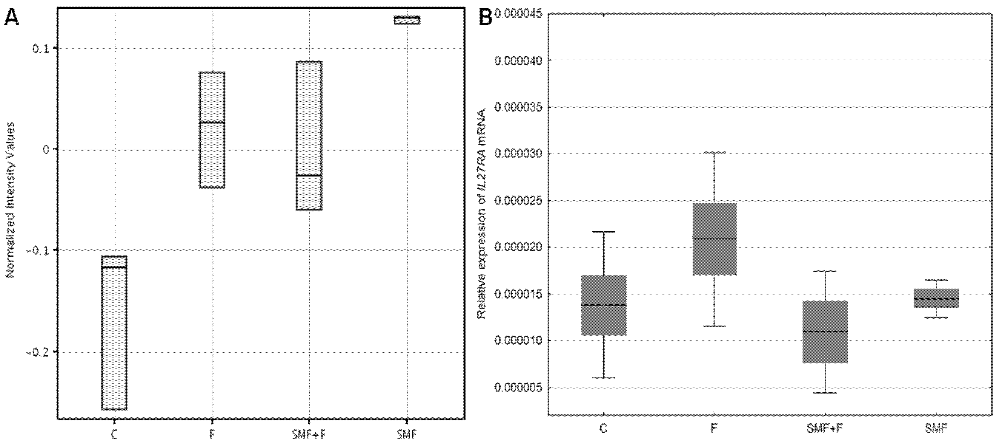
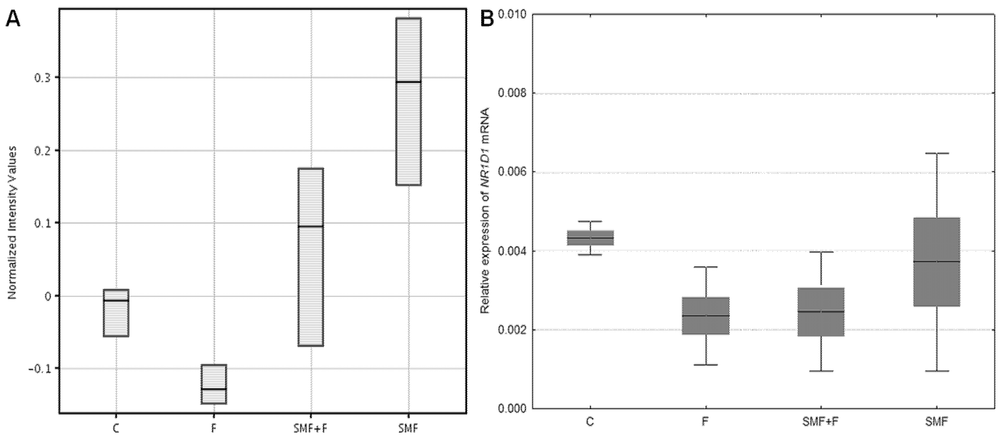
3.1. The Effect of Fluoride and the Static Magnetic Field Action on the Expression of the Melatonin-Associated Genes

The exposure of fibroblasts to the fluoride resulted in a statistically significant change in the expression of one transcript, which had been up-regulated (Tukey post hoc test, p < 0.05, FC > 1.1). In turn, as a result of the action of a static magnetic field alone on the cells, the expression level of five transcripts was statistically significantly higher (Tukey post hoc test, p < 0.05, FC > 1.1). For the skin fibroblasts that had simultaneously been exposed to fluoride and a static magnetic field compared to the F-treated cells, one transcript was differentiated, which had been down-regulated (Tukey post hoc test, p < 0.05, FC > 1.1) (Table 1).
In the next stage, the differentially expressed genes that had been characterized by the greatest change in the FC value (FC ≥ 1.2) were selected to validate the results using the RT-qPCR technique (IL27RA, NR1D1). The RT-qPCR analysis mostly confirmed the direction of the changes in the expression of the IL27RA and NR1D1 genes as a result of the exposure of a cell to a static magnetic field and fluoride (Table 2). In the cells that had simultaneously been exposed to fluoride and a static magnetic field, there was a tendency to compensate for the expression of the differentiating genes that was altered by the presence of fluoride (Figure 1 and Figure 2).
Additionally, the biological importance of the differentiating genes was confirmed by performing an analysis in the PANTHER classification system. This analysis revealed that the proteins that were encoded by the differentiating genes were involved in 58 biological processes (PANTHER overrepresentation test, p < 0.05), which were mainly associated with the response of the cells to various stimuli and the regulation of different cellular processes. When the p-value was expressed as p < 0.01, 22 biological processes were identified (Table 3). This means that these biological processes of differentially expressed genes are overrepresented which confirms that the mechanism of action of static magnetic field at the molecular level is related to genes associated with melatonin activity that play a protective role for cells.

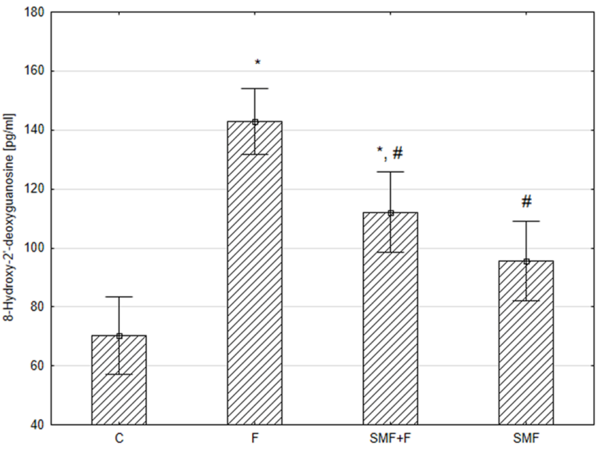
3.2. Effect of Fluoride and the Static Magnetic Field Action on the Concentration of the Oxidative Damage Markers

Because fluoride participates in the free radical processes that lead to the induction of oxidative stress, in the next stage, the way in which the exposure of the cells that had been cultured in the presence of fluoride to a static magnetic field influences the concentration of oxidative damage markers was investigated.
The 8-hydroxy-2′-deoxyguanosine (8-OH-dG) concentration was higher by approximately two-fold in the F group compared with the control group (Tukey post hoc test, p < 0.001). In the cells that had simultaneously been exposed to fluoride and a static magnetic field and in fibroblasts that had been exposed to a static magnetic field alone compared to the F-treated cells, the mean 8-OH-dG concentration was statistically lower by approximately 21.5% and 33.1%, respectively (Tukey post hoc test, p < 0.038, p < 0.002, respectively). Moreover, there was no statistical difference between the SMF-exposed cells and the control group (Figure 3).

4. Discussion

The action of fluoride is associated with the generation of free radicals and the induction of oxidative stress in cells. The biological effects of a static magnetic field are not fully understood, but more and more attention is being paid to its possible therapeutic activity. Earlier studies proved its protective role for normal cells that had been exposed to the factors that disrupt the redox balance against the induction of oxidative stress and apoptosis [16,17]. However, the aspects of the static magnetic field action at the molecular level are still not fully known. Since melatonin can attenuate the action of free radicals and regulate the enzymatic antioxidant system, in this study, we focused on the genes that are associated with its activity in cells that had simultaneously been exposed to fluoride and a moderate-strength static magnetic field. It was shown that both chemical and physical factors influence the expression of these genes, while the change in expression induced by fluoride is counteracted by a static magnetic field. The level of the expression of the selected genes whose expression was changed by fluoride under the influence of a static magnetic field is compensated for.
This study focused on the changes in cells that are caused by fluoride and a static magnetic field primarily at the transcriptional level. The action of melatonin in cells is associated with its immunomodulatory properties not only by affecting the cells of the immune system, but also by regulating the synthesis of the cytokines [26]. Interleukin 27 is a heterodimeric pleiotropic cytokine with pro- and anti-inflammatory properties, which are mediated by the IL27 receptor, which is composed of two subunits: an alpha subunit and glycoprotein 130. Its opposite effect depends, among others, on the type of cells. As was shown, the alpha subunit of IL27RA is necessary for the transcriptional activation of STAT1 and STAT3 (a signal transducer and an activator of transcription) and its expression can promote various types of tumor growth [27,28]. The possibility of IL27 action on non-hematopoietic cells such as fibroblasts or keratinocytes should also be considered. In some diseases, an increased expression of IL27RA in fibroblasts has been demonstrated [27]. Moreover, in our experiment, an increase in the expression of the receptor alpha subunit was observed under the influence of fluoride, which activates the expression of the inflammatory cytokines [29]. However, a static magnetic field can regulate cytokine disturbances, as is suggested by the obtained results. In turn, the NR1D1 gene encodes the protein that play the role of a transcriptional repressor, which coordinates the circadian rhythm and the metabolic pathways of lipids, glucose and proteins. Modifications in its expression are often seen during the development of various diseases [30]. Hence, equalizing the expression by a static magnetic field is beneficial and will result in the restoration of its correct function. Another very important gene that is modified by both fluoride and a static magnetic field is HIST1H2BD, which encodes the histone H2B type 1-D. Histones are responsible for nucleosome structure and play a key role in modulating the transcription process, whereas the histones that are present in the cytoplasm or extracellularly may contribute to promoting the inflammatory response of cells [31,32].
PANTHER analysis of target genes was performed to better understand the function of differentially expressed genes associated with melatonin pathways and explain the mechanism of protective role of static magnetic field on fluoride-treated cells. This analysis confirmed the participation of the differentially expressed genes primarily in the biological processes that are associated with the cellular response to various factors and the regulation of cellular processes, mainly in the cells of the immune system, which ensures a body’s defense against a variety of factors, such as toxic substances, viruses, and bacteria. These categories of target genes were overrepresented in fluoride and static magnetic field-treated cells. This indicates a protective function for the cells of the proteins that are encoded by the differentially expressed genes, which is essential from the point of view of the mechanism of action of a static magnetic field. Moreover, conducting this analysis eliminated the problem of the genes whose differences in their expression and statistical significance did not translate into the influence of a specific gene on the cells.
As a screening technique, oligonucleotide microarrays enable genome-scale expression analysis. Therefore, the genes with the greatest change in expression, which is expressed as a fold change were selected for the next steps in order to validate the obtained microarray results. The RT-qPCR technique showed that the direction of changes in the expression of the studied genes was in accordance with the microarray results. We did not statistically confirm the up-regulation of the NR1D1 gene in the cells that had only been exposed to a static magnetic field compared to the control, but the tendency to increase its expression under the influence of an exposure to a static magnetic field was also visible in RT-qPCR results. The lack of a confirmation of the changes in its expression may be due to the separation between the location of the PCR primers and the microarray probes [33]. However, it should be noted that the research showed a high correlation between these two techniques [34].
In order to confirm the protective role of a static magnetic field for fibroblasts, we determined the concentration of the DNA/RNA oxidative damage markers. It is considered that 8-OH-dG is a biomarker for oxidative damage to the nucleic acids. Higher levels of 8-OH-dG are observed in various diseases whose pathogenesis is played by oxidative stress, for example, in cancers, diabetes or neurodegenerative diseases. At the same time, it was shown earlier that many antioxidant compounds can reduce the 8-OH-dG level [35]. In our study, it was revealed that a moderate-strength static magnetic field has a beneficial effect on cells by lowering the concentration of the DNA/RNA oxidative damage markers in fluoride-treated cells.
In the cells that had simultaneously been exposed to fluoride and a static magnetic field, there was a tendency to compensate for expression of the differentially expressed genes that was altered by the presence of fluoride, which suggests a protective role of a static magnetic field against the factors that cause changes in the cell signaling cascades. However, it should be taken into account that the limitation of this study is the selection of the fluoride concentration, which is not observed in vivo in non-fluoridated areas.

5. Conclusions

In conclusion, the protective role of a moderate-strength static magnetic field on normal human dermal fibroblasts that had been exposed to fluoride was demonstrated, and its mechanism of action is associated with the melatonin-dependent pathways.

Author Contributions

Conceptualization, M.K.-D.; methodology, M.K.-D. and C.K.-R.; formal analysis, M.K.-D. and C.K.-R.; investigation, M.K.-D., A.S.-W. and C.K.-R.; writing—original draft preparation, M.K.-D. and A.S.-W.; writing—review and editing, C.K.-R.; supervision, J.G. All authors have read and agreed to the published version of the manuscript.

Funding

This study was supported by grant PCN-1-121/N/0/I from the Medical University of Silesia, Katowice, Poland.

Conflicts of Interest

The authors declare no conflict of interest.

References

  1. Guth, S.; Hüser, S.; Roth, A.; Degen, G.; Diel, P.; Edlund, K.; Eisenbrand, G.; Engel, K.H.; Epe, B.; Grune, T.; et al. Toxicity of fluoride: Critical evaluation of evidence for human developmental neurotoxicity in epidemiological studies, animal experiments and in vitro analyses. Arch. Toxicol. 2020, 94, 1375–1415. [Google Scholar] [CrossRef]
  2. Johnston, N.R.; Strobel, S.A. Principles of fluoride toxicity and the cellular response: A review. Arch. Toxicol. 2020, 94, 1051–1069. [Google Scholar] [CrossRef] [PubMed]
  3. Strunecka, A.; Strunecky, O. Mechanisms of fluoride toxicity: From enzymes to underlying integrative networks. Appl. Sci. 2020, 10, 7100. [Google Scholar] [CrossRef]
  4. Pandi-Perumal, S.R.; Trakht, I.; Srinivasan, V.; Spence, D.W.; Maestroni, G.J.; Zisapel, N.; Cardinali, D.P. Physiological effects of melatonin: Role of melatonin receptors and signal transduction pathways. Prog. Neurobiol. 2008, 85, 335–353. [Google Scholar] [CrossRef] [PubMed]
  5. Slominski, A.T.; Zmijewski, M.A.; Skobowiat, C.; Zbytek, B.; Slominski, R.M.; Steketee, J.D. Sensing the environment: Regulation of local and global homeostasis by the skin neuroendocrine system. Adv. Anat. Embryol. Cell Biol. 2012, 212. [Google Scholar]
  6. Tarocco, A.; Caroccia, N.; Morciano, G.; Wieckowski, M.R.; Ancora, G.; Garani, G.; Pinton, P. Melatonin as a master regulator of cell death and inflammation: Molecular mechanisms and clinical implications for newborn care. Cell Death Dis. 2019, 10, 317. [Google Scholar] [CrossRef] [PubMed] [Green Version]
  7. Chuffa, L.G.A.; Carvalho, R.F.; Justulin, L.A.; Cury, S.S.; Seiva, F.R.F.; Jardim-Perassi, B.V.; Zuccari, D.A.P.C.; Reiter, R.J. A meta-analysis of microRNA networks regulated by melatonin in cancer: Portrait of potential candidates for breast cancer treatment. J. Pineal Res. 2020, 69, e12693. [Google Scholar] [CrossRef] [PubMed]
  8. Amini, H.; Rezabakhsh, A.; Heidarzadeh, M.; Hassanpour, M.; Hashemzadeh, S.; Ghaderi, S.; Sokullu, E.; Rahbarghazi, R.; Reiter, R.J. An Examination of the Putative Role of Melatonin in Exosome Biogenesis. Front. Cell Dev. Biol. 2021, 9, 686551. [Google Scholar] [CrossRef]
  9. Cunningham, J.E.A.; McCague, H.; Malin, A.J.; Flora, D.; Till, C. Fluoride exposure and duration and quality of sleep in a Canadian population-based sample. Environ. Health 2021, 20, 16. [Google Scholar] [CrossRef] [PubMed]
  10. Jarosz, M. Normy żywienia dla populacji polskiej–nowelizacja. Instytut Żywności i Żywenia 2012, 137–140. [Google Scholar]
  11. Buzalaf, M.A.R. Review of Fluoride Intake and Appropriateness of Current Guidelines. Adv. Dent. Res. 2018, 29, 157–166. [Google Scholar] [CrossRef] [PubMed]
  12. Bharti, V.K.; Giri, A.; Kumar, K. Fluoride sources, toxicity and its amelioration: A review. Ann. Environ. Sci. Toxicol. 2017, 2, 21–32. [Google Scholar] [CrossRef]
  13. Markov, M.S. Therapeutic application of static magnetic fields. Environmentalist 2007, 27, 457–463. [Google Scholar] [CrossRef]
  14. Colbert, A.P.; Wahbeh, H.; Harling, N.; Connelly, E.; Schiffke, H.C.; Forsten, C.; Gregory, W.L.; Markov, M.S.; Souder, J.J.; Elmer, P.; et al. Static magnetic field therapy: A critical review of treatment parameters. Evid. Based Complement. Altern. Med. 2009, 6, 133–139. [Google Scholar] [CrossRef]
  15. Zmyślony, M.; Politański, P. Zdrowotne skutki ekspozycji na stałe pole magnetyczne–przegląd piśmiennictwa [Health effects of exposure to static magnetic field—A review of literature]. Med. Pr. 2019, 70, 107–120. [Google Scholar] [PubMed]
  16. Kimsa-Dudek, M.; Synowiec-Wojtarowicz, A.; Derewniuk, M.; Gawron, S.; Paul-Samojedny, M.; Kruszniewska-Rajs, C.; Pawłowska-Góral, K. Impact of fluoride and a static magnetic field on the gene expression that is associated with the antioxidant defense system of human fibroblasts. Chem. Biol. Interact. 2018, 287, 13–19. [Google Scholar] [CrossRef]
  17. Kimsa-Dudek, M.; Synowiec-Wojtarowicz, A.; Krawczyk, A.; Kruszniewska-Rajs, C.; Gawron, S.; Paul-Samojedny, M.; Gola, J. Anti-apoptotic effect of a static magnetic field in human cells that had been treated with sodium fluoride. J. Environ. Sci. Health A 2020, 55, 1141–1148. [Google Scholar] [CrossRef] [PubMed]
  18. Driessen, S.; Bodewein, L.; Dechent, D.; Graefrath, D.; Schmiedchen, K.; Stunder, D.; Kraus, T.; Petri, A.K. Biological and health-related effects of weak static magnetic fields (≤ 1 mT) in humans and vertebrates: A systematic review. PLoS ONE 2020, 15, e0230038. [Google Scholar] [CrossRef] [PubMed]
  19. Glinka, M.; Gawron, S.; Sieroń, A.; Pawłowska-Góral, K.; Cieślar, G.; Sieroń-Stołtny, K. Test chambers for cell culture in static magnetic field. J. Magn. Mater. 2013, 331, 208–215. [Google Scholar] [CrossRef]
  20. Gawron, S.; Glinka, M.; Wolnik, T. Magnetyczna komora badawcza dedykowana do hodowli komórek. Zeszyty Problemowe Maszyny Elektryczne 2012, 4, 11–16. [Google Scholar]
  21. Kimsa-Dudek, M.; Synowiec-Wojtarowicz, A.; Derewniuk, M.; Paul-Samojedny, M.; Pawłowska-Góral, K. The effect of simultaneous exposure of human fibroblasts to fluoride and moderate intensity static magnetic fields. Int. J. Radiat. Biol. 2019, 95, 1581–1587. [Google Scholar] [CrossRef] [PubMed]
  22. Dini, L.; Abbro, L. Bioeffects of moderate-intensity static magnetic fields on cell cultures. Micron 2005, 36, 195–217. [Google Scholar] [CrossRef] [PubMed]
  23. Schmittgen, T.D.; Livak, K.J. Analyzing real-time PCR data by the comparative C(T) method. Nat. Protoc. 2008, 3, 1101–1108. [Google Scholar] [CrossRef] [PubMed]
  24. St Laurent, G.; Shtokalo, D.; Tackett, M.R.; Yang, Z.; Vyatkin, Y.; Milos, P.M.; Seilheimer, B.; McCaffrey, T.A.; Kapranov, P. On the importance of small changes in RNA expression. Methods 2013, 63, 18–24. [Google Scholar] [CrossRef] [PubMed]
  25. Mi, H.; Muruganujan, A.; Casagrande, J.T.; Thomas, P.D. Large-scale gene function analysis with the PANTHER classification system. Nat. Protoc. 2013, 8, 1551–1566. [Google Scholar] [CrossRef]
  26. Mańka, S.; Majewska, E. Immunoregulatory action of melatonin. The mechanism of action and the effect on inflammatory cells. Postepy Hig. Med. Dosw. (Online) 2016, 70, 1059–1067. [Google Scholar] [CrossRef] [PubMed]
  27. Hall, A.O.; Silver, J.S.; Hunter, C.A. The immunobiology of IL-27. Adv. Immunol. 2012, 115, 1–44. [Google Scholar] [PubMed]
  28. Jones, G.W.; Hill, D.G.; Cardus, A.; Jones, S.A. IL-27: A double agent in the IL-6 family. Clin. Exp. Immunol. 2018, 193, 37–46. [Google Scholar] [CrossRef] [PubMed] [Green Version]
  29. Chen, L.; Kuang, P.; Liu, H.; Wei, Q.; Cui, H.; Fang, J.; Zuo, Z.; Deng, J.; Li, Y.; Wang, X.; et al. Sodium Fluoride (NaF) Induces Inflammatory Responses Via Activating MAPKs/NF-κB Signaling Pathway and Reducing Anti-inflammatory Cytokine Expression in the Mouse Liver. Biol. Trace Elem. Res. 2019, 189, 157–171. [Google Scholar] [CrossRef] [PubMed]
  30. Wang, S.; Li, F.; Lin, Y.; Wu, B. Targeting REV-ERBα for therapeutic purposes: Promises and challenges. Theranostics 2020, 10, 4168–4182. [Google Scholar] [CrossRef] [PubMed]
  31. Chen, R.; Kang, R.; Fan, X.G.; Tang, D. Release and activity of histone in diseases. Cell Death Dis. 2014, 5, e1370. [Google Scholar] [CrossRef] [PubMed] [Green Version]
  32. Hoeksema, M.; van Eijk, M.; Haagsman, H.P.; Hartshorn, K.L. Histones as mediators of host defense, inflammation and thrombosis. Future Microbiol. 2016, 11, 441–453. [Google Scholar] [CrossRef] [PubMed] [Green Version]
  33. Etienne, W.; Meyer, M.H.; Peppers, J.; Meyer, R.A., Jr. Comparison of mRNA gene expression by RT-PCR and DNA microarray. Biotechniques 2004, 36, 618–620. [Google Scholar] [CrossRef] [PubMed] [Green Version]
  34. Dallas, P.B.; Gottardo, N.G.; Firth, M.J.; Beesley, A.H.; Hoffmann, K.; Terry, P.A.; Freitas, J.R.; Boag, J.M.; Cummings, A.J.; Kees, U.R. Gene expression levels assessed by oligonucleotide microarray analysis and quantitative real-time RT-PCR—How well do they correlate? BMC Genom. 2005, 6, 59. [Google Scholar] [CrossRef] [PubMed] [Green Version]
  35. Kasai, H.; Kawai, K. 8-Hydroxyguanine, an Oxidative DNA and RNA Modification. In Modified Nucleic Acids in Biology and Medicine; RNA Technologies; Jurga, S., Erdmann, V., Barciszewski, J., Eds.; Springer: Cham, switzerland, 2016. [Google Scholar]
Figure 1. Expression of the IL27RA gene in control cells (C), F-treated cells (F), cells that had been exposed to a static magnetic field (SMF), and cells that had simultaneously been exposed to a static magnetic field and F (SMF + F) that was determined using the oligonucleotide microarray technique (A) and the RT-qPCR technique (B). The results are presented as the mean ± SE (box) and SD (whisker).
Applsci 11 08810 g001
Figure 2. Expression of the NR1D1 gene in the control cells (C), F-treated cells (F), cells that had been exposed to a static magnetic field (SMF), and cells that had simultaneously been exposed to a static magnetic field and F (SMF + F) that was determined using the oligonucleotide microarray technique (A) and the RT-qPCR technique (B). The results are presented as the mean ± SE (box) and SD (whisker).
Applsci 11 08810 g002
Figure 3. The DNA/RNA oxidative damage marker concentration in the control cells (C), F-treated cells (F), cells that had been exposed to a static magnetic field (SMF) and cells that had simultaneously been exposed to a static magnetic field and F (SMF + F). The results are presented as the mean ± SD; statistical significance: * p < 0.05 vs. C; # p < 0.05 vs. F.
Applsci 11 08810 g003
Table 1. List of differentially expressed genes determined by the oligonucleotide microarray technique.
ID mRNAGene SymbolGene NameF vs. CSMF vs. CSMF + F vs. F
FC
205926_atIL27RAinterleukin 27 receptor subunit alpha1.13↑ *1.22↑ *1.01↓
204760_s_atNR1D1nuclear receptor subfamily 1 group D member 11.08↓1.23↑ *1.14↑
202937_x_atRRP7Aribosomal RNA processing 7 homolog A1.05↑1.19↑ *1.04↓
33307_atRRP7Aribosomal RNA processing 7 homolog A1.02↑1.11↑ *1.03↓
214733_s_atYIPF1yip1 domain family member 11.05↑1.11↑ *1.02↓
222067_x_atHIST1H2BDhistone H2B type 1-D1.03↑1.03↑1.14↓ #
C—control cells, F—F-treated cells; SMF—cells that had been exposed to a static magnetic field; SMF + F—cells that had simultaneously been exposed to a static magnetic field and F; FC—fold change; ↑, ↓—up- or down-regulation of gene; statistical significance: * p < 0.05 vs. C; # p < 0.05 vs. F.
Table 2. Validation of the microarray data using RT-qPCR.
Gene SymbolGene NameF vs. CSMF vs. CSMF + F vs. F
FC
IL27RAinterleukin 27 receptor subunit alpha1.51↑1.05↑1.91↓
NR1D1nuclear receptor subfamily 1 group D member 11.84↓1.16↓1.05↑
C—control cells, F—F-treated cells; SMF—cells that had been exposed to a static magnetic field; SMF + F—cells that had simultaneously been exposed to a static magnetic field and F; FC—fold change; ↑, ↓—up- or down-regulation of gene.
Table 3. The biological processes in which the differentially expressed genes are involved (PANTHER overrepresentation test, p < 0.01).
Biological Processesp-Value
cellular response to a chemical stimulus (GO:0070887)0.0047
cellular response to an organic substance (GO:0071310)0.0030
hormone-mediated signaling pathway (GO:0009755)0.0079
leukocyte cell–cell adhesion (GO:0007159)0.0076
leukocyte proliferation (GO:0070661)0.0089
lymphocyte proliferation (GO:0046651)0.0089
mononuclear cell proliferation (GO:0032943)0.0089
positive regulation of cell adhesion (GO:0045785)0.0064
positive regulation of cell–cell adhesion (GO:0022409)0.0047
positive regulation of leukocyte cell–cell adhesion (GO:1903039)0.0041
positive regulation of T cell activation (GO:0050870)0.0041
positive regulation of T cell proliferation (GO:0042102)0.0023
regulation of cell–cell adhesion (GO:0022407)0.0085
regulation of leukocyte cell–cell adhesion (GO:1903037)0.0068
regulation of leukocyte proliferation (GO:0070663)0.0056
regulation of lymphocyte proliferation (GO:0050670)0.0056
regulation of mononuclear cell proliferation (GO:0032944)0.0056
regulation of T cell activation (GO:0050863)0.0072
regulation of T cell proliferation (GO:0042129)0.0045
response to a chemical (GO:0042221)0.0091
response to an organic substance (GO:0010033)0.0041
T cell proliferation (GO:0042098)0.0045
Publisher’s Note: MDPI stays neutral with regard to jurisdictional claims in published maps and institutional affiliations.

