Article

Thymus vulgaris Essential Oil and Hydro-Alcoholic Solutions to Counteract Wooden Artwork Microbial Colonization

1
Dipartimento di Scienze e Tecnologie Biologiche, Chimiche e Farmaceutiche, Università degli Studi di Palermo, Via Archirafi 38, 90123 Palermo, Italy
2
EPO Srl, Via Stadera 19, 20141 Milano, Italy
*
Author to whom correspondence should be addressed.
Appl. Sci. 2021, 11(18), 8704; https://doi.org/10.3390/app11188704
Submission received: 29 August 2021 / Revised: 9 September 2021 / Accepted: 15 September 2021 / Published: 18 September 2021
(This article belongs to the Special Issue Microbial Communities in Cultural Heritage and Their Control)

Abstract

:

Featured Application

We improve the knowledge on plant extracts application assuming their use as bio-pesticides, safe for both humans and the environment from the point of view of a green strategy.

Abstract

Aromatic plants represent a source of natural products with medicinal properties, and are also utilized in the food and pharmaceutical industries. Recently, the need for eco-compatible and non-toxic products, safe for both the environment and human health, have been proposed for the sustainable conservation of historic–artistic artifacts. In this study, in order to counteract microbial colonization (Aspergillus sp., Streptomyces sp., Micrococcus sp.) on wooden artwork surfaces, Thymus vulgaris L. (Lamiaceae) essential oil (EO) and hydro-alcoholic (HA) solutions were applied in a polyphasic approach. The antimicrobial activities of EO and HA solutions were preliminarily assessed by agar disc diffusion (ADD) and well plate diffusion (WPD) in vitro methods, defining the specific concentration useful for bacterial and fungal genera, identified by optical microscopies, in vitro cultures (nutrient or Sabouraud agar), and DNA base molecular biology investigations. Specifically, the microbial patina was directly removed by a hydro-alcoholic solution (while evaluating the potential colorimetric change of the artwork’s surface) combined with exposure to EO volatile compounds, performed in a dedicated “clean chamber”. This study proposes, for the first time, the combined use of two plant extracts to counteract microbial development on wooden artworks, providing supplementary information on these products as bio-agents.

1. Introduction

This study concerned demo-ethno-anthropological assets from a conservative point of view, proposing an integrated approach as much as was possibly safe for the health of the operators and the environment, from a green restoration perspective. The B731 wooden sculpture used in this study refers to a female subject made from a single block of wood, and part of the Sogo Bò collection at the Museo Internazionale delle Marionette “Pasqualino Noto” in Palermo, Italy (Figure 1), which features ancient artworks of unknown ages.
Sogo Bò is a very old theatre linked to local Mali folklore. Traditionally, to build the puppets, a group of young boys, and, only in recent times, young girls (not yet married) come together in cooperative groups called ton. Sogo Bò theatre is intrinsically linked to music, so much so, that no character or show can be identified without the relevant songs. The construction of the puppets and the staging of the shows are strongly linked to religions and to a spiritual path reserved only for actors and initiates [1,2,3]. In order to respect the intrinsic value of the works, experimental studies were developed to have a more complete awareness of the materials, thus guaranteeing the realization of a coherent intervention, even when using innovative materials.
The conservation and sustainable fruition of cultural assets must be based on evaluating physical, chemical, and biological factors that accelerate the deterioration process [4,5,6,7]. Concerning biological wood, decay, bacteria, fungi, and insects represent a threat to the conservation, acting by bioactive agents (such as enzymes), and hydrolysing the chemical constituents of wood (cellulose, hemicelluloses, and lignin). Moreover, several other biological systems can use wood as a supportive substrate where nutrient molecules (carbohydrates and proteins) can easily be found. Although different degradation steps can be distinguished in relation to woody plant species and preservation strategies, a relevant role is played by temperature, relative humidity, and lighting, particularly in indoor environments, such as museums, libraries, and archives [8,9,10,11,12,13,14,15]. Environmental conditions are closely related to microbial colonization and insect infestation. Insect larvae utilize wood as a source of food and fungi such as Alternaria, Aspergillus, Botrytis, Cladosporium, Fusarium, and Penicillium, have a deteriorative effect due to their metabolic activities. The wood decay process can also be related to bacteria of the genera Bacillus, Micrococcus, and Streptomyces. Moreover, even if bacterial and fungal species are not directly involved in wood deterioration, they can play the role of “first colonizer”, facilitating the spreading of wood decay microorganisms (ecological niche succession) [16,17,18,19,20]. To counteract microbial colonization, a biocidal treatment is generally applied.
Although commercial synthetic biocides are easy to apply and are effective against a wide range of microorganisms, they do not have a single biological target, and can induce several negative side effects such as persistent residues in the environment, which are detrimental to human health. The use of commercial biocides has been taking place for many years, but there is still no detailed information on their effect over time on both historical–artistic artifacts, human health, and the environment [21,22,23,24,25,26].
Plant antimicrobial extracts generally correspond to hydro-alcoholic or essential oil solutions, these are mainly used in food, pharmaceutical and medical industries since they have a marked activity against bacteria and fungi [27,28,29,30,31,32]. They have recently been utilized in the conservation of cultural heritage [33,34,35,36,37], where bioactive molecules were isolated from marine organisms [38] to replace synthetic biocides.
In our previous studies, Thymus vulgaris L. (Lamiaceae family) essential oil was proposed as an alternative to commercial biocides [39,40,41,42]. In this work, the application of hydro-alcoholic solutions (HA) was combined with exposure to essential oil (EO) volatile compounds. Specifically, T. vulgaris HA and EO solutions were utilized to counteract microbial colonization, spread over the surface of a kapok wood (Ceiba pentrada L. Gaertn) sculpture (Sogo Bò theatre, Mali, Africa, artifact B731).

2. Materials and Methods

2.1. Wooden Artwork

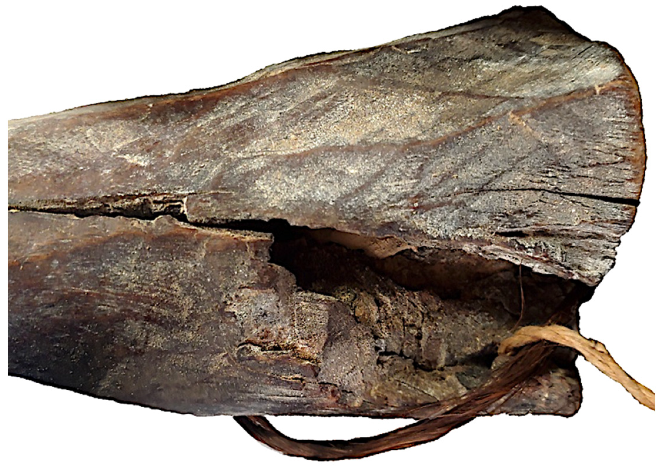
The tradition of puppet theatre in Africa has very ancient origins and an exhaustive historical–geographical path is very difficult to define [1,2]. Originating in Sogo Bò theatre, the wooden sculptures were made of kapok wood (Figure 2), Ceiba pentrada L. Gaertn, traditionally used by Mali population. The artifact shows a dark-brown color, attributable to treatments by hot spatulas in order to induce a superficial combustion of the wood, creating a brown, smooth surface, as suggested by Mary Jo Arnoldi’s personal communication [3]. Samples have been collected from a fracture in the wooden structure with flexible razor blades.

2.2. Microbial Taxa Identification

A spread microbiological patina was clearly distinguished on the artwork’s surface, (Figure 3) where three sampling areas (6 cm × 6 cm) were selected. Sampling was carried out by sterile swabs used to inoculate nutrient or Sabouraud agar plates (OXOID). After incubation at 30 ± 1 °C for 16–48 h, bacterial and fungal colonies were isolated. Fungal mycelia, fruiting structures, and spores were collected by a gentle pressing of small pieces of transparent adhesive tape on isolated colonies. Morphology of conidia and conidiophore structures were observed by optical microscopy (Leica, Wetzlar, Germany), after Lugol’s iodine staining (Lugol’s reactive = 1 g of iodine and 1 g of potassium iodide in 100 mL of distilled water [43]). Moreover, isolated bacteria were taxonomically characterized on the base of their 16S target sequences [44,45]; universal ITS1-ITS 4 primers were utilized for the identification of isolated fungi [46,47]. PCR products were purified according to the manufacturer’s protocol using a GenElute kit (Sigma-Aldrich, Darmstadt, Germany), and were sequenced by Eurofins Genomics’ service (https://www.eurofinsgenomics.eu/ Ebersberg, Germany) based on the Sanger PCR-sequencing method. Sequences were analyzed (% of similarity), referring to genomic databases (EMBL-Germany, NIH-USA), with the BLAST platform [48].

2.3. In Vitro Antimicrobial Activity Assays

Bacterial (Bacillus sp., Streptococcus sp.) and fungal (Penicillium sp., Aspergillus sp.) colonies were preliminarily grown as a liquid culture (nutrient or Sabouraud broth), incubating for 16–36 h at 30 ± 1 °C. The antimicrobial activity of the EO and HA solutions was evaluated by agar diffusion disc (ADD) or well plate diffusion (WPD) in vitro methods (Figure 4). Aliquots (10–20 μL) of bacterial or fungal liquid cultures were uniformly spread by a sterile Drigalsky spatula on nutrient or Sabouraud surface media, and the surface was allowed to dry. The assays were performed, in duplicate, using bacterial or fungal liquid cultures, normalized to the concentration of 1 × 10⁶ CFU/mL and 1 × 10⁴ conidia/mL, respectively.
In the ADD assay, 10 μL aliquots of T. vulgare essential oil (50%, 25%,12.5%) solutions were dropped on a sterile paper disc (6 mm in diameter, Dutscher papier, FR) centred on a nutrient or Sabouraud agar medium (9 cm Petri dishes) surface, previously sown with microbial cultures. Incubating at 30 ± 1 °C, confluent microbial growth was clearly observed except in the inhibition halo (i.h.) area, the diameter (mm) of which was related to the antimicrobial activity of the main components. In control assays, paper discs were soaked with 70% ethanol or 3% (vol/vol) benzalkonium chloride solutions.
In the WPD assay, a hole 4 mm in diameter was drilled aseptically on the agar, and a 10 μL aliquot of T. vulgare HA solution was loaded. After incubation at 30 ± 1 °C, the diameters (mm) of the growth inhibition halos were measured.
The antimicrobial activity vs. the microbial colony was outlined as: a sensitive strain = i.h. > 9 mm; a resistant strain = i.h. < 6 mm. As summarized in Table 1, both EO and HA solutions were able to produce inhibition halos of at least 9 mm, showing good antimicrobial action. Reduced halos in diameter were revealed for benzalkonium-Cl (>6 mm) and for Et-OH (almost no activity).
Biocide or biostatic activity was also evaluated, distinguishing minimum inhibitory concentration (MIC) and minimum biocidal concentration (MBC). MIC corresponds to any visible microbial growth at the lowest concentration after incubation at 30 + 1 °C, and MFC was measured by an antimicrobial-free sub-culture, defining the lowest concentration of essential oil able to kill 99.5% of the original inoculum. In 96-well microtiters, 30 microliter aliquots of 12.5%, 25%, 50%, and 100% EO solutions were mixed with an equal volume of microbial cultures and a liquid nutritive medium. Microbial growth after 18–36 h of incubation at 30 + 1 °C, and the absorbance at 500–600 nm, were measured; as the control, the commercial biocide benzalkonium chloride (3%, v/v) was utilized [48]. Assays were performed in duplicate.

2.4. T. vulgaris EO and HA Solutions

The chemical compounds of the Thymus vulgaris L. essential oil were identified by GC–MS (retention indices on the HP 5MS column) [40], highlighting the richness in carvacrol (64.96%), thymol (8.25%), and their biogenetic precursor p-cymene (11.29%). The hydro-alcoholic solution, supplied by EPO Srl., Milano, had been assessed by gas-chromatography according to the European Council Pharmacopoeia: carvacrol = 0.0010%; thymol = 0.12%; camphor and eucalyptol <0.0010% [49].

2.5. Artwork Surface Analysis

In order to identify the finishing layers/binders on the artwork’s surface, wood samples were analyzed by Fourier-transform infrared spectroscopy (FTIR) and environmental scanning electron microscopy equipped with an X-ray energy dispersive system (ESEM-EDS) [50].
The FTIR was performed in ATR mode, using a Perkin Elmer Spectrum 2 spectroscope, an ATR module with a diamond cell, and a spectral resolution 4 cm−1 (measurement was performed four times/sample, range 400–4000 cm−1). Furthermore, to evaluate any variation on the artwork’s surface after the T. vulgaris HA application, FTIR and ESEM-EDS (FEI QUANTA 600 ESEM equipped with an FEG electron gun and an EDAX –EDS microprobe) investigations were carried out.
ESEM-EDS samples were observed in a low-vacuum mode, i.e., 0.4 mbar of water vapor pressure, in order to observe the samples without gilding.

2.6. Microbial Colonies’ Direct Removal by HA Solution

In order to improve the biocidal effects, the microbial colonization on the artwork’s surface was preliminarily removed with HA T. vulgaris solutions, by cotton swab or a soft brush, in relation to surface conservation conditions (mechanical action was gently carried out for few seconds).
Potential variation in the color, before and after surface treatment, was evaluated by a portable colorimeter, PCE CSM 7.

2.7. Exposure to T. vulgaris EO Volatile Compounds

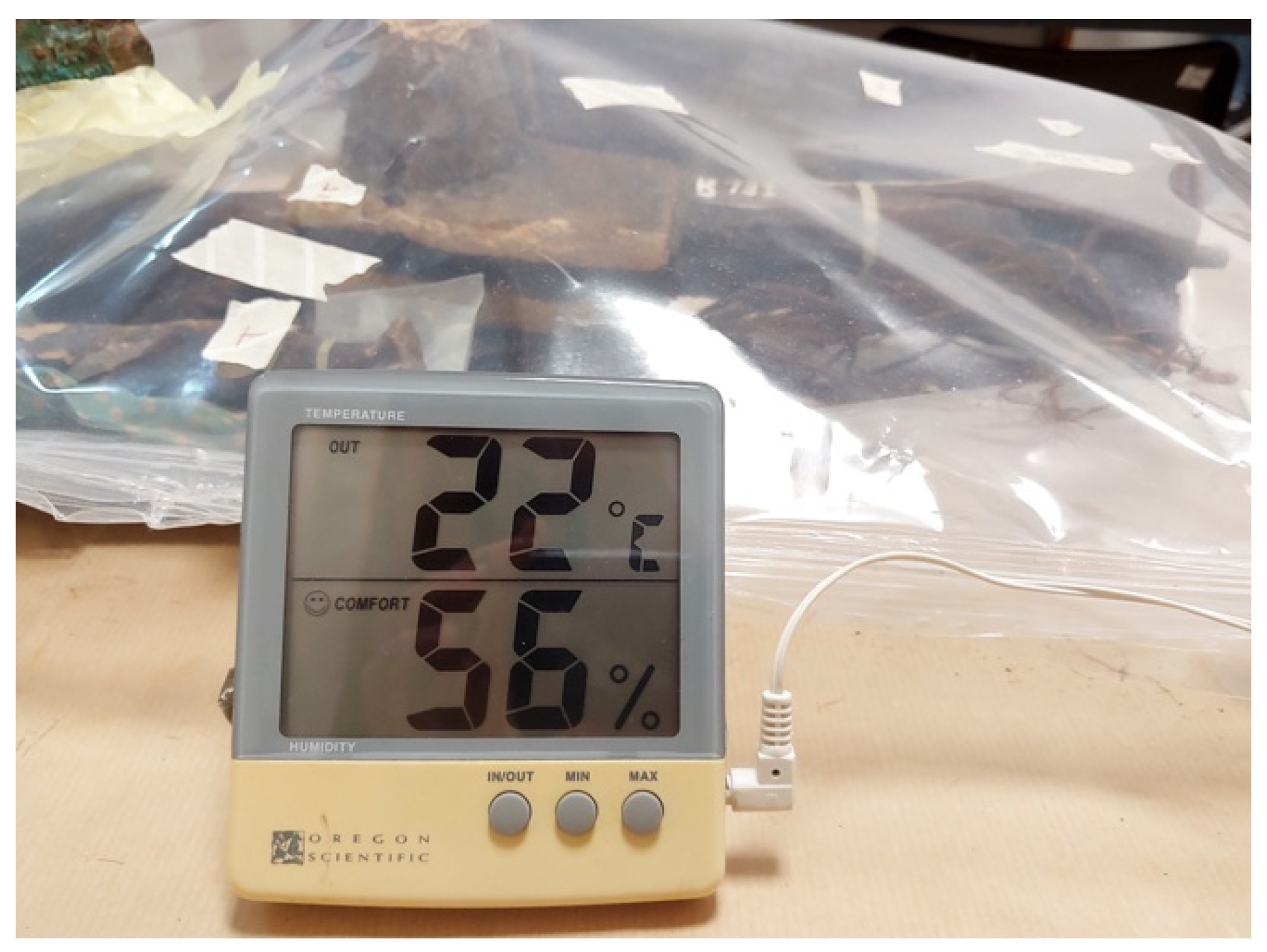
As in our previous studies, artifacts were exposed to EO volatile compounds in ad hoc assembled “clean chambers” with gas-barrier thermo-sealed film, in environmental conditions [40,42]. In order to maintain a saturate atmosphere, 0.9 mL/1000 cm3 of T. vulgaris EO was dispensed in three small glass containers, placed at equidistant points. The thermo-hygrometric parameters (inside–outside, min–max temperature, and relative humidity) were continuously monitored by an Oregon Scientific datalogger equipped with a microprobe. Treatments were carried out at temp = 22 + 2 °C and U.R. = 56 + 2% values for seven weeks.

2.8. Microbial Monitoring

Microbial growth monitoring was performed, assessing the total microbial load in the three areas selected on the artwork’s surface. Sampling was performed before, during, and at the end (seven weeks) of treatment, with sterile cotton swabs.
Before combined treatment, the total microbial load was high in all selected.
Keeping one area as the control, identified as T. vulgaris EO exposure without surface treatment (orange line), the surface of the other two selected areas was treated with a T. vulgaris HA or Et-OH solution.
During exposure, sampling on the selected areas was performed by making small cuts on the gas-barrier film with a sterile scalpel, through which a sterile swab was easily introduced without changing the environmental conditions; immediately after sampling, barrier film cuts were thermo-sealed. Each swab was used to inoculate nutrient and Sabouraud agar plates.
Microbial load was assessed as the total number of bacterial and fungal colonies grown on nutrient and Sabouraud agar plates per sample.

3. Results

Thin sections of the sculpture’s wood were analyzed with a Leitz Laborlux Pol 12 petrographic microscope (Figure 2), and images were acquired using a Leica MD170HD camera and Leica Application Suite v4.3 software polarizing filters (used in parallel mode). Specifically, vessels were gathered in radial chains, often placed side by side, with decreasing dimensions according to the growth period of the tree. The perforation plates showed a simple organization. Multi-seriate rays, composed of fewer than 10 uniform cells with different heights, were observed. These characteristics, in agreement with M.J. Arnoldi’s suggestion, allowed us to consider Ceiba sp. as the original plant, Angiosperm (hardwood).
On the artwork’s surface was a clearly distinguishable whitish biological patina (Figure 3). Microscopy observations, in vitro cultures, and molecular investigations allowed us to identify the bacterial (Bacillus sp., Streptococcus sp.) and fungal (Penicillium sp., Aspergillus sp.) colonies. In our previous studies, the Thymus vulgaris EO was successful when applied as an antimicrobial agent to control microbial colonization, acting on bacterial and fungal colonies, as well as on complex biofilm [40,41,42,51].
Several studies showed that compounds of natural origin, such as plant extracts, display antimicrobial and antioxidant activity that differ in relation to several factors: organic/inorganic artefacts; indoor/outdoor environments; bacterial or fungal colonizer; biological activity of the EO and HA solutions and mechanisms of action [51]. The antimicrobial actions of essential oil and hydro-alcoholic solutions have been preliminarily evaluated in vitro, using ADD and WPD methods, against both bacterial- and fungal-isolated colonies.
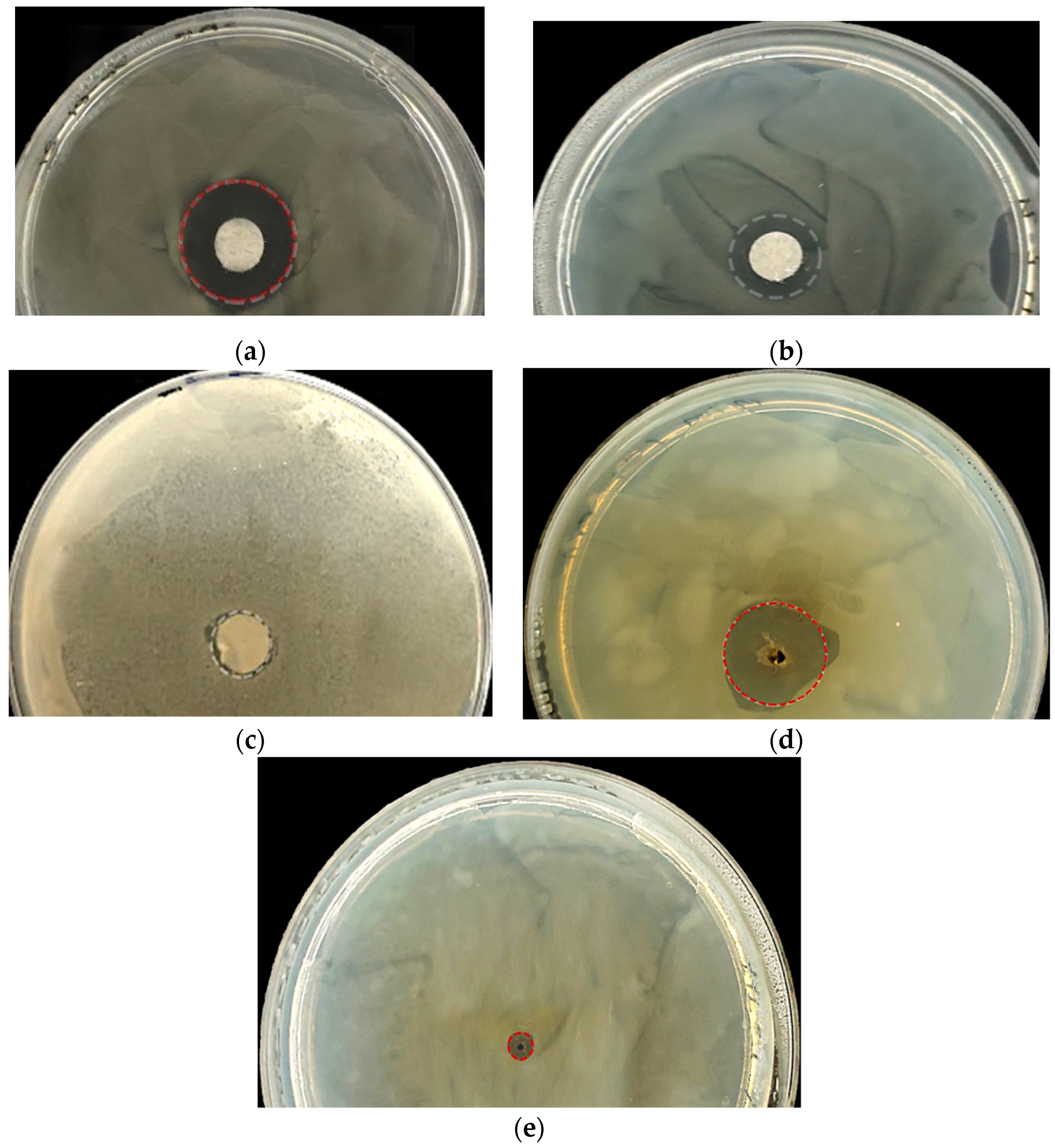
Shown in Figure 4 is the antimicrobial activity of the 12.5% T. vulgaris EO (Figure 4a), the control solution 3% v/v benzalkonium chloride (Figure 4b), and the 70% v/v Et-OH (Figure 4c) assayed vs. Streptomyces sp. colonies with the ADD method. Different inhibition halo (i.h.) diameters were distinguishable: (a) i.h. > 9 mm; (b) i.h. > 6 mm; (c) i.h. = almost not observable.
For the T. vulgare hydro-alcoholic solution, the antimicrobial activity was evaluated by the WPD method: the 100% HA solution produced an i.h. > 9 mm (Figure 4d); i.h. < 4 mm was obtained with the 70% Et-OH solution (Figure 4e).
In the ADD and WPD assays, 3% v/v benzalkonium chloride and 70% v/v Et-OH solutions represent control solutions; the benzalkonium chloride (phase) solution was diluted in water to achieved a final concentration equal to 3% (vol/vol).
Assays were performed in duplicate.
Results of the ADD and WPD assays performed in this study, as shown vs. the Streptomyces sp. colonies in Figure 4, are summarized in Table 1. Although the EO showed increased antimicrobial activity, the HA solution was able to produce inhibition halos of at least 9 mm. A reduced diameter is shown for the benzalkonium-Cl and Et-OH control solutions, producing halos less than 6 mm in diameter.
The minimum inhibitory concentration (MIC) and minimum bactericidal/fungicidal concentration (MBC/MFC) were assessed with the microdilution method, performed in a 96-well microtiter. The minimum inhibitory concentration (MIC) corresponds to the lowest concentration of the sample at which the tested microorganisms did not demonstrate any visible growth. The minimum bactericidal/fungicidal (MBC/MFC) concentrations were determined as the lowest concentration of the antimicrobial able to kill the 99.5% of the original inoculum in the sub-culture (antimicrobial-free media) [39].
In 96-well microtiters, 30 microliter aliquots of 12.5%, 25%, 50%, and 100% EO solutions were mixed with an equal volume of a microbial culture and a liquid nutritive medium, measuring the absorbance at 500–600 nm, after 18–36 h of incubation at 30 + 1 °C. As the control, the commercial biocide benzalkonium chloride (3%, v/v) was utilized. The T. vulgaris EO showed a good, but different, inhibitory effect vs both bacterial and fungal colonies; MBC/MFC values measured are closely related to phenolic compounds [48,51,52].
A reduced variance was revealed, which could be related to different factors, such as oil solubility or the different susceptibility of the microbes to the substances, because all microbes present differences in cell wall structure, lipid and protein composition of the cytoplasmic membrane, as well as in specific physiological processes.
The values of inhibition halos (mm) in Table 1 were derived from ADD and WPD assays, performed in duplicate for each antimicrobial and control solution. A variable activity of EO and HA solutions was distinguished, and attributable to the different concentrations of chemical compounds such as carvacrol and thymol. Furthermore, different activity on microbial systems could be related to difference on cell wall structures, lipid/protein composition of cytoplasmic membranes, and to potential influence on specific physiological processes [53,54,55].
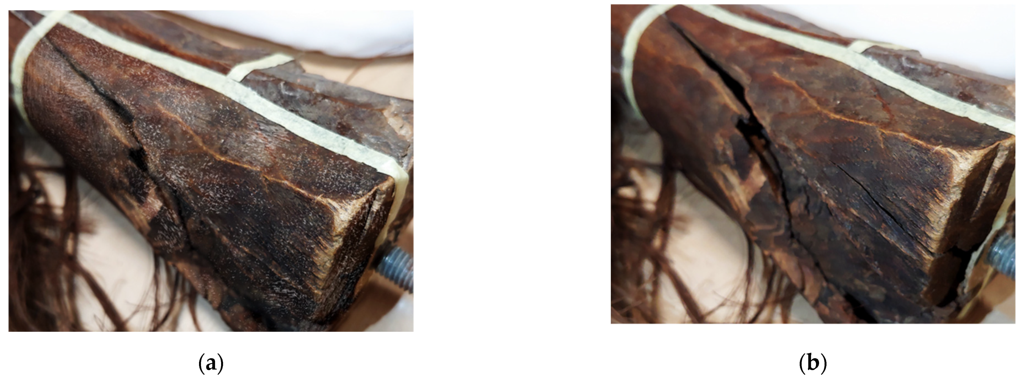
A survey to assess possible changes pre- and post-removal by HA solution application was carried out, performing colorimetric measurement on selected points of the artwork’s surface (Figure 5). The ΔE results were lower than 4.0 (3.821 on the lighter area and 2.75 on the darker area, respectively), corresponding to a variation not perceptible by the human eye, as shown in Figure 6. The measurement did not show any significant variation in all three parameters (L*, a*, b*), both on the darkened (surface) and lighter (lacuna) areas.
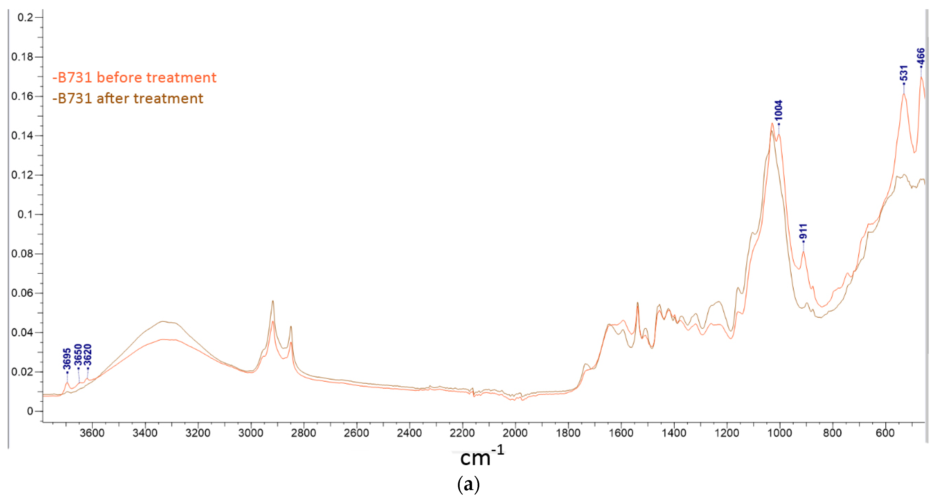
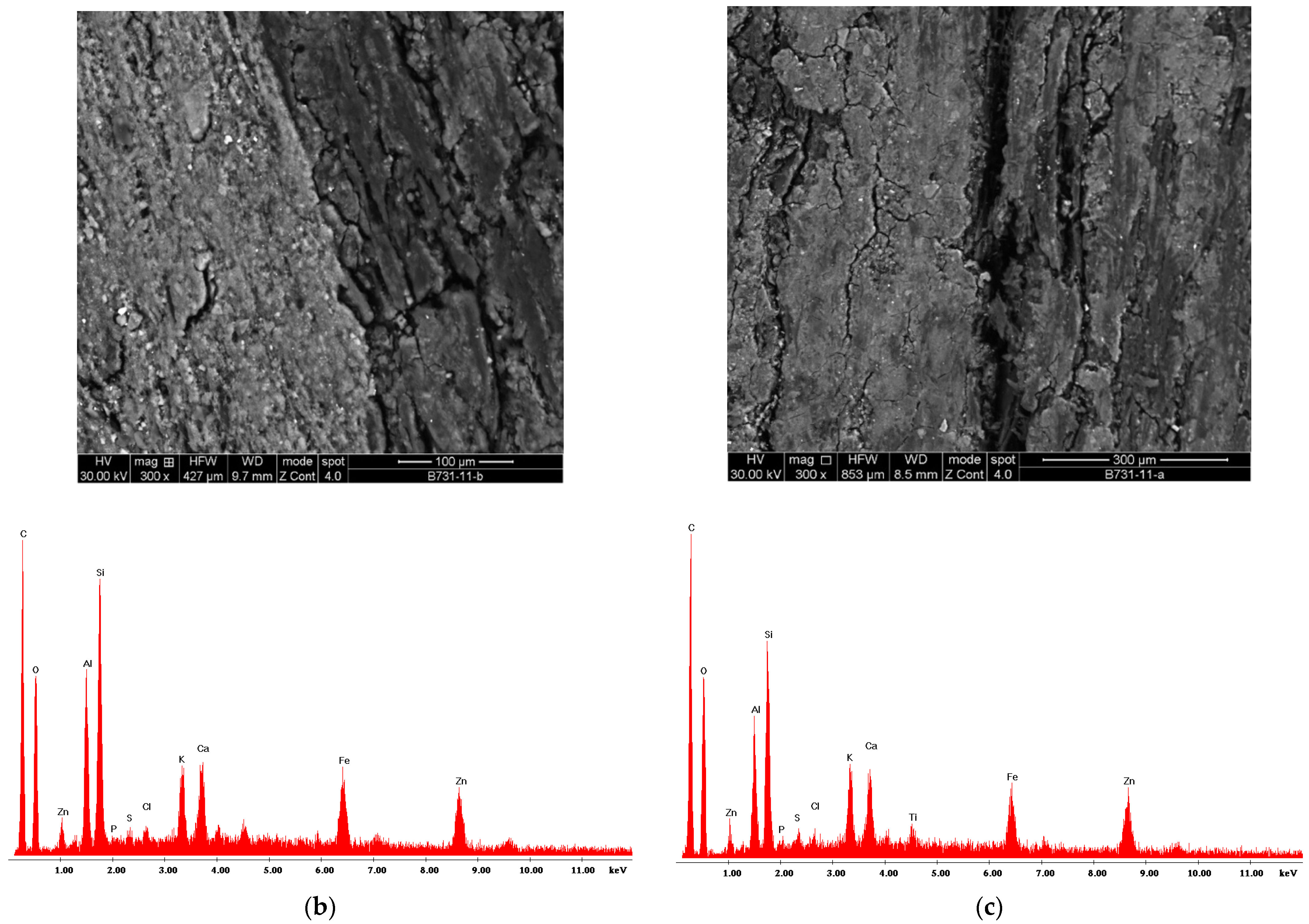
Removal action by HA solution, was preceded by FTIR and ESEM-EDS analyses of the wooden sculpture’s surface, in order to exclude the presence of binder or finishing layers. As seen in Figure 6a, the FTIR spectrum revealed a silico-aluminous layer classified as a soiled contaminant due to conservation conditions, identified by the highlighted peaks. Furthermore, an E-SEM analysis of the wood surface was performed, pre- and post-treatment by the HA solution, and any changes have been highlighted (Figure 6b,c). Particularly E-SEM images showed no increase in fractures or changes in the elemental composition of the surface.
To test the hypothesis that the antimicrobial efficiency of T. vulgaris extracts can be implemented performing a cooperative action of EO exposure and HA solutions, a dedicated “clean chamber” was assembled. Specifically, after the microbial colonies’ removal by the HA solution, the wooden artwork was exposed to EO volatile compounds (Figure 7). During the seven weeks of permanence in the “clean chamber”, a monitoring of microbial colonization was carried out weekly. Sampling on the selected surface areas was performed with sterile swabs, utilized to inoculate nutrient and Sabouraud agar media in an in vitro microbial culture. The monitoring continued for another ten weeks, after which the artifact was brought back to the museum’s exhibition hall.
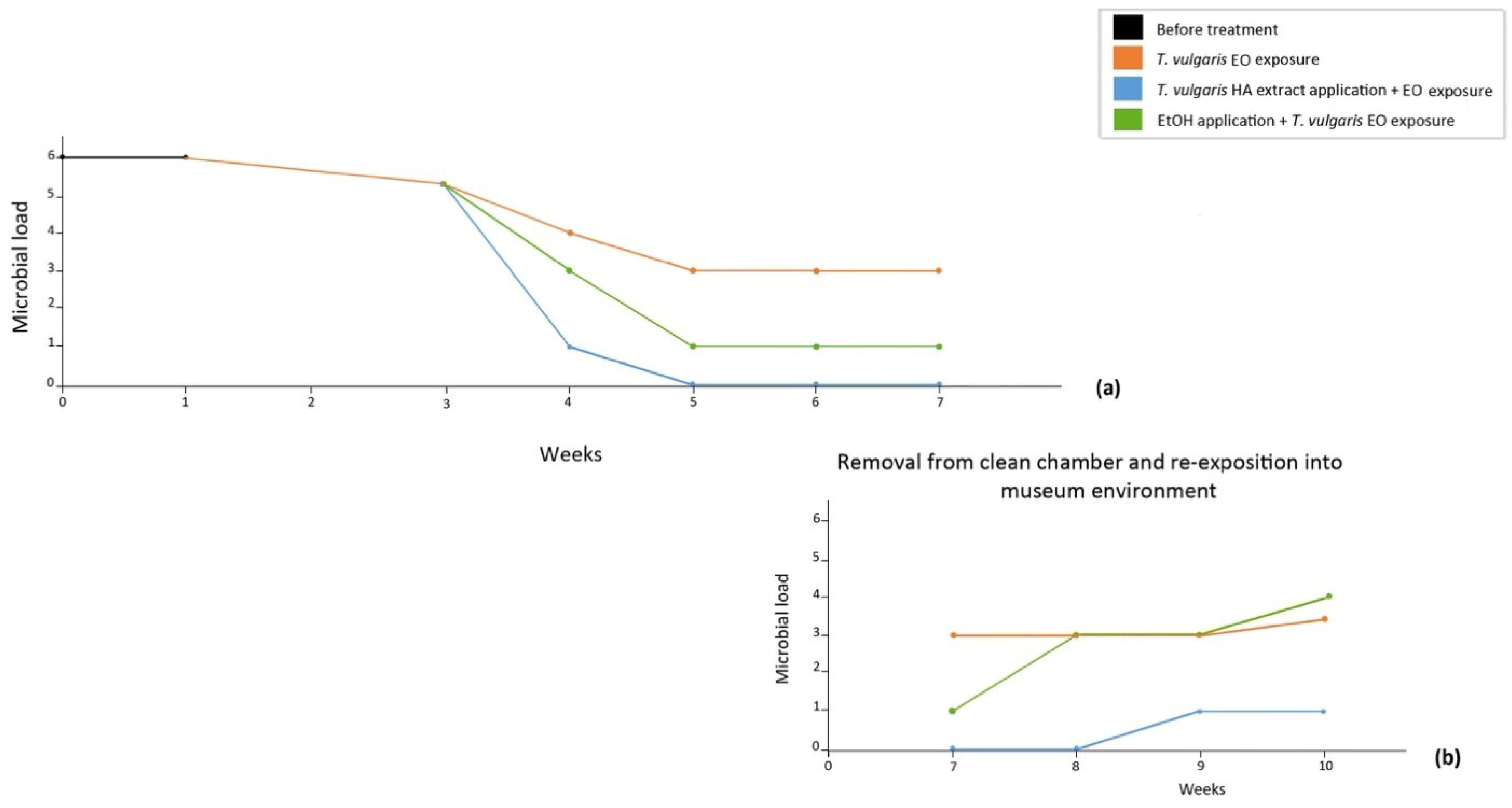
Results of the microbial growth monitoring, performed for total of seventeen weeks, are shown in Figure 8a,b, evaluating the total microbial load (TML). In the first seven weeks: (a) TML initially revealed on artwork’s surface (black line), slightly decreased after exposure to EO volatile compounds (orange line). Alternatively, if, before EO volatile compounds exposure, a treatment with 70% Et-OH (green line) or an HA solution (blue line) was carried out, a reduced or very low microbial load was recognizable, and the microbial load reached zero when the cooperative action of T. vulgaris solutions (HA–EO) was achieved (blue line).
Moreover, microbial load was monitored for a further ten weeks after the re-exposition of the wooden artifact into the museum’s exposure-hall environment. As shown in (b), the best results can still be seen for the combined HA–EO treatment (blue line).

4. Discussion

Generally, to control and eradicate microbial colonization, synthetic biocides are applied, but several factors, such as environmental pollution and toxicity to humans, need to be evaluated [23,24,25,26]. In recent times, the choice of sustainable methods to counteract microbial colonization on cultural assets is focused on plant bioactive compounds, representing valid alternatives to synthetic preservatives [56,57,58,59,60,61]. Specifically, essential oils can certainly be considered antimicrobial agents, containing several chemical compounds (phenols, quinines, tannins, etc.) acting on microbial systems through different mechanisms and their potential synergic effect [31]. Undoubtedly, the main limit of the use of plant extracts is the method of application on cultural assets, as avoiding direct contact may induce unpredictable modifications on surface layers (varnishing, pigments, or other substances). In this study, Thymus vulgaris essential oil and hydro-alcoholic solutions are proposed as a novel protocol to counteract microbial-colonized Sogo Bò wooden sculptures.
Gas-chromatography analysis showed that carvacrol and thymol are the main compounds in both EO and HA solutions; the antimicrobial activity of both is reported in the literature [62,63].
In this combined treatment, the wooden sculpture’s surface was preliminarily treated with a T. vulgaris HA solution, followed by exposure to EO volatile compounds in a dedicated “clean chamber”, under environmental conditions. On the bigger section of the wooden sculpture, three areas were selected to monitor the action of thymus products on microbial colonization. As summarized in Figure 8a, before treatment, a high microbial load was revealed, slightly decreasing after exposure to EO volatile compounds in the ad hoc assembled “clean chamber”. Total microbial load was further reduced when combining the exposure to EO volatile compounds with an application of 70% Et-OH, becoming zero when the T. vulgaris HA solution was applied.
Monitoring was continued for a further ten weeks after the artifacts were re-exhibited in the museum hall, which highlighted that the microbial load was still very low.

5. Conclusions

Historical–artistic assets represent an inestimable heritage whose value is not only related to artistic characteristics, but also to past history, culture, and traditions. These significant treasures need to be protected from damage induced by air pollution, physical and chemical agents, and microbial colonization.
Aiming to set up specific protocols to increase the efficacy of exposure to EO volatile compounds, in our previous study, the exposition of microbial colonized parchment artworks to Thymus vulgaris L. and Crithmum maritimum L. EOs was performed under vacuum conditions that significantly reduced the exposure time [64].
Evaluating that plant bioactive compounds are extracted as EO or HA solutions, in the present work, for the first time, a synergic effect of T. vulgaris extracts (essential oil and hydro-alcoholic solutions) was proposed for a specific wooden sculpture, performing the exposure under environmental conditions.
This strategy turned out to be ideal for the Sogo Bò theatre (Mali, Africa) artifact, the finishing treatment of which is not present on the sculpture’s surface.
In our hypothesis, after alcohol evaporation (HA solution), the T. vulgaris antimicrobial compound should remain on the artwork’s surface, enhancing the antimicrobial activity of EO volatile compounds, applied in an ad hoc assembled “clean chamber”.
This is a further applicative protocol, respectful of both operators and the environment, based on green conservation strategy, which allowed us to hypothesize its use in replacing synthetic biocides in a sustainable conservation of cultural assets.

Author Contributions

Conceptualization, S.S., G.N. and F.P.; methodology, S.S., T.F., G.G., B.M. and B.G.; software, S.S. and F.P.; investigation and data curation, S.S., B.M., B.B. and G.G.; writing—original draft preparation, S.S., B.B. and F.P.; writing—review and editing, S.S., G.N. and F.P. All authors have read and agreed to the published version of the manuscript.

Funding

This research received no external funding. Publication fee was supported by EPO Srl, Milano, Italy.

Institutional Review Board Statement

The study did not involve humans or animals.

Acknowledgments

The authors especially thank the Director and the staff of “Museo Internazionale delle Marionette A. Pasqualino” Palermo, Italy, for yielded collaboration, and M.J. Arnoldi for the precious suggestions on wooden species. Results on application of T. vulgaris essential oil and hydro-alcoholic solution are part of S. Sparacello’s final examination of the Master’s degree in Conservation and Restoration of Cultural Heritage at the University of Palermo (Italy), graduated cum laude and qualified as an Italian Restorer of Cultural Heritage (MiC, Italian Ministry of Culture).

Conflicts of Interest

The authors declare no conflict of interest.

References

  1. Arnoldi, M.J. Playing the Puppets: Innovation and Rivalry in Bamana Youth Theatre of Mali. Drama Rev. 1988, 32, 2. [Google Scholar] [CrossRef]
  2. Linking, W.W. Marionnettes du Mali, Paris. 1980. Available online: http://www.gourcuff-gradenigo.com/mali.html (accessed on 24 September 2020).
  3. Arnoldi, M.J.; Curator Emerita, Africa Department of Anthropology, Smithsonian Institution, Washington, DC, USA. Personal communication, 2021.
  4. Pournou, A. Biology of wood deteriogens. In Biodeterioration of Wooden Cultural Heritage; Springer Nature: Basingstoke, UK, 2020; Chapter 3; p. 99. [Google Scholar]
  5. Allsopp, D.; Seal, K.J.; Gaylarde, C.C. Introduction to Biodeterioration; Cambridge University Press: Cambridge, UK, 2004. [Google Scholar]
  6. Dakal, T.C.; Cameotra, S.S. Microbially induced deterioration of architectural heritages: Routes and mechanisms involved. Environ. Sci. Eur. 2012, 24, 1–13. [Google Scholar] [CrossRef] [Green Version]
  7. Ranalli, G.; Zanardini, E.; Sorlini, C. Biodeterioration—Including Cultural Heritage, in Encyclopedia of Microbiology. In Encyclopedia of Microbiology, 4th ed.; Schmidt, T.M., Ed.; Academic Press: Cambridge, MA, USA, 2019. [Google Scholar]
  8. Di Carlo, E.; Barresi, G.; Palla., F. Biodeterioration. In Biotechnology and Conservation of Cultural Heritage; Palla, F., Barresi, G., Eds.; Springer International Publishing: Basel, Switzerland, 2017; pp. 1–30. [Google Scholar]
  9. Blanchette, R.A.; Nilsson, T.; Daniel, G.; Abad, A. Biological degradation of wood. In Archaeological Wood; Rowell, R.M., Barbour, R.J., Eds.; American Chemical Society: Washington, DC, USA, 1990; Volume 74, pp. 141–174. [Google Scholar]
  10. Reinprect, L. Wood Deterioration, Protection and Maintenance; John Wiley & Sons, Ltd.: Chichester, UK, 2016. [Google Scholar]
  11. Ljaljević Grbić, M.; Stupar., M.; Vukojević, J.; Maričić, I.; Bungur, N. Molds in Museum Environments: Biodeterioration of Art Photographs and Wooden Sculptures. Arch. Biol. Sci. 2013, 65, 955–962. [Google Scholar] [CrossRef]
  12. Valentin, N. Microbial contamination in museum collections: Organic materials. In Molecular Biology and Cultural Heritage; Saiz-Jimenez, C., Ed.; Swets & Zeitlinger: Lisse, The Netherlands, 2003; pp. 85–91. [Google Scholar]
  13. Palla, F. Biological risks for the conservation and exhibition of historical artistic artefacts in confined spaces. In Risk Management in the Cultural Heritage Sector: Museums, Libraries and Archives; Lorusso, S., Natali, A., Palla, F., Eds.; MIMESIS Edizioni: Milano, Italy, 2015; pp. 95–108. [Google Scholar]
  14. Biological Deterioration & Damage to Furniture & Wooden Objects. Available online: https://www.si.edu/mci/english/learn_more/taking_care/biodetwood.html (accessed on 29 March 2018).
  15. Tornari, V.; Basset, T.; Andrianakis, M.; Kosma, K. Impact of Relative Humidity on Wood Sample: A Climate Chamber Experimental Simulation Monitored by Digital Holographic Speckle Pattern Interferometry. J. Imaging 2019, 5, 65. [Google Scholar] [CrossRef] [PubMed] [Green Version]
  16. Clausen, A.C. Bacterial association with decaying wood: A review. Int. Biodeterior. Biodegrad. 1996, 37, 101–107. [Google Scholar] [CrossRef]
  17. Blanchette, R.A. A review of microbial deterioration found in archaeological wood from different environments. Int. Biodeterior. Biodegrad. 2000, 46, 189–204. [Google Scholar] [CrossRef]
  18. Pangallo, D.; Šimonovičová, A.; Chovanová, K.; Ferianc, P. Wooden art objects and the museum environment: Identification and biodegradative characteristics of isolated microflora. Lett. Appl. Microbiol. 2007, 45, 87–94. [Google Scholar] [CrossRef]
  19. Romero, S.; Giudicessi, S.L.; Vitale, R.G. Is the fungus Aspergillus a threat to cultural heritage? J. Cult. Her. 2021, 51, 107–124. [Google Scholar] [CrossRef]
  20. Hyde, K.D.; Jones, E.B.G. Introduction to fungal succession. Fungal Divers. 2002, 10, 241–253. [Google Scholar]
  21. Chojnackah, M.K. Biocides. In Encyclopedia of Toxicology, 3rd ed.; Biomedical Sciences, Elsevier: Amsterdam, The Netherlands, 2014; pp. 461–463. [Google Scholar]
  22. Young, M.E.; Alakomi, H.L.; Fortune, A.A.; Goburshina, A.A.; Krumbein, W.E.; Maxwell, I.; McCullagh, C.; Robertson, P.; Saarela, M.; Valero, J.; et al. Development of a biocidal treatment regime to inhibit biological growths on cultural heritage: BIODAM. Environ. Geol. 2008, 56, 631–641. [Google Scholar] [CrossRef]
  23. Marcotte, S.; Estel, L.; Leboucher, S.; Minchin, S. Occurrence of organic biocides in the air and dust at the Natural History Museum of Rouen, France. J. Cult. Herit. 2014, 15, 68–72. [Google Scholar] [CrossRef]
  24. Arias-Estévez, M.; López-Periago, E.; Martínez-Carballo, E.; Simal-Gándara, J.; Juan-Mejuto, J.C.; García-Río, L. The mobility and degradation of pesticides in soils and the pollution of groundwater resources. Agric. Ecosyst. Environ. 2008, 123, 247–260. [Google Scholar] [CrossRef]
  25. Hernández, A.F.; Parron, T.; Tsatsakis, M.A.; Requena, M.; Alarcón, R.; López-Guarnido, O. Toxic effects of pesticide mixtures at molecular level: Their relevance to human health. Toxicology 2013, 307, 136–145. [Google Scholar] [CrossRef]
  26. Hernández, A.F.; Gil, F.; Lacasagña, M. Toxicological interactions of pesticide mixtures: An update. Arch. Toxicol. 2017, 91, 3211–3223. [Google Scholar] [CrossRef]
  27. Petrovska, B.B. Historical review of medicinal plants usage. Pharmacogn. Rev. 2012, 6, 1–5. [Google Scholar] [CrossRef] [Green Version]
  28. Xie, Y.; Wang, Z.; Huang, Q.; Zhang, D. Antifungal activity of several essential oils and major components against wood-rot fungi. Ind. Crop. Prod. 2017, 108, 278–285. [Google Scholar] [CrossRef]
  29. Fernandez-Lopes, J.; Viuda-Martos, M. Application of essential oils in food systems. Foods 2018, 7, 1–4. [Google Scholar]
  30. Abu-Shanab, B.; Adwan, G.; Abu-Safiya, D.; Jarrar, N.; Adwan, K. Antibacterial Activities of Some Plant Extracts Utilized in Popular Medicine in Palestine. Turk. J. Biol. 2004, 28, 99–102. [Google Scholar]
  31. Bakkali, F.; Averbeck, S.; Averback, D.; Idaomar, M. Biological effects of essential oils—A review. Food Chem. Toxicol. 2008, 46, 446–475. [Google Scholar] [CrossRef]
  32. Reichling, J.; Schnitzler, P.; Suschke, U.; Saller, R. Essential oils of aromatic plants with antibacterial, antifungal, antiviral, and cytotoxic properties—An overview. Complement. Med. Res. 2009, 16, 79–90. [Google Scholar] [CrossRef] [Green Version]
  33. Afifi, H.A.M. Comparative efficacy of some plant extracts against fungal deterioration of stucco ornaments in the Mihrab of Mostafa Pasha, Ribat, Cairo, Egypt. Am. J. Biochem. Mol. Biol. 2012, 2, 40–47. [Google Scholar] [CrossRef] [Green Version]
  34. Gatti, L.; Troiano, F.; Vacchini, V.; Cappitelli, F.; Balloi, A. An In Vitro Evaluation of the Biocidal Effect of Oregano and Cloves’ Volatile Compounds against Microorganisms Colonizing an Oil Painting—A Pioneer Study. Appl. Sci. 2021, 11, 78. [Google Scholar] [CrossRef]
  35. Fierascu, I.; Ion, R.M.; Radu, M.; Bunghez, I.R.; Avramescu, S.M.; Fierascu, R.C. Comparative study of antifungal effect of natural extracts and essential oils of Ocimum basilicum on selected artefacts. Rev. Roum. Chim. 2014, 59, 207–211. [Google Scholar]
  36. Liu, Z.; Zhang, Y.; Zhang, F.; Hu, C.; Liu, G.; Pan, J. Microbial community analyses of the deteriorated storeroom objects in the Tianjin Museum using culture-independent and culture-dependent approaches. Front. Microbiol. 2018, 9, 802. [Google Scholar] [CrossRef] [PubMed]
  37. Rakotonirainy, M.S.; Lavédrine, B. Screening for antifungal activity of essential oils and related compounds to control the biocontamination in libraries and archives storage areas. Int. Biodeter. Biodegrad. 2005, 55, 141–147. [Google Scholar] [CrossRef]
  38. Barresi, G.; Di Carlo, E.; Trapani, M.R.; Parisi, M.G.; Chille, C.; Mulè, M.F.; Cammarata, M.; Palla, F. Marine organisms as source of bioactive molecules applied in restoration projects. Herit. Sci. 2015, 3, 17. [Google Scholar] [CrossRef] [Green Version]
  39. Rotolo, V.; Barresi, G.; Di Carlo, E.; Giordano, A.; Lombardo, G.; Crimi, E.; Costa, E.; Bruno, M.; Palla, F. Plant extracts as green potential strategies to control the biodeterioration of cultural heritage. Int. J. Conserv. Sci. 2016, 7, 839–846. [Google Scholar]
  40. Palla, F.; Bruno, M.; Mercurio, F.; Tantillo, A.; Rotolo, V. Essential oil as natural biocides in conservation of cultural heritage. Molecules 2020, 25, 730. [Google Scholar] [CrossRef] [PubMed] [Green Version]
  41. Ebadollahi, A.; Ziaee, M.; Palla, F. Essential oils extracted from different species of the Lamiaceae plant family as prospective bioagents against several detrimental pests. Molecules 2020, 25, 1556. [Google Scholar] [CrossRef] [Green Version]
  42. Palla, F.; Caruana, E.; Di Carlo, E.; Rotolo, V. Plant essential oils in controlling fungal colonization on wooden substrate. Borziana 2021, 2, 5–14. [Google Scholar]
  43. Di Carlo, E.; Chisesi, R.; Barresi, G.; Barbaro, S.; Lombardo, G.; Rotolo, V.; Sebastianelli, M.; Travagliato, G.; Palla, F. Fungi and bacteria in indoor cultural heritage environments: Microbial related risks for artworks and human health. Environ. Ecol. Res. 2016, 4, 257–264. [Google Scholar] [CrossRef]
  44. Frank, J.A.; Reich, C.I.; Sharma, S.; Weisbaum, J.S.; Wilson, B.A.; Olsen, G.J. Critical evaluation of two primers commonly used for amplification of bacterial 16S rRNA genes. Appl. Environ. Microbiol. 2008, 74, 2461–2470. [Google Scholar] [CrossRef] [Green Version]
  45. Cardinale, M.; Brusetti, L.; Quatrini, P.; Borin, S.; Puglia, A.M.; Rizzi, A.; Zanardini, E.; Sorlini, C.; Corselli, C.; Daffonchio, D. Comparison of different primer sets for use in automated ribosomal intergenic spacer analysis of complex bacterial communities. Appl. Environ. Microbiol. 2004, 70, 6147–6156. [Google Scholar] [CrossRef] [PubMed] [Green Version]
  46. Gardes, M.; Bruns, T.D. ITS primers with enhanced specificity for basidiomycetes-application to the identification of mycorrhizae and rusts. Mol. Ecol. 1993, 2, 113–118. [Google Scholar] [CrossRef] [PubMed]
  47. Martin, K.J.; Rygiewicz, P.T. Fungal-specific PCR primers developed for analysis of the ITS region of environmental DNA extracts. BMC Microbiol. 2005, 5, 28. [Google Scholar] [CrossRef] [PubMed] [Green Version]
  48. Barresi, G.; Cammarata, M.; Palla, F. Biocide. In Biotechnology and Conservation of Cultural Heritage; Palla, F., Barresi, G., Eds.; Springer International Publishing: Basel, Switzerland, 2017; pp. 49–66. [Google Scholar]
  49. European Pharmacopoeia 10th Edition, 10.2 n.0865, July 2020. Available online: www.edqm.eu (accessed on 24 September 2020).
  50. Timofeeff, M.N.; Lowestein, T.K.; Blackburn, W.H. ESEM-EDS: An improved technique for major element chemical analysis of fluid inclusions. Chem. Geol. 2000, 164, 171–181. [Google Scholar] [CrossRef]
  51. Rotolo, V.; De Caro, M.L.; Giordano, A.; Palla, F. Solunto archaeological park in Sicily: Life under tesserae. Flora Medit 2018, 28, 233–245. [Google Scholar]
  52. Barry, A.L. The Antimicrobic Susceptibility Test: Principles and Practices; Lea and Febiger: Philadelphia, PA, USA, 1976. [Google Scholar]
  53. Reichling, J.; Suschke, U.; Schneele, J.; Geiss, H.K. Antibacterial activity and irritation potential of selected essential oil components structure activity relationship. Nat. Prod. Commun. 2006, 1, 1003–1012. [Google Scholar] [CrossRef]
  54. Popovici, R.A.; Vaduva, D.A.; Pinzaru, I.; Dehelean, C.A.; Farcas, C.G.; Coricovac, D.; Danciu, C.; Popescu, I.; Alexa, E.; Lazureanu, V.; et al. A comparative study on the biological activity of essential oil and total hydro-alcoholic extract of Satureja hortensis L. Exp. Ther. Med. 2019, 18, 932–942. [Google Scholar] [CrossRef]
  55. Saad, N.; Muller, C.D.; Lobstein, A. Major bioactivities and mechanism of action of essential oil and their components. Fragance Flavours J. 2013, 28, 269–279. [Google Scholar] [CrossRef]
  56. Cappitelli, F.; Cattò, C.; Villa, F. The control of cultural heritage microbial deterioration. Microorganisms 2020, 8, 1542. [Google Scholar] [CrossRef] [PubMed]
  57. Fidanza, M.R.; Caneva, G. Natural biocides for the conservation of stone cultural heritage: A review. J. Cult. Herit. 2019, 38, 271–286. [Google Scholar] [CrossRef]
  58. Lavin, P.; Gómez de Saravia, S.; Guiamet, P. Scopulariopsis sp. and Fusarium sp. in the Documentary Heritage: Evaluation of their biodeterioration ability and antifungal effect of two essential oils. Microb. Ecol. 2016, 71, 628–633. [Google Scholar] [CrossRef] [PubMed]
  59. Nuță, D.C.; Limban, C.; Ioniță, C.C.; Nicolau, I.; Zarafu, I. The Use of Essential Oils as a Strategy to Combat Microbial Biofilms. A Review. Processes 2021, 9, 537. [Google Scholar] [CrossRef]
  60. Borrego, S.; Valdés, O.; Vivar, I.; Lavin, P.; Guiamet, P.; Battistoni, P.; Gómez de Saravia, S.G.; Borges, P. Essential oils of plants as biocides against microorganisms isolated from Cuban and Argentine documentary heritage. ISRN Microbiol. 2012, 2012, 826786. [Google Scholar] [CrossRef] [Green Version]
  61. Sakr, A.A.; Ghaly, M.F.; El-Sayed, F.; Abdel-Haliem, M. The efficacy of specific essential oils on yeasts isolated from the royal tomb paintings at Tanis, Egypt. Int. J. Conserv. Sci. 2012, 3, 87–92. [Google Scholar]
  62. Gavaric, N.; Mozina, S.S.; Kladar, N.; Bozin, B. Chemical profile, antioxidant and antibacterial activity of Thyme and Oregano essential oils, Thymol and Carvacrol and their possible synergism. J. Essent. Oil Bear. Plants 2015, 18, 1013–1021. [Google Scholar] [CrossRef]
  63. Klarić, M.Š.; Kosaleć, I.; Mastelić, J.; Piecková, E.; Pepeljnak, S. Antifungal activity of thyme (Thymus vulgaris L.) essential oil and thymol against moulds from damp dwellings. Lett. Appl. Microbiol. 2007, 44, 36–42. [Google Scholar] [CrossRef]
  64. D’Agostino, G.; Giambra, B.; Palla, F.; Bruno, M.; Badalamenti, N. The Application of the Essential Oils of Thymus vulgaris L. and Crithmum maritimum L. as Biocidal on Two Tholu Bommalu Indian Leather Puppets. Plants 2021, 10, 1508. [Google Scholar] [CrossRef]
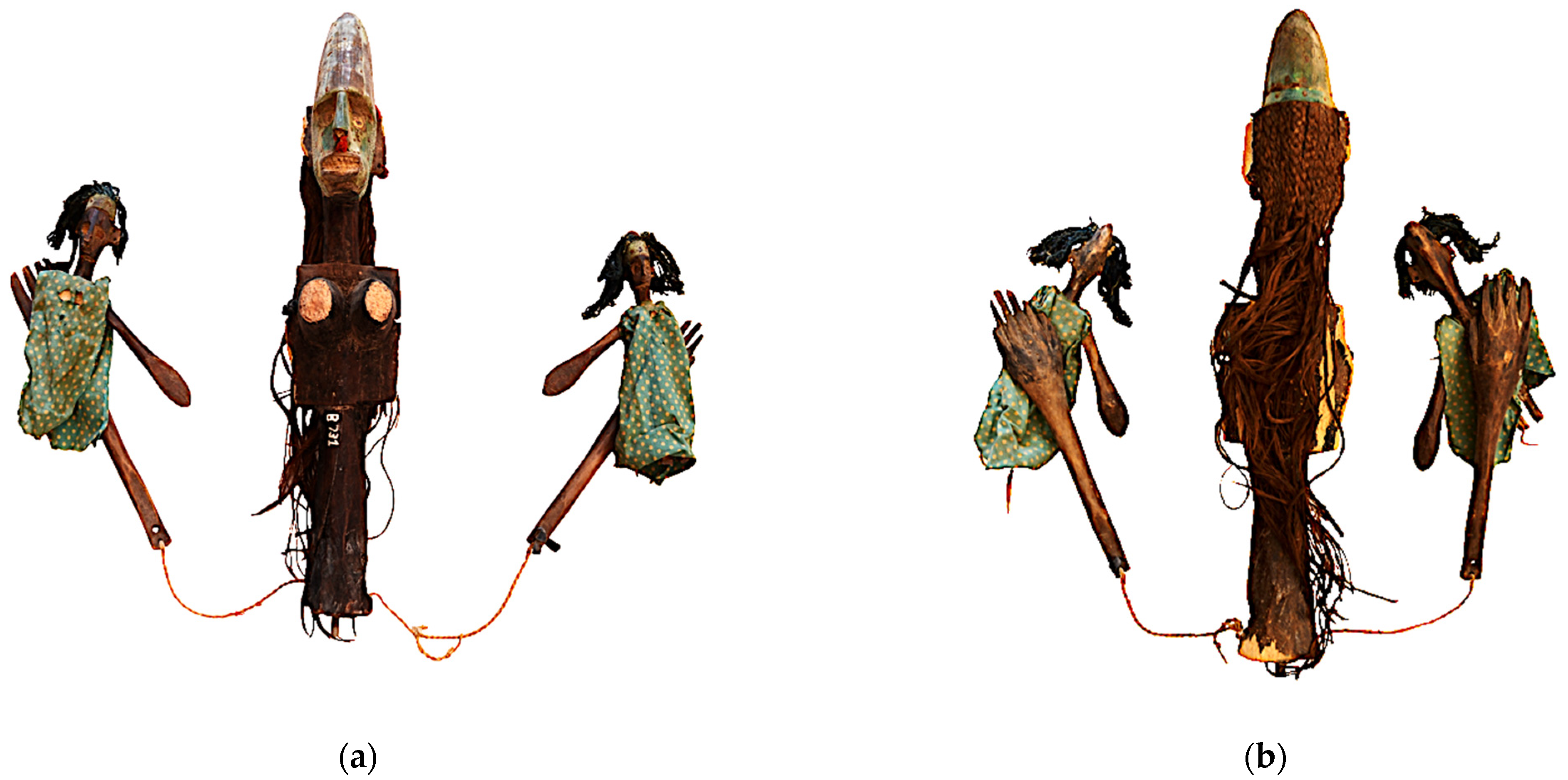
Figure 1. The Sogo Bò theatre wooden sculpture (B731) from the International Puppet Museum, Palermo, Italy: (a) recto; (b) verso. The largest central sculpture is 64.5 cm in height and 25.0 cm in width; the two small ones on the sides are 20.0 cm in height and 12.0 cm in width.
Applsci 11 08704 g001
Figure 2. Microscopy observation of the thin wood sections, three anatomical directions: (a) transverse; (b) tangential; (c) radial. The bars correspond to 100 μm.
Applsci 11 08704 g002
Figure 3. The Sogo Bò sculpture’s surface, whitish spots are recognizable.
Applsci 11 08704 g003
Figure 4. Antimicrobials vs. Streptomyces sp. colonies and related inhibition halo assayed with the ADD method: (a) T. vulgaris essential oil (12.5%); (b) benzalkonium chloride (3% v/v); (c) Et-OH (70% v/v). The diameter of >9 mm in (a,b) confirms antibacterial activity in these solutions. A very small diameter (almost not observable) is seen in (c), the 70% Et-OH solution. WPD method: whereas an inhibition halo is clearly recognizable in (d) related to the T. vulgare hydro-alcoholic solution (100%), the i.h. is barely detectable for (e), the 70% Et-OH solution.
Applsci 11 08704 g004
Figure 5. Selected area of the wood sculpture surface pre- (a) and post- (b) microbial colony removal by the T. vulgaris hydro-alcoholic solution. Notable is the complete removal of whitish spots and any distinguishable change in color in the brownish and light areas (lacuna) respectively.
Applsci 11 08704 g005
Figure 6. Evaluation of potential changes on the artwork’s surface pre- and post- treatment, respectively: (a) comparison of FTIR spectra; ESEM-EDS analysis pre (b) and post treatment (c).
Applsci 11 08704 g006aApplsci 11 08704 g006b
Figure 7. “Clean chamber” assembled for exposure (7 weeks) of the wooden sculpture to T. vulgaris EO volatile compounds. Inside/outside temperature (22 + 1 °C) and relative humidity (56 + 2%) values were continuously monitored by an Oregon thermo-hygrometer equipped with a microprobe.
Applsci 11 08704 g007
Figure 8. Monitoring of the microbial load on selected areas of the artwork’s surface, pre- and-post treatment, performed (a) for seventeen weeks and (b) for ten weeks after re-exposition into museum exposition hall.
Applsci 11 08704 g008
Table 1. Antimicrobial activity of the T. vulgaris and control solutions vs. microbial taxa isolated in this study. Values of inhibition halos (mm) were derived from duplicate assays.
Biocide
Solution
Microbial TaxaADD Method Inhibition Halo (mm)
E.O.
12.5%
Bacillus sp.21.5–22.0
Streptococcus sp.17.4–18.0
Aspergillus sp.8.5–9.0
Penicillum sp.12.0–12.5
WPD method Inhibition halo (mm)
H.A.
100%.
Bacillus sp.11.0–11.0
Streptococcus sp.9.3–10.0
Aspergillus sp.8.7–9.0
Penicillum sp.8.1–9.0
Control
solutions
All bacterial and fungal isolated coloniesADD or
WPD method Inhibition halo (mm)
Et-OH
70%
1.8–2.0
0.4–0.5
Benz. Clor.
3%
5.5–6.0
3.8–4.0
Publisher’s Note: MDPI stays neutral with regard to jurisdictional claims in published maps and institutional affiliations.