Review

A Review on Potential Antimutagenic Plants of Saudi Arabia

by
Syed Mohammed Basheeruddin Asdaq
1,*,
Syed Imam Rabbani
2,
Mohd. Imran
3,
Amani A. Alanazi
4,
Ghada Y. Alnusir
4,
Abdullah A. Al-Shammari
4,
Feras H. Alsubaie
4 and
Abdulkhaliq J. Alsalman
5
1
Department of Pharmacy Practice, College of Pharmacy, AlMaarefa University, Dariyah, Riyadh 13713, Saudi Arabia
2
Department of Pharmacology and Toxicology, College of Pharmacy, Qassim University, Buraydah 51452, Saudi Arabia
3
Department of Pharmaceutical Chemistry, Faculty of Pharmacy, Northern Border University, Rafha 91911, Saudi Arabia
4
Faculty of Pharmacy, Northern Border University, Rafha 91911, Saudi Arabia
5
Department of Clinical Pharmacy, Faculty of Pharmacy, Northern Border University, Rafha 91911, Saudi Arabia
*
Author to whom correspondence should be addressed.
Appl. Sci. 2021, 11(18), 8494; https://doi.org/10.3390/app11188494
Submission received: 18 August 2021 / Revised: 4 September 2021 / Accepted: 7 September 2021 / Published: 13 September 2021
(This article belongs to the Special Issue Medicinal and Aromatic Plants: Pharmacy, Food and Nutrition)

Abstract

:
Mutagenic complications can cause disease in both present as well as future generations. The disorders are caused by exogenous and endogenous agents that damage DNA beyond the normal repair mechanism. Rapid industrialization and the population explosion have contributed immensely to changes in the environment, leading to unavoidable exposure to mutagens in our daily life. As it is impossible to prevent exposure, one of the better approaches is to increase the intake of anti-mutagenic substances derived from natural resources. This review summarizes some of the important plants in Saudi Arabia that might have the potential to exhibit anti-mutagenic activity. The data for the review were retrieved from Google scholar, NCBI, PUBMED, EMBASE and the Web of Science. The information in the study has importance since one of the major reasons for mutation is viral infection. Considering the pandemic situation due to novel coronavirus and its aftermath, the native plants of Saudi Arabia could become an important source for reducing mutagenic complications associated with exogenous agents, including viruses.

1. Introduction

The alteration of DNA leading to a heritable change in the nucleotide sequence is called mutation. The agents that cause these alterations are referred to as mutagens and are derived from sources of endogenous and exogenous origin [1]. Some of the important endogenous reactions that cause the production of mutagens include oxidation, methylation, deamination, and depurination. Although the body has mechanisms to repair damaged DNA, oxidative damages are not perfectly repaired, leading to mutations [2].
The mutagens derived from exogenous sources include food products, environmental pollutants, drugs, pesticides, viruses, and irradiation [1]. The human diet is known to contain a great variety of natural mutagens and carcinogens. Some of the important food mutagens identified are pyrrolizidine alkaloids, polycyclic aromatics, aflatoxins, nitrosamines, and heterocyclic amines [3,4].
Mutations can be classified as genomic, point and chromosomal. Genomic mutations refer to changes in the number of chromosomes, such as loss or gain of a single chromosome. Point mutation means changes in the nucleotide sequence in one or a few codons and includes base substitution (one base is substituted by another), deletion or addition of one or more bases from one or more codons [5]. If additions or deletions change the reading frame of the DNA then they are known as frameshift mutations. Chromosomal mutations are identified as morphological alterations in the gross structure of chromosomes and are analysed by microscopic examination of cells at metaphase [6].
The end-results of mutation can be observed in both germ cells and somatic cells. If germ cells are mutated, then the disorders are related to point and chromosomal mutations. Germ cell mutations are reported to cause genetic disorders such as Klinefelter’s syndrome, Down’s syndrome, Edward’s syndrome [5,7]. On the other hand, somatic mutation is known to be the major cause of carcinogenesis. In addition, somatic mutation can also cause diabetes, neurological disorders, heart ailments, and aging [8]. Further, lifestyle parameters such as cigarette smoking, alcohol intake and inadequate physical activity have been reported to amplify the deleterious effects of mutation [8,9].
In experimental studies, DNA- as well as RNA-containing viruses have induced chromosomal changes and were found to be highly mutagenic. The virus has modified the loci and the mutation rates of the virus were found to be extraordinary. The mutagenic potential of the virus was reported to be the same as that observed with other exogenous chemicals known to induce DNA defects in mammalian cells [10]. In the viral components, the nucleic acid has shown the ability to damage DNA, while the protein components did not have this potential in experimental studies. Point mutation was found to be the most frequent and it occurred due to insertion of viral nuclear component into the host cells. In this context, the pandemic due to coronavirus assumes prime importance and it is essential to follow the outcomes of infection in recovered patients [11].
Substances derived from natural resources are reported to benefit against diseases caused by environmental chemicals. These agents exert their action from multiple pathways. The phytochemicals present in a crude drug preparation have been reported to play vital roles in pharmacological actions [12]. Oxidative stress is an important cause of mutation. The antioxidant property exhibited by natural substances is reported to reduce DNA damage [2]. This review summarizes the important plants of Saudi Arabia that have antioxidant properties that might possess the potential to prevent mutations and their complications.

Search Method

An online review of literature was conducted on PubMed, Google Scholar and Science Direct websites using key words such as ‘Mutation’, ‘Plants’, ‘Antioxidant’, ‘Saudi Arabia’, ‘Phytochemicals’ and ‘Mechanism’. The review was conducted between the months of April and May, 2021. The review resulted in 1080 articles in total. However, only 45 articles were selected for the present study. The authors independently reviewed the titles, abstracts, and text of the articles. The information such as English language, scientific content, study design, route of administration, chemical analysis, biochemical estimation, and statistical tests were considered the critical parameters for evaluating the content. Only articles having this information were included for review [13].

2. How Does Oxidative Stress Contribute to Mutation?

Normal oxidative metabolism in the cells causes the formation of reactive oxygen species (ROS) and they are known to play a role in both physiological and pathological changes in the body. The beneficial action of ROS occurs when cells are exposed to unpleasant situations such as noxia and infection. These responses include activation of the cellular signalling system and usually take place at a low/moderate level of ROS. The harmful effect of these species is termed oxidative stress and could damage biological systems due to over-production of free radicals [14].
Damaging effects were found to occur in almost all the cellular components, including nucleic acids, proteins, and lipids. Ageing, metabolic diseases, cancer and inflammatory reactions are a few of the important pathological changes that occur due to oxidative modification of biomolecules [15].
As has been reported, lipid peroxidation could be both a free radical-mediated process as well as a source of secondary free radicals. These free radicals are known to perform the function of second messengers, directly react with other organelles and/or diffuse into surrounding tissues to spread the lesion [16]. Previous research indicated that lipid peroxidation is associated with the destruction of lipid membranes and is found to be high in patients with diabetes, ischemic reperfusion injury, Alzheimer’s disease, and autoimmune diseases [17].
The oxidative damage of proteins is reported to occur in three pathways: reaction with oxidative products of lipids and carbohydrates, amino acid residue modification and peptide cleavage [18]. Proteins containing arginine, cysteine, methionine, cystine, tryptophane, tyrosine and histidine are considered to be the most vulnerable to oxidative damage. Occurrences of age-related diseases such as arthritis and Alzheimer’s have been strongly linked to the oxidative damage of proteins [19].
Several studies conducted in the past have demonstrated that nuclear components such as DNA and RNA are vulnerable to oxidative damage. Damage to RNA will interfere with protein synthesis, whereas DNA injuries are reported to be a major cause of age-related diseases and cancer [20]. The studies suggest that DNA damage includes strand breaks (single/double), cross linking of bases, modification of purine and pyrimidine bases. The most common free radicals that are known to damage DNA include hydroxyl radical (OH), superoxide anions (O2•−), hydrogen peroxide (H2O2), malondialdehyde (MDA), xanthine oxidase (XO) and reactive nitrogen species (RNS) [21].
Earlier studies showed that O2•− interferes with enzyme functions, increases the proliferation of B-lymphocytes, and causes peroxidation of unsaturated lipids. H2O2 is a potent oxidant, possessing the ability to kill any cell, and can induce changes in the proliferation of cells by activating the NF-κ B [22].
One of the most unstable free radicals is OH, which can react with any molecule in the cell, such as nucleic acids, proteins, polysaccharides, and lipids. While MDA is formed from lipid peroxidation and prostaglandin biosynthesis, its production is reported to be associated with carcinogenesis [23]. Further, XO is found to be cytotoxic and produce a considerable amount of O2•− that has the tendency to cause DNA-strand breaks [24]. In addition, inflamed tissues have been reported to produce nitric oxide and its derivatives. These components can contribute to carcinogenesis by inducing DNA breaks and can also act as a source of mutagens [25].
It is observed that almost all cells will undergo DNA damage after exposure to oxidative stress. All the damage need not necessarily lead to mutation/cancer. The damage of lower magnitude is reported to be efficiently repaired while highly damaged cell undergoes apoptosis. However, the intermediate level of damage to DNA is the most likely source of mutagenesis since the host system neither repairs it efficiently nor kills the cells [26].
Several mechanisms have been proposed for nuclear damage by free radicals. In one of the accepted theories, it is suggested that H2O2 has the capacity to penetrate easily the nuclear membrane and can form OH by reacting with copper and iron ions. Although OH is very reactive, it does not have the ability to diffuse within the cell. Hence, this mechanism is only applicable when OH is generated from H2O2 by metallic reaction and must be close to the nuclear components [27].
In another theory, it is speculated that oxidative DNA damage activates the nuclease enzymes through a series of metabolic events. The activation of these enzymes has the capacity to damage the cytoskeleton of DNA [28]. Homocysteine-mediated DNA damage has also been implicated due to excess formation of ROS causing too much incorrect inclusion of uracil during the process of DNA methylation. Abnormal methylated DNA has been reported to play a role in carcinogenic progression [29].

3. What Is the Role of Antioxidants in Mutagenic Complications?

Excessive oxidative stress diminishes the antioxidant level in the body by exhausting or reducing their synthesis in the host cell. The antioxidant status in the body is maintained by macromolecules through enzymatic and non-enzymatic reactions. The antioxidant enzymes are superoxide dismutase (SOD), catalase (CAT), glutathione peroxidase (GPx), while the non-enzymatic action is performed by reduced glutathione, vitamin C, vitamin E, flavonoids, carotenoids, thiol antioxidants, etc. [30].
According to the literature, SOD is a family of enzymes containing metallic ions and they convert O2•− to H2O2 [31]. The enzyme can be found practically in all aerobic life-forms and can be classified into four families; Cu-SOD, Cu-Zn-SOD, Mn-SOD, and Fe-SOD. The Cu-Zn-SOD enzyme is the human form of SOD [32]. O2•− is the only known substrate for SOD and the enzyme reacts by taking its electron. The enzyme is produced in response to oxidative stress. SOD can be identified in a huge number of tissues and organisms and is found to defend cells from harmful effects caused by O2•−. SOD catalyzes the conversion of O2•− to H2O2 in biological systems, and the enzyme works in combination with H2O2-removing enzymes, such as CAT and GPx [33].
CAT, another abundant enzyme, catalyses the breakdown of hydrogen peroxide into water and oxygen. CAT, a heme-containing proteins is localized primarily in mitochondria and in sub-cellular respiratory organelles. One of the first lines of defence against oxidative stress is the GPx enzyme, and it requires glutathione as a cofactor. GPx is found to catalyse the oxidation of GSH to GSSH at the cost of H2O2. GPx is also reported to contend with CAT for H2O2 as a substrate and is the chief defence against small levels of oxidative stress. Chemically, glutathione is a tripeptide and is considered as the major thiol antioxidant. It is one of the components of the intracellular non-enzymatic antioxidant system. GSH is abundant in the cytosol, nuclei and mitochondria and is the key water-soluble antioxidant in these cellular compartments [34].
The reduced type of glutathione is known as GSH, and the oxidized form is called GSSH (glutathione disulphide). The oxidized form of glutathione will remain within the cells and the ratio of GSH/GSSH is considered to be an excellent measure of oxidative stress. Extremely high concentrations of oxidized glutathione are found to damage many enzymes oxidatively. On the other hand, glutathione reductase catalyses the NADPH dependent reduction of oxidized glutathione, serving to preserve intracellular glutathione supplies and favouring the redox position [35]. In addition to these enzymes, estimation of malondialdehyde is also believed to be a pointer of oxidative stress. Malondialdehyde is one of the end products of lipid peroxidation known as a thiobarbituric acid reactive substance (TBARS) [36].

4. What Is the Role of Virus in Oxidative Stress and Mutations?

Viral infection is known to cause oxidative stress in the host cells. Earlier studies have reported that the activation of phagocytes causes the generation of several types of ROS. In addition, different pro-oxidant cytokines (TNF-α and IL-1) were found to be released by phagocytes as well as the infected host cells in response to the viral infection [37]. Further, viral infection was found to alter the balance between oxidant/antioxidant status in the host cells. The infection can enhance the cellular pro-oxidants levels of iron and nitric oxide and diminish the synthesis of antioxidant enzymes. The enzymes most frequently affected were SOD, GPx and CAT. As reported earlier, ROS has the ability to damage every organelle in the cell, including the nucleus [38].
Several studies conducted in the past have demonstrated that after chronic viral infection (DNA/RNA virus), the integrity of the host cells is modified. The virus targets several genes that regulate cell growth, cell differentiation, and cell-cycle and these are linked to the alteration of host genomic components. These modifications are reported to contribute to mutation and occurrences of several diseases, such as cancer [39]. A study conducted to determine the etiology of mutation-related complications indicated that 20% of human cancers are associated with viral infection. Several common viruses that cause infection in humans were found to be linked to this [40]. The most important among them are Epstein Barr virus (EBV), hepatitis C virus (HCV), human T cell lymphotrophic type-1 (HTLT-1), hepatitis B virus (HBV), human herpes type 8 (HH8)/Kaposi’s sarcoma herpes virus (KSHV), human immunodeficiency virus (HIV) and human papilloma virus (HPV) [41].
Proto-oncogenes formed after viral infection are reported to be converted into oncogenes through mutation. Studies have indicated that viral oncogenes augment this process by different mechanisms such as addition, deletion, and/or substitution of nucleic acids [42]. Both DNA and RNA virus are known to contain these oncogenes. Other conditions, such as chronic inflammation that follows viral infection, were found to play a key role in the mutation. Hyper-activation of the cytokines system during chronic inflammation can induce DNA damage in the host cells and could contribute to mutation [43]. Furthermore, the activation of oncogenes, inhibition of tumor suppressor genes, and altered function of immune cells in detecting and destroying mutated cells accelerate the process of mutation-related diseases [44].
To date, the research conducted on coronavirus has not suggested mutations in host cells. However, considering the fact that the RNA virus has the ability to modify the integrity and function of the host nucleus, it is essential to devise plans before-hand to reduce mutation-related problems in the future.

5. What Are the Strategies for Minimizing Mutagenic Complications?

One of the approaches to preventing mutation-related complications is to avoid exposure to mutagens or to increase the intake of substances that possess antimutagenic properties. Since oxidative stress is the chief reason for mutation, compounds having the ability to increase antioxidant status are reported to exhibit anti-mutagenic potential [45]. These substances are known to balance the outcome of ROS through enzymatic and non-enzymatic pathways. The substances provide excellent defences as they directly remove free radicals from the biological system [46].
The literature suggests that methods by which the anti-mutagens wield their effects are complex and often involve multiple activities. The important activities reported for dietary and endogenous antioxidants include pharmacokinetic alterations in absorption, protein binding, metabolism (detoxification), activation of mutagens and DNA repair processes [47]. Interference with the P450-dependent biotransformation of mutagens is one of the most specific mechanisms by which dietary components exert their effects [48].
Some of the known antioxidants, such as vitamins, have been reported to reduce DNA damage. Their action involves breaking down a chain of events essential for mutagenesis and can contribute to DNA repair mechanisms [30]. According to literature, one of the main damaging intracellular ROS is hydroxyl radical (•OH) and vitamin E has reduced H2O2-induced OH production and successive DNA base pair adjustment in host cells. Vitamin E is also reported to provide an inhibitory effect against the peroxynitrite mediated DNA damage that is produced by immune cells during inflammation [49].
In addition to this, Vitamin E administration during radiation therapy to bone marrow polychromatic erythrocytes, reduced oxidative stress-induced micronucleus development. These inhibitory effects were reportedly due to the antioxidant potential as well as the modulation of DNA repair structures and exclusion of damaged DNA from host cells [50]. In another study, it was observed that Vitamin A or retinol exhibited antimutagenic activity due to its antioxidant properties. It was found to attenuate the oxidative stress-induced DNA defects produced by benzo (a) pyrene, cyclophosphamide, aflatoxin B and 3-methyl cholanthrene. Furthermore, the anti-mutagenic effects of vitamin A were also reported against N-nitrosoamine compounds, quinoline derivatives, methyl methane sulfonate (MMS) and bovine papilloma virus. The methods used to identify the anti-mutagenic property were DNA fragments, sister-chromatid exchanges, micronuclei frequency and chromosomal aberrations in different types of rodent cells [51]. Retinol, a known dietary antioxidant, exhibited these effects by scavenging the chemical mutagens and their metabolites. In addition, other mechanisms suggested for anti-mutagenic activity include DNA repair, prevention of conversion of oncogenic metabolites, and enhanced elimination of chemical mutagens [52]. Further, the deficiency of vitamin A in some patients has been associated with higher incidences of breast cancer [53].
The research conducted on vitamin C/ascorbic acid suggests that it possesses antioxidant properties against a variety of free radicals such as ONOO-, NO2, NO and hypochlorous acid. Vitamin C has been tested extensively against mutagenic difficulties induced by oxidative stress and was shown to mitigate the changes induced by gamma-irradiation [54]. In addition, vitamin C supplementation was found to rejuvenate other antioxidants such as glutathione and carotenes. The ability of vitamin C to prevent mutagenic complications has been linked to reduced chances of carcinogenesis [55].

6. How Can Plants Be Used as Preventive Medicine in Mutagenic Complications?

Plant resources from ancient times have been extensively studied and used for treating various ailments. The extracts and several active ingredients of these natural sources are reported to possess antioxidant potential. The important phytoconstituents known to produce antioxidant activity are phenolic compounds, flavonoids, tannins, carotenoids, diterpenoids, coumarins, anthraquinones, saponins, and xanthones [54,55]. Evidence from previous studies suggest a strong relationship between antioxidant property and anti-mutagenic potential [50,51,52,53]. Interestingly, some dietary components, such as tomatoes, carrots, spinach, turmeric, mustard oil, and guava, were found to possess anti-mutagenic potential due to antioxidant action [56]. Table 1 summarizes the important antioxidant phytoconstituents reported for anti-mutagenic activity.

7. Which Are the Medicinal Plants of Saudi Arabia That Might Demonstrate Anti-Mutagenesis?

Saudi Arabia is known as one of the oldest human habitats. The vast regions of the country are known to contain several varieties of plant. Different provinces of Saudi Arabia have different climatic conditions that support the growth of several rare categories of fauna and flora [72]. Traditionally, the population of the country has used plant-based medicine for treating various conditions, such as pain, inflammation, ulcers, helminthiasis, bronchoconstriction, depression, anaemia, and dysmenorrhoea [73]. These drugs are commonly referred to in the Arabian Peninsula as ‘prophetic’ medicines. The therapeutic activities of some of them, like black seeds, costus, miswak, and fenugreek, have been proven by scientific studies. However, many others are yet to be documented with supporting evidence [74].
Traditional medicines derived from natural resources are typically devoid of major side effects and are cost-effective. The Arabian Peninsula comprises Saudi Arabia, Kuwait, Bahrain, Yemen, Qatar, United Arab Emirates and Oman. These regions have different tribes and are known to practice ancient methods of healing diseases [75]. Saudi Arabia is the largest country in terms of geography in the region and is comprised of mountains, valleys, lava areas, meadows, and rocky deserts. The climatic variation supports the growth of several species of fauna and flora that have medicinal properties [76].
According to one study, Saudi Arabia produces more than 900 genera comprising 2500 species of plants. More than 20% of them were found to be new and uncommon plants, particularly native to this part of the world [77]. Another study suggested that the practice of using traditional medicine is transferred from one generation to another [78]. This is evident from the use of myrrh, black seeds and kawajawa in different natural remedies still popular among the local population. Some of these substances are also commonly used in the preparation of culinary and traditional dietary supplements [79].
A substance that has the ability to inhibit the function of oxidizing enzymes, prevent the cellular damaging action of oxidizing agents, or repair the damage caused by oxidizing agents can be referred to as an antioxidant [53]. Antioxidants exert their action by multiple pathways, such as decreasing the localized concentration of oxygen free radicals, scavenging the free radicals, decomposing the lipid peroxide radicals, converting free radicals into non-radical products, and breaking the process involved in free radical-mediated cellular damage [55]. Several plant-isolated phytochemicals such as flavonoids, tannins, saponins, and phenolic compounds have exhibited antioxidant potential. Natural substances having these phytoconstituents could play a vital role in minimizing oxidative damage [54]. Identifying such natural sources not only provides scientific knowledge but also helps in exploring their health benefits.
This review was planned with the intention to highlighting the rare and uncommon plants that have been reported to possess medicinal properties. The information listed in the following sections briefly summarizes the biological data that might provide a direction for future studies to evaluate the therapeutic potential of plants (Figure 1) native to Saudi Arabia.

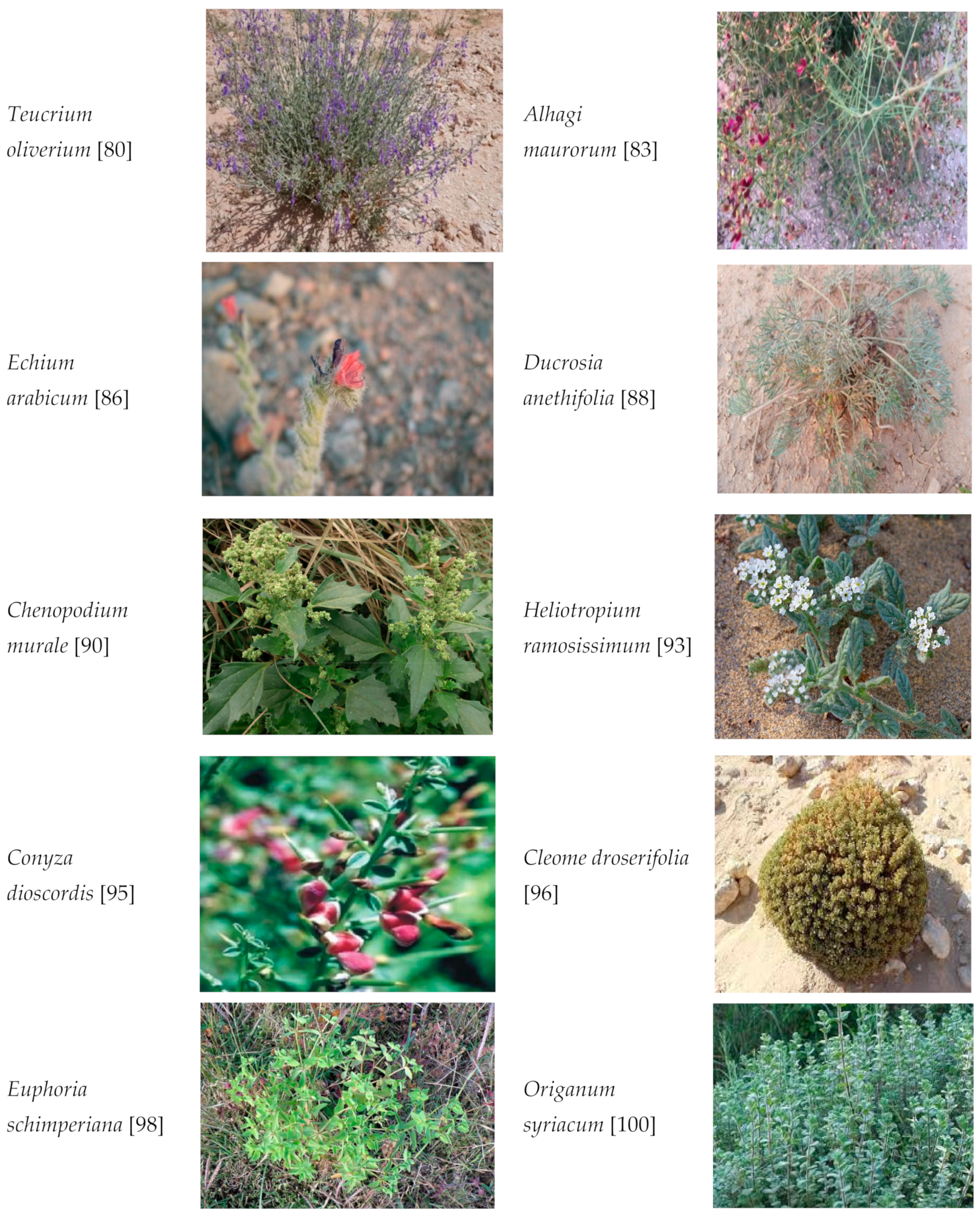
7.1. Teucrium oliverium

The genus Teucrium contains several species that grow widely in the desert regions. Teucrium oliverium (George Bentham, 1835) is a perennial plant which mostly grows in the rocky runnels bearing sand layers. The plant belongs to the family Lamiaceae and bears bright purple flowers [80]. Extracts of the plant are found to exhibit numerous pharmacological activities, such as anti-diabetic, anti-nociceptive, hypolipidemic, anti-cancer and antioxidant activities. Several important phytoconstituents have been isolated and identified, including teucrolivins (A, B, C, D, E, and F) (Figure 2).
These components, as well as the flavonoids, sterol, tannins, saponins, and coumarins, are speculated to be responsible for the medicinal properties, including antioxidant effects. The antioxidant activity was evaluated by the In-vitro 2,2-diphenyl-1-picryl hydrazyl (DPPH) free radical scavenging method [81].

7.2. Alhagi maurorum

Alhagi maurorum (Friedrich Medikus, 1787) is one of the common folk medicines used in different regions of Saudi Arabia. It is an under-shrub, globrous plant belonging to the family Fabaceae. The plant grows extensively in the deserts of the region and is used as camel feed [82]. The plant is traditionally used as an appetizer, aphrodisiac, tonic, anti-asthmatic, anti-rheumatic, diuretic, antipyretic, demulcent, expectorant, etc. Even among the local population, the plant is famous as a laxative and is used in the management of liver, gastrointestinal and urinary tract diseases. The literature suggests that extracts of the plant contain several active ingredients like flavonoids, phenolic acids, sitosterols, triterpenes, and proanthocyanidines [83]. Two flavonoids called isorhamnetin-3-O-[alpha-1-rhamnopyranosyl-beta-D-glucopyranoside and 3-O-methylorobol isolated from the extract were found to be possess antioxidant properties in the DPPH free radical method [84].

7.3. Echium arabicum

The genus Echium belongs to the family Boraginaceae. Several species of the family are used traditionally for medicinal purposes. Echium arabicum (Fitter, 1974) is a woody shrub, an annual biennial or perennial flowering plant. In several places, the plant is used as an ornamental plant and in gardens. Since the plant is hardy, it adapts well to the harsh climatic conditions of Saudi Arabia [85]. Predominantly, the plant is popular for its sedative, anti-inflammatory, antioxidant, and anxiolytic properties, and can be used for skin abrasions and insect/snake bites. As reported, the plant is known to contain several vital components responsible for its medicinal properties, and among them are naphthoquinones, flavonoids, terpenoids, and phenols [86]. The flavonoids isolated from E. Arabicum extract such as luteolin-7-O-glucosides, myricetin and quercetin have shown antioxidant activity in the DPPH assay [87].

7.4. Ducrosia anethifolia

Ducrosia (Francois Ducros, 1822) species belong to the Apiaceae family and are endemic to the Persian and Arab regions. The plant is a biennial with white flowers and can be found in the deserts of the Arar valley in Saudi Arabia [88]. Several phytochemical ingredients have been isolated and identified. Important among them are alkaloids, tannins, flavonoids, essential oils, glycosides and phenolic derivatives. The major constituents reported to possess antioxidant activity by the DPPH method is alpha-pinene, beta-myrcene, beta-pinene and limonene. Pharmacological studies suggest that the plant has therapeutic properties such as antioxidant, anti-cancer, anti-diabetic, anxiolytic, anti-inflammatory, antifungal, antiviral and may be active against neurological diseases [89].

7.5. Chenopodium murale

Chenopodium murale (Carl Linnaeus, 1778) is an annual herb that is conventionally used in the treatment of liver diseases. The plant is a member of the Chenopodiaceae and can be found in the Taif region of Saudi Arabia [90]. Phytochemical investigation of the extract has shown the presence of phenols, flavonoids, tannins, saponnins, terpenoids, caumarins, steroids, and anthraquinones. These components have been linked to the medicinal properties of the plant and the most important among them are anti-bacterial, digestive problems, sterility, anxiolytic, and anti-hypertensive. The antioxidant potential of the plant has been linked to the presence of tannins such as gallic acid and the tests were conducted by DPPH and 2,2′-azo-bis (3-ethyl benzothiazoline-6-sulphonic acid (ABTS) free radical scavenging methods [88,91].

7.6. Heliotropium ramosissimum

Heliotropium ramosissimum (Drubrovinskii, 1947) is a member of the Boraginaceae, commonly found in the moderate and tropical areas of Saudi Arabia. Heliotropium is derived from ‘heliotrope’, meaning the leaves of the plant turn in the direction of the sun [92]. Several pharmacological activities have been reported for the plant, such as antibacterial, anti-inflammatory, anti-nociceptive, anti-convulsive, anti-neoplastic, anti-diuretic, nephroprotective, anti-fertility, and analgesic. The important active constituents identified are alkaloids, phenolic compounds, terpenoids, quinines, and flavonoids. The antioxidant property has been linked to the presence of phenols, dimethoxy flavones, methyl galangin, chlologenic acid, and filifolinols. The methods used to determine the antioxidant property were lipid peroxidation, hydrogen peroxide and nitric oxide scavenging in vitro tests [93].

7.7. Conyza dioscordis

Conyza dioscordis (Christian Lessing. 1832) is a wild plant that grows in several places in the Middle Eastern regions, including Saudi Arabia. The plant belongs to the Asteraceae family and is frequently used as a folk medicine. The phyto-ingredients identified are phenols, flavonoids, glucosides, alkaloids, sterols, triterpenes, beta-sitosterol, alpha-amyrin, etc. [94]. Screening for therapeutic activity indicated that the plant’s extract showed promising effects in treating rheumatic pain, epilepsy, colic pain, ulcers, colds, diarrhoea, diabetes, and microbial infections. Antioxidant studies indicated that extracts of the plant reduced ROS-mediated changes and were attributed to the presence of components such as gallic acid, quercetin and kemperol. The in vitro DPPH free radical scavenging method was employed to determine the antioxidant property [94,95].

7.8. Cleome droserifolia

According to literature, the Cleome (Ali, 1977) genus is the largest member of the family Cleomaceae. The plant is widely distributed in the dry and semi-arid regions of Saudi Arabia and other Middle Eastern countries. The plant is a perennial, aromatic shrub, popular among several tribes of the region [96]. Traditionally, the plant is used for the treatment of stomach aches, skin allergies and open wounds. Several important phytochemicals have been isolated and characterized. The plant is reported to contain high concentrations of flavonoids, terpenes, glucosinolates, anthocyanin alkaloids and polyphenols. Pharmacological screening indicated that extracts of the herb possess anti-diabetic, antioxidant, hepatoprotective and anti-cancer properties. These activities are linked to the phyto-components present in the plant. In vitro DPPH and ABTS tests were carried out to determine the antioxidant property. The important constituents identified were rutin, naringenin, kaempferol and p-coumaric acid [97].

7.9. Euphorbia schimperiana

The family Euphorbiaceae contains several plants that range from woody trees to simple weeds. Different species of the Euphorbia plant (Scheele, 1844) have been known since ancient times for their medicinal activities. The plant grows well in moderate temperate areas and can be found in the western provinces of Saudi Arabia (98). The plant and its extract are reported to exhibit a range of pharmacological actions, such as analgesic, anti-ulcer, antioxidant, anti-inflammatory, antiviral, anti-spasmodic, skin allergy and anti-cancer actions. The important active ingredients that were identified for the above activities are triterpenes, flavonoids, alkaloids, tannins, and sterols. The presence of specific diterpenes such as phorbids, ingenane, tigliane, and daphnane are reported for their potential pharmacological activities [98]. In addition, the phenolic components such as quercetin-7-O-β-D-glucoronside and 3-methyl-quercetin-7-O-β-D-glucoronside were identified for their antioxidant activity by the DPPH free radical assay [99].

7.10. Origanum syriacum

These are aromatic plants (Nathan Ben Abraham, 1977) belonging to the family Lamiaceae. The plant is widely grow in the Mediterranean region and can be found in several places in Saudi Arabia. The medical uses of the plant are popular among different tribes of the country and are used for treating pain, infection, and diseases of the skin [100]. Pharmacological screening suggests that the plant possesses blood cholesterol lowering, hypoglycaemic, antibacterial, anti-carcinogenesis, anti-inflammatory, antioxidant, antiviral and anti-cardiovascular disease properties. These activities have been strongly related to the essential secondary metabolites of the plant. Some of the important among them are polyphenols, flavonoids, volatile oils, sterols, and terpenes. The antioxidant activity is linked to catechin, gallic acid, quercetin, and hesperidin and the assay was undertaken by the DPPH method [101].

8. Conclusions

The present review summarized the mutation and related complications of host cells. DNA damage due to oxidative stress is the major cause of mutation-induced diseases such as cancer. Antioxidants have an important role in minimizing mutagenic defects. Medicinal plants native to Saudi Arabia such as Teucrium oliverium, Alhagi maurorum, Echium arabicum, Ducrosia anethifolia, Chenopodium murale, Heliotropium ramosissimum, Conyza dioscordis, Cleome droserifolia, Euphoria schimperiana and Origanum syriacum have shown significant antioxidant properties. These plants have the potential to exhibit antimutagenic effects since they contain the vital phytoconstituents essential for activity. Future research on these plants might explore their true potential and the possibility of using them as prophylactic agents in reducing the complications of exogenous mutagens such as viruses.

Author Contributions

The manuscript writing, editing, and original draft preparation were done by S.I.R., and S.M.B.A. The data search and literature were extracted by A.A.A., F.H.A., and A.J.A. The conception and design were done by A.A.A.-S., and G.Y.A. The review and final editing of the manuscript was carried out by S.M.B.A. and M.I. The supervision of the manuscript writing was done by S.I.R. All authors have read and agreed to the published version of the manuscript.

Funding

This research received no external funding.

Institutional Review Board Statement

Not applicable.

Informed Consent Statement

Not applicable.

Acknowledgments

The authors are thankful to AlMaarefa University, Riyadh for providing support to do this review article.

Conflicts of Interest

The authors declare no conflict of interest.

References

  1. Drakvik, E.; Altenburger, R.; Aoki, Y.; Backhaus, T.; Bahadori, T. Statement on advancing the assessment of chemical mixtures and their risks for human health and the environment. Environ. Int. 2020, 134, 105267. [Google Scholar] [CrossRef]
  2. Munnia, A.; Giese, R.W.; Polvani, S.; Galli, A.; Cellai, F.; Peluso, M.E. Bulky DNA Adducts, Tobacco Smoking, Genetic Susceptibility, and Lung Cancer Risk. Adv. Clin. Chem. 2017, 81, 231–277. [Google Scholar]
  3. Matthew, D.W.; Fradet-Turcotte, A. Virus DNA Replication and the Host DNA Damage Response. Ann. Rev. Virol. 2018, 5, 141–164. [Google Scholar]
  4. Cellai, F.; Capacci, F.; Sgarrella, C.; Poli, C.; Arena, L.; Tofani, L.; Giese, R.W.; Peluso, M.A. Cross-Sectional Study on 3-(2-Deoxy-d-Erythro-Pentafuranosyl) Pyrimido[1,2-Purin-10(3H)-One Deoxyguanosine Adducts among Woodworkers in Tuscany, Italy. Int. J. Mol. Sci. 2019, 20, 2763. [Google Scholar] [CrossRef] [Green Version]
  5. Anderson, G.P.; Bozinovski, S. Acquired somatic mutations in the molecular pathogenesis of COPD. Trends Pharmacol. Sci. 2003, 24, 71–76. [Google Scholar] [CrossRef]
  6. Baer, C.F.; Miyamoto, M.M.; Denver, D.R. Mutation rate variation in multicellular eukaryotes: Causes and consequences. Natl. Rev. Genet. 2007, 8, 619–631. [Google Scholar] [CrossRef]
  7. Bae, T.; Tomasini, L.; Mariani, J.; Zhou, B.; Roychowdhury, T.; Franjic, D. Different mutational rates and mechanisms in human cells at pregastrulation and neurogenesis. Science 2018, 359, 550–555. [Google Scholar] [CrossRef] [Green Version]
  8. Arvanitis, D.A.; Flouris, G.A.; Spandidos, D.A. Genomic rearrangements on VCAM1, SELE, APEG1 and AIF1 loci in atherosclerosis. J. Cell. Mol. Med. 2005, 9, 153–159. [Google Scholar] [CrossRef]
  9. Andreassi, M.G. Coronary atherosclerosis and somatic mutations: An overview of the contributive factors for oxidative DNA damage. Mutat. Res. 2003, 543, 67–86. [Google Scholar] [CrossRef]
  10. Gershenson, S.M. Viruses as environmental mutagenic factors. Mutat. Res. 1986, 167, 203–213. [Google Scholar] [CrossRef]
  11. Gershenzon, S.M. Do viruses participate in the origin of group outbursts of gene mutations occurring in natural Drosophila populations? Tsitol. Genet. 1998, 32, 94–102. [Google Scholar]
  12. Shruthi, S.; Vijayalaxmi, K.K. Antigenotoxic effects of a polyherbal drug septilin against the genotoxicity of cyclophosphamide in mice. Toxicol. Rep. 2016, 3, 563–571. [Google Scholar] [CrossRef] [Green Version]
  13. Adepu, A.; Narala, S.; Ganji, A.; Chilvalvar, S. A review on natural plant: Aerva lanata. Int. J. Pharm. Sci. 2013, 3, 398–402. [Google Scholar]
  14. Formation of Free Radicals from Different Sources. 2020. Available online: https://maeterna.com/the-truth-what-are-antioxidants-free-radicals/ (accessed on 18 April 2021).
  15. Marnett, L.J. Oxy radicals, lipid peroxidation and DNA damage. Toxicology 2002, 181, 219–222. [Google Scholar] [CrossRef]
  16. Adam-Vizi, V. Production of reactive oxygen species in brain mitochondria: Contribution by electron transport chain and non-electron transport chain sources. Antioxid. Redox Signal. 2005, 7, 1140–1149. [Google Scholar] [CrossRef] [PubMed]
  17. Avery, S.V. Molecular targets of oxidative stress. Biochem. J. 2011, 434, 201–210. [Google Scholar]
  18. Hamanishi, T.; Furuta, H.; Kato, H.; Doi, A.; Tamai, M.; Shimomura, H.; Sakagashira, S.; Nishi, M.; Sasaki, H.; Sanke, T.; et al. Functional variants in the glutathione peroxidase-1 (GPx-1) gene are associated with increased intima-media thickness of carotid arteries and risk of macrovascular diseases in Japanese type 2 diabetic patients. Diabetes 2004, 53, 2455–2460. [Google Scholar] [CrossRef] [Green Version]
  19. Liu, D.; Wen, J.; Liu, J.; Li, L. The roles of free radicals in amyotrophic lateral sclerosis: Reactive oxygen species and elevated oxidation of protein, DNA, and membrane phospholipids. FASEB J. 1999, 13, 2318–2328. [Google Scholar] [CrossRef]
  20. Kong, G.K.; Miles, L.A.; Crespi, G.A.N.; Morton, C.J.; Ng, H.L. Copper binding to the Alzheimer’s disease amyloid precursor protein. Eur. Biophys. J. 2008, 37, 269–279. [Google Scholar] [CrossRef] [Green Version]
  21. Roth, E.F., Jr.; Ruprecht, R.M.; Schulman, S.; Vandersberg, J.; Olson, J.A. Ribose metabolism and nucleic acid synthesis in normal and glucose-6-phosphate deficient human erythrocytes infected with Plasmodium falciparum. J. Clin. Investig. 1986, 77, 1129–1235. [Google Scholar] [CrossRef]
  22. Goldstein, I.M.; Kaplan, H.B.; Edelson, H.S.; Weissmann, G. Ceruloplasmin: An acute phase reactant that scavenges oxygen-derived free radicals. Ann. New York Acad. Sci. 1982, 389, 368–379. [Google Scholar] [CrossRef] [PubMed]
  23. Gongora, M.C.; Harrison, D.G. Sad heart from no SOD. Hypertension 2008, 51, 28–30. [Google Scholar] [CrossRef] [Green Version]
  24. Souliotis, V.L.; Vlachogiannis, N.I.; Pappa, M.; Argyriou, A.; Ntouros, P.A.; Sfikakis, P.P. DNA Damage Response and Oxidative Stress in Systemic Autoimmunity. Int. J. Mol. Sci. 2020, 21, 55. [Google Scholar] [CrossRef] [PubMed] [Green Version]
  25. Lightfoot, T.J.; Skibola, C.F.; Smith, A.G.; Forrest, M.S. Polymorphisms in the oxidative stress genes superoxide dismutase, glutathione peroxidase and catalase and risk of non-Hodgkin’s lymphoma. Haematologica 2006, 91, 1222–1227. [Google Scholar] [PubMed]
  26. Lubos, E.; Handy, D.E.; Loscalzo, J. Role of oxidative stress and nitric oxide in atherothrombosis. Front. Biosci. 2008, 13, 5323–5344. [Google Scholar] [CrossRef] [Green Version]
  27. Nakabeppu, Y.; Kajitani, K.; Sakamoto, K.; Yamaguchi, H.; Tsuchimoto, D. MTH1, an oxidized purine nucleoside triphosphatase, prevents the cytotoxicity and neurotoxicity of oxidized purine nucleotides. DNA Repair 2006, 5, 761–772. [Google Scholar] [CrossRef]
  28. Monteiro, H.P.; Winterbourn, C.C. Release of iron from ferritin by divicine, isouramil, acid-hydrolyzed vicine, and dialuric acid and initiation of lipid peroxidation. Arch. Biochem. Biophys. 1989, 271, 536–545. [Google Scholar] [CrossRef]
  29. Thorpe, G.W.; Fong, C.S.; Alic, N.; Higgins, V.J.; Dawes, I.W. Cells have distinct mechanisms to maintain protection against different reactive oxygen species: Oxidative-stress-response genes. Proc. Natl. Acad. Sci. USA 2004, 101, 6564–6569. [Google Scholar] [CrossRef] [Green Version]
  30. Lubos, E.; Loscalzo, J.; Handy, D.E. Homocysteine and glutathione peroxidase-1. Antioxid. Redox Signal. 2007, 9, 1923–1940. [Google Scholar] [CrossRef] [PubMed]
  31. Saez, G.T.; Estan-Capell, N. Antioxidant enzymes. In Encyclopaedia of Cancer; Schwab, M., Ed.; Springer: Berlin, Germany, 2014. [Google Scholar]
  32. Devasangayam, T.P.A.; Tilak, J.C.; Boloor, K.K.; Sane, K.S.; Ghaskadbi, S.S.; Lele, R.D. Free radicals and antioxidant in human health: Current status and future prospects. J. Assoc. Physicians India 2004, 52, 794–804. [Google Scholar]
  33. Orrell, R.W. Amyotrophic lateral sclerosis: Copper/zinc superoxide dismutase (SOD1) gene mutations. Neuromuscul. Disord. 2000, 10, 63–68. [Google Scholar] [CrossRef]
  34. Furukawa, Y.; Torres, A.S.; O’Halloran, T.V. Oxygen-induced maturation of SOD1: A key role for disulfide formation by the copper chaperone CCS. Eur. Mol. Biol. Organ. J. 2004, 23, 2872–2881. [Google Scholar] [CrossRef] [PubMed]
  35. Crystal, R.G. Oxidants and antioxidants. Am. J. Med. 1991, 91, 3S–10S. [Google Scholar]
  36. Tomas-Barberan, F.A.; Robins, R.J. Phytochemistry of Fruits and Vegetables; Calendon Press: New York, NY, USA, 1997. [Google Scholar]
  37. Ahuja, R.; Jamal, A.; Nosrati, N. Human oncogenic viruses and cancer. Curr. Sci. 2014, 107, 768–785. [Google Scholar]
  38. Herzer, K.; Gerken, G.; Hofmann, T.G. Hepatitis Cassociated liver carcinogenesis: Role of PML nuclear bodies. World J. Gastroenterol. 2014, 20, 12367. [Google Scholar] [CrossRef] [PubMed]
  39. Hoshida, Y.; Fuchs, B.C.; Bardeesy, N. Pathogenesis and prevention of hepatitis C virus-induced hepatocellular carcinoma. J. Hepatol. 2014, 61, S79–S90. [Google Scholar] [CrossRef] [Green Version]
  40. Lim, H.; Park, Y.S.; Lee, J.H. Features of gastric carcinoma with lymphoid stroma associated with Epstein-Barr virus. Clin. Gastroenterol. Hepatol. 2015, 13, 1738–1744. [Google Scholar] [CrossRef] [Green Version]
  41. Schiller, J.T.; Lowy, D.R. Virus infection and human cancer: An overview. Recent Results Cancer Res. 2014, 193, 1–10. [Google Scholar] [PubMed]
  42. Sxevik, M. Oncogenic viruses and mechanisms of oncogenesis. Turk. J. Vet. Anim. Sci. 2012, 36, 323–329. [Google Scholar]
  43. Macarthur, M.; Hold, G.L.; El-Omar, E.M. Inflammation and Cancer II. Role of chronic inflammation and cytokine gene polymorphisms in the pathogenesis of gastrointestinal malignancy. Am. J. Physiol. Gastrointest. Liver Physiol. 2004, 286, G515–G520. [Google Scholar] [CrossRef]
  44. Tian, Y.; Yang, W.; Song, J. Hepatitis B virus X protein induced aberrant epigenetic modifications contributing to human hepatocellular carcinoma pathogenesis. Mol. Cell Biol. 2013, 33, 2810–2816. [Google Scholar] [CrossRef] [Green Version]
  45. Moriel, P.; Plavnik, F.L.; Zazella, M.T.; Bertolami, M.C.; Abdalla, D.S.P. Lipid peroxidation and antioxidants in hyperlipidemia and hypertension. Biol. Res. 2000, 33, 105–111. [Google Scholar] [CrossRef] [PubMed]
  46. Słoczynska, K.; Powroznik, B.; Pękala, E. Antimutagenic compounds and their possible mechanisms of action. J. Appl. Genet. 2014, 55, 273–285. [Google Scholar] [CrossRef] [PubMed] [Green Version]
  47. Abdel-Wahhab, M.A.; Ahmed, H.H. Protective effect of Korean Panax ginseng against chromium VI toxicity and free radicals generation in rats. J. Ginseng Res. 2004, 28, 11–17. [Google Scholar]
  48. Li, J.; Yu, H.; Wang, S. Natural products, an important resource for discovery of multitarget drugs and functional food for regulation of hepatic glucose metabolism. Drug Des. Dev. Ther. 2018, 12, 121–135. [Google Scholar] [CrossRef] [PubMed] [Green Version]
  49. Edenharder, R.; Worf-Wandelburg, A.; Decker, M.; Platt, K.L. Antimutagenic effects and possible mechanisms of action of vitamins and related compounds against genotoxic heterocyclic amines from cooked food. Mutat. Res. 1999, 444, 235–248. [Google Scholar] [CrossRef]
  50. de Oliveira, M.R. The neurotoxic effects of vitamin A and retinoids. Ann. Braz. Acad. Sci. 2015, 87, 1361–1373. [Google Scholar] [CrossRef] [Green Version]
  51. Naziroglu, M.; Cay, M. Protective role of intraperitoneally administered vitamin E and selenium on the oxidative defense mechanisms in rats with diabetes induced by streptozotocin. Biol. Trace Elem. Res. 2011, 79, 149–159. [Google Scholar] [CrossRef]
  52. Rabbani, S.I.; Devi, K.; Khanam, S. Role of Pioglitazone with Metformin or Glimepiride on Oxidative Stress-induced Nuclear Damage and Reproductive Toxicity in Diabetic Rats. Malays. J. Med. Sci. 2010, 17, 3–11. [Google Scholar]
  53. Alpsoy, L.; Yildirim, A.; Agar, G. The antioxidant effects of vitamin A, C, and E on aflatoxin B1-induced oxidative stress in human lymphocytes. Toxicol. Ind. Health 2009, 25, 121–127. [Google Scholar] [CrossRef] [PubMed]
  54. Sarma, L.; Kesavan, P.C. Protective effect of vitamin C and E against gamma-ray induced chromosomal damage in mouse. Int. J. Radiat. Biol. 1993, 759, 339–345. [Google Scholar]
  55. Konapacka, M.; Widel, M.; Rzeszowska-Wolny, J. Modifying effect of vitamins C, E and beta-carotene against gamma-ray-induced DNA damage in mouse cells. Mutat. Res. 1998, 417, 85–91. [Google Scholar] [CrossRef]
  56. Brewer, M.S. Natural antioxidants: Sources, compounds, mechanisms of action and potential applications. Complim. Rev. Food Saf. 2011, 10, 221–246. [Google Scholar] [CrossRef]
  57. Nayeem, N.; Asdaq, S.M.; Alamri, A.S.; Alsanie, W.F.; Alhomrani, M.; Mohzari, Y.; Alrashed, A.A.; Alotaibi, N.; Alharbi, M.A.; Aldhawyan, N.N.; et al. Wound healing potential of Dodonaea viscosa extract formulation in experimental animals. J. King Saud Univ. Sci. 2021, 33, 101476. [Google Scholar] [CrossRef]
  58. Esposito, E.; Ratilio, D.; Di Matteo, V.; Di Glulia, C.; Cicchino, M.; Algeri, S. A review on specific dietary antioxidant and the effects on biochemical mechanism related to neurodegenerative processes. Neurobiol. Aging 2002, 23, 719–735. [Google Scholar] [CrossRef]
  59. Chulasiri, M.; Bunyapraphatsara, N.; Moonkarndi, P. Mutagenicity and antimutagenicity of hispidulin and hortensin, the flavonoids from Millingtonia hortensis L. Environ. Mol. Mutagenesis 1992, 20, 307–312. [Google Scholar] [CrossRef]
  60. Sharma, R.; Singh, B.; Singh, D.; Chandrawat, P. Ethnomedicinal, pharmacological properties and chemistry of some medicinal plants of Boraginaceae in India. J. Med. Plant Res. 2009, 3, 1153–1175. [Google Scholar]
  61. Arshadullah, M.; Rasheed, M.; Mahmood, I.A. Niazboo (Ocimum basilicum) as medicinal plant establishes against salinity and sodicity. Adv. Biotechnol. Microbiol. 2019, 15, 1–8. [Google Scholar]
  62. Geetha, T.; Garg, A.; Chopra, K.; Kaur, I.P. Delineation of antimutagenic activity of catechin, epicatechin and green tea extract. Mutat. Res. 2004, 556, 65–74. [Google Scholar] [CrossRef] [PubMed]
  63. Soudamini, K.K.; Unnikrishnan, M.C.; Sukumaran, K.; Kuttan, R. Mutagenicity and antimutagenicity of some selected spices. Indian J. Physiol. Pharmacol. 1995, 39, 347–353. [Google Scholar]
  64. Kuo, M.; Lee, K.; Lin, J. Genotoxicities of nitropyrenes and their modulation by apigenin, tannic acid, ellagic acid and indole-3-carbinol in the Salmonella and CHO systems. Mutat. Res. 1992, 270, 87–95. [Google Scholar] [CrossRef]
  65. Ali, A.; Akhtar, N.; Khan, B.A.; Khan, M.S. Acacia nilotica: A plant of multipurpose medicinal uses. J. Med. Plant Res. 2012, 6, 1492–1496. [Google Scholar]
  66. Khalaf, I.A. Antimutagenic effect of carrot (Daucus carota) on induction of streptomycin and rifampicin resistant mutants in bacterial systems. Mesop. J. Agric. 2008, 36, 94–104. [Google Scholar] [CrossRef] [Green Version]
  67. Wang, S.; Zhangxin, Y.U.; Wang, C.; Chongming, W.U.; Go, P.; Wei, J. Chemical Constituents and Pharmacological Activity of Agarwood and Aquilaria Plants. Molecules 2018, 23, 342. [Google Scholar] [CrossRef] [Green Version]
  68. Guo, J.; Weng, X.; Wu, H.; Li, Q.; Bi, K. Antioxidants from a Chinese medicinal herb—Psoralea corylifolia L. Food Chem. 2005, 91, 287–292. [Google Scholar]
  69. Wall, M.E.; Wani, M.C.; Manukumar, G.; Hughes, T.J. Plant antimutagenic agents, 3. Coumarins. J. Nat. Prod. 1988, 51, 1148–1152. [Google Scholar] [CrossRef]
  70. Shankel, D.M.; Pillai, S.P.; Telidepalli, H.; Menon, S.R.; Pillai, C.A.; Mitsche, L.A. Role of antimutagenic/anticarcinogens in cancer prevention. BioFactors 2000, 12, 113–121. [Google Scholar] [CrossRef]
  71. Geetha, T.; Saini, A.; Kaur, I.P. Ginseng extract exhibits antimutagenic activity against induced mutagenesis in various strains of Salmonella typhimurium. Indian J. Exp. Biol. 2006, 44, 838–841. [Google Scholar] [PubMed]
  72. Monache, F.D.; MacQuhae, M.M.; Monache, G.D.; Bettolo, G.B.M.; De Lima, R.A. Xanthones, xanthonolignoids and other constituents of the roots of Vismia guaramirangae. Phytochemistry 1983, 22, 227–232. [Google Scholar] [CrossRef]
  73. Rabbani, S.I.; Devi, K.; Khanam, S.; Zahra, N. Citral, a component of lemongrass oil inhibits the clastogenic effect of nickel chloride in mouse micronucleus test system. Pak. J. Pharm. Sci. 2006, 19, 108–113. [Google Scholar]
  74. Mossa, S.; AI-Yahya, M.A.; AI-Meshal, I.A. Medicinal Plants of Saudi Arabia; King Fahad National Library Publication: Riyadh, Saudi Arabia, 2000; Volume 2, pp. 1–355. [Google Scholar]
  75. Alfarhan, A.H.; Chaudhary, S.A.; Thomas, J. Notes on the flora of Saudi Arabia. J. King Saud Univ. 1998, 10, 31–40. [Google Scholar]
  76. Abdallah, E.M.; El-Ghazali, G. Screening for antimicrobial activity of some plants from Saudi folk medicine. Glob. J. Res. Med. Plants Indig. Med. 2013, 2, 210–218. [Google Scholar]
  77. Al-Sodany, Y.M.; Bazaid, S.A.; Mosallam, H.A. Medicinal plants in Saudi Arabia: I. Sarrwat Mountains at Taif, KSA. Acad. J. Plant Sci. 2013, 6, 134–145. [Google Scholar]
  78. El-Ghazali, G.E.; Al-Khalifa, K.S.; Saleem, G.A.; Abdallah, E.M. Traditional medicinal plants indigenous to Al-Rass province, Saudi Arabia. J. Med. Plants Res. 2010, 4, 2680–2683. [Google Scholar]
  79. Al-asmari, A.; Manthiri, R.A.; Abdo, N.; AL-duaiji, F.A.; Khan, H.A. Saudi medicinal plants for the treatment of scorpion sting envenomation. Saudi J. Biol. Sci. 2017, 24, 1204–1211. [Google Scholar] [CrossRef] [Green Version]
  80. Abdel-Sattar, E.; Harraz, F.M.; Al-Ansari, S.M.A.; El-Mekkawy, S.; Ichino, C.; Kiyohara, H. Antiplasmodial and antirypanosomal activity of plants from the Kingdom of Saudi Arabia. J. Nat. Med. 2009, 63, 232–239. [Google Scholar] [CrossRef]
  81. Alkofahi, A.; Atta, A.H. Pharmacological screening of the anti-ulcerogenic effects of some Jordanian medicinal plants in rats. J. Ethnopharmacol. 1999, 67, 341–345. [Google Scholar] [CrossRef]
  82. Al-Saleem, M.S.; Al-Wahaibi, L.H.; Abdel-Mageed, W.M.; Gouda, Y.G.; Sayed, H.M. Antioxidant flavonoids from Alhagi maurorum with hepatoprotective effect. Pharmacogn. Mag. 2019, 15, 592–599. [Google Scholar]
  83. Muhammad, G.; Hussain, M.A.; Anwar, F.; Ashraf, M.; Gilani, A.H. Alhagi: A plant genus rich in bioactives for pharmaceuticals. Phytother. Res. 2015, 29, 1–13. [Google Scholar] [CrossRef]
  84. Ahmad, S.; Riaz, M.; Saleem, M. Antioxidant flavonoids from Alhagi maurorum. J. Asian Nat. Prod. 2010, 12, 138–143. [Google Scholar] [CrossRef]
  85. Abbaszadeh, S.; Radjabian, T.; Taghizadeh, M. Antioxidant Activity, Phenolic and Flavonoid Contents of Echium Species from Different Geographical Locations of Iranian. J. Medincal Plants Prod. 2013, 2, 23–31. [Google Scholar]
  86. Kuete, V.; Wiench, B.; Alsaid, M.S.; Alyahya, M.A.; Fankam, A.G.; Shahat, A.A. Cytotoxicity, mode of action and antibacterial activities of selected Saudi Arabian medicinal plants. BMC Complement. Altern. Med. 2013, 13, 354–359. [Google Scholar] [CrossRef] [Green Version]
  87. Jin, J.; Boersch, M.; Nagarajan, A.; Davey, A.K. Antioxidant and reported ethnomedicinal use of the genus Echium. Antioxidants 2020, 9, 722. [Google Scholar] [CrossRef] [PubMed]
  88. Elsharkawy, E.R.; Abdallah, E.M.; Shiboob, M.H.; Alghanem, S. Phytochemical, antioxidant and antibacterial potential of Ducrosia anethifolia in northern border region of Saudi Arabia. J. Pharm. Res. Int. 2019, 31, 1–6. [Google Scholar] [CrossRef]
  89. Mottaghipisheh, J.; Nové, M.; Spengler, G.; Kúsz, N.; Hohmann, J.; Csupor, D. Antiproliferative and cytotoxic activities of furocoumarins of Ducrosia anethifolia. Pharm. Biol. 2018, 56, 658–664. [Google Scholar] [CrossRef] [PubMed] [Green Version]
  90. Majrashi, A.; Khandaker, M.M. Survey of chenopodiaceae family from Taif, Saudi Arabia. Plant Arch. 2020, 2, 5958–5964. [Google Scholar]
  91. Khan, N.; Ahmed, M.; Khan, R.A.; Gul, S. Antioxidant, cytotoxicity activity and phytochemical analysis of Chenopodium murale L. Int. J. Bot. Stud. 2019, 4, 25–28. [Google Scholar]
  92. Alwahibi, M. Anatomical study of four species of Heliotropium, L. (Boraginaceae) from Saudi Arabia. Afr. J. Plant Sci. 2013, 7, 35–42. [Google Scholar] [CrossRef] [Green Version]
  93. Sharma, V.; Agrawal, R.C.; Shrivastava, V.K. Assessment of median lethal dose and anti-mutagenic effects of Glycyrrhiza glabra root extract against chemically induced micronucleus formation in Swiss albino mice. Int. J. Basic Clin. Pharmacol. 2014, 3, 292–298. [Google Scholar] [CrossRef] [Green Version]
  94. Alqasoumi, S.I. Phytochemical study of the aerial parts of Conyza discoridis growing in Saudi Arabia. Nat. Prod. Sci. 2009, 15, 66–70. [Google Scholar]
  95. El-Hamoul, M.M.A.; Ibrahim, M.T. GC/MS analysis of the volatile constituents of individual organs of Conyza dioscoridis (L.) Desf. growing in Egypt. Alex. J. Pharm. Sci. 2003, 17, 75–80. [Google Scholar]
  96. Shtalwi, M.H.; Rawi, S.M.; Alshibly, N.M.; Al-Hazimi, M. Chemical screening and antihyperglycemic property of Cleome droserifolia ethanol extract. J. Med. Plant Res. 2013, 7, 2769–2776. [Google Scholar]
  97. Hashem, N.M.; Shehata, M.G. Antioxidant and antimicrobial activity of Cleome droserifolia (Forssk.) Del. and its biological effects on redox status, immunity and gut microflora. Animals 2021, 11, 1929. [Google Scholar] [CrossRef] [PubMed]
  98. Shaker, K.H.; Al Shehri, B.M.; Oteef, M.D.Y.; Mahmoud, M.F. Antioxidant compounds from Euphorbia schimperiana Scheele in Areer region, Saudi Arabia. Int. J. Pharm. Sci. Rev. Res. 2015, 32, 117–122. [Google Scholar]
  99. Shi, Q.W.; Su, X.H.; Kiyota, H. Chemical and Pharmacological Research in the plants in genus Euphorbia. Chem. Rev. 2008, 108, 4295. [Google Scholar] [CrossRef] [PubMed]
  100. Alraey, D.A.; Haroun, S.A.; Omar, M.N.; Abd-ElGawad, A.M.; El-Shobaky, A.M.; Mowafy, A.M. Fluctuation of essential oil constituents in Origanum syriacum subsp. Sinaicum in response to plant growth promoting bacteria. J. Essent. Bear. Plants 2019, 22, 18–23. [Google Scholar] [CrossRef]
  101. Afify, A.M.R.; Esawy, S.H.; El-Hadidy, E.M.; Abdel-Salam, M.A.L. Antioxidant and cytotoxicity of Origanum syriacum L. Adv. Food Sci. 2014, 36, 58–64. [Google Scholar]
Figure 1. Diagrammatic representation of potential antimutagenic plants of Saudi Arabia.
Applsci 11 08494 g001
Figure 2. Structure of teucrolivin B isolated from T. oliverium [81].
Applsci 11 08494 g002
Table 1. Some important plants having antioxidant and anti-mutagenic properties.
PhytoconstituentsMode of ActionSourceActive IngredientsAntimutagenic TestReferences
Flavonoids Neutralizes and scavenges all types of oxidizing radicals.Millingtonia hortensisHispidulin, hortensinS. typhimurium strain (In-vitro)[57]
Glycyrrhiza glabraQuercetin, myricetinIn vivo mouse micronucleus[58]
Ocimum basilicumHerperidineS. typhimurium strain (In-vitro)[59]
Phenolic compoundsScavenges all types of free radicals.Camellia sinensisEpicatechin, epigallocatechinIn vivo mouse lymphoma cell[60]
Curcuma longaCurcuminIn vivo rat micronucleus[61]
Syzygium aromaticumEugenol In vivo mouse micronucleus
TanninsScavenges reactive oxygen species and inhibits lipid peroxidation.Acacia niloticaTannic acid, ellagic acid, gallic acidIn vivo mouse micronucleus,
In-vitro S. typhi strain
[62,63]
Carotenoids Protects cell from free radical attack and improves the antioxidant status.Daucus carotaBeta-carotene, canthaxanthineIn vitro callus culture cell lines[64]
Diterpenoids Counteracts against oxidative stress-induced lipid peroxidation.Aquillaria agallochaErythroxydiol In vivo rodent spleen DNA damage[65]
Coumarins Reduces the effects of oxidative stress.Psoralea corylifoliaPsoralen In vivo mouse micronucleus[66]
Selinum monniereImperatorin S. typhimurium strain (In-vitro)[67]
Anthraquinones Improves the status of antioxidants.Aloe barborescenceAloe-emodin-anthraquinone,
anthrone
D. melanogaste phenotype test[68]
Saponins Scavenges free radicals and improves the functioning of antioxidant enzymes.Panax ginsengGinseng saponinS. typhimurium strain (In-vitro)[69]
Xanthones Acts against oxygen-derived free radicals.Visma amazonicaEuxanthone S. typhimurium strain (In-vitro)[70]
Monoterpenoids Protects against NADPH induced oxidative injury.Cymbopogan citratesCitral In vivo mouse micronucleus[71]
Publisher’s Note: MDPI stays neutral with regard to jurisdictional claims in published maps and institutional affiliations.

