Review

Anomalous Origin of the Right Vertebral Artery

Department of Radiology, Faculty of Medicine, Oita University, 1-1 Idaigaoka, Hasama, Yufu, Oita 879-5593, Japan
*
Author to whom correspondence should be addressed.
Appl. Sci. 2021, 11(17), 8171; https://doi.org/10.3390/app11178171
Submission received: 11 July 2021 / Revised: 28 August 2021 / Accepted: 31 August 2021 / Published: 3 September 2021
(This article belongs to the Special Issue Applications of Image Processing in Anatomy)

Abstract

:
An anomalous origin of the right vertebral artery (VA) is a rare anomaly that is much rarer than that of the left VA. It can be divided into a few patterns, including aortic origin, right carotid or brachiocephalic arterial origin, and duplicated origin. In embryological development, the VA is made up of a longitudinal anastomosis between cervical segments. The mechanism of the anomalous origin of the right VA can be explained by the persistence of the cervical segmental artery and the regression point of the 4th right aortic arch. Although the anomaly is usually found incidentally on imaging modalities, it can be a potential cause of complication during surgical and interventional procedures. However, there are a lot of reports about the radiomics of the anomaly. Therefore, we discuss the potential relationship between the anomalous origin of the right VA and radiomics. As the take-home message, understanding several patterns of anomalous origin of the right VA with their embryology and imaging findings is important for surgical and endovascular interventions to avoid intraprocedural complications.

1. Introduction

Some authors have reported variations in the anomalous origin of the vertebral artery (VA) and their embryological mechanisms, which account for 4.6% of VAs [1,2,3]. The anomalous origin of the right vertebral artery (RVA) occurs much less frequently (0.7%) than that of the left VA (4%), so it has not yet been well recognized [1]. Although the majority of patients with this anomaly are asymptomatic, it can be a potential cause of complications during surgical or interventional procedures in the upper thoracic and cervical regions and during endovascular procedures for cerebrovascular lesions. Therefore, the knowledge of the normal development and vascular anomaly of the VA is very important for vascular surgery and endovascular treatment to avoid this complication. Additionally, understanding the development of the VA is very useful for recognizing the vascular anomaly. Computed tomography (CT) and magnetic resonance imaging (MRI) are non-invasive imaging modalities that are widely used in daily practice. According to recent technological advancements in imaging modalities, more precise images for thoracic and cervical vessels can be obtained, and therefore, the opportunity to encounter rare vascular anomalies has increased. In this pictorial review, we describe the embryological development and the normal anatomy of the RVA, as well as several types of anomalous origins of the RVA with CT and MRI images from an embryological perspective.

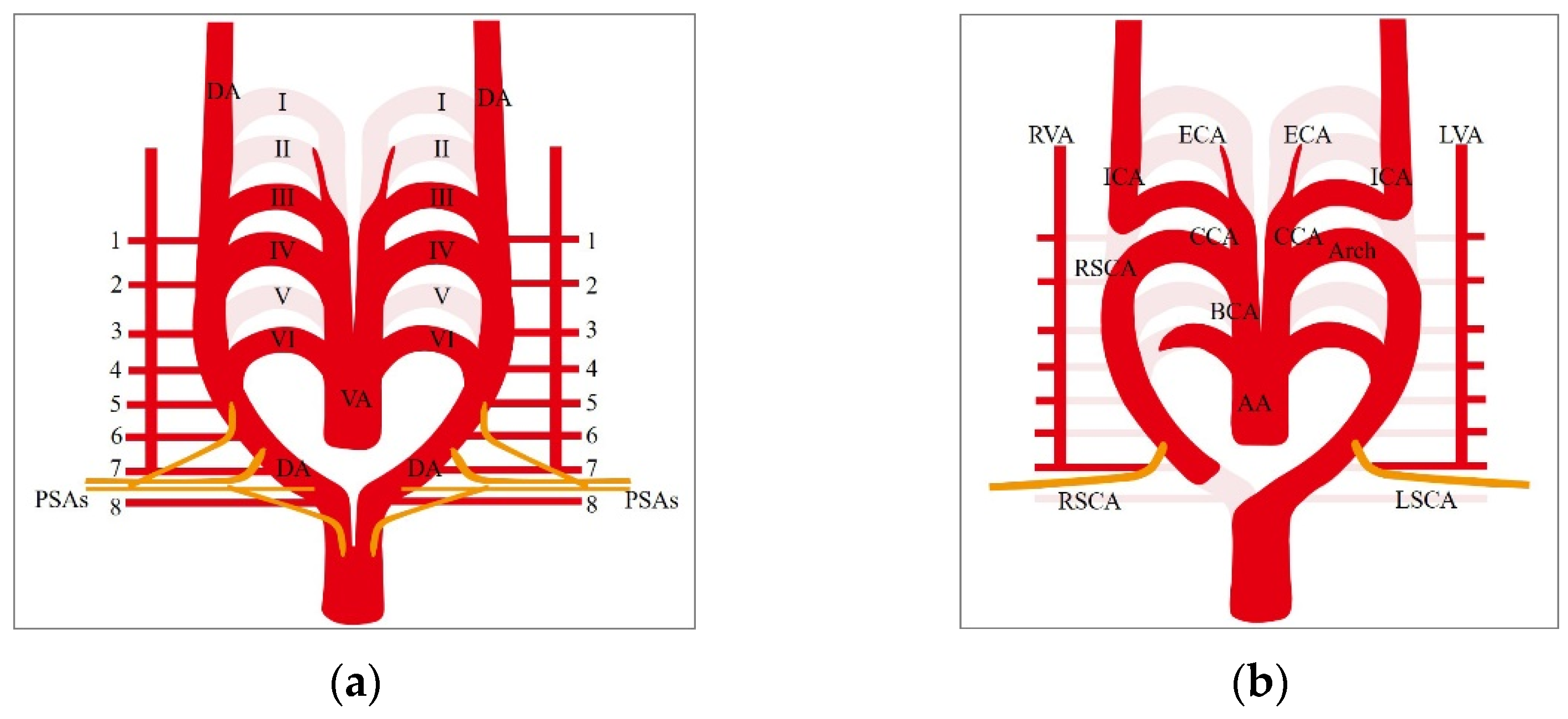
2. Embryological Development and Normal Anatomy of the Proximal Part of the Right Vertebral Artery

At the early stage of embryological development of the arterial system, six aortic arches arise from the truncus arteriosus on each side and connect to the paired dorsal aorta. These aortic arches selectively regress to form the adult-type aortic arch and its major branches. Among the arches, the paired 3rd arches form the common carotid artery, the origin of the internal carotid artery, and the cranial part of the dorsal aorta beyond the 3rd arch become the primitive internal carotid artery. The 4th left arch forms the distal main part of the adult aortic arch. The dorsal aorta gives off the paired segmental arteries from the cervical to the sacral region to supply the developing somites and spinal cord [4]. At the 7-mm stage (day 32), seven cervical segmental arteries (CSAs) appear, originating from each of the paired dorsal aortas accompanying segmental cervical nerves 2–8. According to a study by Aizawa et al. on the development of the subclavian artery in the rat embryo, a few arterial trunks of the forelimbs (primary subclavian artery) arise from the lateral surface of the dorsal aorta at the 6th–10th segmental levels to form a vascular network (Figure 1a) [5]. The primary subclavian arteries anastomose with neighboring segmental arteries, and then the origins of those segmental arteries regress. One of the primary subclavian arteries at the level of the 7th segment becomes thicker, and the others regress. Then, the anastomosis between the primary subclavian artery and the 7th CSA forms the proximal part of the future vertebral artery. The paired dorsal aorta begins to fuse at the midline to form a single dorsal aorta. The level of fusion of the dorsal aorta transitionally reaches the most cranial point near the third CSA level but shifts down to the origin of the level of the 7th CSA. The caudal part of the right dorsal aorta (just before its confluence with the left dorsal aorta) regresses, and the right subclavian artery (RSCA) is formed by the remnant part of the right dorsal aorta and the primary subclavian artery. The left subclavian artery is formed by the left primary subclavian artery at the 7th CSA level. At the 7-mm stage, the primitive VA appears, and its development is usually complete by the 14- to 17-mm stage [4,5,6]. At the 10- to 12-mm stage, the postcostal longitudinal anastomosis develops between the CSAs (developing bilaterally from the 1st to the 7th CSAs) and becomes the VA [7]. At the 14- to 17-mm stage, the aortic origins of the first 6 CSAs regress, and the 7th CSA becomes the branches of the SCA. This developmental process results in a normal pattern of development of the VA from the SCA (Figure 1b). As a result of normal development of the arterial system, the right vertebral artery (RVA) usually arises from the right subclavian artery (RSCA) as its first branch at the upper part, just proximal to the origin of the thyrocervical trunk. The RVA enters the transverse foramen of the 6th cervical spine and runs upward into the posterior fossa through the foramen magnum. The RVA gives off segmental arteries that supply the corresponding vertebrae, cervical nerve, dura mater, and paravertebral muscles. Some of them give off the anterior and/or posterior radiculomedullary artery supplying the spinal cord.

3. Anomalous Origin of the Right Vertebral Artery

Several types of anomalous origins of the RVA have been reported [1,2,3,8], but they can generally be divided into three types: (1) aortic origin (RVA originating directly from the aorta), (2) carotid/brachiocephalic origin (RVA originating from the common carotid arteries or the brachiocephalic artery), and (3) dual origin (RVA has two origins, usually from the SCA). Lazaridis et al. reported that the incidences of those types were 0.05%, 0.18–0.37%, and 0.04–0.4%, respectively. Unusually persistent CSAs and the obliteration point of the right aortic arch are associated with the embryological development of an anomalous-origin RVA (Figure 2a–d).

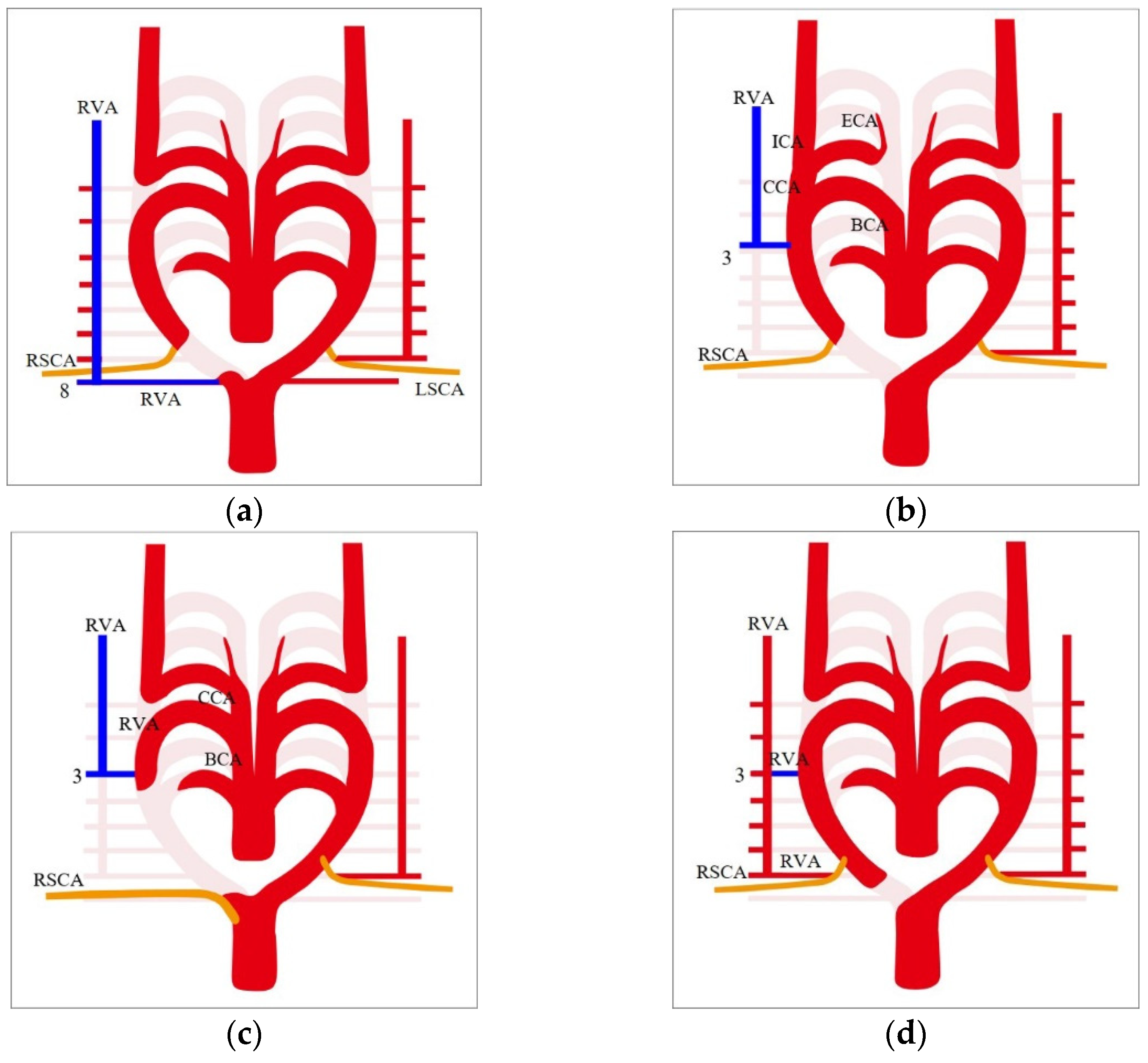
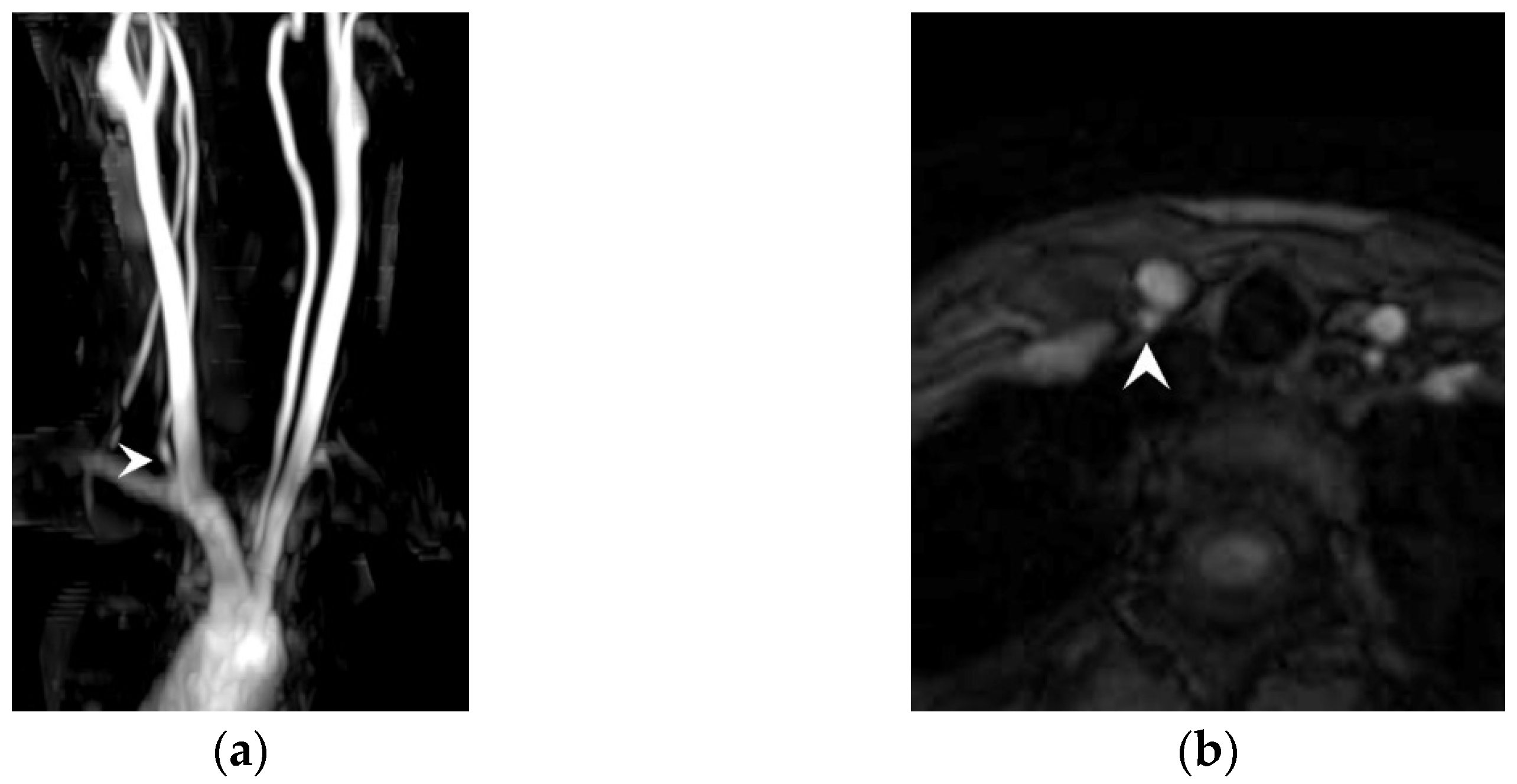
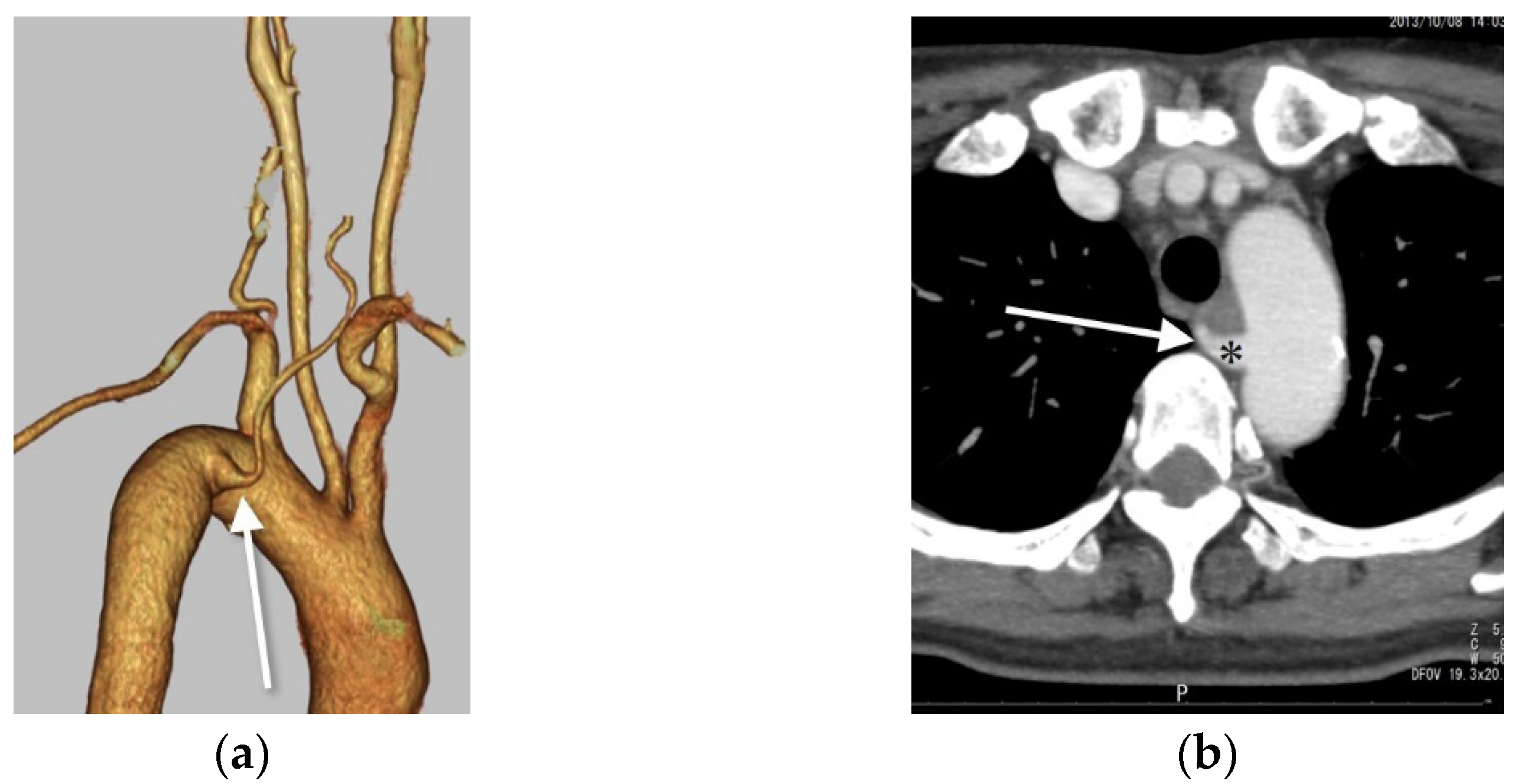
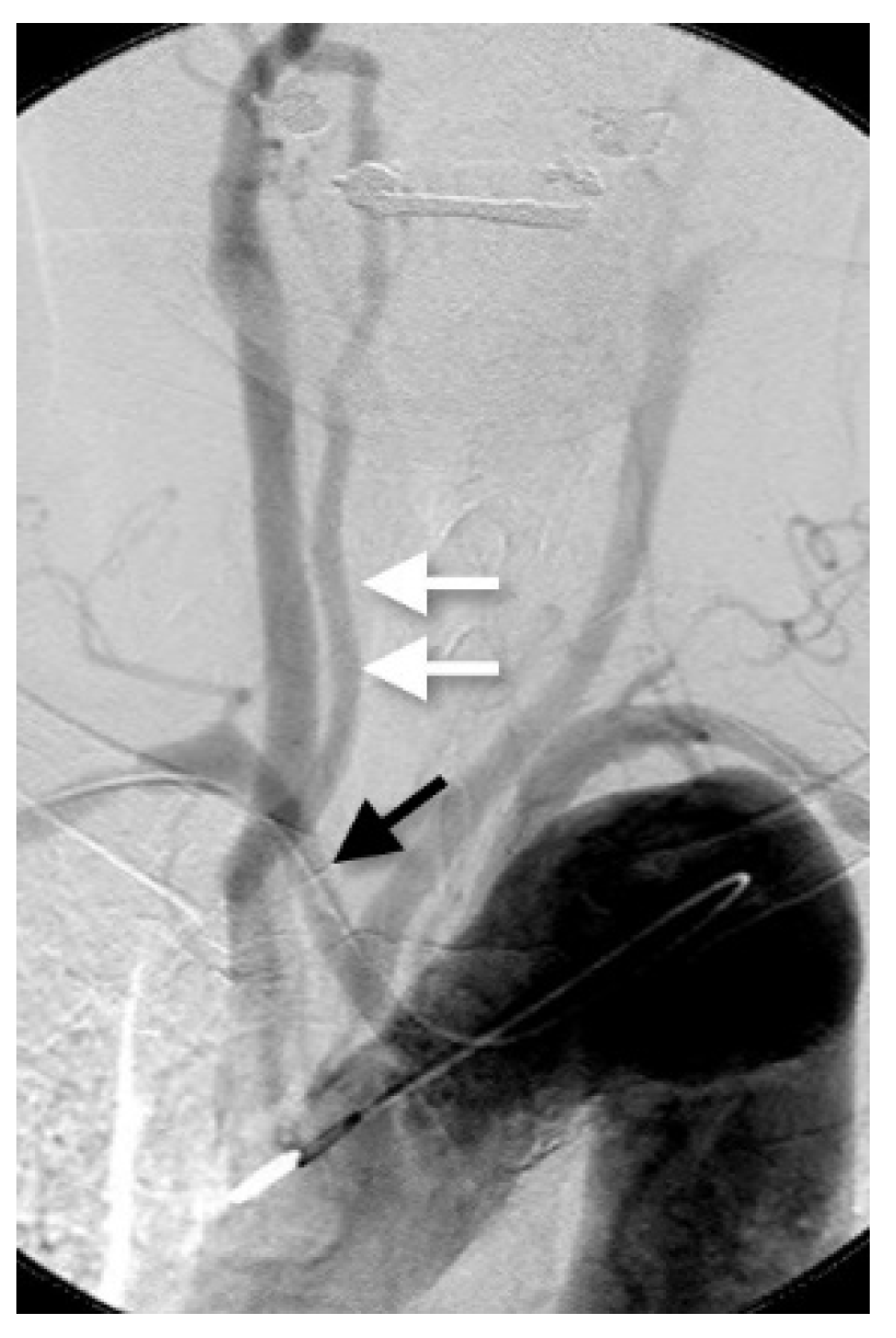
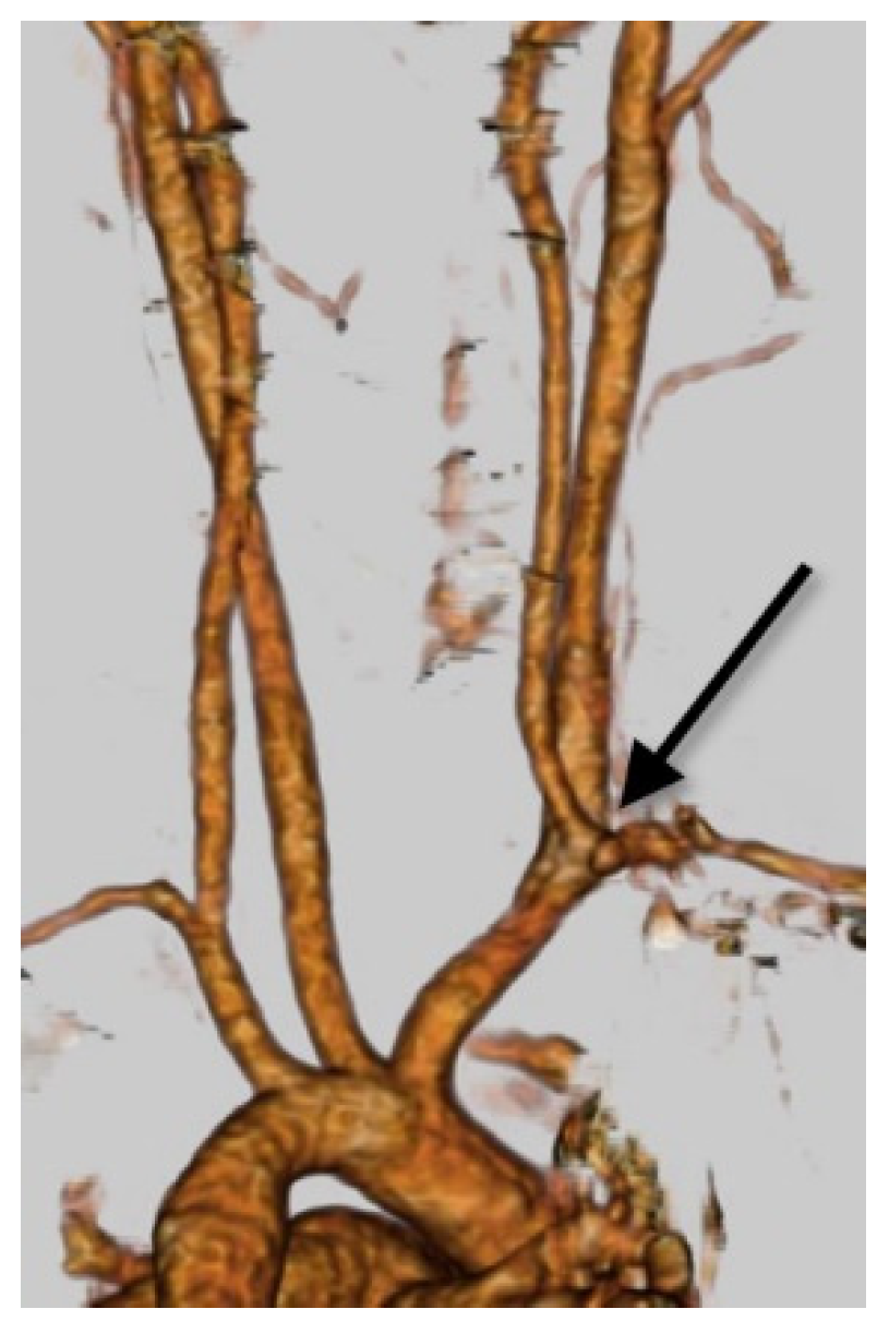
3.1. Right VA Originating from the Aortic Arch (Aortic Origin)

There are some points where the RVA can arise directly from the aorta [1,2,3]. The most common site of the aortic origin of the RVA is the distal portion of the aortic arch beyond the origin of the LSCA, which accounts for 85.7% of aortic-origin RVAs (Figure 3a,b). Other sites of aortic origin of the RVA are rare and include the site between the left common carotid artery (LCCA) and LSCA, at the ascending aorta, and between the RSCA and the right common carotid artery (RCCA) with a missing brachiocephalic artery [1,2,8,9,10]. Lemke et al. presumed that the embryotic mechanism of an aortic origin of the RVA was from the distal portion to the LSCA. According to their hypothesis, it is caused by the RVA originating from the persistent right 8th CSA [8] and the physiologic obliteration of the right dorsal aorta distal from the 7th CSA (Figure 2a). This type of aortic origin of the RVA is occasionally associated with the diverticulum of Kommerell, which is thought to be a remnant of the distal part of the right dorsal aorta [11] (Figure 4a,b). The embryologic mechanisms of the other types of aortic origin of the right VA may be more complicated processes involving fusion and division of the dorsal aorta, regression of the primary subclavian arteries and cervical segmental arteries and their anastomoses.

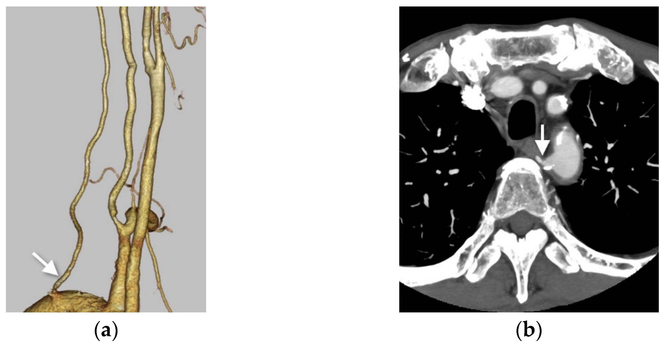
3.2. Right VA Originating from the Common Carotid Artery or the Brachiocephalic Artery

Cases of RVA originating from the RCCA or BCT are frequently associated with aberrant RSCA (Figure 5). Few cases of RVA originating from the RCCA alone or with missing brachiocephalic arteries have been reported [3,12] (Figure 6). The embryological mechanism of this anomaly can be explained by the persistent origin of one of the CSAs (3rd–6th segmental levels) and the right dorsal aorta between the 3rd and 4th aortic arch with abnormal regression of the proximal portion of the 3rd aortic arch CA. A persistent origin of the CSA forms the anomalous origin of the RVA from the right CCA or the right BCT (Figure 2b). When the right dorsal aorta regresses above the origin of the right primary subclavian artery, an aberrant RSCA also arises (Figure 2c) [13].

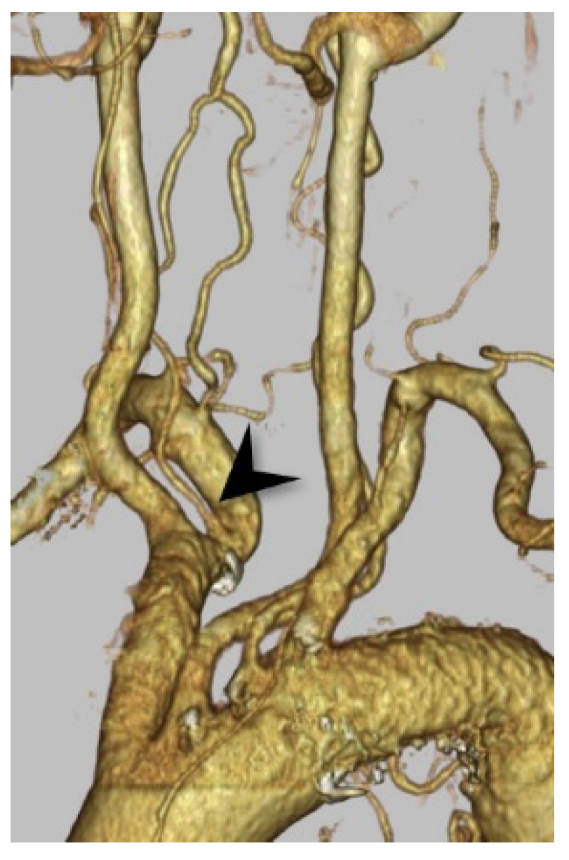
3.3. The Dual Origin of the Right Vertebral Artery

The VA has two origins, with fusion at a variable level of the neck. The dual origin of the VA has been confused with a fenestration of the VA, which has a single origin and has two parallel segments anywhere along its course. The dual origin of the VA is much rarer than fenestration. This variation occurs due to the remnant of another CSA with a normal vertebral artery or the remnant of another anastomotic channel between the primary subclavian artery and the lower CSAs (Figure 2d). Some patterns of the dual origin of the RVA have been reported; in the most popular pattern, both origins are from the RSCA (Figure 7), while in the rarer patterns, the RVA arises from the BCT, the RCCA (Figure 8), the right thyrocervical trunk, or the aortic arch [1,2,14,15].

4. Discussion

In almost all cases, the anomalous origin of the VA is an incidental finding. In rare cases, patients are complicated by dizziness and have a correlation with an anomalous origin of the VA [16]. Although conclusive evidence has not been established between the anomalous origin of the RVA and cerebrovascular disease, some authors have suggested that alterations in the large vessels can change the cerebral hemodynamics to possibly cause cerebral vascular disease, including aortic dissection, aneurysm, and arteriovenous fistula [17]. In recent years, the advancements in imaging technology have been remarkable. In the near future, it may be shown that hemodynamic changes brought about by vascular anomalies are the cause of cerebrovascular disease. Especially, as some authors suggest, the relation between the cerebrovascular events and the radiomics [18,19,20]; however, there have not been any reported relations about the anomalous origin of the right VA and radiomics. Therefore, radiomics are a possible application to clarify the potential relationships in the future. The clinical importance of recognizing the anomalous origins of the RVA are to prevent complications in cervico-thoracic surgery and endovascular treatments as well as to avoid misdiagnosing the anomaly as other vascular pathologies. For example, the duplicated origins of the VA are often misdiagnosed as dissections. The VR images are very useful to evaluate the branching pattern of great vessels. Therefore, surgeons are also required to evaluate the VR image before surgery.
In conclusion, typical imaging features of several anomalous origin types of the RVA were demonstrated with their embryological development. This knowledge is very important for safe and successful procedures in vascular surgery and endovascular treatment.

Author Contributions

For research articles with several authors, conceptualization and editing are credited to K.T. and H.K. Acquisition was attributed to H.B. Supervision, review and editing were accomplished by H.K. and Y.A. All authors have read and agreed to the published version of the manuscript.

Funding

This research received no external funding.

Institutional Review Board Statement

Not applicable.

Informed Consent Statement

Not applicable.

Conflicts of Interest

The authors declare no conflict of interest.

References

  1. Lazaridis, N.; Piagkou, M.; Loukas, M.; Piperaki, E.T.; Totlis, T.; Noussiou, G.; Natsis, K. A systematic classification of the vertebral artery variable origin: Clinical and surgical implications. Surg. Radiol. Anat. 2018, 40, 779–797. [Google Scholar] [CrossRef] [PubMed]
  2. Maiti, T.K.; Konar, S.K.; Bir, S.; Nanda, A.; Cuellar, H. Anomalous Origin of the Right Vertebral Artery: Incidence and Significance. World Neurosurg. 2016, 89, 601–610. [Google Scholar] [CrossRef] [PubMed]
  3. Uchino, A.; Saito, N.; Takahashi, M.; Okada, Y.; Kozawa, E.; Nishi, N.; Mizukoshi, W.; Nakajima, R.; Watanabe, Y. Variations in the origin of the vertebral artery and its level of entry into the transverse foramen diagnosed by CT angiography. Neuroradiology 2013, 55, 585–594. [Google Scholar] [CrossRef] [PubMed]
  4. Sadler, T.W. Chapter 13 cardiovascular system. In Vascular Development in Langman′s Medical Embryology, 12th ed.; Wolters Kluwer Health/Lippincott Williams & Wilkins: Philadelphia, PA, USA, 2012; pp. 185–191. [Google Scholar]
  5. Padget, D.H. The development of the cranial arteries in the human embryo. Contrib. Embryol. 1948, 32, 207–261. [Google Scholar]
  6. Aizawa, Y.; Isogai, S.; Izumiyama, M.; Horiguchi, M. Morphogenesis of the primary arterial trunks of the forelimb in the rat embryos: The trunks originate from the lateral surface of the dorsal aorta independently of the intersegmental arteries. Anat. Embryol. 1999, 200, 573–584. [Google Scholar] [CrossRef] [PubMed]
  7. Gailloud, P.; Gregg, L.; Pearl, M.S.; San, M.D. Ascending and Descending Thoracic Vertebral Arteries. AJNR Am. J. Neuroradiol. 2017, 38, 327–335. [Google Scholar] [CrossRef] [PubMed] [Green Version]
  8. Lemke, A.J.; Benndorf, G.; Liebig, T.; Felix, R. Anomalous origin of the right vertebral artery: Review of the literature and case report of right vertebral artery origin distal to the left subclavian artery. AJNR Am. J. Neuroradiol. 1999, 20, 1318–1321. [Google Scholar] [PubMed]
  9. Wasserman, B.A.; Mikulis, D.J.; Manzione, J.V. Origin of the right vertebral artery from the left side of the aortic arch proximal to the origin of the left subclavian artery. AJNR Am. J. Neuroradiol. 1992, 13, 355–358. [Google Scholar] [PubMed]
  10. Akdeniz, B.; Yilmaz, E.; Pekel, N.; Ergul, B.U. Anomalous origin of the right vertebral artery from the ascending aorta in the presence of an aberrant right subclavian artery. Int. J. Cardiovasc. Imaging 2007, 23, 39–42. [Google Scholar] [CrossRef] [PubMed]
  11. Goldbach, A.; Dass, C.; Surapaneni, K. Aberrant Right Vertebral Artery with a Diverculum of Kommerell: Review of a Rare Aortic Arch Anomaly. J. Radiol. Case Rep. 2018, 12, 19–26. [Google Scholar] [PubMed]
  12. Vitošević, F.; Vitošević, Z.; Rasulić, L. The right vertebral artery arising from the right common carotid artery: Report of a rare case. Case Reports. Surg. Radiol. Anat. 2020, 42, 1263–1266. [Google Scholar] [CrossRef] [PubMed]
  13. Kesler, W.W.; Sabat, S.B. Isolated anomalous origin of the vertebral artery from the common carotid artery. Case Reports. Interact. Cardiovasc. Thorac. Surg. 2018, 27, 615–616. [Google Scholar] [CrossRef] [PubMed]
  14. Baik, J.; Baek, H.J.; Shin, H.S.; Choi, K.H. Duplication of the right vertebral artery: MRA findings and review of the literature. Springerplus 2016, 5, 1123. [Google Scholar] [CrossRef] [PubMed] [Green Version]
  15. Motomura, M.; Watanabe, K.; Tabira, Y.; Iwanaga, J.; Matsuuchi, W.; Yoshida, D.; Saga, T.; Yamaki, K. A Case of Duplicated Right Vertebral Artery. Kurume. Med. J. 2018, 64, 69–73. [Google Scholar] [CrossRef] [PubMed] [Green Version]
  16. Nishijima, M.; Harada, J.; Akai, T.; Endo, S.; Tkaku, A. Operative correction of kinking and coiling at the origin of vertebral artery and stellate ganglionectomy in patients with severe vertigo and dizziness. No Shinkei Geka. 1989, 32, 356–359. [Google Scholar]
  17. Yuan, S.M. Aberrant Origin of Vertebral Artery and its Clinical Implications. Braz. J. Cardiovasc. Surg. 2016, 31, 52–59. [Google Scholar] [PubMed]
  18. Le, E.P.V.; Rundo, L.; Tarkin, J.M.; Evans, N.R.; Chowdhury, M.M.; Coughlin, P.A.; Pavey, H.; Wall, C.; Zaccagna, F.; Gallagher, F.A.; et al. Assessing robustness of carotid artery CT angiography radiomics in the identification of culprit lesions in cerebrovascular events. Sci. Rep. 2021, 11, 3499. [Google Scholar] [CrossRef] [PubMed]
  19. Zhang, R.; Zhang, Q.; Ji, A.; Lv, P.; Zhang, J.; Fu, C.; Lin, J. Identification of high-risk carotid plaque with MRI-based radiomics and machine learning. Eur. Radiol. 2021, 31, 3116–3126. [Google Scholar] [CrossRef] [PubMed]
  20. Zaccagna, F.; Ganeshan, B.; Arca, M.; Rengo, M.; Napoli, A.; Rundo, L.; Groves, A.M.; Laghi, A.; Carbone, I.; Menezes, L.J. CT texture-based radiomics analysis of carotid arteries identifies vulnerable patients: A preliminary outcome study. Neuroradiology 2021, 63, 1043–1052. [Google Scholar] [CrossRef] [PubMed]
Figure 1. Drawing of the embryologic development of the aorta and brachiocephalic vessels. (a) Basic pattern of the development of vertebral artery and major branches of the aortic arch (b) Common branching pattern of major aortic branches and the vertebral arteries in the human adult. I–VI, the first to sixth aortic arches; 1–8, the first to eighth segmental artery; VA, ventral aorta; DA, dorsal aorta; PSAs, primary subclavian arteries; ECA, external carotid artery; ICA, internal carotid artery; CCA, common carotid artery; R (L)VA, right (left) vertebral artery; R (L) SCA, right (left) subclavian artery; BCA, brachiocephalic artery; AA, ascending aorta; Arch, aortic arch.
Applsci 11 08171 g001
Figure 2. Drawing of the variations of the anomalous origin of the RVA. (a) Aortic origin of the RVA originating at the distal segment of the aortic arch beyond the origin of the LSCA. (b) Common carotid or brachiocephalic arterial origin of the RVA. (c) Common carotid or brachiocephalic arterial origin of the RVA with aberrant RSCA. (d) Double origin of the RVA.
Applsci 11 08171 g002
Figure 3. Right VA originating from the aortic arch. (a) Right anterior oblique view on volume-rendered computed tomography (VR-CT) angiography shows the RVA directly arising from the aortic arch distal to the left subclavian artery (white arrow). (b) Axial contrast-enhanced CT shows the right vertebral artery (white arrow) arising from the aortic arch as the fourth branch. The Ziostation 2 (Ziosoft Inc., Tokyo, Japan) was used to obtain the VR image.
Applsci 11 08171 g003
Figure 4. Right VA originating from the aortic arch with a diverticulum of Kommerell. (a) Right posterior oblique view on VR-CT angiography shows the RVA directly arising from the aortic arch (long white arrow) distal to the left subclavian artery with a diverticulum of Kommerell. (b) Axial contrast-enhanced CT shows the right vertebral artery (white long arrow) arising from the aortic arch as the fourth branch with a diverticulum of Kommerell (*). The Ziostation 2 (Ziosoft Inc., Tokyo, Japan) was used to obtain the VR image.
Applsci 11 08171 g004
Figure 5. Right VA originating from the common carotid artery with RSCA. Anteroposterior view on aortography shows the RVA arising from the right common carotid artery (double white arrow). An aberrant RSCA is also detected (black arrow).
Applsci 11 08171 g005
Figure 6. Right VA originating from the BCT. Right posterior oblique view on VR-CT angiography shows the RVA arising from the BCT (black long arrow). The SYNAPSE VINCENT software 6.3 (Fujifilm Holdings Corp., Tokyo, Japan) was used to obtain the VR image.
Applsci 11 08171 g006
Figure 7. The dual origin of the RVA. Left anterior oblique view on VR-CT angiography shows the dual origin of the RVA. The abnormal VA originates from the RSCA (black arrowhead). The Ziostation 2 (Ziosoft Inc., Tokyo, Japan) was used to obtain the VR image.
Applsci 11 08171 g007
Figure 8. The dual origin of the RVA. (a) Right anterior oblique view of the magnetic resonance angiography (MRA) shows the dual origin of the RVA. The abnormal VA originates from the RCCA (white arrowhead). (b) Axial views of the source image of the MRA show the abnormal VA (white arrowhead) originating from the common carotid artery and the normal vessels from the RSCA.
Applsci 11 08171 g008
Publisher’s Note: MDPI stays neutral with regard to jurisdictional claims in published maps and institutional affiliations.

