Article

Risk Factors of Avulsion Fracture after Iliac Crestal Flap for Jaw Reconstruction

1
Department of Oral and Maxillofacial Surgery, Chosun University Dental Hospital, Swangju 61452, Korea
2
Department of Oral and Maxillofacial Surgery, Collage of Dentistry, Chosun University, Gwanju 61452, Korea
*
Author to whom correspondence should be addressed.
Appl. Sci. 2021, 11(17), 8081; https://doi.org/10.3390/app11178081
Submission received: 9 August 2021 / Revised: 23 August 2021 / Accepted: 26 August 2021 / Published: 31 August 2021
(This article belongs to the Section Applied Dentistry and Oral Sciences)

Abstract

:
The iliac crestal flap is often used in maxillofacial reconstruction surgery because it has a similar bone contour to jaw bone. However, complications occur frequently in this flap, and the avulsion fracture of the anterior superior iliac spine (ASIS) occurs only in rare cases. The purpose of this study is to evaluate the risk factors affecting fractures that may occur after iliac crestal flap harvesting. In this study, we performed preoperative and postoperative CT scans of the iliac bones of 22 patients who underwent iliac crestal flaps between February 2013 and July 2019. Three of these patients had fractures within 1 month after surgery. After converting DICOM files to a three-dimensional model using Mimics software, some reference points were defined for analysis. The patients were classified into fracture and non-fracture groups. The patients’ age and sex, harvested bone, residual bone and absolute bed rest (ABR) periods were investigated. The patients’ age and sex were not associated with fracture occurrence. There was a statistically significant difference in the width of the anterior–superior part of residual bone. The fracture group showed statistically significantly shorter ABR periods. In the case of the iliac crestal flap, it was found that the factors that can affect the occurrence of fractures are the width of the residual bone and the ABR period. Iliac avulsion fracture can be prevented when the remaining bone is sufficient and the ABR period is prolonged sufficiently.

1. Introduction

Bone grafts are frequently used for reconstruction in maxillofacial surgery, such as in cancer surgery, trauma, and severe bone resorption, and for the correction of congenital deformities [1]. The reconstruction of the jaw can be performed using microvascular composite free flaps with the scapula, iliac crest, and fibula [2,3]. The surgeon chooses the donor site based on several factors such as the size and location of the defect and the condition of the patient [4]. The iliac crest has been a common donor site for mandible and maxilla reconstruction because it provides the highest concentration of osteocompetent cells and available volume [1,5,6,7,8,9,10,11,12]. Furthermore, the iliac crest can be accessed with a reasonable surgical method [7,8,13]. For these reasons, iliac crestal bone has been used for bone grafts for various defects as the gold standard [7,8,14,15], and it is suitable for both free non-vascularized and vascularized bone grafts [16]. In addition, compared to the fibula flap, the iliac crestal flap has benefits in terms of the bone contour for the reconstruction of the jaw, such as adequate bone height and width for dental implantation and optimal soft tissue reconstruction using internal oblique muscle without the need for a skin flap [3]. Another important factor to consider when selecting a donor site is its morbidity after grafting [4]. Urken et al. reported minimal donor-site morbidity after over 1 year of follow-up when using the iliac crestal osseomyocutaneous microvascular free flap [17].
Conventionally, the anterior iliac crestal flap has been harvested as a bicortical form, which can lead to an abnormal hip contour, hernia, severe bleeding tendency, and gait disturbance [3]. The monocortical iliac crestal flap may reduce the donor-site morbidity after harvesting a large amount of bone segment [3]. Recently, computer-assisted surgery has become popular in oral and maxillofacial surgery. Most reconstructive surgeries are performed by preoperative simulation and surgical guide fabrication. Therefore, it is important to determine the optimal bone harvesting site in the anterior iliac crest for successful reconstruction during simulation for preoperative planning. Serious complications related to the iliac crestal flap are rare, but when they do occur, they cause serious problems [14]. Major complications have been reported, such as chronic pain, neurological or vascular injury, infection, hematoma, intestinal prolapse, cosmetic defect, and iliac crest avulsion fracture [5,6,8,11,14,15,18,19,20]. Among these complications, fractures of the anterior iliac crest following bone graft are very rare [5,21,22]; only a few cases have been reported in the literature [21,23,24,25,26,27]. The stress fracture of the anterior iliac crest after the harvesting of the iliac crestal flap has been reported in a few works, which may cause significant problems and increase morbidity [18]. Although the avulsion fracture of the anterior superior iliac spine (ASIS) after bone grafting is not common, the fracture could occur more commonly in older patients than reported in the literature [21]. The treatment of fractures usually focuses on walking assistance after a short break until the fracture heals, but sometimes additional surgical treatment is required to remove avulsed bone or open reduction and internal fixation [21]. Previous studies have reported that the iliac crestal flap in elderly patients with osteoporosis may increase the risk of iliac crest fractures [21,23,28]. Some authors have reported that harvesting sites and surgery methods also could affect the fracture risk [18,29,30,31,32]. An understanding of the bone harvesting techniques and management of the risk factors for fracture could help prevent iliac crest fractures after bone grafting.
The aim of this study was to evaluate the risk factors associated with avulsion fracture after iliac crestal flap harvesting for jaw reconstructions.

2. Patients and Methods

2.1. Patients

This study was conducted with 22 patients who underwent iliac crestal flap surgery for jaw reconstruction from February 2013 to July 2019. The patient’s medical records and pre- and post-operative CT scan data of iliac bones were evaluated. Within one month after surgery, fracture of the iliac bone occurred in three patients, of which two patients required surgical reduction and one patient was treated with conservative treatment by maintaining bed rest for six weeks.
Patient 1 was a 70-year-old male who underwent reconstruction surgery using the iliac crestal flap for mandibular reconstruction. The patient had absolute bed rest for 6 days, then conducted a wheelchair ambulation after the 7th postoperative day (POD) and was discharged on the 18th POD. About a month after the operation, he was complaining of pain in the donor site while walking downhill. An x-ray of the pelvis was taken and he was diagnosed with an anterior superior iliac spine (ASIS) avulsion fracture (Figure 1a). He underwent open reduction and internal fixation. There was no gait disturbance after surgery during 18 months follow-up.
Patient 2 was a 78-year-old female who had a partial maxillectomy with verrucous carcinoma and reconstructive surgery with a DCIA flap. She complained of pain in the donor site on the 16th POD when moving out of bed. There was a fracture of the ASIS on the donor side (Figure 1b). She needed ABR for about a month or more and was treated using symptomatic treatment and gait training with physical therapy in the department of rehabilitation medicine. At 32 days after the fracture, callus formation was observed on x-ray and CT images. Due to her old age and the decrease of muscle, she had 4 weeks of walking practice. She was on follow-up for more than 2 years and she did not have difficulties in walking at her final check-up.
Patient 3 was a 53-year-old male with reconstruction surgery using an iliac crestal flap after segmental mandibulectomy. After the third POD, he was allowed to move using a wheelchair, and after the fifth POD, gait practice was performed with support from the nursing staff. He complained about the sound from the donor-side pelvis bone at the 24th POD. An avulsion fracture of the ASIS was founded in pelvis x-ray and CT scan images (Figure 1c). A reductive operation was performed with a titanium mesh. He was discharged after hospitalization for about one month, and no gait disturbance was observed in follow-up checks for more than 3 years.

2.2. Operation and Postoperative Care

All surgeries were performed by a surgeon of the oral and maxillofacial surgery. The anterior approach was used for the harvesting of iliac crest grafts. Prophylactic antibiotics were given intravenously after the induction of general anesthesia. The iliac bone and internal oblique muscle flap were harvested from the iliac crestal area with deep circumflex iliac vessels. A vacuum drain was used in the all cases to prevent unnecessary hematoma.
The patients were instructed to stabilize for a certain period of time after surgery, and the range of activities was gradually increased, including the period of movement using assistive devices such as wheelchairs, depending on the patient’s condition.

2.3. Measurement

Iliac CT scan data of patients were organized into DICOM files. After converting DICOM files to a three-dimensional model and photomapping the postoperative pelvic radiographs using Mimics software, we defined some reference points for analysis.
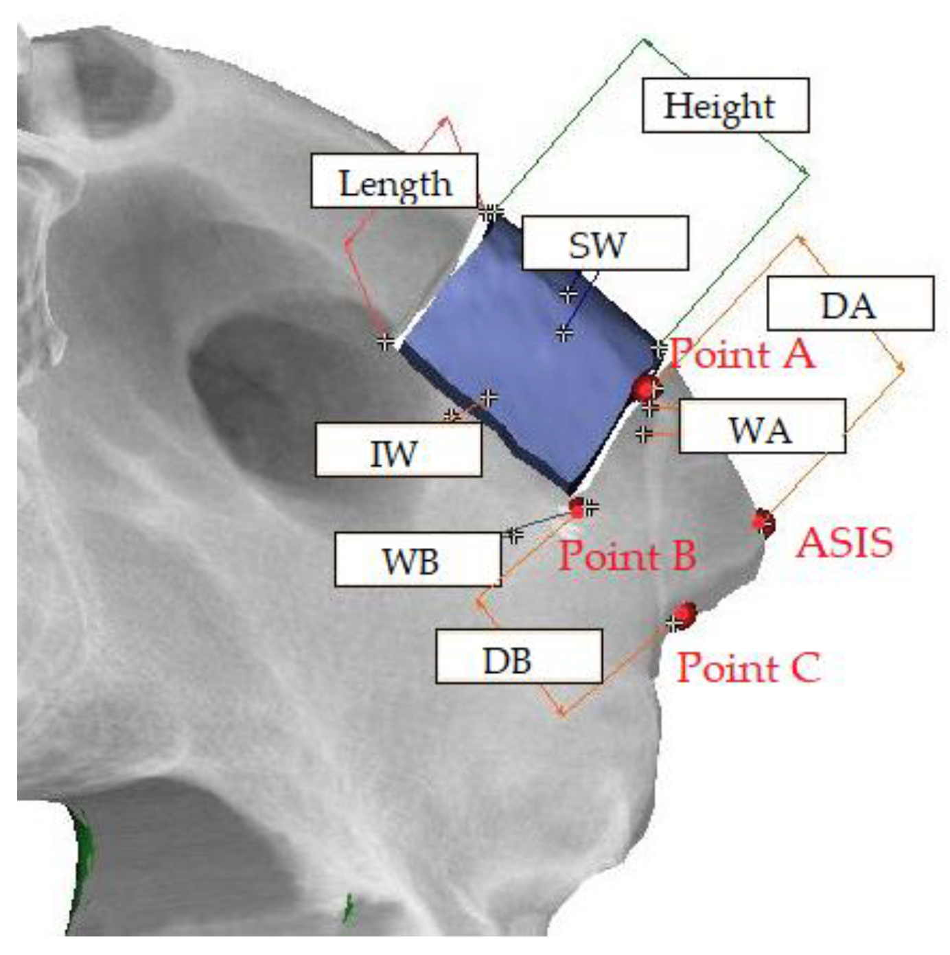
Point A was defined as the contact point by drawing a vertical line on the boundary of the harvested bone segment from the ASIS. Point B was defined as the contact point by drawing a vertical line on the boundary of the harvested bone segment from point C. Point C was defined as the most concave point between the ASIS and anterior inferior iliac spine. Then, we measured the distance between the ASIS and Point A (DA), between Point B and Point C (DB) and the width at Point A (WA) and Point B (WB) using Mimics software. We also measured the midpoint width of the superior and inferior part (SW, IW), height and length of the harvested bone segment (Figure 2). The age and sex of the patients and the ABR period were investigated. We set up the fracture group and the non-fracture group to statistically analyze the difference between groups.

2.4. Statistical Methods

The Mann–Whitney test and the independent t-test were used to compare the samples according to the age, sex, ABR period and characteristics of the donor site and harvested bone. Statistical computation was performed with SPSS 20.0 (IBM, New York, NY, USA) using of Windows 10 Pro (Microsoft, Redmond, WA, USA).

3. Results

3.1. Patient Characteristics

The gender makeup of the patients was 11 males and 11 females, and this factor was not associated with fracture incidence (p = 0.557, Pearson’s correlation analysis) (Table 1).
The mean (S.D) age of the patients at the time of operation was 55.9 (19.30) (range 15–80, median 62). There was no correlation between fracture incidence and age (p = 0.295, Pearson’s correlation analysis) (Table 2).

3.2. Anatomical Characteristics

The average length of DA was 16.88 ± 4.54 mm in the non-fracture group; it was 22.28 ± 9.80 mm in the fracture group, and there was no statistically significant difference. WA was 13.63 ± 1.58 mm on average in the non-fracture group and 17.05 ± 3.71 mm in fracture group, and there was a statistically significant difference (p = 0.032, independent t-test). DB averaged 17.29 ± 4.44 mm in the non-fracture group and 20.95 ± 7.66 mm in the fracture group, and WB was 10.74 ± 1.63 mm in the non-fracture group and 13.38 ± 5.52 mm in the fracture group. Each result showed no statistically significant difference (Table 3, Table 4, Table 5 and Table 6).

3.3. Postoperative Care

When the ABR period was investigated, ambulation was performed after an average of 10.50 ± 3.61 days in the non-fracture group and 5.67 ± 2.31 days in the fracture group. This period showed a statistically significant difference (Table 7).

4. Discussion

Harvesting autogenous bone from the iliac crest has been a reliable and well-established technique in oral and maxillofacial surgery in recent decades [33,34,35]. However, complications at the donor site are common [19]. The incidence of minor complications following autogenous iliac crest bone graft harvest has been reported to be from 0% to 48% [8,36,37,38,39,40,41] and the rate of major complications is up to 11.9% [42]. Deep infections, hemorrhages, hernias, ilium fractures and pelvic instability, as well as vascular and nervous lesions, are reported as major complications [11,14].
Fracture is caused by the contraction of the sartorius muscle and other immediate causes [43]. The harvesting of anterior iliac crest bone grafts can occur too close to the ASIS, thereby causing the fracture of the ASIS from the iliac wing in a few studies [25,44]. Therefore, most of the literature suggested that iliac grafts 3–4 cm posterior to the ASIS provide maximum support for the sartorius and tensor fascia latae muscles. Barone et al. placed the anterior osteotomy 2 cm distal to the ASIS to avoid any weakening of the pelvic ring [35]. Additionally, Hu et al. showed that the removal of bone 30 mm posterior to the ASIS preserves 2.4 times more strength of the iliac crest compared with the harvesting of a bone graft 15 mm posterior to the ASIS [23]. The downward pulling of the sartorius muscle and tensor fascia latae muscle can lead to a late stress fracture after harvesting the anterior iliac crest, so the base of ASIS must be wide enough to reduce the risk of stress fracture due to the downward pulling of the sartorius muscle [5].
In this study, there was no statistically significant difference in the distance from the ASIS to the harvesting margin between the fracture group and non-fracture group. This is because the operation was performed maintaining a sufficient distance from the ASIS of at least 2 cm to prevent the avulsion fracture of the ASIS.
In order to reduce the complications of the pelvis, Jones et al. reported that an osteotome-harvested graft had an increased incidence of propagations of fracture compared with saw-harvested bone grafts on fresh frozen cadavers [29]. Burstein et al. evaluated the morbidity of grafts by comparing traditional iliac bone grafts with minimally invasive techniques [45]. They reported that a minimally invasive technique was statistically superior for pain control, operation time and mean incision length. Eufinger et al. reported that the short-term morbidity (pain and scar at the donor site) in the traditional open osteotomy group was slightly greater than the closed harvesting group [46]. Conversely, long-term morbidity showed a negligible difference in both groups. Sandor et al. reported a minimally invasive trephine method [33] which is a safe method and minimizes morbidity. Brawley et al. reported that there no patients experienced morbidity at the harvesting site when using a small acetabular reamer [47].
Most of the iliac crest fractures reported in the literature occurred within 2 weeks after the harvesting procedure [1,21,25,26,44,48]. Since a forceful activity with contraction of the sartorius muscle should cause the avulsion of the weakened bone part, a sufficient ABR period is needed to stabilize the donor site. In this study, there was a statistically significant difference in ABR periods between the fracture group and non-fracture group.
Clinical studies suggest that old age, obesity, osteoporosis, co-morbidities and improper harvesting techniques are the risk factors for the fracture of the iliac crest after bone grafting [5,21,23].
The treatment of iliac crest fracture is usually symptomatic and involves a short rest period with assisted ambulation and the usage of analgesics until the fracture has healed [23,49]. However, for a displacement of the fragment by more than 3 cm infero-laterally, non-union and patients that need a short rest period such as athletes, comorbidities are indications of the open reduction and internal fixation for the iliac avulsion fracture [5].
In summary, the location of the harvesting bone (distance from the ASIS), harvesting method (bi-cortical and mono-cortical) and a short ABR period can increase the risk of avulsion fracture at the anterior iliac crest.
The measurement reference point may vary depending on the person who measures it, and there is a problem that the bone density test or investigation is insufficient. There is also a limited number of cases, which makes the measurement statistically more prone to errors. This should be addressed in future research.

5. Conclusions

Although an iliac bone graft has many advantages, it may result in donor site complications. Fracture of the iliac crest is a rare complication, but while rare, the surgeon should be fully aware of the risk that the patient may suffer from a fracture of the donor site after bone grafting. When harvesting the iliac crestal flap, a sufficient distance posteriorly from the ASIS of at least 2 cm should be maintained; however, if 2 cm of residual bone cannot be preserved, the patient should maintain a sufficient ABR period to minimize avulsion fracture.

Author Contributions

Conceptualization, S.-Y.M.; methodology, S.-Y.M. and Y.-J.J.; software, Y.-J.J. and J.-S.C.; validation, J.-A.B., Y.-J.J. and J.-S.C.; formal analysis, Y.-J.J.; investigation, Y.-J.J. and J.-S.C.; resources, S.-Y.M.; data curation, Y.-J.J.; writing—original draft preparation, Y.-J.J.; writing—review and editing, S.-Y.M. and J.K.; visualization, Y.-J.J. and J.-S.C.; supervision, S.-Y.M.; project administration, S.-Y.M.; funding acquisition, S.-Y.M. All authors have read and agreed to the published version of the manuscript.

Funding

This study was supported by a research fund from Chosun University, 2021.

Institutional Review Board Statement

The study was conducted according to the guidelines of the Declaration of Helsinki and was approved by the Institutional Review Board of Chosun university dental hospital (CUDHIRB 1902008).

Informed Consent Statement

Informed consent was obtained from all subjects involved in the study.

Data Availability Statement

No new data were created or analyzed in this study. Data sharing is not applicable to this article.

Conflicts of Interest

The authors declare no conflict of interest.

References

  1. Arrington, E.D.; Smith, W.J.; Chambers, H.G.; Bucknell, A.L.; Davino, N.A. Complications of iliac crest bone graft harvesting. Clin. Orthop. Relat. Res. 1996, 329, 300–309. [Google Scholar] [CrossRef]
  2. Möhlhenrich, S.C.; Kniha, K.; Elvers, D.; Ayoub, N.; Goloborodko, E.; Hölzle, F.; Modabber, A. Intraosseous stability of dental implants in free revascularized fibula and iliac crest bone flaps. J. Cranio Maxillofac. Surg. 2016, 44, 1935–1939. [Google Scholar] [CrossRef] [PubMed]
  3. Moon, S.-Y. Monocortical deep circumflex iliac artery flap in jaw reconstruction. J. Craniofac. Surg. 2015, 26, 1294–1298. [Google Scholar] [CrossRef] [PubMed]
  4. Kim, H.J.; Moon, S.Y. The Deep Circumflex Iliac Artery Flap for Mandibular Reconstruction and Donor Site Reconstruction with a Patient-Specific Implant: A Case Report. Appl. Sci. 2020, 10, 1587. [Google Scholar] [CrossRef] [Green Version]
  5. İliak, K.G.A.T.A.; Kırığı, K.K. Avulsion fracture of the anterior iliac crest after bone graft harvest: Case report and review of techniques, risk factors and treatment. Turk. Neurosurg. 2011, 21, 423–426. [Google Scholar]
  6. Joshi, A.; Kostakis, G. An investigation of post-operative morbidity following iliac crest graft harvesting. Br. Dent. J. 2004, 196, 167–171. [Google Scholar] [CrossRef] [PubMed] [Green Version]
  7. Westrich, G.H.; Geller, D.S.; O’Malley, M.J.; Deland, J.T.; Helfet, D.L. Anterior iliac crest bone graft harvesting using the corticocancellous reamer system. J. Orthop. Trauma 2001, 15, 500–506. [Google Scholar] [CrossRef]
  8. Ahlmann, E.; Patzakis, M.; Roidis, N.; Shepherd, L.; Holtom, P. Comparison of anterior and posterior iliac crest bone grafts in terms of harvest-site morbidity and functional outcomes. JBJS 2002, 84, 716–720. [Google Scholar] [CrossRef]
  9. Hill, N.M.; Geoffrey Horne, J.; Devane, P.A. Donor site morbidity in the iliac crest bone graft. Aust. N. Z. J. Surg. 1999, 69, 726–728. [Google Scholar] [CrossRef] [PubMed]
  10. Ghassemi, A.; Ghassemi, M.; Modabber, A.; Knobe, M.; Fritz, U.; Riediger, D.; Gerressen, M. Functional long-term results after the harvest of vascularised iliac bone grafts bicortically with the anterior superior iliac spine included. Br. J. Oral Maxillofac. Surg. 2013, 51, e47–e50. [Google Scholar] [CrossRef]
  11. Nodarian, T.; Sariali, E.; Khiami, F.; Pascal-Mousselard, H.; Catonné, Y. Iliac crest bone graft harvesting complications: A case of liver herniation. Orthop. Traumatol. Surg. Res. 2010, 96, 593–596. [Google Scholar] [CrossRef] [Green Version]
  12. Kalk, W.W.; Raghoebar, G.M.; Jansma, J.; Boering, G. Morbidity from iliac crest bone harvesting. J. Oral Maxillofac. Surg. 1996, 54, 1424–1429. [Google Scholar] [CrossRef]
  13. Skaggs, D.L.; Samuelson, M.A.; Hale, J.M.; Kay, R.M.; Tolo, V.T. Complications of posterior iliac crest bone grafting in spine surgery in children. Spine 2000, 25, 2400–2402. [Google Scholar] [CrossRef]
  14. Goulet, J.A.; Senunas, L.E.; DeSilva, G.L.; Greenfield, M.L.V. Autogenous iliac crest bone graft: Complications and functional assessment. Clin. Orthop. Relat. Res. 1997, 339, 76–81. [Google Scholar] [CrossRef] [Green Version]
  15. Schaaf, H.; Lendeckel, S.; Howaldt, H.-P.; Streckbein, P. Donor site morbidity after bone harvesting from the anterior iliac crest. Oral Surg. Oral Med. Oral Pathol. Oral Radiol. Endodontol. 2010, 109, 52–58. [Google Scholar] [CrossRef] [PubMed]
  16. Ghassemi, A.; Ghassemi, M.; Riediger, D.; Hilgers, R.-D.; Gerressen, M. Comparison of donor-site engraftment after harvesting vascularized and nonvascularized iliac bone grafts. J. Oral Maxillofac. Surg. 2009, 67, 1589–1594. [Google Scholar] [CrossRef]
  17. Urken, M.L.; Vickery, C.; Weinberg, H.; Buchbinder, D.; Lawson, W.; Biller, H.F. The internal oblique-iliac crest osseomyocutaneous free flap in oromandibular reconstruction: Report of 20 cases. Arch. Otolaryngol. Head Neck Surg. 1989, 115, 339–349. [Google Scholar] [CrossRef]
  18. Ebraheim, N.A.; Yang, H.; Lu, J.; Biyani, A.; Yeasting, R.A. Anterior iliac crest bone graft: Anatomic considerations. Spine 1997, 22, 847–849. [Google Scholar] [CrossRef] [PubMed]
  19. Kessler, P.; Thorwarth, M.; Bloch-Birkholz, A.; Nkenke, E.; Neukam, F. Harvesting of bone from the iliac crest—Comparison of the anterior and posterior sites. Br. J. Oral Maxillofac. Surg. 2005, 43, 51–56. [Google Scholar] [CrossRef] [PubMed]
  20. Babbi, L.; Barbanti-Brodano, G.; Gasbarrini, A.; Boriani, S. Iliac crest bone graft: A 23-years hystory of infection at donor site in vertebral arthrodesis and a review of current bone substitutes. Eur. Rev. Med. Pharmacol. Sci. 2016, 20, 4670–4676. [Google Scholar] [PubMed]
  21. Al-Sayyad, M. Fracture of the anterior iliac crest following autogenous bone grafting. Saudi Med. J. 2006, 27. [Google Scholar]
  22. Zermatten, P.; Wettstein, M. Iliac wing fracture following graft harvesting from the anterior iliac crest: Literature review based on a case report. Orthop.Traumatol. Surg. Res. 2012, 98, 114–117. [Google Scholar] [CrossRef] [PubMed] [Green Version]
  23. Hu, R.W.; Bohlman, H. Fracture at the iliac bone graft harvest site after fusion of the spine. Clin. Orthop. Relat. Res. 1994, 208–213. [Google Scholar]
  24. Samartzis, D.; Shen, F.H. Postoperative iliac-crest avulsion fracture. Cmaj 2006, 175, 475. [Google Scholar] [CrossRef] [Green Version]
  25. Nocini, P.F.; Bedogni, A.; Valsecchi, S.; Trevisiol, L.; Ferrari, F.; Fior, A.; Saia, G. Fractures of the iliac crest following anterior and posterior bone graft harvesting. Review of the literature and case presentation. Minerva Stomatol. 2003, 52, 441–448. [Google Scholar]
  26. Porchet, F.; Jaques, B. Unusual complications at iliac crest bone graft donor site: Experience with two cases. Neurosurgery 1996, 39, 856–858. [Google Scholar] [CrossRef] [PubMed]
  27. Guha, S.; Poole, M. Stress fracture of the iliac bone with subfascial femoral neuropathy: Unusual complications at a bone graft donor site: Case report. Br. J. Plast. Surg. 1983, 36, 305–306. [Google Scholar] [CrossRef]
  28. Fernando, T.L.; Kim, S.S.; Mohler, D.G. Complete pelvic ring failure after posterior iliac bone graft harvesting. Spine 1999, 24, 2101. [Google Scholar] [CrossRef]
  29. Jones, A.; Dougherty, P.; Sharkey, N.; Benson, D. Iliac crest bone graft. Osteotome versus saw. Spine 1993, 18, 2048–2052. [Google Scholar] [CrossRef]
  30. Hu, R.; Hearn, T.; Yang, J. Bone Graft Harvest Site As a Determinant of Iliac Crest Strength. Clin. Orthop. Relat. Res. 1995, 252–256. [Google Scholar] [CrossRef]
  31. Behairy, Y.M.; Al-Sebai, W. A modified technique for harvesting full-thickness iliac crest bone graft. Spine 2001, 26, 695–697. [Google Scholar] [CrossRef] [PubMed]
  32. Steffen, T.; Downer, P.; Steiner, B.; Hehli, M.; Aebi, M. Minimally invasive bone harvesting tools. Eur. Spine J. 2000, 9, S114–S118. [Google Scholar] [CrossRef] [Green Version]
  33. Sàndor, G.K.; Rittenberg, B.N.; Clokie, C.M.; Caminiti, M.F. Clinical success in harvesting autogenous bone using a minimally invasive trephine. J. Oral Maxillofac. Surg. 2003, 61, 164–168. [Google Scholar] [CrossRef]
  34. Nkenke, E.; Weisbach, V.; Winckler, E.; Kessler, P.; Schultze-Mosgau, S.; Wiltfang, J.; Neukam, F. Morbidity of harvesting of bone grafts from the iliac crest for preprosthetic augmentation procedures: A prospective study. Int. J. Oral Maxillofac. Surg. 2004, 33, 157–163. [Google Scholar] [CrossRef] [PubMed]
  35. Barone, A.; Ricci, M.; Mangano, F.; Covani, U. Morbidity associated with iliac crest harvesting in the treatment of maxillary and mandibular atrophies: A 10-year analysis. J. Oral Maxillofac. Surg. 2011, 69, 2298–2304. [Google Scholar] [CrossRef] [PubMed]
  36. Sasso, R.C.; LeHuec, J.C.; Shaffrey, C.; Group, S.I.R. Iliac crest bone graft donor site pain after anterior lumbar interbody fusion: A prospective patient satisfaction outcome assessment. Clin. Spine Surg. 2005, 18, S77–S81. [Google Scholar] [CrossRef] [PubMed]
  37. Swan, M.; Goodacre, T. Morbidity at the iliac crest donor site following bone grafting of the cleft alveolus. Br. J. Oral Maxillofac. Surg. 2006, 44, 129–133. [Google Scholar] [CrossRef]
  38. Robertson, P.A.; Wray, A.C. Natural history of posterior iliac crest bone graft donation for spinal surgery: A prospective analysis of morbidity. Spine 2001, 26, 1473–1476. [Google Scholar] [CrossRef] [Green Version]
  39. Huemer, G.M.; Puelacher, W.; Schoeller, T. Improving the iliac crest donor site by plate insertion after harvesting vascularized bone. J. Cranio Maxillofac. Surg. 2004, 32, 387–390. [Google Scholar] [CrossRef] [PubMed]
  40. Ito, M.; Abumi, K.; Moridaira, H.; Shono, Y.; Kotani, Y.; Minami, A.; Kaneda, K. Iliac crest reconstruction with a bioactive ceramic spacer. Eur. Spine J. 2005, 14, 99–102. [Google Scholar] [CrossRef] [Green Version]
  41. Halsnad, S.; Dhariwal, D.; Bocca, A.; Evans, P.; Hodder, S. Titanium plate reconstruction of the osseous defect after harvest of a composite free flap using the deep circumflex iliac artery. Br. J. Oral Maxillofac. Surg. 2004, 42, 254–256. [Google Scholar] [CrossRef]
  42. Chan, K.; Resnick, D.; Pathria, M.; Jacobson, J. Pelvic instability after bone graft harvesting from posterior iliac crest: Report of nine patients. Skelet. Radiol. 2001, 30, 278–281. [Google Scholar] [CrossRef]
  43. Reale, F.; Gambacorta, D.; Mencattini, G. Iliac crest fracture after removal of two bone plugs for anterior cervical fusion: Case report. J. Neurosurg. 1979, 51, 560–561. [Google Scholar] [CrossRef] [PubMed]
  44. Reynolds, A.F.; Turner, P.T.; Loeser, J.D. Fracture of the anterior superior iliac spine following anterior cervical fusion using iliac crest: Case report. J. Neurosurg. 1978, 48, 809–810. [Google Scholar] [CrossRef] [PubMed]
  45. Burstein, F.D.; Simms, C.; Cohen, S.R.; Work, F.; Paschal, M. Iliac crest bone graft harvesting techniques: A comparison. Plast. Reconstr. Surg. 2000, 105, 34–39. [Google Scholar] [CrossRef] [PubMed]
  46. Eufinger, H.; Leppänen, H. Iliac crest donor site morbidity following open and closed methods of bone harvest for alveolar cleft osteoplasty. J. Cranio Maxillofac. Surg. 2000, 28, 31–38. [Google Scholar] [CrossRef]
  47. Brawley, S.C.; Simpson, R.B. Results of an alternative autogenous iliac crest bone graft harvest method. Orthopedics 2006, 29, 342. [Google Scholar] [CrossRef]
  48. Neo, M.; Matsushita, M.; Morita, T.; Nakamura, T. Pseudoaneurysm of the deep circumflex iliac artery: A rare complication at an anterior iliac bone graft donor site. Spine 2000, 25, 1848–1851. [Google Scholar] [CrossRef]
  49. Friend, K.D.; Koval, K.J.; Mirovsky, Y.; Remer, S.S.; Bloom, N.; Neuwirth, M.G. Fracture of the iliac crest following bone grafting: A case report and literature review. Bull. Hosp. Jt. Dis. 1995, 54, 49–51. [Google Scholar]
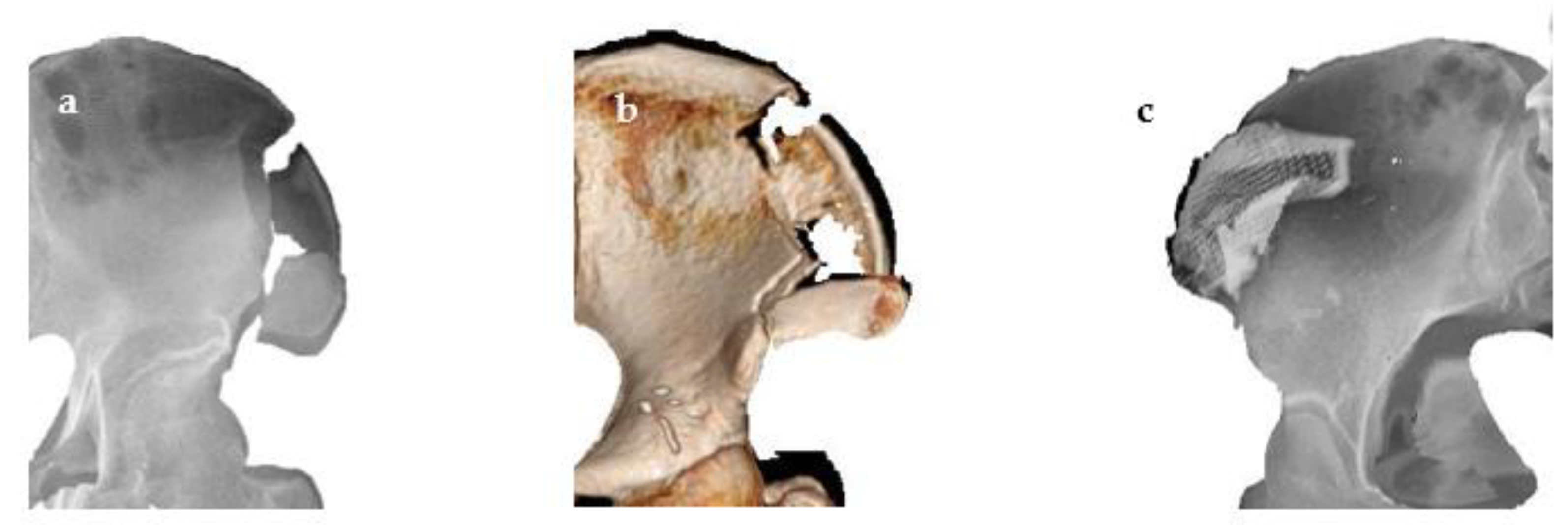
Figure 1. Three-dimensional modeling of fractured patients; (a) the fractured iliac bone images of patient 1. The patient had to undergo open reduction and internal fixation; (b) fractured pelvis of patient 2. Conservative treatment led to the recovery of the fracture site; (c) 3D reconstruction model of patient 3, showing the reduced fracture segment after reduction with a 3D titanium implant.
Applsci 11 08081 g001
Figure 2. Reference points and the distance between reference points of a non-fracture patient. Point A was defined as the contact point by drawing a vertical line on the boundary of the harvested bone segment from the ASIS. Point B was defined as the contact point by drawing a vertical line on the boundary of the harvested bone segment from point C. Point C was defined as the most concave point between the ASIS and anterior inferior iliac spine. (DA: distance between the ASIS and Point A; DB: distance between Point B and C; WA: width at point A; WB: width at point B; SW: midpoint width on the superior part of harvested bone; IW: midpoint width on the inferior part of harvested bone).
Applsci 11 08081 g002
Table 1. Classification of patients by sex.
Non-FractureFractureTotal
Male9211
Female10111
Total19322
p-value0.295
Table 2. Classification of patients by age.
AgeNon-FractureFractureTotal
10–19202
20–29101
30–39101
40–49202
50–59415
60–69707
70–79628
<80101
p-value0.557
Table 3. Distance measurement on fractured patients.
(Mean ± S.D.)
(mm)DADBWAWB
133.3629.1214.8411.15
214.7813.9214.989.32
318.6919.8021.3419.67
Mean22.28 ± 9.8020.95 ± 7.6617.05 ± 3.7113.38 ± 5.52
Table 4. Harvested bone segment on fractured patient.
(Mean ± S.D.)
(mm)HeightLengthSWIW
133.3629.1214.8411.15
214.7813.9214.989.32
318.6919.8021.3419.67
Mean22.28 ± 9.8020.95 ± 7.6617.05 ± 3.7113.38 ± 5.52
Table 5. Comparisons of non-fracture and fracture groups.
(Mean ± S.D.)
(mm)DADBWAWB
Non-fracture16.88 ± 4.5417.29 ± 4.4413.63 ± 1.5810.74 ± 1.63
Fracture22.28 ± 9.8020.95 ± 7.6617.05 ± 3.7113.38 ± 5.52
p-value0.2370.5860.032 *0.705
* There is a statistically significant difference (p < 0.05) (independent t-test).
Table 6. Comparisons of measurement on harvested bone segment.
(Mean ± S.D.)
(mm)HeightLengthSWIW
Non-fracture22.00 ± 5.3711.86 ± 2.0416.75 ± 2.0111.86 ± 2.04
Fracture22.85 ± 5.8310.07 ± 4.3113.65 ± 2.7710.07 ± 4.31
p-value0.9530.4410.1100.441
There is no statistically significant difference (p < 0.05) (independent t-test).
Table 7. Comparisons of ABR period.
(Mean ± S.D.)
ABR Period
Non-fracture10.50 ± 3.61
Fracture5.67 ± 2.31
p-value0.045 *
* There is a statistically significant difference (p < 0.05) (independent t-test).
Publisher’s Note: MDPI stays neutral with regard to jurisdictional claims in published maps and institutional affiliations.