
Recent Advances in Cellulose-Based Structures as the Wound-Healing Biomaterials: A Clinically Oriented Review

 , , , , and
Abstract
:
1. Introduction
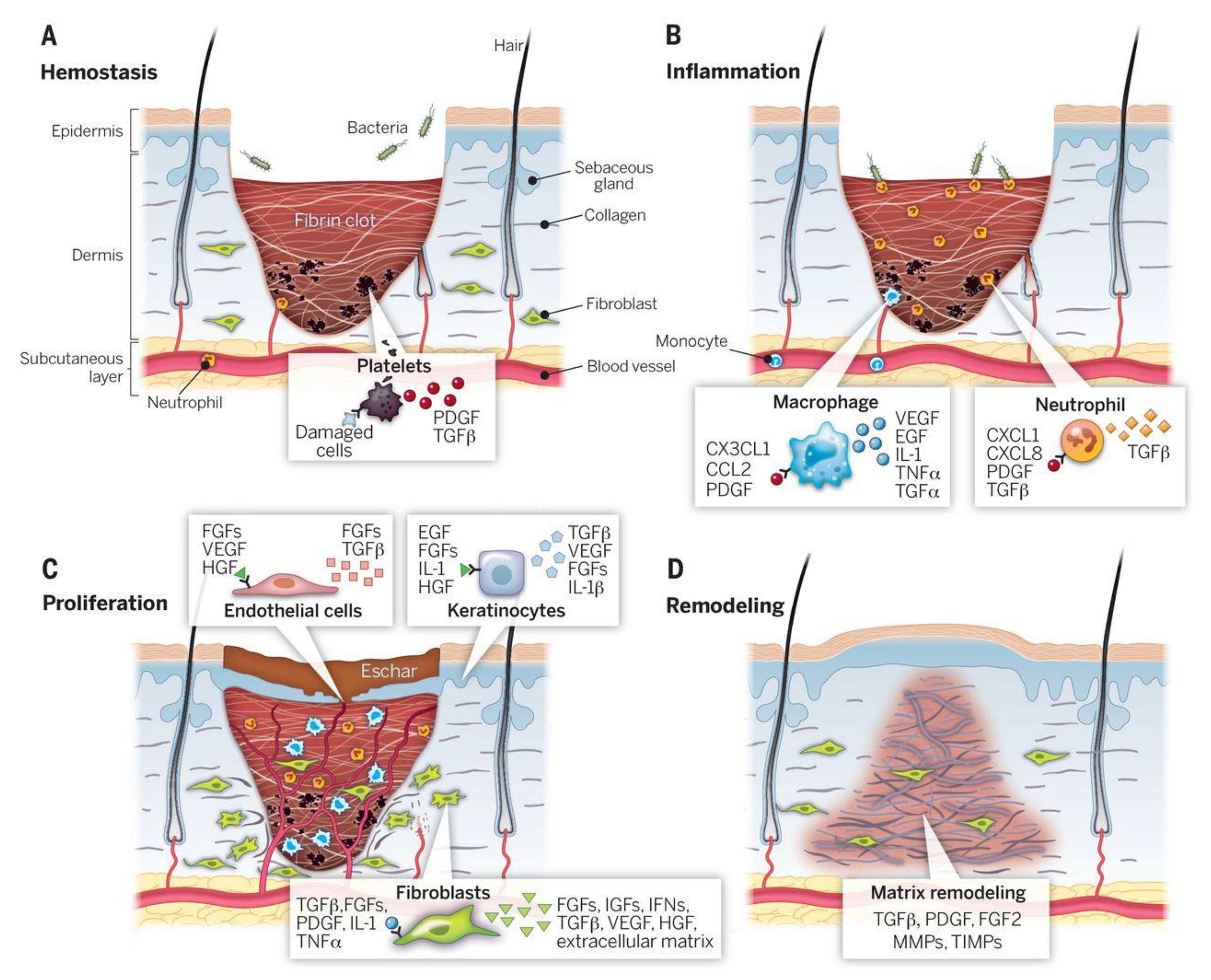
2. Wound Healing Process: The Role of the Wound-Dressing
3. Wound-Healing Challenges in the Clinic
4. The Ideal Wound-Healing/Dressing Materials
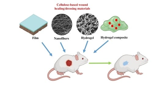
5. Cellulose-Based Biomaterials in Wound-Healing Augmentation
5.1. Cellulose Acetate (CA)
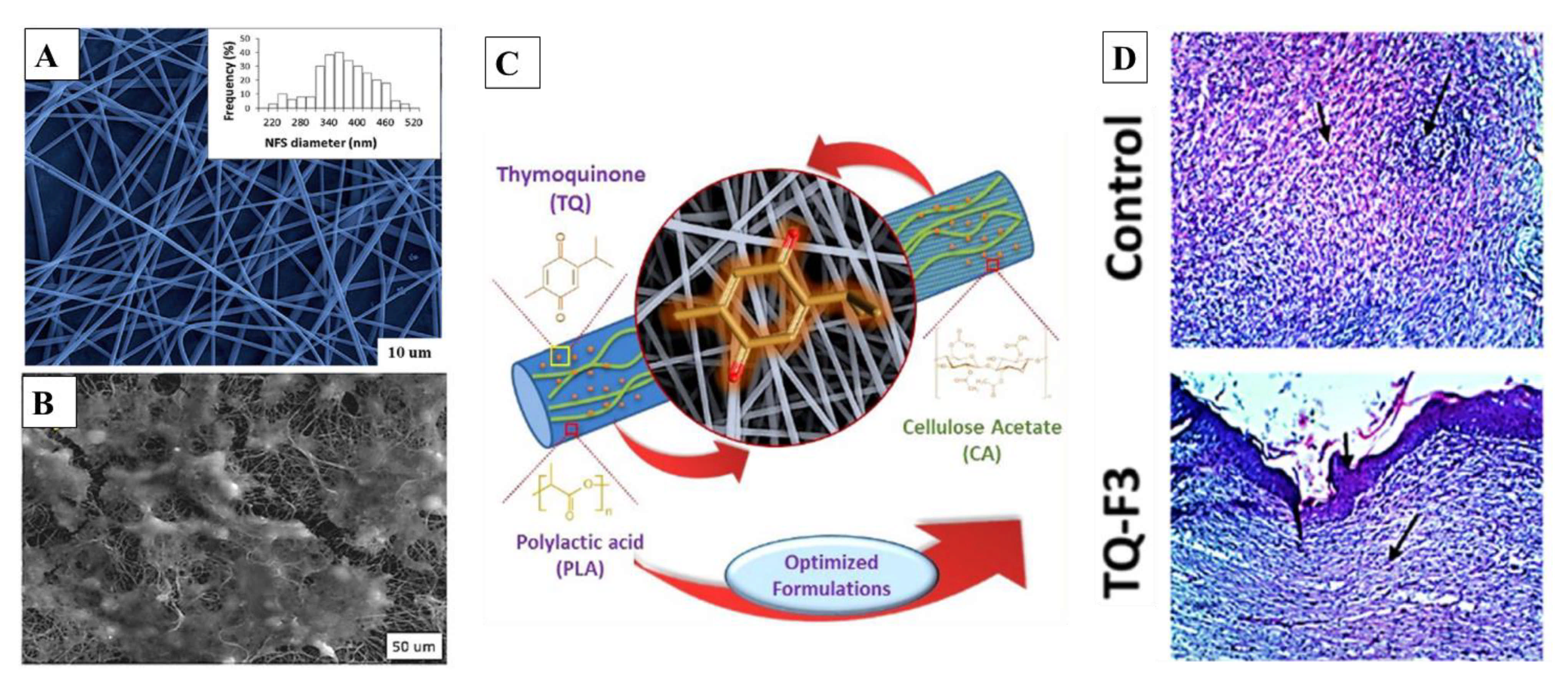
5.2. CA-Based Nanofibers
5.3. CA-Based Hydrogels
6. Carboxymethyl Cellulose (CMC)
6.1. CMC-Based Nanofibers
6.2. CMC-Based Hydrogels
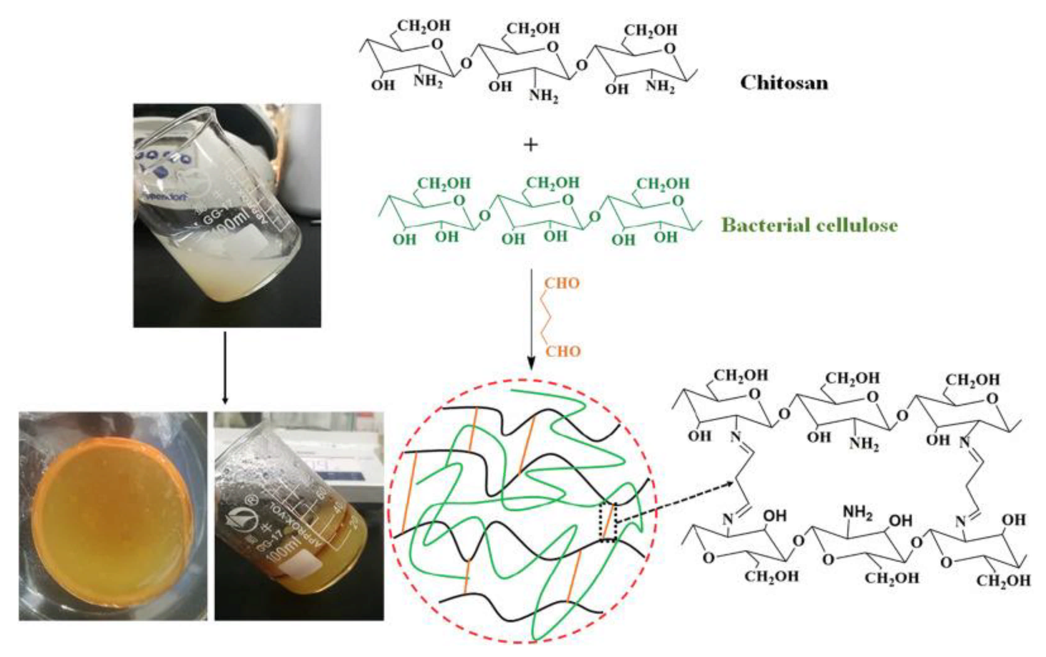
7. Bacterial Cellulose
7.1. BC-Based Nanofibers
7.2. BC-Based Hydrogels
8. Conclusions and Future Prospective
Author Contributions
Funding
Acknowledgments
Conflicts of Interest
References
- Cañedo-Dorantes, L.; Cañedo-Ayala, M. Skin acute wound healing: A comprehensive review. Int. J. Inflamm. 2019, 2019, 3706315. [Google Scholar] [CrossRef]
- Joodaki, H.; Panzer, M.B. Skin mechanical properties and modeling: A review. Proc. Inst. Mech. Eng. Part H 2018, 232, 323–343. [Google Scholar] [CrossRef]
- Gonzalez, A.C.d.O.; Costa, T.F.; Andrade, Z.d.A.; Medrado, A.R.A.P. Wound healing-A literature review. An. Bras. Dermatol. 2016, 91, 614–620. [Google Scholar] [CrossRef] [Green Version]
- Polerà, N.; Badolato, M.; Perri, F.; Carullo, G.; Aiello, F. Quercetin and its natural sources in wound healing management. Curr. Med. Chem. 2019, 26, 5825–5848. [Google Scholar] [CrossRef]
- Nour, S.; Baheiraei, N.; Imani, R.; Khodaei, M.; Alizadeh, A.; Rabiee, N.; Moazzeni, S.M. A review of accelerated wound healing approaches: Biomaterial-assisted tissue remodeling. J. Mater. Sci. Mater. Med. 2019, 30, 120. [Google Scholar] [CrossRef] [PubMed]
- Ehterami, A.; Salehi, M.; Farzamfar, S.; Samadian, H.; Vaez, A.; Sahrapeyma, H.; Ghorbani, S. A promising wound dressing based on alginate hydrogels containing vitamin D3 cross-linked by calcium carbonate/d-glucono-δ-lactone. Biomed. Eng. Lett. 2020, 10, 309. [Google Scholar] [CrossRef] [PubMed]
- Mohammadinejad, R.; Maleki, H.; Larrañeta, E.; Fajardo, A.R.; Nik, A.B.; Shavandi, A.; Sheikhi, A.; Ghorbanpour, M.; Farokhi, M.; Govindh, P. Status and future scope of plant-based green hydrogels in biomedical engineering. Appl. Mater. Today 2019, 16, 213–246. [Google Scholar] [CrossRef] [Green Version]
- Stoica, A.E.; Chircov, C.; Grumezescu, A.M. Nanomaterials for wound dressings: An up-to-date overview. Molecules 2020, 25, 2699. [Google Scholar] [CrossRef]
- Aljghami, M.E.; Saboor, S.; Amini-Nik, S. Emerging innovative wound dressings. Ann. Biomed. Eng. 2019, 47, 659–675. [Google Scholar] [CrossRef] [PubMed]
- Mala, R.; Rani, M.J.; Prasath, N.H.; Celsia, A.R. Nano composite material of chitosan for wound healing: A review. In Proceedings of the AIP Conference Proceedings; AIP Publishing: New York, NY, USA, 2019; p. 020013. [Google Scholar]
- Ranjith, R.; Balraj, S.; Ganesh, J.; Milton, M.J. Therapeutic agents loaded chitosan-based nanofibrous mats as potential wound dressings: A review. Mater. Today Chem. 2019, 12, 386–395. [Google Scholar] [CrossRef]
- Gaspar-Pintiliescu, A.; Stanciuc, A.-M.; Craciunescu, O. Natural composite dressings based on collagen, gelatin and plant bioactive compounds for wound healing: A review. Int. J. Biol. Macromol. 2019, 138, 854–865. [Google Scholar] [CrossRef]
- Obagi, Z.; Damiani, G.; Grada, A.; Falanga, V. Principles of Wound Dressings: A Review. Surg. Technol. Int. 2019, 35, 35. [Google Scholar]
- Zhong, Y.; Xiao, H.; Seidi, F.; Jin, Y. Natural Polymer-Based Antimicrobial Hydrogels without Synthetic Antibiotics as Wound Dressings. Biomacromolecules 2020, 21, 2983–3006. [Google Scholar] [CrossRef]
- Abdul Khalil, H.; Adnan, A.; Yahya, E.B.; Olaiya, N.; Safrida, S.; Hossain, M.; Balakrishnan, V.; Gopakumar, D.A.; Abdullah, C.; Oyekanmi, A. A Review on plant cellulose nanofibre-based aerogels for biomedical applications. Polymers 2020, 12, 1759. [Google Scholar] [CrossRef]
- Rana, A.K.; Frollini, E.; Thakur, V.K. Cellulose nanocrystals: Pretreatments, preparation strategies, and surface functionalization. Int. J. Biol. Macromol. 2021, 182, 1554–1581. [Google Scholar] [CrossRef]
- Suwantong, O.; Ruktanonchai, U.; Supaphol, P. Electrospun cellulose acetate fiber mats containing asiaticoside or Centella asiatica crude extract and the release characteristics of asiaticoside. Polymer 2008, 49, 4239–4247. [Google Scholar] [CrossRef]
- Sun, B.K.; Siprashvili, Z.; Khavari, P.A. Advances in skin grafting and treatment of cutaneous wounds. Science 2014, 346, 941–945. [Google Scholar] [CrossRef] [PubMed]
- Fu, Y.; Wang, L.; Wang, F.; Guan, G.; Hu, X.; Xie, Q.; Wang, W.; King, M.W. Influence of structures on the mechanical and absorption properties of a textile pile debridement material and its biological evaluation. RSC Adv. 2015, 5, 87580–87588. [Google Scholar] [CrossRef]
- FU, Y.-J.; Wang, L.; Wang, F.-J.; Wang, W.-Z.; Meng, S.-Y. Preparation and mechanical properties of a novel textile pad for wound debridement. J. Donghua Univ. 2014, 31, 621–624. [Google Scholar]
- Shi, L.; Carson, D. Collagenase Santyl ointment: A selective agent for wound debridement. J. Wound Ostomy Cont. Nurs. 2009, 36, S12–S16. [Google Scholar] [CrossRef] [PubMed]
- Barrientos, S.; Stojadinovic, O.; Golinko, M.S.; Brem, H.; Tomic-Canic, M. Growth factors and cytokines in wound healing. Wound Repair Regen. 2008, 16, 585–601. [Google Scholar] [CrossRef]
- Grazul-Bilska, A.T.; Johnson, M.L.; Bilski, J.J.; Redmer, D.A.; Reynolds, L.P.; Abdullah, A.; Abdullah, K.M. Wound healing: The role of growth factors. Drugs Today 2003, 39, 787–800. [Google Scholar] [CrossRef]
- Pierce, G.F.; Mustoe, T.A.; Altrock, B.W.; Deuel, T.F.; Thomason, A. Role of platelet-derived growth factor in wound healing. J. Cell. Biochem. 1991, 45, 319–326. [Google Scholar] [CrossRef] [PubMed]
- Flanagan, M. Barriers to the implementation of best practice in wound care. WOUNDS UK 2005, 1, 74. [Google Scholar]
- Demidova-Rice, T.N.; Hamblin, M.R.; Herman, I.M. Acute and impaired wound healing: Pathophysiology and current methods for drug delivery, part 1: Normal and chronic wounds: Biology, causes, and approaches to care. Adv. Skin Wound Care 2012, 25, 304. [Google Scholar] [CrossRef] [Green Version]
- Das, S.; Baker, A.B. Biomaterials and nanotherapeutics for enhancing skin wound healing. Front. Bioeng. Biotechnol. 2016, 4, 82. [Google Scholar] [CrossRef] [PubMed]
- Mahmoudi, M.; Gould, L.J. Opportunities and challenges of the management of chronic wounds: A multidisciplinary viewpoint. Chronic Wound Care Manag. Res. 2020, 7, 27–36. [Google Scholar] [CrossRef]
- Darwin, E.; Tomic-Canic, M. Healing chronic wounds: Current challenges and potential solutions. Curr. Dermatol. Rep. 2018, 7, 296–302. [Google Scholar] [CrossRef] [PubMed]
- Zhao, R.; Liang, H.; Clarke, E.; Jackson, C.; Xue, M. Inflammation in chronic wounds. Int. J. Mol. Sci. 2016, 17, 2085. [Google Scholar] [CrossRef] [PubMed]
- Xu, Z.; Han, S.; Gu, Z.; Wu, J. Advances and impact of antioxidant hydrogel in chronic wound healing. Adv. Healthc. Mater. 2020, 9, 1901502. [Google Scholar] [CrossRef]
- Stewart, P.S.; Bjarnsholt, T. Risk factors for chronic biofilm-related infection associated with implanted medical devices. Clin. Microbiol. Infect. 2020. Available online: https://www.sciencedirect.com/science/article/pii/S1198743X20301312 (accessed on 12 August 2021). [CrossRef] [PubMed]
- Aitzetmüller, M.M.; Machens, H.-G.; Duscher, D. Challenges and Opportunities in Drug Delivery and Wound Healing. In Regenerative Medicine and Plastic Surgery; Springer: Berlin/Heidelberg, Germany, 2019; pp. 27–38. [Google Scholar]
- Rezvani Ghomi, E.; Khalili, S.; Nouri Khorasani, S.; Esmaeely Neisiany, R.; Ramakrishna, S. Wound dressings: Current advances and future directions. J. Appl. Polym. Sci. 2019, 136, 47738. [Google Scholar] [CrossRef] [Green Version]
- Zhao, D.; Zhu, Y.; Cheng, W.; Chen, W.; Wu, Y.; Yu, H. Cellulose-based flexible functional materials for emerging intelligent electronics. Adv. Mater. 2020, 33, 2000619. [Google Scholar] [CrossRef] [PubMed]
- Oprea, M.; Voicu, S.I. Recent advances in composites based on cellulose derivatives for biomedical applications. Carbohydr. Polym. 2020, 247, 116683. [Google Scholar] [CrossRef]
- Atila, D.; Keskin, D.; Tezcaner, A. Cellulose acetate based 3-dimensional electrospun scaffolds for skin tissue engineering applications. Carbohydr. Polym. 2015, 133, 251–261. [Google Scholar] [CrossRef] [PubMed]
- Hamano, F.; Seki, H.; Ke, M.; Gopiraman, M.; Lim, C.T.; Kim, I.S. Cellulose acetate nanofiber mat with honeycomb-like surface structure. Mater. Lett. 2016, 169, 33–36. [Google Scholar] [CrossRef]
- Konwarh, R.; Karak, N.; Misra, M. Electrospun cellulose acetate nanofibers: The present status and gamut of biotechnological applications. Biotechnol. Adv. 2013, 31, 421–437. [Google Scholar] [CrossRef]
- Sultana, N.; Zainal, A. Cellulose acetate electrospun nanofibrous membrane: Fabrication, characterization, drug loading and antibacterial properties. Bull. Mater. Sci. 2016, 39, 337–343. [Google Scholar] [CrossRef] [Green Version]
- Panichpakdee, J.; Pavasant, P.; Supaphol, P. Electrospun cellulose acetate fiber mats containing emodin with potential for use as wound dressing. Chiang Mai J. Sci. 2016, 43, 1249–1259. [Google Scholar]
- Ullah, A.; Ullah, S.; Khan, M.Q.; Hashmi, M.; Nam, P.D.; Kato, Y.; Tamada, Y.; Kim, I.S. Manuka honey incorporated cellulose acetate nanofibrous mats: Fabrication and in vitro evaluation as a potential wound dressing. Int. J. Biol. Macromol. 2020, 155, 479–489. [Google Scholar] [CrossRef] [PubMed]
- Khoshnevisan, K.; Maleki, H.; Samadian, H.; Doostan, M.; Khorramizadeh, M.R. Antibacterial and antioxidant assessment of cellulose acetate/polycaprolactone nanofibrous mats impregnated with propolis. Int. J. Biol. Macromol. 2019, 140, 1260–1268. [Google Scholar] [CrossRef]
- Tsekova, P.B.; Spasova, M.G.; Manolova, N.E.; Markova, N.D.; Rashkov, I.B. Electrospun curcumin-loaded cellulose acetate/polyvinylpyrrolidone fibrous materials with complex architecture and antibacterial activity. Mater. Sci. Eng. C 2017, 73, 206–214. [Google Scholar] [CrossRef]
- Darakhshan, S.; Pour, A.B.; Colagar, A.H.; Sisakhtnezhad, S. Thymoquinone and its therapeutic potentials. Pharmacol. Res. 2015, 95, 138–158. [Google Scholar] [CrossRef] [PubMed]
- Gomaa, S.F.; Madkour, T.M.; Moghannem, S.; El-Sherbiny, I.M. New polylactic acid/cellulose acetate-based antimicrobial interactive single dose nanofibrous wound dressing mats. Int. J. Biol. Macromol. 2017, 105, 1148–1160. [Google Scholar] [CrossRef] [PubMed]
- Unnithan, A.R.; Gnanasekaran, G.; Sathishkumar, Y.; Lee, Y.S.; Kim, C.S. Electrospun antibacterial polyurethane–cellulose acetate–zein composite mats for wound dressing. Carbohydr. Polym. 2014, 102, 884–892. [Google Scholar] [CrossRef] [PubMed]
- Liu, F.; Li, X.; Wang, L.; Yan, X.; Ma, D.; Liu, Z.; Liu, X. Sesamol incorporated cellulose acetate-zein composite nanofiber membrane: An efficient strategy to accelerate diabetic wound healing. Int. J. Biol. Macromol. 2020, 149, 627–638. [Google Scholar] [CrossRef] [PubMed]
- Samadian, H.; Zamiri, S.; Ehterami, A.; Farzamfar, S.; Vaez, A.; Khastar, H.; Alam, M.; Ai, A.; Derakhshankhah, H.; Allahyari, Z. Electrospun cellulose acetate/gelatin nanofibrous wound dressing containing berberine for diabetic foot ulcer healing: In vitro and in vivo studies. Sci. Rep. 2020, 10, 1–12. [Google Scholar] [CrossRef]
- Bajpai, S.; Banger, P. Photopolymerized Cellulose Acetate/Poly (Acrylic Acid) Semi-IPN Films for Wound Dressing Applications. J. Macromol. Sci. A 2014, 51, 756–766. [Google Scholar] [CrossRef]
- Abd El-Mohdy, H. Radiation synthesis of nanosilver/poly vinyl alcohol/cellulose acetate/gelatin hydrogels for wound dressing. J. Polym. Res. 2013, 20, 177. [Google Scholar] [CrossRef]
- Melero, A.M.G.; Senna, A.; Domingues, J.; Hausen, M.; Duek, E.A.R.; Botaro, V. Hydrogel based on cellulose acetate used as scaffold for cell growth. Int. J. Chem. Eng. 2018, 12, 352–362. [Google Scholar]
- Elsayed, M.T.; Hassan, A.A.; Abdelaal, S.A.; Taher, M.M.; Khalaf Ahmed, M.; Shoueir, K.R. Morphological, antibacterial, and cell attachment of cellulose acetate nanofibers containing modified hydroxyapatite for wound healing utilizations. J. Mater. Res. Technol. 2020, 9, 13927–13936. [Google Scholar] [CrossRef]
- Barnthip, N.; Muakngam, A. Preparation of cellulose acetate nanofibers containing centella asiatica extract by electrospinning process as the prototype of wound-healing materials. J. Bionanosci. 2014, 8, 313–318. [Google Scholar] [CrossRef]
- Kurečič, M.; Maver, T.; Virant, N.; Ojstršek, A.; Gradišnik, L.; Hribernik, S.; Kolar, M.; Maver, U.; Kleinschek, K.S. A multifunctional electrospun and dual nano-carrier biobased system for simultaneous detection of pH in the wound bed and controlled release of benzocaine. Cellulose 2018, 25, 7277–7297. [Google Scholar] [CrossRef]
- Ahmed, M.; Menazea, A.; Abdelghany, A. Blend biopolymeric nanofibrous scaffolds of cellulose acetate/ε-polycaprolactone containing metallic nanoparticles prepared by laser ablation for wound disinfection applications. Int. J. Biol. Macromol. 2020, 155, 636–644. [Google Scholar] [CrossRef] [PubMed]
- Farahani, H.; Barati, A.; Arjomandzadegan, M.; Vatankhah, E. Nanofibrous cellulose acetate/gelatin wound dressing endowed with antibacterial and healing efficacy using nanoemulsion of Zataria multiflora. Int. J. Biol. Macromol. 2020, 162, 762–773. [Google Scholar] [CrossRef]
- Gaydhane, M.K.; Kanuganti, J.S.; Sharma, C.S. Honey and curcumin loaded multilayered polyvinylalcohol/cellulose acetate electrospun nanofibrous mat for wound healing. J. Mater. Res. 2020, 35, 600–609. [Google Scholar] [CrossRef]
- López-Calderón, H.D.; Avilés-Arnaut, H.; Galán-Wong, L.J.; Almaguer-Cantú, V.; Laguna-Camacho, J.; Calderón-Ramón, C.; Escalante-Martínez, J.; Arévalo-Niño, K. Electrospun Polyvinylpyrrolidone-Gelatin and Cellulose Acetate Bi-Layer Scaffold Loaded with Gentamicin as Possible Wound Dressing. Polymers 2020, 12, 2311. [Google Scholar] [CrossRef]
- Liu, X.; Lin, T.; Gao, Y.; Xu, Z.; Huang, C.; Yao, G.; Jiang, L.; Tang, Y.; Wang, X. Antimicrobial electrospun nanofibers of cellulose acetate and polyester urethane composite for wound dressing. J. Biomed. Mater. Res. Part B Appl. Biomater. 2012, 100, 1556–1565. [Google Scholar] [CrossRef]
- Bie, X.; Khan, M.Q.; Ullah, A.; Ullah, S.; Kharaghani, D.; Phan, D.-N.; Tamada, Y.; Kim, I.S. Fabrication and characterization of wound dressings containing gentamicin/silver for wounds in diabetes mellitus patients. Mater. Res. Express 2020, 7, 045004. [Google Scholar] [CrossRef]
- Dos Santos, A.E.A.; dos Santos, F.V.; Freitas, K.M.; Pimenta, L.P.S.; de Oliveira Andrade, L.; Marinho, T.A.; de Avelar, G.F.; da Silva, A.B.; Ferreira, R.V. Cellulose acetate nanofibers loaded with crude annatto extract: Preparation, characterization, and in vivo evaluation for potential wound healing applications. Mater. Sci. Eng. C 2020, 118, 111322. [Google Scholar] [CrossRef] [PubMed]
- Elsayed, R.E.; Madkour, T.M.; Azzam, R.A. Tailored-design of electrospun nanofiber cellulose acetate/poly (lactic acid) dressing mats loaded with a newly synthesized sulfonamide analog exhibiting superior wound healing. Int. J. Biol. Macromol. 2020, 164, 1984–1999. [Google Scholar] [CrossRef] [PubMed]
- Wutticharoenmongkol, P.; Hannirojram, P.; Nuthong, P. Gallic acid-loaded electrospun cellulose acetate nanofibers as potential wound dressing materials. Polym. Adv. Technol. 2019, 30, 1135–1147. [Google Scholar] [CrossRef]
- Khan, M.Q.; Kharaghani, D.; Shahzad, A.; Saito, Y.; Yamamoto, T.; Ogasawara, H.; Kim, I.S. Fabrication of antibacterial electrospun cellulose acetate/silver-sulfadiazine nanofibers composites for wound dressings applications. Polym. Test. 2019, 74, 39–44. [Google Scholar] [CrossRef]
- Samadian, H.; Salehi, M.; Farzamfar, S.; Vaez, A.; Ehterami, A.; Sahrapeyma, H.; Goodarzi, A.; Ghorbani, S. In vitro and in vivo evaluation of electrospun cellulose acetate/gelatin/hydroxyapatite nanocomposite mats for wound dressing applications. Artif. Cells Nanomed. Biotechnol. 2018, 46, 964–974. [Google Scholar] [CrossRef] [PubMed] [Green Version]
- Wang, W.; Wang, A. Nanocomposite of carboxymethyl cellulose and attapulgite as a novel pH-sensitive superabsorbent: Synthesis, characterization and properties. Carbohydr. Polym. 2010, 82, 83–91. [Google Scholar] [CrossRef]
- Ninan, N.; Muthiah, M.; Park, I.K.; Elain, A.; Thomas, S.; Grohens, Y. Pectin/carboxymethyl cellulose/microfibrillated cellulose composite scaffolds for tissue engineering. Carbohydr. Polym. 2013, 98, 877–885. [Google Scholar] [CrossRef] [PubMed]
- Singh, B.N.; Panda, N.N.; Mund, R.; Pramanik, K. Carboxymethyl cellulose enables silk fibroin nanofibrous scaffold with enhanced biomimetic potential for bone tissue engineering application. Carbohydr. Polym. 2016, 151, 335–347. [Google Scholar] [CrossRef]
- Joy, J.; Pereira, J.; Aid-Launais, R.; Pavon-Djavid, G.; Ray, A.R.; Letourneur, D.; Meddahi-Pellé, A.; Gupta, B. Gelatin—Oxidized carboxymethyl cellulose blend based tubular electrospun scaffold for vascular tissue engineering. Int. J. Biol. Macromol. 2018, 107, 1922–1935. [Google Scholar] [CrossRef]
- Tripathy, J.; Raichur, A.M. Designing carboxymethyl cellulose based layer-by-layer capsules as a carrier for protein delivery. Colloids Surf. B Biointerfaces 2013, 101, 487–492. [Google Scholar] [CrossRef]
- Amalraj, A.; Gopi, S.; Thomas, S.; Haponiuk, J.T. Cellulose Nanomaterials in Biomedical, Food, and Nutraceutical Applications: A Review. Macromol. Symp. 2018, 380, 1–9. [Google Scholar] [CrossRef]
- Pettignano, A.; Charlot, A.; Fleury, E. Carboxyl-functionalized derivatives of carboxymethyl cellulose: Towards advanced biomedical applications. Polym. Rev. 2019, 59, 510–560. [Google Scholar] [CrossRef]
- Ali, N.H.; Amin, M.C.I.M.; Ng, S.-F. Sodium carboxymethyl cellulose hydrogels containing reduced graphene oxide (rGO) as a functional antibiofilm wound dressing. J. Biomater. Sci. Polym. 2019, 30, 629–645. [Google Scholar] [CrossRef] [PubMed]
- Beele, H.; Meuleneire, F.; Nahuys, M.; Percival, S.L. A prospective randomised open label study to evaluate the potential of a new silver alginate/carboxymethylcellulose antimicrobial wound dressing to promote wound healing. Int. Wound J. 2010, 7, 262–270. [Google Scholar] [CrossRef]
- Rakhshaei, R.; Namazi, H. A potential bioactive wound dressing based on carboxymethyl cellulose/ZnO impregnated MCM-41 nanocomposite hydrogel. Mater. Sci. Eng. C 2017, 73, 456–464. [Google Scholar] [CrossRef]
- Walker, M.; Hobot, J.A.; Newman, G.R.; Bowler, P.G. Scanning electron microscopic examination of bacterial immobilisation in a carboxymethyl cellulose (AQUACEL®) and alginate dressings. Biomaterials 2003, 24, 883–890. [Google Scholar] [CrossRef]
- Capanema, N.S.V.; Mansur, A.A.P.; de Jesus, A.C.; Carvalho, S.M.; de Oliveira, L.C.; Mansur, H.S. Superabsorbent crosslinked carboxymethyl cellulose-PEG hydrogels for potential wound dressing applications. Int. J. Biol. Macromol. 2018, 106, 1218–1234. [Google Scholar] [CrossRef] [PubMed]
- Namazi, H.; Rakhshaei, R.; Hamishehkar, H.; Kafil, H.S. Antibiotic loaded carboxymethylcellulose/MCM-41 nanocomposite hydrogel films as potential wound dressing. Int. J. Biol. Macromol. 2016, 85, 327–334. [Google Scholar] [CrossRef]
- Cai, X.; Hu, S.; Yu, B.; Cai, Y.; Yang, J.; Li, F.; Zheng, Y.; Shi, X. Transglutaminase-catalyzed preparation of crosslinked carboxymethyl chitosan/carboxymethyl cellulose/collagen composite membrane for postsurgical peritoneal adhesion prevention. Carbohydr. Polym. 2018, 201, 201–210. [Google Scholar] [CrossRef]
- Fan, L.; Peng, M.; Zhou, X.; Wu, H.; Hu, J.; Xie, W.; Liu, S. Modification of carboxymethyl cellulose grafted with collagen peptide and its antioxidant activity. Carbohydr. Polym. 2014, 112, 32–38. [Google Scholar] [CrossRef] [PubMed]
- Wiegand, C.; Hipler, U.C. Polymer-based biomaterials as dressings for chronic stagnating wounds. Macromol. Symp. 2010, 294, 1–13. [Google Scholar] [CrossRef]
- Moura, L.I.F.; Dias, A.M.A.; Carvalho, E.; De Sousa, H.C. Recent advances on the development of wound dressings for diabetic foot ulcer treatment–A review. Acta Biomater. 2013, 9, 7093–7114. [Google Scholar] [CrossRef] [PubMed] [Green Version]
- Onofrei, M.; Filimon, A. Cellulose-based hydrogels: Designing concepts, properties, and perspectives for biomedical and environmental applications. Polym. Sci. 2016, 108–120. [Google Scholar]
- Gopiraman, M.; Jatoi, A.W.; Hiromichi, S.; Yamaguchi, K.; Jeon, H.Y.; Chung, I.M.; Ick Soo, K. Silver coated anionic cellulose nanofiber composites for an efficient antimicrobial activity. Carbohydr. Polym. 2016, 149, 51–59. [Google Scholar] [CrossRef]
- Lee, J.H.; Lim, S.-J.; Oh, D.H.; Ku, S.K.; Li, D.X.; Yong, C.S.; Choi, H.-G. Wound healing evaluation of sodium fucidate-loaded polyvinylalcohol/sodium carboxymethylcellulose-based wound dressing. Arch. Pharm. Res. 2010, 33, 1083–1089. [Google Scholar] [CrossRef]
- Maver, T.; Kurečič, M.; Pivec, T.; Maver, U.; Gradišnik, L.; Gašparič, P.; Kaker, B.; Bratuša, A.; Hribernik, S.; Kleinschek, K.S. Needleless electrospun carboxymethyl cellulose/polyethylene oxide mats with medicinal plant extracts for advanced wound care applications. Cellulose 2020, 27, 4487–4580. [Google Scholar] [CrossRef]
- Alipour, R.; Khorshidi, A.; Shojaei, A.F.; Mashayekhi, F.; Moghaddam, M.J.M. Silver Sulfadiazine-loaded PVA/CMC Nanofibers for the Treatment of Wounds Caused by Excision. Fibers Polym. 2019, 20, 2461–2469. [Google Scholar] [CrossRef]
- Almasian, A.; Najafi, F.; Eftekhari, M.; Ardekani, M.R.S.; Sharifzadeh, M.; Khanavi, M. Polyurethane/carboxymethylcellulose nanofibers containing Malva sylvestris extract for healing diabetic wounds: Preparation, characterization, in vitro and in vivo studies. Mater. Sci. Eng. C 2020, 14, 111039. [Google Scholar] [CrossRef] [PubMed]
- Nada, A.A.; Ali, E.A.; Soliman, A.A.; Shen, J.; Abou-Zeid, N.Y.; Hudson, S.M. Multi-layer dressing made of laminated electrospun nanowebs and cellulose-based adhesive for comprehensive wound care. Int. J. Biol. Macromol. 2020, 162, 629–644. [Google Scholar] [CrossRef]
- Shi, D.; Wang, F.; Lan, T.; Zhang, Y.; Shao, Z. Convenient fabrication of carboxymethyl cellulose electrospun nanofibers functionalized with silver nanoparticles. Cellulose 2016, 23, 1899–1909. [Google Scholar] [CrossRef]
- Maver, T.; Kurečič, M.; Smrke, D.M.; Kleinschek, K.S.; Maver, U. Electrospun nanofibrous CMC/PEO as a part of an effective pain-relieving wound dressing. J. Sol-Gel Sci. Technol. 2016, 79, 475–486. [Google Scholar] [CrossRef]
- Kim, S.; Park, S.G.; Kang, S.W.; Lee, K.J. Nanofiber-Based Hydrocolloid from Colloid Electrospinning Toward Next Generation Wound Dressing. Macromol. Mater. Eng. 2016, 301, 818–826. [Google Scholar] [CrossRef]
- Esmaeili, A.; Haseli, M. Optimization, synthesis, and characterization of coaxial electrospun sodium carboxymethyl cellulose-graft-methyl acrylate/poly (ethylene oxide) nanofibers for potential drug-delivery applications. Carbohydr. Polym. 2017, 173, 645–653. [Google Scholar] [CrossRef] [PubMed]
- Joorabloo, A.; Khorasani, M.T.; Adeli, H.; Mansoori-Moghadam, Z.; Moghaddam, A. Fabrication of heparinized nano ZnO/poly (vinylalcohol)/carboxymethyl cellulose bionanocomposite hydrogels using artificial neural network for wound dressing application. J. Ind. Eng. Chem. 2019, 70, 253–263. [Google Scholar] [CrossRef]
- Liu, J.; Zhang, C.; Miao, D.; Sui, S.; Deng, F.; Dong, C.; Zhang, L.; Zhu, P. Preparation and characterization of carboxymethylcellulose hydrogel fibers. J. Eng. Fibers Fabr. 2018, 13, 155892501801300302. [Google Scholar] [CrossRef] [Green Version]
- De Lima, G.G.; de Lima, D.W.; de Oliveira, M.J.; Lugão, A.B.; Alcântara, M.T.; Devine, D.M.; de Sá, M.J. Synthesis and in vivo behavior of PVP/CMC/agar hydrogel membranes impregnated with silver nanoparticles for wound healing applications. ACS Appl. Bio Mater. 2018, 1, 1842–1852. [Google Scholar] [CrossRef]
- Oliveira, R.N.; Moreira, A.P.D.; Thiré, R.M.d.S.M.; Quilty, B.; Passos, T.M.; Simon, P.; Mancini, M.C.; McGuinness, G.B. Absorbent polyvinyl alcohol–sodium carboxymethyl cellulose hydrogels for propolis delivery in wound healing applications. Polym. Eng. Sci. 2017, 57, 1224–1233. [Google Scholar] [CrossRef]
- Gao, Z.; Yu, Z.; Huang, C.; Duan, L.; Gao, G.H. Carboxymethyl cellulose reinforced poly (vinyl alcohol) with trimethylol melamine as a chemical crosslinker. J. Appl. Polym. Sci. 2017, 134, 134. [Google Scholar] [CrossRef]
- Park, J.-S.; An, S.-J.; Jeong, S.-I.; Gwon, H.-J.; Lim, Y.-M.; Nho, Y.-C. Chestnut honey impregnated carboxymethyl cellulose hydrogel for diabetic ulcer healing. Polymers 2017, 9, 248. [Google Scholar] [CrossRef] [PubMed] [Green Version]
- Vinklárková, L.; Masteiková, R.; Vetchý, D.; Doležel, P.; Bernatonienė, J. Formulation of novel layered sodium carboxymethylcellulose film wound dressings with ibuprofen for alleviating wound pain. Biomed Res. Int. 2015, 2015, 1–11. [Google Scholar] [CrossRef] [Green Version]
- Li, D.; Ye, Y.; Li, D.; Li, X.; Mu, C. Biological properties of dialdehyde carboxymethyl cellulose crosslinked gelatin–PEG composite hydrogel fibers for wound dressings. Carbohydr. Polym. 2016, 137, 508–514. [Google Scholar] [CrossRef]
- Das, A.; Kumar, A.; Patil, N.B.; Viswanathan, C.; Ghosh, D. Preparation and characterization of silver nanoparticle loaded amorphous hydrogel of carboxymethylcellulose for infected wounds. Carbohydr. Polym. 2015, 130, 254–261. [Google Scholar] [CrossRef]
- Kawasaki, T.; Nakaji-Hirabayashi, T.; Masuyama, K.; Fujita, S.; Kitano, H. Complex film of chitosan and carboxymethyl cellulose nanofibers. Colloids Surf. B Biointerfaces 2016, 139, 95–99. [Google Scholar] [CrossRef] [Green Version]
- Esmaeili, A.; Haseli, M. Electrospinning of thermoplastic carboxymethyl cellulose/poly(ethylene oxide) nanofibers for use in drug-release systems. Mater. Sci. Eng. C 2017, 77, 1117–1127. [Google Scholar] [CrossRef]
- Teixeira, M.A.; Paiva, M.C.; Amorim, M.T.P. Electrospun Nanocomposites Containing Cellulose and Its Derivatives Modified with Specialized Biomolecules for an Enhanced Wound Healing. Nanomaterials 2020, 10, 557. [Google Scholar] [CrossRef] [PubMed] [Green Version]
- Liu, M.; Duan, X.-P.; Li, Y.-M.; Yang, D.-P.; Long, Y.-Z. Electrospun nanofibers for wound healing. Mater. Sci. Eng. C 2017, 76, 1413–1423. [Google Scholar] [CrossRef] [PubMed]
- Farbodi, M.; Khoshkbar Sadeghi, A.R. Preparation of Carboxymethyl Cellulose/Calcium Alginate/Polyvinyl Alcohol/Silver Nanocomposite by Electrospinning Method and Its Performance as Wound Dressing. Modares J. Biotechnol. 2018, 9, 593–601. [Google Scholar]
- Hebeish, A.; Sharaf, S. Novel nanocomposite hydrogel for wound dressing and other medical applications. RSC Adv. 2015, 5, 103036–103046. [Google Scholar] [CrossRef]
- Agarwal, R.; Alam, M.S.; Gupta, B. Polyvinyl alcohol-polyethylene oxide-carboxymethyl cellulose membranes for drug delivery. J. Appl. Polym. Sci. 2013, 129, 3728–3736. [Google Scholar] [CrossRef]
- Agarwal, R.; Sarwar Alam, M.; Gupta, B. Preparation of curcumin loaded poly(Vinyl Alcohol)-poly(Ethylene Oxide)-carboxymethyl cellulose membranes for wound care application. J. Biomater. Tissue Eng. 2013, 3, 273–283. [Google Scholar] [CrossRef]
- Salama, A.; El-Sakhawy, M.; Kamel, S. Carboxymethyl cellulose based hybrid material for sustained release of protein drugs. Int. J. Biol. Macromol. 2016, 93, 1647–1652. [Google Scholar] [CrossRef] [PubMed]
- Huang, Y.; Zhu, C.; Yang, J.; Nie, Y.; Chen, C.; Sun, D. Recent advances in bacterial cellulose. Cellulose 2014, 21, 1–30. [Google Scholar] [CrossRef]
- Picheth, G.F.; Pirich, C.L.; Sierakowski, M.R.; Woehl, M.A.; Sakakibara, C.N.; de Souza, C.F.; Martin, A.A.; da Silva, R.; de Freitas, R.A. Bacterial cellulose in biomedical applications: A review. Int. J. Biol. Macromol. 2017, 104, 97–106. [Google Scholar] [CrossRef]
- Sulaeva, I.; Henniges, U.; Rosenau, T.; Potthast, A. Bacterial cellulose as a material for wound treatment: Properties and modifications: A review. Biotechnol. Adv. 2015, 33, 1547–1571. [Google Scholar] [CrossRef] [PubMed]
- Fu, L.; Zhang, J.; Yang, G. Present status and applications of bacterial cellulose-based materials for skin tissue repair. Carbohydr. Polym. 2013, 92, 1432–1442. [Google Scholar] [CrossRef] [PubMed]
- Czaja, W.; Krystynowicz, A.; Bielecki, S.; Brown, R.M. Microbial cellulose–The natural power to heal wounds. Biomaterials 2006, 27, 145–151. [Google Scholar] [CrossRef] [PubMed]
- Helenius, G.; Bäckdahl, H.; Bodin, A.; Nannmark, U.; Gatenholm, P.; Risberg, B. In vivo biocompatibility of bacterial cellulose. J. Biomed. Mater. Res. A 2006, 76, 431–438. [Google Scholar] [CrossRef] [PubMed]
- Dahman, Y. Nanostructured biomaterials and biocomposites from bacterial cellulose nanofibers. J. Nanosci. Nanotechnol. 2009, 9, 5105–5122. [Google Scholar] [CrossRef]
- Kucińska-Lipka, J.; Gubanska, I.; Janik, H. Bacterial cellulose in the field of wound healing and regenerative medicine of skin: Recent trends and future prospectives. Polym. Bull. 2015, 72, 2399–2419. [Google Scholar] [CrossRef]
- Abeer, M.M.; Mohd Amin, M.C.I.; Martin, C. A review of bacterial cellulose-based drug delivery systems: Their biochemistry, current approaches and future prospects. J. Pharm. Pharmacol. 2014, 66, 1047–1061. [Google Scholar] [CrossRef] [PubMed]
- Eslahi, N.; Mahmoodi, A.; Mahmoudi, N.; Zandi, N.; Simchi, A. Processing and properties of nanofibrous bacterial cellulose-containing polymer composites: A review of recent advances for biomedical applications. Polym. Rev. 2020, 60, 144–170. [Google Scholar] [CrossRef]
- Gorgieva, S.; Trček, J. Bacterial Cellulose: Production, Modification and Perspectives in Biomedical Applications. Nanomaterials 2019, 9, 1352. [Google Scholar] [CrossRef] [PubMed] [Green Version]
- Maneerung, T.; Tokura, S.; Rujiravanit, R. Impregnation of silver nanoparticles into bacterial cellulose for antimicrobial wound dressing. Carbohydr. Polym. 2008, 72, 43–51. [Google Scholar] [CrossRef]
- Wu, J.; Zheng, Y.; Song, W.; Luan, J.; Wen, X.; Wu, Z.; Chen, X.; Wang, Q.; Guo, S. In situ synthesis of silver-nanoparticles/bacterial cellulose composites for slow-released antimicrobial wound dressing. Carbohydr. Polym. 2014, 102, 762–771. [Google Scholar] [CrossRef] [PubMed]
- Yuan, N.; Xu, L.; Zhang, L.; Ye, H.; Zhao, J.; Liu, Z.; Rong, J. Superior hybrid hydrogels of polyacrylamide enhanced by bacterial cellulose nanofiber clusters. Mater. Sci. Eng. C 2016, 67, 221–230. [Google Scholar] [CrossRef]
- Sureshkumar, M.; Siswanto, D.Y.; Lee, C.K. Magnetic antimicrobial nanocomposite based on bacterial cellulose and silver nanoparticles. J. Mater. Chem. 2010, 20, 6948–6955. [Google Scholar] [CrossRef]
- Pal, S.; Nisi, R.; Stoppa, M.; Licciulli, A. Silver-Functionalized Bacterial Cellulose as Antibacterial Membrane for Wound-Healing Applications. ACS Omega 2017, 2, 3632–3639. [Google Scholar] [CrossRef] [Green Version]
- Azarniya, A.; Eslahi, N.; Mahmoudi, N.; Simchi, A. Effect of graphene oxide nanosheets on the physico-mechanical properties of chitosan/bacterial cellulose nanofibrous composites. Compos. A Appl. Sci. Manuf. 2016, 85, 113–122. [Google Scholar] [CrossRef] [Green Version]
- Jiang, F.; Yin, L.; Yu, Q.; Zhong, C.; Zhang, J. Bacterial cellulose nanofibrous membrane as thermal stable separator for lithium-ion batteries. J. Power Sources 2015, 279, 21–27. [Google Scholar] [CrossRef]
- Morena, A.G.; Roncero, M.B.; Valenzuela, S.V.; Valls, C.; Vidal, T.; Pastor, F.I.J.; Diaz, P.; Martínez, J. Laccase/TEMPO-mediated bacterial cellulose functionalization: Production of paper-silver nanoparticles composite with antimicrobial activity. Cellulose 2019, 26, 8655–8668. [Google Scholar] [CrossRef]
- Hu, W.; Chen, S.; Li, X.; Shi, S.; Shen, W.; Zhang, X.; Wang, H. In situ synthesis of silver chloride nanoparticles into bacterial cellulose membranes. Mater. Sci. Eng. C 2009, 29, 1216–1219. [Google Scholar] [CrossRef]
- Khalid, A.; Khan, R.; Ul-Islam, M.; Khan, T.; Wahid, F. Bacterial cellulose-zinc oxide nanocomposites as a novel dressing system for burn wounds. Carbohydr. Polym. 2017, 164, 214–221. [Google Scholar] [CrossRef] [PubMed]
- Lin, S.P.; Kung, H.N.; Tsai, Y.S.; Tseng, T.N.; Hsu, K.D.; Cheng, K.C. Novel dextran modified bacterial cellulose hydrogel accelerating cutaneous wound healing. Cellulose 2017, 24, 4927–4937. [Google Scholar] [CrossRef]
- Favi, P.M.; Benson, R.S.; Neilsen, N.R.; Hammonds, R.L.; Bates, C.C.; Stephens, C.P.; Dhar, M.S. Cell proliferation, viability, and in vitro differentiation of equine mesenchymal stem cells seeded on bacterial cellulose hydrogel scaffolds. Mater. Sci. Eng. C 2013, 33, 1935–1944. [Google Scholar] [CrossRef] [PubMed]
- Fricain, J.C.; Granja, P.L.; Barbosa, M.A.; de Jéso, B.; Barthe, N.; Baquey, C. Cellulose phosphates as biomaterials. In vivo biocompatibility studies. Biomaterials 2002, 23, 971–980. [Google Scholar] [CrossRef]
- Loh, E.Y.X.; Mohamad, N.; Fauzi, M.B.; Ng, M.H.; Ng, S.F.; Mohd Amin, M.C.I. Development of a bacterial cellulose-based hydrogel cell carrier containing keratinocytes and fibroblasts for full-thickness wound healing. Sci. Rep. 2018, 8, 1–12. [Google Scholar] [CrossRef] [PubMed] [Green Version]
- Wahid, F.; Hu, X.H.; Chu, L.Q.; Jia, S.R.; Xie, Y.Y.; Zhong, C. Development of bacterial cellulose/chitosan based semi-interpenetrating hydrogels with improved mechanical and antibacterial properties. Int. J. Biol. Macromol. 2019, 122, 380–387. [Google Scholar] [CrossRef]
- Li, Y.; Jiang, H.; Zheng, W.; Gong, N.; Chen, L.; Jiang, X.; Yang, G. Bacterial cellulose-hyaluronan nanocomposite biomaterials as wound dressings for severe skin injury repair. J. Mater. Chem. B 2015, 3, 3498–3507. [Google Scholar] [CrossRef]
- Zmejkoski, D.; Spasojević, D.; Orlovska, I.; Kozyrovska, N.; Soković, M.; Glamočlija, J.; Dmitrović, S.; Matović, B.; Tasić, N.; Maksimović, V. Bacterial cellulose-lignin composite hydrogel as a promising agent in chronic wound healing. Int. J. Biol. Macromol. 2018, 118, 494–503. [Google Scholar] [CrossRef] [Green Version]
- Cam, M.E.; Crabbe-Mann, M.; Alenezi, H.; Hazar-Yavuz, A.N.; Ertas, B.; Ekentok, C.; Ozcan, G.S.; Topal, F.; Guler, E.; Yazir, Y. The comparision of glybenclamide and metformin-loaded bacterial cellulose/gelatin nanofibres produced by a portable electrohydrodynamic gun for diabetic wound healing. Eur. Polym. J. 2020, 134, 109844. [Google Scholar] [CrossRef]
- Azarniya, A.; Tamjid, E.; Eslahi, N.; Simchi, A. Modification of bacterial cellulose/keratin nanofibrous mats by a tragacanth gum-conjugated hydrogel for wound healing. Int. J. Biol. Macromol. 2019, 134, 280–289. [Google Scholar] [CrossRef]
- Aydogdu, M.O.; Altun, E.; Crabbe-Mann, M.; Brako, F.; Koc, F.; Ozen, G.; Kuruca, S.E.; Edirisinghe, U.; Luo, C.; Gunduz, O. Cellular interactions with bacterial cellulose: Polycaprolactone nanofibrous scaffolds produced by a portable electrohydrodynamic gun for point-of-need wound dressing. Int. Wound J. 2018, 15, 789–797. [Google Scholar] [CrossRef]
- Qiu, Y.; Qiu, L.; Cui, J.; Wei, Q. Bacterial cellulose and bacterial cellulose-vaccarin membranes for wound healing. Mater. Sci. Eng. C 2016, 59, 303–309. [Google Scholar] [CrossRef] [PubMed]
- Ifuku, S.; Tsuji, M.; Morimoto, M.; Saimoto, H.; Yano, H. Synthesis of silver nanoparticles templated by TEMPO-mediated oxidized bacterial cellulose nanofibers. Biomacromolecules 2009, 10, 2714–2717. [Google Scholar] [CrossRef]
- Tsai, Y.-H.; Yang, Y.-N.; Ho, Y.-C.; Tsai, M.-L.; Mi, F.-L. Drug release and antioxidant/antibacterial activities of silymarin-zein nanoparticle/bacterial cellulose nanofiber composite films. Carbohydr. Polym. 2018, 180, 286–296. [Google Scholar] [CrossRef] [PubMed]
- Ardila, N.; Medina, N.; Arkoun, M.; Heuzey, M.-C.; Ajji, A.; Panchal, C.J. Chitosan–bacterial nanocellulose nanofibrous structures for potential wound dressing applications. Cellulose 2016, 23, 3089–3104. [Google Scholar] [CrossRef]
- Chuah, C.; Wang, J.; Tavakoli, J.; Tang, Y. Novel bacterial cellulose-poly (acrylic acid) hybrid hydrogels with controllable antimicrobial ability as dressings for chronic wounds. Polymers 2018, 10, 1323. [Google Scholar] [CrossRef] [Green Version]
- Volova, T.; Shumilova, A.; Nikolaeva, E.; Kirichenko, A.; Shishatskaya, E. Biotechnological wound dressings based on bacterial cellulose and degradable copolymer P (3HB/4HB). Int. J. Biol. Macromol. 2019, 131, 230–240. [Google Scholar] [CrossRef] [Green Version]
- Spaic, M.; Small, D.P.; Cook, J.R.; Wan, W. Characterization of anionic and cationic functionalized bacterial cellulose nanofibres for controlled release applications. Cellulose 2014, 21, 1529–1540. [Google Scholar] [CrossRef]
- Ciechanska, D. Multifunctional bacterial cellulose/chitosan composite materials for medical applications. Fibres Text. East. Eur. 2004, 12, 69–72. [Google Scholar]


| Components and Type | Fabrication Method | Bioactive Substance | Test Model | Significant Findings | Ref. |
|---|---|---|---|---|---|
| Cu-HAP@CA nanofibers | Electrospinning | Cu-modified HA | Human HFB4 osteoblast S. aureus E. coli | Significant antibacterial activates Proper cell adhesion and proliferation | [53] |
| CA nanofibers | Electrospinning | Centella Asiatica extract | NA | Excellent water uptake kinetic | [54] |
| CA-Zein nanofibers | Electrospinning | Sesamol | STZ-induced diabetic C57BL/6 J clean-grade male mice | Promoted wound-healing in diabetic mice Myofibroblasts’ formation through enhancing TGF-β signaling pathway transduction Enhanced keratinocyte proliferation | [48] |
| CA nanofibers | Electrospinning | PAC CACE | Normal human dermal fibroblasts | Modified release profile Biocompatible wound-dressing | [17] |
| CA/PVP nanofibers | One-pot or dual spinneret electrospinning | Curcumin | S. aureus | Higher hydrophilicity and faster release through the addition of PVP Dual spinneret electrospinning provide more complex architecture, beneficial for easily modulating the release profile and proper antibacterial activities | [44] |
| CA/MH nanofibers | Electrospinning | MH | NIH 3T3 fibroblast cell S. aureus E. coli | Biocompatible, antioxidant, and antibacterial wound-dressing | [42] |
| CA nanofibers | Electrospinning | BZC BCG | Human skin fibroblast cells | Biocompatible nanofibrous mats with an in situ pH-detecting capability pH-dependent release of BZC | [55] |
| CA/PCL nanofibers | Electrospinning | ZnO, Ag and CuO NPs | Human osteoblast cell line HFB4 S. aureus E. coli | Biocompatible and antibacterial wound-dressing | [56] |
| CA/Gel nanofibers | Electrospinning | ZM | Fibroblast L929 Cells S. aureus E. coli Male Wistar rats | Biocompatible and antibacterial wound-dressing Accelerated sound healing in animal model | [57] |
| Multilayered CA/PVA nanofibers | Electrospinning | Curcumin Honey | S. aureus | Antioxidant and antibacterial wound-dressing with proper swelling performance | [58] |
| Multilayered Gel/PVP/CA nanofibers | Electrospinning | Gentamicin | P. aeruginosa S. aureus | Excellent antibacterial activities | [59] |
| PEU/CA nanofibers | One-pot or dual spinneret electrospinning | PHMB | Rat skin fibroblast E. coli Male Wistar rats | Strong antibacterial activities Proper biocompatibility and significant wound-healing outcomes | [60] |
| CA/Ag NPs nanofibers | Electrospinning and Simple facile wetting process | Gentamicin | B. Subtilis E. coli | Significant antibacterial properties Controlled release | [61] |
| CA nanofibers | Electrospinning | Annatto extract | Mouse WT fibroblasts Male Wistar rats | Biocompatible wound-dressing with dose/response inflammatory effects on mast cells | [62] |
| CA/PLA/PEO nanofibers | Electrospinning | Sulfonamide HBSP analog | Human dermal fibroblast cells (HFB4) S. aureus E. coli Bulb/c male mice | Proper biocompatibility Strong antibacterial activities Significant wound-healing | [63] |
| CA nanofibers | Electrospinning | GA | S. aureus | Antioxidant and antibacterial wound-dressing | [64] |
| CA nanofibers | Electrospinning | SDD | B. Subtilis E. coli | Strong antibacterial activities | [65] |
| CA/HA nanofibers | Electrospinning | HA | L929 murine fibroblastic cell Male Wistar rats | Biocompatible and bioactive wound-dressing Significant wound-healing | [66] |
| CA/PCL nanofibers | Electrospinning | Propolis | S. aureus S. epidermidis E. coli P. aeruginosa | Significant antioxidant and antibacterial activities | [43] |
| CA/AAc Semi-IPN hydrogel | BP-induced free radical photo-polymerization | NA | A. flavus E. coli | Proper mechanical properties with permeable to moisture and O2 gas Acceptable antibacterial and antifungal activities | [50] |
| CA/PVA/Gel Hydrogel | Gamma radiation-induced crosslinking | Ag NPs | A. fumigatus G. candidum C. albicans S. racemosum B. subtilis S. aureus P. aeruginosa E. coli | Well-dispersed and spherical Ag NPs within hydrogel matrix Strong antibacterial and antifungal activities | [51] |
| Type of Structure | Chemical Modification | Fabrication Method | Bioactive Substance | The Test Model | Significant Findings | Ref. |
|---|---|---|---|---|---|---|
| Nanofibers | ||||||
| CMC/PEO | Co-blending | Needleless electrospinning | Plant extracts: Achillea millefolium Calendula officinalis Matricaria chamomilla Echinacea purpurea | S. aureus E. coli Human skin fibroblasts (SF) Human skin keratinocytes (HACAT) | Proper antioxidant activity Acceptable antibacterial effect Good biocompatibility | [87] |
| CMC/PAV | Co-blending | Electrospinning | SSD | S. aureus E. coli P. aeruginosa New Zealand white rabbits HSF-PI 18 fibroblast-like cells | Significant antibacterial effects Proper biocompatibility Accelerated wound-healing | [88] |
| CMC/PU | Co-blending | Malva sylvestris extract | S. aureus E. coli Mesenchymal stem cells Male Wister rats | Significant antibacterial and antioxidant activities Cell proliferative effect Reduced acute and chronic Inflammations Promoted wound closure Increased collagen deposition and neo-vascularization | [89] | |
| MMA- EHA-CMC PVA | Graft copolymerization with MMA and EHA | Casting Electrospinning | Diclofenac sodium CI Capsaicin Gentamicin | human skin fibroblasts (HFB4) Male wistar rats | Insignificant adverse effects on cells Accelerated wound-healing from 21 to 7 days | [90] |
| CMC/Ag NPs | Co-blending | Electrospinning | Ag NPs | S. aureus E. coli | Uniform distribution of nanoparticles and complete antibacterial efficiency | [91] |
| CMC/PEO | Co-blending | Electrospinning | Diclofenac | Automated Transdermal Diffusion Cells Sampling System | Similarity with extracellular matrix and accelerated wound-healing compared to commercial Aquacel® | [92] |
| CMC/TPU | Co-blending | Colloid electrospinning | NA | ICR mouse | High surface area, proper wettability, and open wound-healing improvement | [93] |
| NaCMC-MA-PEO | Graft polymerization with MA and PEO | Coaxial electrospinning | TCH | Human fibroblast (HDF) S. aureus E. coli | Proper drug incorporation, antibacterial activity, and high cell viability | [94] |
| Hydrogels | ||||||
| PVA-CMC-PEG hydrogels | Hydrogen bond interaction | Phase separation | NA | L929 cells New Zealand white rabbits | Proper mechanical properties Acceptable hemocompatiblity and biocompatibility Enhanced wound-healing | [58] |
| Heparinized Nano ZnO/PVA/CMC | Hydrogen bond interaction | Co-blending | Heparinized Nano ZnO | Mouse fibroblast cells (L-929) Human dermal fibroblast (HDF) S. aureus E. coli | Enhanced mechanical properties Strong antibacterial effects Acceptable biocompatibility | [95] |
| CMC hydrogel fibers | Aluminum sulfate crosslinking | Solution spinning | NA | NA | Excellent moisture absorption performance CMC-dependent mechanical properties | [96] |
| CMC/PEG/PVP/Ag NPs | - | Gamma-rays crosslinking | Ag NPs | L929 cell line New Zealand white rabbits | Monodisperse NPs synthesis No signs of toxicity in vitro Enhanced wound-healing through the high intensity of fibroblasts and neo-vascularization | [97] |
| CMC/Ag NPs/mesoporous silica | - | Citric acid crosslinking | Zinc oxide impregnated mesoporous silica Tetracycline | Adipose tissue-derived stem cells (ADSCs) S. aureus E. coli | Flexible nanocomposite hydrogel films with improved tensile strength, swelling, erosion, and gas permeability performances Excellent biocompatibility | [76] |
| NaCMC/PVA | - | Freeze–thawed | Propolis | S. aureus | Flexible hydrogel with proper antioxidant, antibacterial, and biocompatibility properties | [98] |
| CMC-PVA | - | Trimethylol melamine crosslinking | NA | NA | Hydrogels with high mechanical behavior and homogeneous porous structure | [99] |
| NaCMC | - | Gamma-rays crosslinking | Chestnut Honey | S. aureus E. coli db/db mice | Acceptable antibacterial activities Enhanced wound-healing | [100] |
| CMC | - | Hydrogel-solvent casting | Ibuprofen | Compartmentalized in vitro drug release system | Adequate mechanical and swelling properties, proper pH, and controllable drug release | [101] |
| CMC/mesoporous silica | - | Hydrogel-sonication | Tetracycline Methylene blue | S. aureus | Improved swelling, water vapor, and oxygen permeability, and a significant antibacterial activity | [79] |
| DCMC/Gel/PEG | - | Hydrogel-gel spinning and crosslinking | NA | L929 fibroblasts | High mechanical property and low toxicity | [102] |
| CMC/Ag NPs | - | Hydrogel in situ reduction of silver nitrate | Ag NPs | Primary fibroblasts and keratinocytes | High saline absorbance, moisture donation, proper cytocompatibility, and anti-microbial activity | [103] |
| CMC | Hydrogel-freeze-drying | Sericin | Human keratinocytes | Adequate release of growth factor, no cytotoxicity, and low inflammatory response | [103] | |
| NaCMC/rGO | - | Hydrogel-ionic interactions | NA | C. elegans S. aureus P. aeruginosa Primary human dermal fibroblast cells (HDF) | Cell biocompatibility and effective antibiofilm activity | [74] |
| Type of Structure | Fabrication Method | Bioactive Substance | The Test Model | Significant Findings | Ref. |
|---|---|---|---|---|---|
| BC/Gel Nanofibers | Portable, point of need electrospinning set up | GB Met | L929 fibroblast Adult male and female Sprague- Dawley rats | The BC/Gel/GB group induced a stronger wound-healing role compared to the BC/Gel/Met group The highest decrease of TNF-α level induced by BC/Gel/GB | [141] |
| BC/keratin/TG Nanofibers | Electrospinning Nonionic triblock copolymers conjugation | NA | L929 fibroblast | The hydrogel particles were uniformly embedded into fibrous network nanofibers with improved the hydrophilicity (~23%), module of elasticity (~31%), tensile strength (~35%), and ductility (~23%) | [142] |
| BC/PCL Nanofibers | Portable, point of need electrospinning set up | NA | Saos-2 cell | Good biocompatibility with ability to control the proliferation by adjusting the ratio of polymers | [143] |
| BC/BC-Vac Nanofibers | Bacterial synthesis by Gluconacetobacter xylinum | Vaccarin | L929 fibroblast ICR male mice | Proper biocompatibility Enhanced wound-healing | [144] |
| BC/Ag NPs Nanofibers | In situ Ag NPs formation | Ag NPs | S. aureus P. aeruginosa E. coli Westar fetal rats | Anti-bacterial and biocompatible wound-dressing Enhanced wound-healing with the lowest inflammation | [125] |
| BC/Ag NPs Nanofibers | TEMPO-mediated oxidation of BC | Ag NPs | NA | Narrow size NPs properly dispersed with high density on nanofibers | [145] |
| BC/Ag NPs Nanofibers | Nanofiber-pH adjusting | Ag NPs | E. coli B. subtilis | Strong anti-microbial activity | [127] |
| BC/Zein Nanofibers | Bacterial biosynthesis and immersion in colloidal suspensions | SMN | S. aureus E. coli | Significant antioxidant and antibacterial activities | [146] |
| BC/chitosan Nanofibers | Coaxial and simultaneous electrospinning | NA | E. coli | Coaxial system produced desired nanofibrous mat with antibacterial activites | [147] |
| BC/PAA Hydrogel | EDC/NHS crosslinking | Amoxicillin | NA | pH-responsive wound-dressing with proper mechanical properties | [148] |
| BC/lignin Hydrogel | Hydrogel-mixing and air-drying | Lignin | S. typhimurium P. aeruginosa L. monocytogenes S. aureus | Successful in vitro gradual release of lignin as a strong antibacterial substance | [140] |
| BC/hyaluronan Hydrogel | Solution impregnation method | NA | Primary human fibroblasts Female Wistar rats | Water uptake capability, breathable structure, cell biocompatibility, and optimized in vivo wound-healing at 0.1% hyaluronan | [139] |
| BC/P (3HB/4HB) Hydrogel | - | Actovegin | Adipose-derived MSCs Female Wistar rats | Significant accelerated wound-healing | [149] |
| Oxidized and aminated BC Hydrogel | Hydrogel-TEMPO-catalyzed oxidation | NA | NA | Superior colloidal stability and pH-dependent water-retention property | [150] |
| Semi-IPN BC/chitosan Hydrogel | Hydrogel-slurry blending and chemical crosslinking | S. aureus E. coli | High mechanical properties and BC/chitosan ratio-dependent antibacterial properties | [138] | |
| BC/chitosan Hydrogel | DNS method | N.A | S. aureus E. coli | Good mechanical properties in wet state, High moisture-keeping properties under lysozyme action Strong antibacterial activities | [151] |
| BC/PAA Hydrogel | Hydrogel-freeze-drying | NA | Epidermal keratinocytes Dermal fibroblasts Male athymic mice | Excellent cell attachment, maintained cell viability with limited migration, and allowed cell transfer accelerated wound-healing | [137] |
| BC/dextran Hydrogel | Hydrogel-freeze-drying | NIH/3T3 mouse fibroblasts C57BL/6 mice | Enhanced cell proliferation, no cytotoxicity, and accelerated in vivo wound-healing | [134] |
Publisher’s Note: MDPI stays neutral with regard to jurisdictional claims in published maps and institutional affiliations. |
© 2021 by the authors. Licensee MDPI, Basel, Switzerland. This article is an open access article distributed under the terms and conditions of the Creative Commons Attribution (CC BY) license (https://creativecommons.org/licenses/by/4.0/).
Share and Cite
Abazari, M.F.; Gholizadeh, S.; Karizi, S.Z.; Birgani, N.H.; Abazari, D.; Paknia, S.; Derakhshankhah, H.; Allahyari, Z.; Amini, S.M.; Hamidi, M.; et al. Recent Advances in Cellulose-Based Structures as the Wound-Healing Biomaterials: A Clinically Oriented Review. Appl. Sci. 2021, 11, 7769. https://doi.org/10.3390/app11177769
Abazari MF, Gholizadeh S, Karizi SZ, Birgani NH, Abazari D, Paknia S, Derakhshankhah H, Allahyari Z, Amini SM, Hamidi M, et al. Recent Advances in Cellulose-Based Structures as the Wound-Healing Biomaterials: A Clinically Oriented Review. Applied Sciences. 2021; 11(17):7769. https://doi.org/10.3390/app11177769
Chicago/Turabian StyleAbazari, Mohammad Foad, Shayan Gholizadeh, Shohreh Zare Karizi, Nazanin Hajati Birgani, Danya Abazari, Simin Paknia, Hossein Derakhshankhah, Zahra Allahyari, Seyed Mohammad Amini, Masoud Hamidi, and et al. 2021. "Recent Advances in Cellulose-Based Structures as the Wound-Healing Biomaterials: A Clinically Oriented Review" Applied Sciences 11, no. 17: 7769. https://doi.org/10.3390/app11177769
APA StyleAbazari, M. F., Gholizadeh, S., Karizi, S. Z., Birgani, N. H., Abazari, D., Paknia, S., Derakhshankhah, H., Allahyari, Z., Amini, S. M., Hamidi, M., & Delattre, C. (2021). Recent Advances in Cellulose-Based Structures as the Wound-Healing Biomaterials: A Clinically Oriented Review. Applied Sciences, 11(17), 7769. https://doi.org/10.3390/app11177769