
Effect of Adhesive Application Method on the Enamel Bond Durability of a Two-Step Adhesive System Utilizing a Universal Adhesive-Derived Primer

 ,
,
Abstract
1. Introduction
2. Materials and Methods
2.1. Study Materials
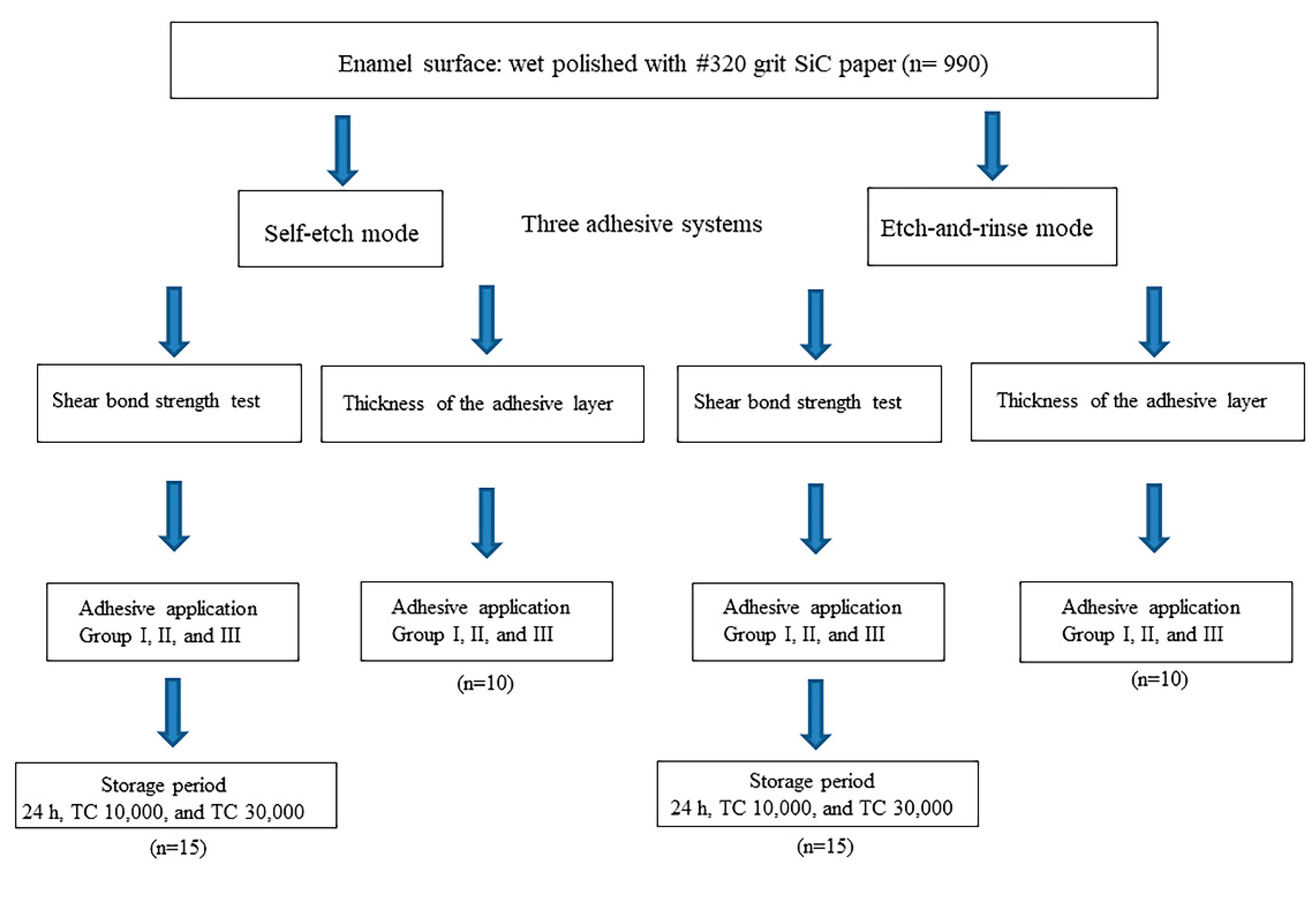
2.2. Specimen Preparation
2.3. Adhesive Application Protocol and Thermal Cycling
2.4. SBS Tests
2.5. Thickness of the Adhesive Layer
2.6. Scanning Electron Microscopy Observations
2.7. Statistical Analysis
3. Results
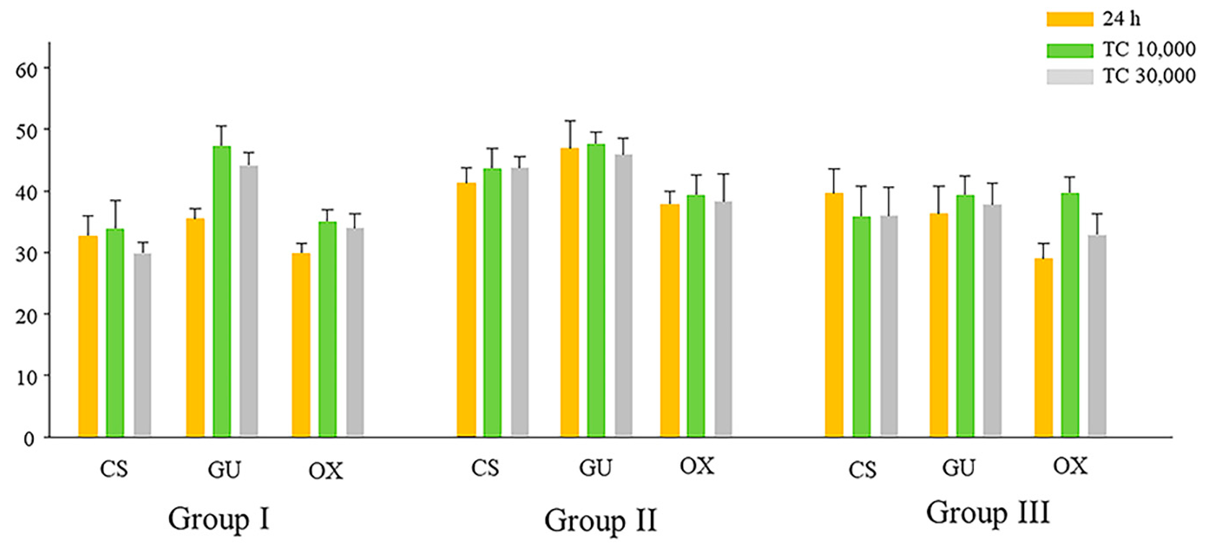
3.1. SBS in SE Mode
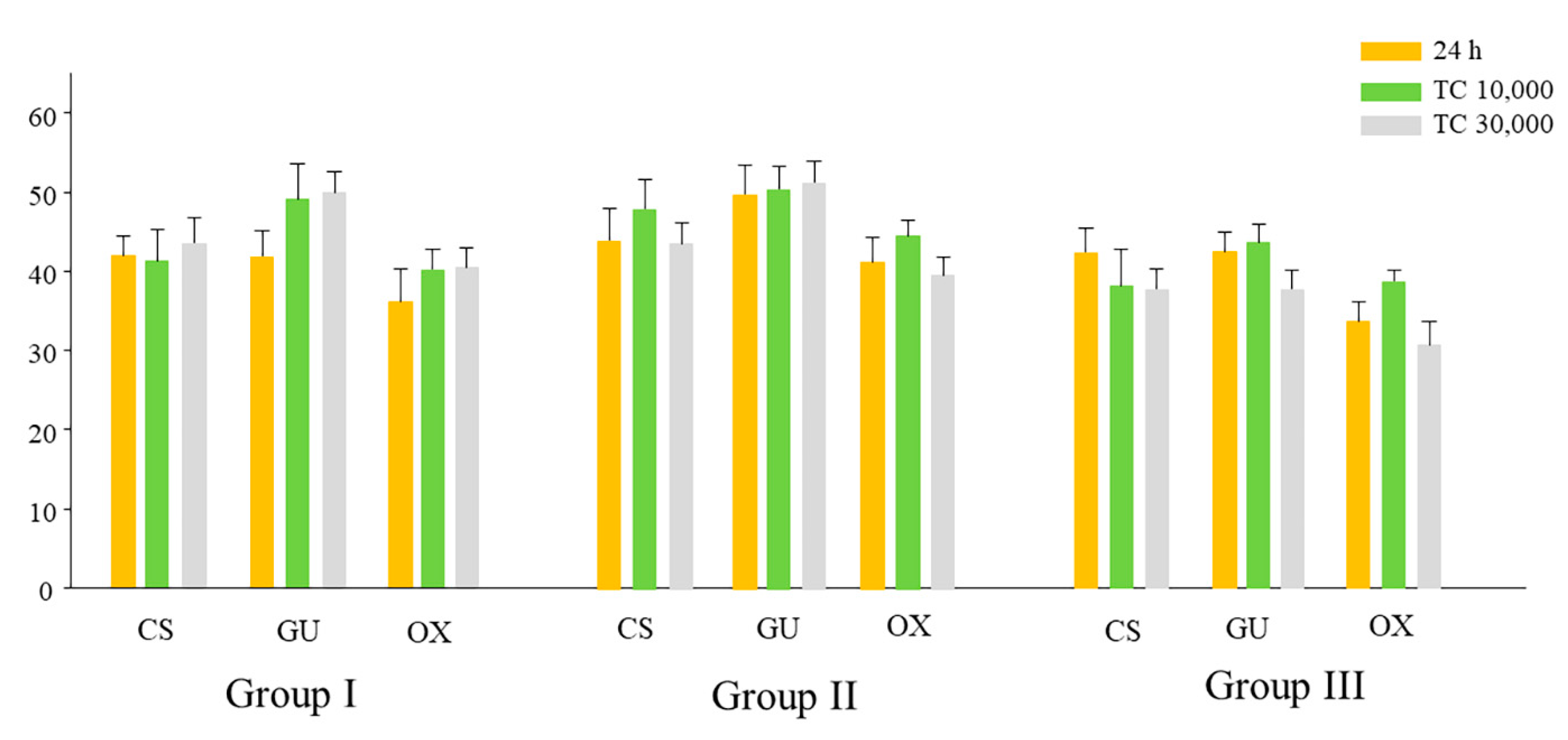
3.2. SBS in ER Mode
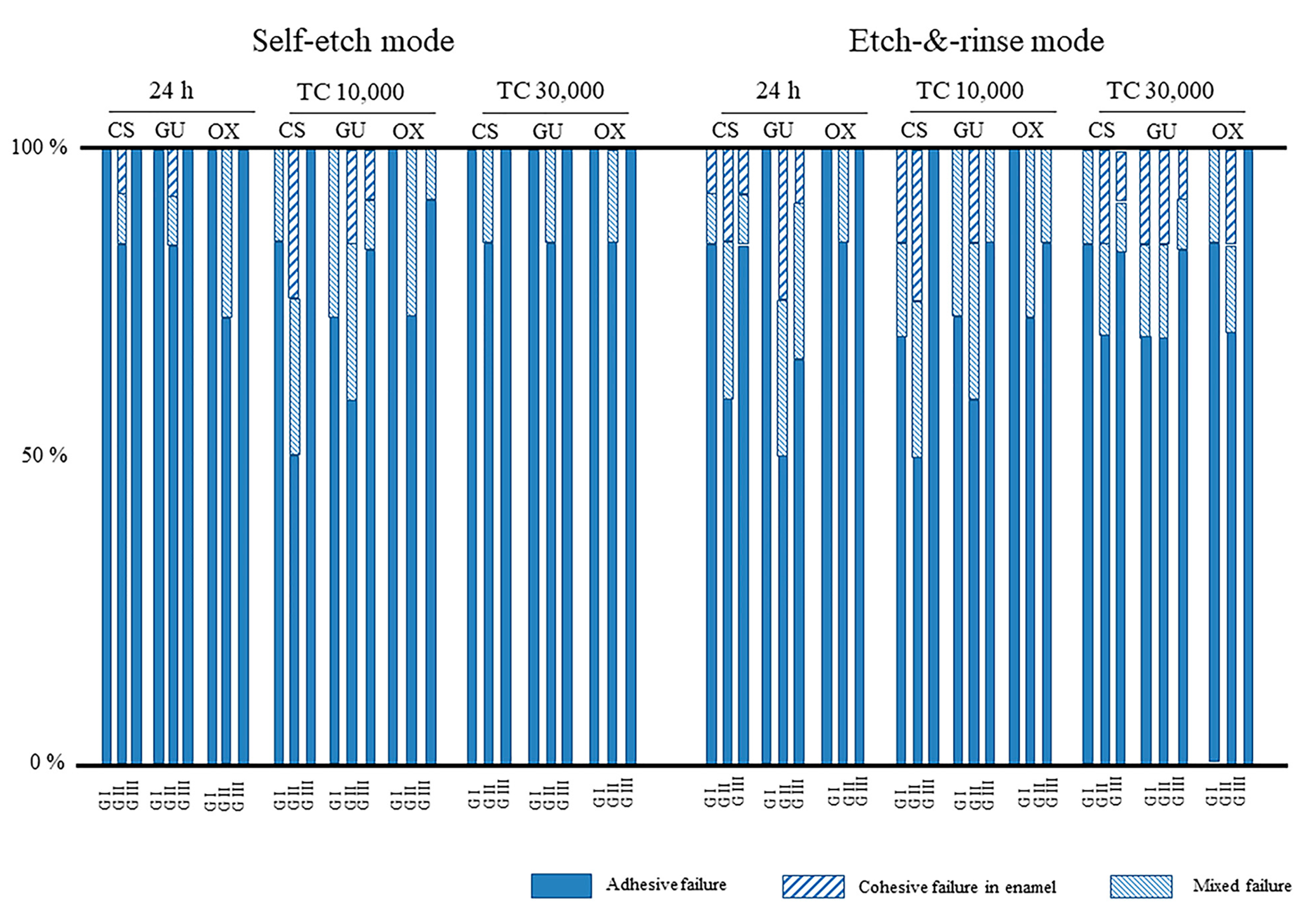
3.3. Failure Mode Analysis of Debonded Specimens after SBS
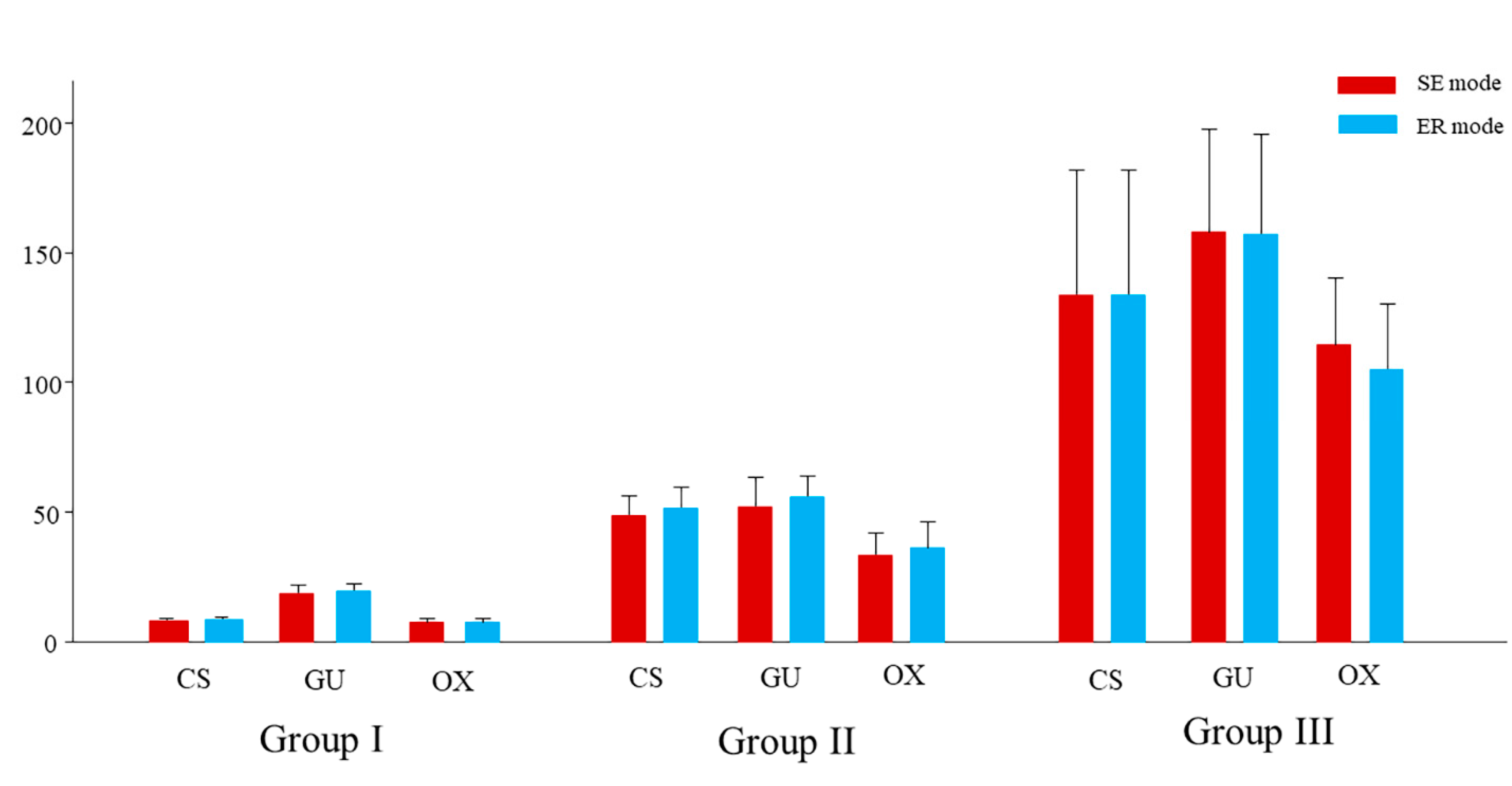
3.4. Thickness of the Adhesive Layer
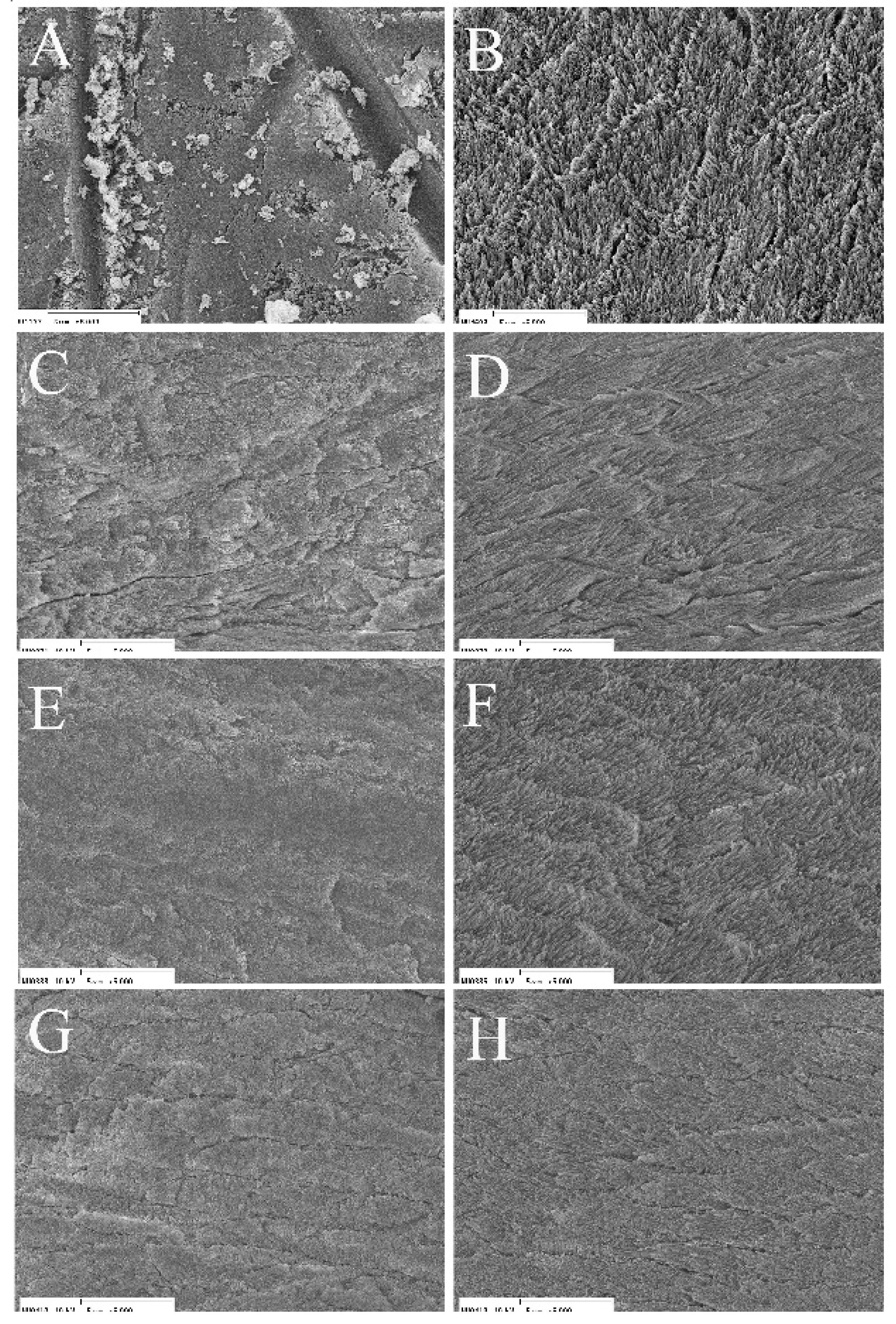
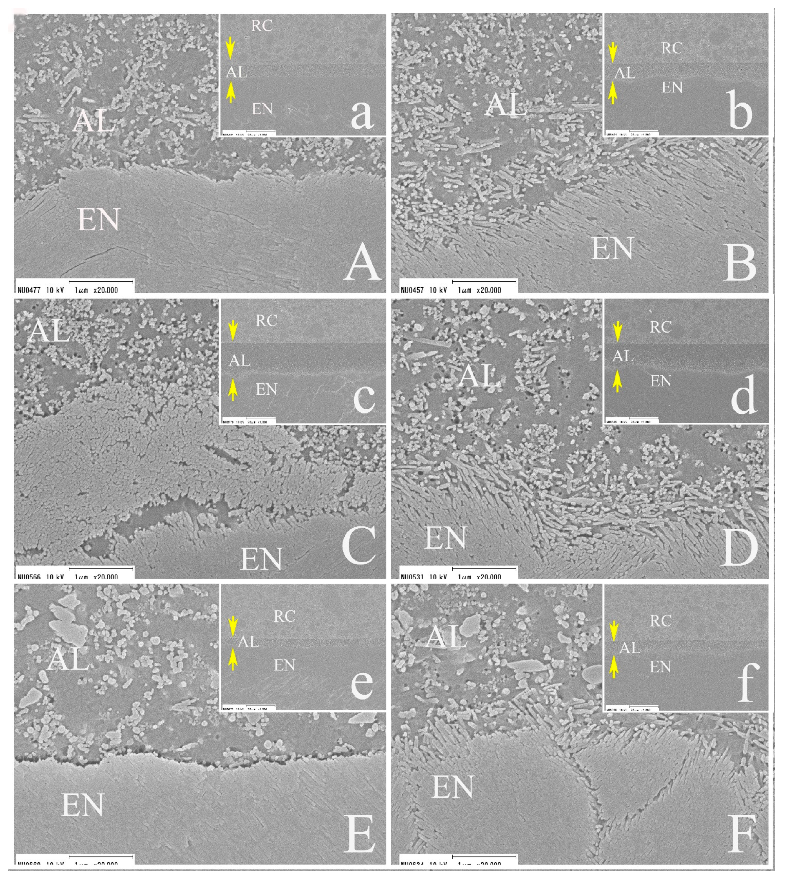
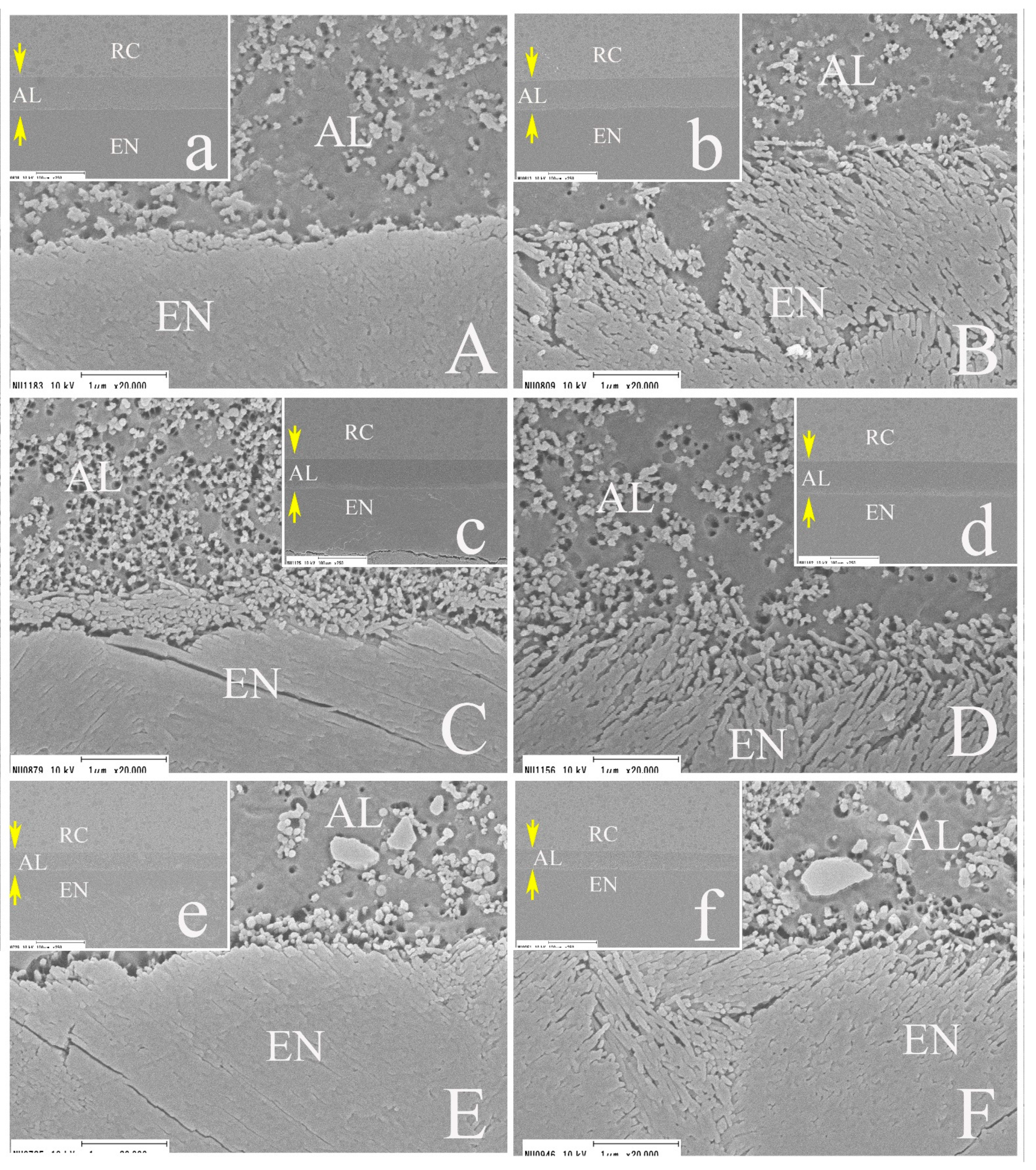
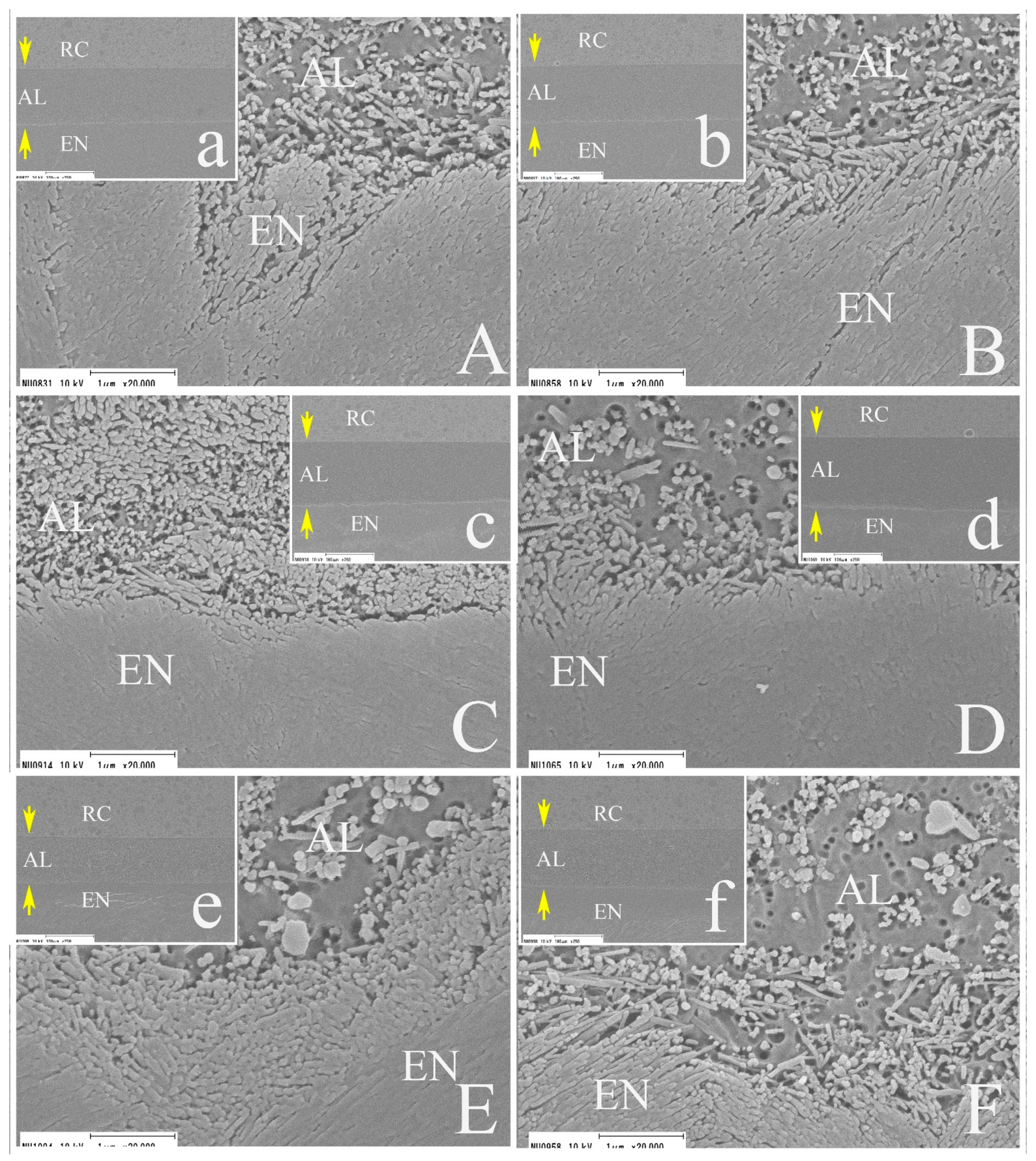
3.5. SEM Observations
4. Discussion
5. Conclusions
- All the factors (adhesive application method, TC period, and adhesive system) significantly influenced the SBS values in SE mode (p < 0.001).
- All the factors (adhesive application method, TC period, and adhesive system) significantly influenced the SBS values in ER mode (p < 0.001).
- Although the application method and adhesive systems significantly influenced the thickness of the adhesive layer (p < 0.001), the etching mode did not have any influence (p = 0.974).
- Although GU and OX showed significantly higher SBS values in ER mode than in SE mode at 24 h, no significant differences in the SBS values of the three adhesives were observed between the SE and ER modes at TC 30,000 in Group III.
- The application method in Group II, which conformed to the manufacturers’ recommendations, resulted in an adhesive layer that was approximately 40–60 μm in thickness and appeared to be optimal for effective enamel bonding, regardless of the type of adhesive system or etching mode.
- Within the limitations of this study, the new-generation two-step SE adhesive, which adopts a universal adhesive-derived primer and a hydrophobic bonding agent, showed superior bond performance to the conventional two-step adhesive systems.
Author Contributions
Funding
Conflicts of Interest
References
- Perdigão, J.; Muñoz, M.A.; Sezinando, A.; Luque-Martinez, I.V.; Staichak, R.; Reis, A.; Loguercio, A.D. Immediate adhesive properties to dentin and enamel of a universal adhesive associated with a hydrophobic resin coat. Oper. Dent. 2014, 39, 489–499. [Google Scholar] [CrossRef] [PubMed]
- Saikaew, P.; Chowdhury, A.F.M.A.; Fukuyama, M.; Kakuda, S.; Carvalho, R.M.; Sano, H. The effect of dentine surface preparation and reduced application time of adhesive on bonding strength. J. Dent. 2016, 47, 63–70. [Google Scholar] [CrossRef]
- Shiratsuchi, K.; Tsujimoto, A.; Takamizawa, T.; Furuichi, T.; Tsubota, K.; Kurokawa, H.; Miyazaki, M. Influence of warm air-drying on enamel bond strength and surface free-energy of self-etch adhesives. Eur. J. Oral. Sci. 2013, 121, 370–376. [Google Scholar] [CrossRef]
- Takamizawa, T.; Barkmeier, W.W.; Tsujimoto, A.; Scheidel, D.D.; Erickson, R.L.; Latta, M.A.; Miyazaki, M. Effect of phosphoric acid pre-etching on fatigue limits of self-etching adhesives. Oper. Dent. 2015, 40, 379–395. [Google Scholar] [CrossRef]
- Imai, A.; Takamizawa, T.; Sai, K.; Tsujimoto, A.; Nojiri, K.; Endo, H.; Barkmeier, W.W.; Latta, M.A.; Miyazaki, M. Influence of application method on surface free energy and bond strength of universal adhesive systems to enamel. Eur. J. Oral Sci. 2017, 125, 385–395. [Google Scholar] [CrossRef]
- Moritake, N.; Takamizawa, T.; Ishii, R.; Tsujimoto, A.; Barkmeier, W.W.; Latta, M.A.; Miyazaki, M. Effect of active application on bond durability of universal adhesives. Oper. Dent. 2019, 44, 188–199. [Google Scholar] [CrossRef]
- Fujiwara, S.; Takamizawa, T.; Barkmeier, W.W.; Tsujimoto, A.; Imai, A.; Watanabe, H.; Erickson, R.L.; Latta, M.A.; Nakatsuka, T.; Miyazaki, M. Effect of double-layer application on bond quality of adhesive systems. J. Mech. Behav. Biomed. Mater. 2018, 77, 501–509. [Google Scholar] [CrossRef]
- Hirokane, E.; Takamizawa, T.; Kasahara, Y.; Ishii, R.; Tsujimoto, A.; Barkmeier, W.W.; Latta, M.A.; Miyazaki, M. Effect of double-layer application on the early enamel bond strength of universal adhesives. Clin. Oral. Investig. 2021, 25, 907–921. [Google Scholar] [CrossRef]
- Yokoyama, M.; Takamizawa, T.; Tamura, T.; Namura, Y.; Tsujimoto, A.; Barkmeier, W.W.; Latta, M.A.; Miyazaki, M. Influence of different application methods on the bonding effectiveness of universal adhesives to the dentin in the early phase. J. Adhes. Dent. 2021, in press. [Google Scholar]
- Shirai, K.; De Munk, J.; Yoshida, Y.; Inoue, S.; Lambrechts, P.; Suzuki, K.; Shintani, H.; Van Meerbeek, B. Effect of cavity configuration and aging on the bonding effectiveness of six adhesives to dentin. Dent. Mater. 2005, 21, 110–124. [Google Scholar] [CrossRef]
- Cunha, L.G.; Alonso, R.C.B.; Pfeifer, C.S.C.; Correr-Sobrinho, L.; Ferracane, J.L.; Shihoreti, M.A.C. Contraction stress and physical properties development of a resin-based composite irradiated using modulated curing methods at two C-factor levels. Dent. Mater. 2008, 24, 392–398. [Google Scholar] [CrossRef]
- Takamizawa, T.; Imai, A.; Hirokane, E.; Tsujimoto, A.; Barkmeier, W.W.; Erickson, R.L.; Latta, M.A.; Miyazaki, M. SEM observation of novel characteristic of the dentin bond interfaces of universal adhesives. Dent. Mater. 2019, 35, 1791–1804. [Google Scholar] [CrossRef]
- Hashimoto, M.; Sano, H.; Yoshida, E.; Hori, M.; Kaga, M.; Oguchi, H.; Pashely, D.H. Effects of multiple adhesive coatings on dentin bonding. Oper. Dent. 2004, 29, 416–423. [Google Scholar]
- Takamizawa, T.; Barkmeier, W.W.; Tsujimoto, A.; Endo, H.; Tsuchiya, K.; Erickson, R.L.; Latta, M.A.; Miyazaki, M. Influence of pre-etching times on fatigue strength of self-etch adhesives to enamel. J. Adhes. Dent. 2016, 18, 501–511. [Google Scholar] [CrossRef]
- Yamanaka, A.; Mine, A.; Matsumoto, M.; Hagino, R.; Yumitate, M.; Ban, S.; Ishida, M.; Miura, J.; Van Meerbeek, B.; Yatani, H. Back to the multi-step adhesive system: A next-generation two-step system with hydrophobic bonding agent improves bonding effectiveness. Dent. Mater. J. 2021. [Google Scholar] [CrossRef]
- Tamura, T.; Takamizawa, T.; Ishii, R.; Hirokane, E.; Tsujimoto, A.; Barkmeier, W.W.; Latta, M.A.; Miyazaki, M. Influence of a primer resembling universal adhesive on the bonding effectiveness of an experimental two-step self-etch adhesive. J. Adhes. Dent. 2020, 22, 635–646. [Google Scholar] [CrossRef]
- International Organization for Standardization. Dentistry—Adhesion—Notched-Edge Shear Bond Strength Test; ISO 29022 TR: Geneva, Switzerland, 2013. [Google Scholar]
- Ausiello, P.; Apicella, A.; Davidson, C.L. Effect of adhesive layer properties on stress distribution in composite restorations-a 3D finite element analysis. Dent. Mater. 2002, 18, 295–303. [Google Scholar] [CrossRef]
- Albuquerque, M.; Pegoraro, M.; Mattei, G.; Loguercio, A.D. Effect of double-application or the application of a hydrophobic layer for improved efficacy of one-step self-etch systems in enamel and dentin. Oper. Dent. 2008, 33, 564–570. [Google Scholar] [CrossRef]
- Wakasa, K.; Yamaki, M.; Matsui, A. Calculation models for average stress and plastic deformation zone size of bonding area in dentine bonding systems. Dent. Mater. J. 1995, 14, 152–165. [Google Scholar] [CrossRef] [PubMed]
- Chowdhury, A.F.M.A.; Saikaew, P.; Alam, A.; Sun, J.; Carvalho, R.M.; Sano, H. Effects of double application of contemporary self-etch adhesives on their bonding performance to dentin with clinically relevant smear layers. J. Adhes. Dent. 2019, 21, 59–66. [Google Scholar] [CrossRef] [PubMed]
- Osorio, R.; Osorio, E.; Aguilera, F.S.; Tay, F.R.; Pinto, A.; Toledano, M. Influence of application parameters on bond strength of an “all in one” water-based self-etching primer/adhesive after 6 and 12 months of water aging. Odontology 2010, 98, 117–125. [Google Scholar] [CrossRef]
- De Neves, A.A.; Coutinho, E.; Poitevin, A.; Van der Sloten, J.; Van Meerbeek, B.; Van Oostereyck, H. Influence of joint component mechanical properties and adhesive layer thickness on stress distribution in micro-tensile bond strength specimens. Dent. Mater. 2009, 25, 4–12. [Google Scholar] [CrossRef]
- D’Arcangelo, C.; Vanini, L.; Prosperi, G.D.; Di Bussolo, G.; De Angelis, F.; D’Amario, M.; Caputi, S. The influence of adhesive thickness on the microtensile bond strength of three adhesive systems. J. Adhes. Dent. 2009, 11, 109–115. [Google Scholar] [CrossRef]
- Ermis, R.B.; Temel, U.B.; Cellik, E.U.; Kam, O. Clinical performance of a two-step self-etch adhesive with additional enamel etching in Class III cavities. Oper. Dent. 2010, 35, 147–155. [Google Scholar] [CrossRef] [PubMed]
- Suzuki, M.; Takamizawa, T.; Hirokane, E.; Ishii, R.; Tsujimoto, A.; Barkmeier, W.W.; Latta, M.A.; Miyazaki, M. Bond durability of universal adhesives to intact enamel surface in different etching modes. Eur. J. Oral. Sci. 2021, 129, e12768. [Google Scholar] [CrossRef]


| Code | Adhesive System (Lot. No.) | Main Components | pH (Primer) | Manufacturer |
|---|---|---|---|---|
| CS | Clearfil SE Bond 2 (Primer: 5852494) (Adhesive: 5847004) | Primer: MDP, HEMA, water, initiators Adhesive: MDP, HEMA, bis-GMA, initiators, microfiller | 2.0 | Kuraray Noritake Dental, Tokyo, Japan |
| GU | G2-Bond Universal (Primer: 190711) (Adhesive: 190711) | Primer: 4-MET, MDP, MDTP, dimethacrylate monomer, acetone, water, photoinitiator, filler Adhesive: dimethacrylate monomer, bis-GMA, filler, photoinitiator | 1.5 | GC, Tokyo, Japan |
| OX | OptiBond eXTRa Universal (Primer: 58470004) (Adhesive: 5852494) | Primer: GPDM, HEMA, acetone, ethyl alcohol Adhesive: GPDM, HEMA, glycerol dimethacrylate, ethyl alcohol, sodium hexafluorosilicate | 1.6 | Kerr, Brea, CA, USA |
| Resin Composite | Main Components | Manufacturer | ||
| Clearfil AP-X (N416713) | bis-GMA, TEGDMA, silane barium glass filler, silane silica filler, silanated colloidal silica, CQ, pigments, others | Kuraray Noritake Dental | ||
| Etching-Mode | Pre-Etching Protocol | |
| SE (self-etch) | Phosphoric acid pre-etching was not performed. | |
| ER (etch-and-rinse) | Enamel surfaces were phosphoric acid etched for 15 s. Etched surface was rinsed with water for 15 s and air-dried. | |
| Adhesive | Group | Adhesive Application Protocol |
| CS | I | Primer was applied to air-dried enamel surface for 20 s followed by medium air pressure for 5 s. Bonding agent was then applied to the primed surface and a strong stream of air was applied over the bonding agent for 5 s. Light irradiated for 10 s. |
| II | Primer was applied to air-dried enamel surface for 20 s followed by medium air pressure for 5 s. Bonding agent was then applied to the primed surface and was gently air thinned for 5 s. Light irradiated for 10 s. | |
| III | After bonding procedure of Group II without light irradiation, another layer of bonding agent was applied and was gently air thinned for 5 s. Bonding agent was light irradiated for 10 s. | |
| GU | I | Primer was applied to air-dried enamel surface for 10 s and then a strong stream of air was applied over the liquid primer for 5 s. Bonding agent was then applied to the primed surface and a strong stream of air was applied over the bonding agent for 5 s. Light irradiated for 10 s. |
| II | Primer was applied to air-dried enamel surface for 10 s and then a strong stream of air was applied over the primer for 5 s. Bonding agent was then applied to the primed surface and was gently air thinned for 5 s. Light irradiated for 10 s. | |
| III | After bonding procedure of Group II without light irradiation, another layer of bonding agent was applied and was gently air thinned for 5 s. Light irradiated for 10 s. | |
| OX | I | Primer was applied to air-dried enamel surface with rubbing action for 20 s. Medium air pressure was applied to the surface for 5 s. Bonding agent was applied to the primed surface with rubbing action for 15 s, and then a strong stream of air was applied over the bonding agent for 5 s. Light irradiated for 10 s. |
| II | Primer was applied to air-dried enamel surface with rubbing action for 20 s. Medium air pressure was applied to the surface for 5 s. Bonding agent was applied to the primed surface with rubbing action for 15 s and then gently air thinned for 5 s. Light irradiated for 10 s. | |
| III | After bonding procedure of Group II without light irradiation another layer of bonding agent was applied and was gently air thinned for 5 s. Light irradiated for 10 s. | |
| Group I | Group II | Group III | |||||||
|---|---|---|---|---|---|---|---|---|---|
| 24 h | TC 10,000 | TC 30,000 | 24 h | TC 10,000 | TC 30,000 | 24 h | TC 10,000 | TC 30,000 | |
| CS | 32.8 (3.2) bCD | 33.9 (4.5) bCD | 29.9 (1.8) cD | 41.2 (2.5) bA | 43.7 (3.2) bA | 43.8 (1.7) aA | 39.6 (3.9) aAB | 35.9 (4.9) bBC | 36.0 (4.5) abBC |
| GU | 35.5 (1.6) aB | 47.4 (3.1) aA | 44.2 (2.1) aA | 46.9 (4.4) aA | 47.7 (1.8) aA | 45.9 (2.7) aA | 36.3 (4.5) aB | 39.4 (3.0) aB | 37.8 (3.5) aB |
| OX | 29.9 (1.5) cD | 35.1 (1.8) bBC | 34.0 (2.2) bC | 37.9 (2.0) cAB | 39.4 (3.1) cA | 38.3 (4.4) bAB | 29.0 (2.4) bD | 39.7 (2.6) aA | 32.9 (3.4) bCD |
| Group I | Group II | Group III | |||||||
|---|---|---|---|---|---|---|---|---|---|
| 24 h | TC 10,000 | TC 30,000 | 24 h | TC 10,000 | TC 30,000 | 24 h | TC 10,000 | TC 30,000 | |
| CS | * 42.0 (2.5) aBC | * 41.3 (3.9) bBC | * 43.6 (3.2) bB | * 44.3 (4.0) bAB | * 48.3 (3.7) aA | 43.9 (2.6) bAB | 42.6 (3.1) aBC | 38.4 (4.6) bCD | 38.0 (2.5) aD |
| GU | * 41.9 (3.2) aBC | 49.1 (4.5) aA | * 50.0 (2.5) aA | * 50.1 (3.7) aA | * 50.8 (2.8) aA | * 51.6 (2.7) aA | * 42.7 (2.5) aB | * 43.9 (2.3) aB | 38.0 (2.3) aC |
| OX | * 36.2 (4.1) bCD | * 40.3 (2.5) bB | * 40.5 (2.5) cB | * 41.6 (3.1) bAB | * 44.9 (1.9) bA | 39.9 (2.3) cB | * 33.9 (2.5) bDE | 39.0 (1.4) bBC | 30.9 (2.9) bE |
| Group I | Group II | Group III | ||||
|---|---|---|---|---|---|---|
| SE | ER | SE | ER | SE | ER | |
| CS | 8.9 (0.7) bC [7.9%] | 9.2 (0.7) bC [7.6%] | 49.6 (7.1) aB [14.3%] | 52.5 (7.5) aB [14.3%] | 134.4 (48.1) abA [35.8%] | 134.6 (47.7) abA [35.4%] |
| GU | 19.2 (3.1) aC [15.6%] | 20.2 (2.7) aC [13.4%] | 52.9 (10.9) aB [20.6%] | 56.9 (7.7) aB [13.5%] | 158.7 (39.4) aA [24.8%] | 157.8 (38.5) aA [24.4%] |
| OX | 8.2 (1.3) bC [15.9%] | 8.2 (1.2) bC [14.6%] | 34.4 (8.2) bB [23.8%] | 37.0 (9.9) bB [26.8%] | 115.1 (25.9) bA [22.5%] | 105.9 (25.0) bA [23.6%] |
Publisher’s Note: MDPI stays neutral with regard to jurisdictional claims in published maps and institutional affiliations. |
© 2021 by the authors. Licensee MDPI, Basel, Switzerland. This article is an open access article distributed under the terms and conditions of the Creative Commons Attribution (CC BY) license (https://creativecommons.org/licenses/by/4.0/).
Share and Cite
Takamizawa, T.; Yokoyama, M.; Sai, K.; Shibasaki, S.; Barkmeier, W.W.; Latta, M.A.; Tsujimoto, A.; Miyazaki, M. Effect of Adhesive Application Method on the Enamel Bond Durability of a Two-Step Adhesive System Utilizing a Universal Adhesive-Derived Primer. Appl. Sci. 2021, 11, 7675. https://doi.org/10.3390/app11167675
Takamizawa T, Yokoyama M, Sai K, Shibasaki S, Barkmeier WW, Latta MA, Tsujimoto A, Miyazaki M. Effect of Adhesive Application Method on the Enamel Bond Durability of a Two-Step Adhesive System Utilizing a Universal Adhesive-Derived Primer. Applied Sciences. 2021; 11(16):7675. https://doi.org/10.3390/app11167675
Chicago/Turabian StyleTakamizawa, Toshiki, Munenori Yokoyama, Keiichi Sai, Sho Shibasaki, Wayne W. Barkmeier, Mark A. Latta, Akimasa Tsujimoto, and Masashi Miyazaki. 2021. "Effect of Adhesive Application Method on the Enamel Bond Durability of a Two-Step Adhesive System Utilizing a Universal Adhesive-Derived Primer" Applied Sciences 11, no. 16: 7675. https://doi.org/10.3390/app11167675
APA StyleTakamizawa, T., Yokoyama, M., Sai, K., Shibasaki, S., Barkmeier, W. W., Latta, M. A., Tsujimoto, A., & Miyazaki, M. (2021). Effect of Adhesive Application Method on the Enamel Bond Durability of a Two-Step Adhesive System Utilizing a Universal Adhesive-Derived Primer. Applied Sciences, 11(16), 7675. https://doi.org/10.3390/app11167675