Communication

Biohydrogel Based on Dynamic Covalent Bonds for Wound Healing Applications

by
Chukwuma O. Agubata
1,
Cynthia C. Mbaoji
1,
Ifeanyi T. Nzekwe
2,
César Saldías
3 and
David Díaz Díaz
4,5,6,*
1
Department of Pharmaceutical Technology and Industrial Pharmacy, University of Nigeria, Nsukka 410001, Enugu State, Nigeria
2
Department of Pharmaceutics and Pharmaceutical Technology, Nnamdi Azikiwe University, Awka 420110, Anambra State, Nigeria
3
Departamento de Química Física, Facultad de Química y de Farmacia, Pontificia Universidad Católica de Chile, Santiago 7820436, Chile
4
Departamento de Química Orgánica, Universidad de La Laguna, Avda, Astrofísico Francisco Sánchez S/N, 38206 La Laguna, Tenerife, Spain
5
Instituto Universitario de Bio-Orgánica Antonio González, Universidad de La Laguna, Avda, Astrofísico Francisco Sánchez 2, 38206 La Laguna, Tenerife, Spain
6
Institut für Organische Chemie, Universität Regensburg, Universitätsstr. 31, 93053 Regensburg, Germany
*
Author to whom correspondence should be addressed.
Appl. Sci. 2021, 11(15), 6945; https://doi.org/10.3390/app11156945
Submission received: 29 June 2021 / Revised: 20 July 2021 / Accepted: 24 July 2021 / Published: 28 July 2021
(This article belongs to the Special Issue Hydrogel Composites for Bioengineering Applications)

Abstract

:
In this work, a biohydrogel based on alginate and dynamic covalent B-O bonds, and derived composites, has been evaluated for wound healing applications. In particular, a phenylboronic acid–alginate (PBA-Alg) complex was synthesized by coupling 3-aminophenylboronic acid onto alginate, and used to prepare varied concentrations of hydrogels and silicate-based nanocomposites in PBS. The resulting hydrogels were characterized in terms of interfacial tension, moisture uptake and loss, interaction with fresh acid-soluble collagen, self-healing ability, effects on blood clotting and wound healing. The interfacial tension between the hydrogels and biorelevant fluids was low and moisture loss of 55–60% was evident without uptake from the environment. The components of the hydrogels and their mixtures with collagen were found to be compatible. These hydrogels showed efficient self-healing and thixotropic behavior, and the animals in the treatment groups displayed blood clotting times between 9.1 min and 10.7 min. In contrast, the composites showed much longer or shorter clotting times depending on the silicate content. A significant improvement in wound healing was observed in 3% w/v PBA-Alg formulations. Overall, the PBA-Alg hydrogels exhibit self-healing dynamic covalent interactions and may be useful in dressings for incision wounds.

1. Introduction

When the body is injured, it activates a sequence of processes to heal, entailing hemostasis, inflammatory, proliferative, and maturation phases [1]. Synthetic biomaterials around the wound can regenerate and heal after damage via sealing or reparative processes. This process may involve the presence of sacrificial bonds between molecules which break and reform, allowing improved adaptation of the material to external conditions [2,3], or may involve the release of encapsulated sealing agent [4].
Dynamic covalent chemistries offer interesting paths for the preparation of soft materials that offer the advantages of both strength and reversibility [5]. These dynamic materials are affected by external conditions, including pH and temperature, as well as compositional variables, such as concentration; thus, these materials are stimulus responsive [6]. Among different dynamic bonds [5], B-O bonds constitute one of the most versatile examples because their dynamic behavior in trigonal planar boronic ester can be achieved in numerous ways, including hydrolysis/dehydration, transesterification, or metathesis reactions [6]. In particular, the reversible condensation reactions between derivatives of boronic acid and cis-1,2 or cis-1,3 diols (polyols) to form cyclic esters have been explored to create self-healing materials at room temperature [7,8]. Another advantage of the B-O dynamic bonds has been ascribed to their role in the modulation of cell attachment and differentiation by using dynamic platforms in the presence of phenylboronic acids, which can interact with cell surface polysaccharides [9]. These qualities of dynamic B-O bonds have been applied for tissue engineering and drug delivery [10]. Moreover, natural polysaccharides such as sodium alginate can be conveniently functionalized with covalent bonds [11] to form biodegradable and biocompatible [12] self-healing soft materials [13,14] with biomedical potential.
On the other hand, nanocomposites involve mixtures of materials in which at least one physical dimension is reduced to the nanoscale range [15]. In particular, nanocomposite hydrogels are basically hydrated polymeric networks containing nanomaterials, which appear to have unique properties different from those of the individual constituents. Indeed, composite hydrogels contain multifunctional entities that can improve efficiency of the polymeric network. The properties of these heterogeneous and reinforced matrices are affected by the constituents and their properties, structure, and interfacial interactions [16]. Within this context, inorganic materials such as silicates show a high aspect ratio and are being increasingly used for the preparation of nanocomposites with enhanced properties, which are mainly attributed to the many surface interactions of polymeric networks and silicate particles [17]. Thus, the combination of silicate particles with hydrogel networks based on B-O bonds could also provide a versatile approach to modulate the functional properties of the gels.
Wounds resulting from physical, chemical, or thermal agents can become infected if left open, leading to a need to achieve rapid wound closure. Wound healing processes entail cellular and biointerfacial interactions, involving fibroblasts, keratinocytes, and endothelial cells [1], whose mobilization and arrangements may be influenced by certain biomimetic materials. Different stages of wound healing occur in sequence, and hydrogels can affect any of the steps. Provision of a dynamic scaffold to facilitate regeneration of cells at wound site may enhance wound closure. Hydrogels can help maintain the necessary moist wound environment and their physico-mechanical properties are similar to those of soft tissues, making them compatible at the site of action. Furthermore, hydrogel materials may be useful in chronic cases where the pH of the wound zone increases, becoming alkaline. A novel application of PBA-Alg hydrogels would be based on its auto-renewal of degenerated surfaces in contact with the wound and this would avoid the need for multiple replacements of wound dressings as usually observed with other common products.
Inspired by all these considerations, the aim of this study was to prepare and evaluate the use of phenylboronic acid–alginate (PBA-Alg) hydrogels and related composites for wound healing.

2. Materials and Methods

3-Aminophenylboronic acid, sodium alginate (Sigma-Aldrich, Burlington, MA, USA), magnesium aluminometasilicates (Neusilin® FH2, Fuji Chemical Industry Co., Minato, Tokyo, Japan), Savlon (Johnson and Johnson, New Brunswick, NJ, USA), and neomycin sulfate Vaseline gauze (Unilever, Englewood Cliffs, NJ, USA) were used as received. Acid-soluble collagen was obtained from the Department of Biochemistry, University of Nigeria. PBA-Alg conjugate was synthesized and characterized as we have previously reported [13,14].

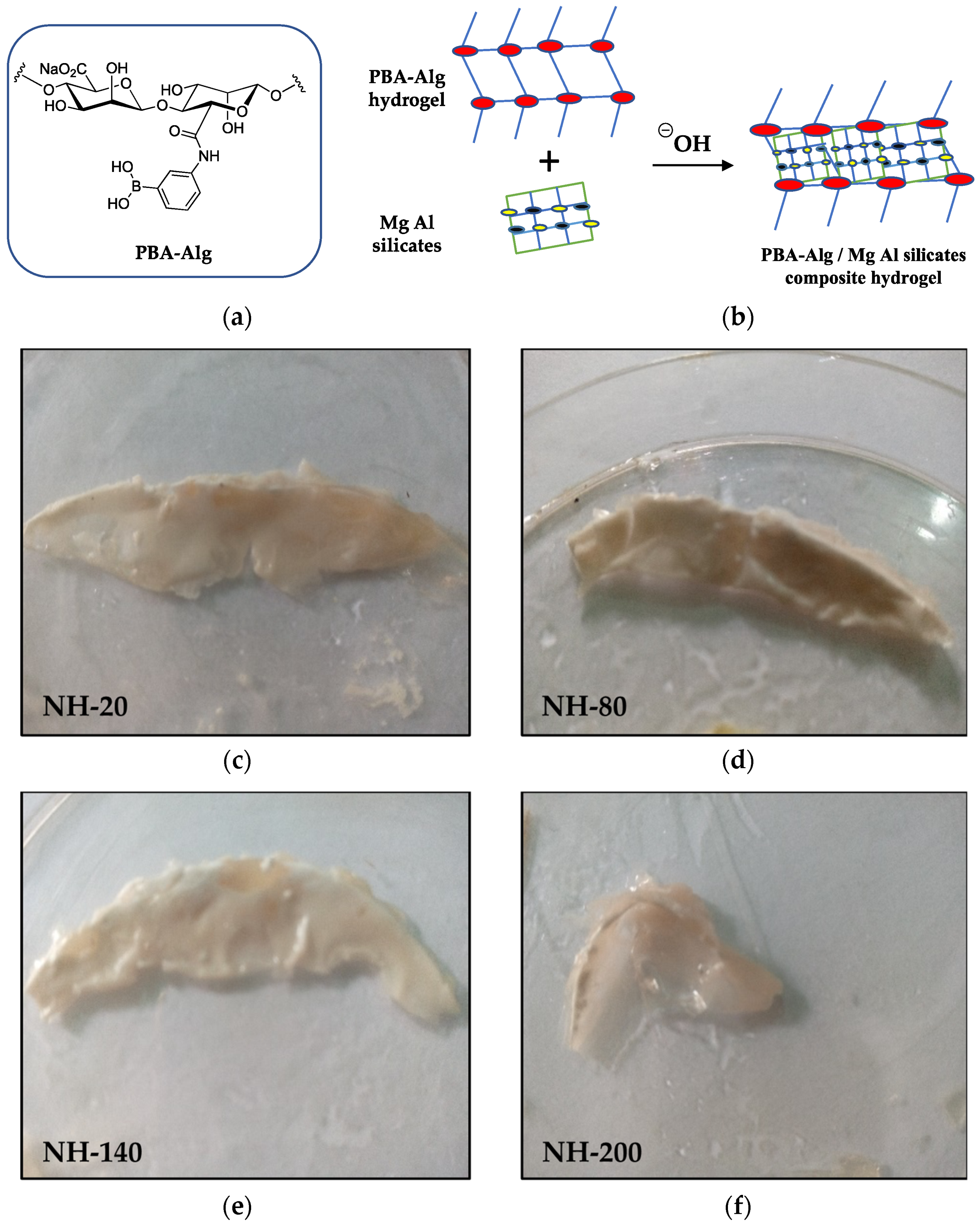
2.1. Synthesis of Phenyl Boronic Acid–Alginate Complex

The PBA-Alg complex was synthesized by coupling alginate with 3-aminophenylboronic acid in the presence of 1-(3-dimethylaminopropyl)-3-ethylcarbodiimide hydrochloride, as previously described [13,18], albeit with minor modifications. Sodium alginate (5 g, 25 mmol) was dissolved in 500 mL of deionized water, to which 4.8 g (25 mmol) of EDC·HCl and 1.95 g (12.5 mmol) of 3-aminophenylboronic acid were added for a 1:1:0.5 molar mixture. The mixture was stirred at ambient temperature for 24 h, after which it was dialyzed against distilled water for 7 d, replacing the water several times, and subsequently lyophilized.

2.2. Preparation of PBA-Alg Hydrogel

Dry PBA-Alg (30 mg) was dissolved in 0.1 M PBS buffer (pH = 7.4) to obtain 1 mL (3 wt%). Base (25 µL 1 M NaOH) was added to the solution with continuous vigorous stirring. After preparation, samples were equilibrated at room temperature overnight. Using the same procedure, 4% w/w hydrogels were prepared using an initial 40 mg of dry sample, and 3% w/w PBA-Alg gel containing Savlon antiseptic (based on cetrimide and chlorhexidine gluconate) was prepared using the described method but with 10% v/v of liquid Savlon added. A 3% w/w sodium alginate hydrogel was also prepared in PBS.

2.3. Preparation of Nanocomposite Hydrogels of PBA-Alg and Magnesium Aluminosilicate (Neusilin® FH2)

Four samples of 3 wt% PBA-Alg solutions were prepared by dissolving PBA-Alg (50 mg) in 0.1 M PBS buffer. Different amounts of magnesium aluminometasilicate (20 mg, 80 mg, 140 mg, and 200 mg) were added to each PBA-Alg solution under continuous stirring to obtain uniform dispersions. Particle size reduction (nanonization) was achieved by vigorous wet milling. A 1 M NaOH solution (25 µL) was added to each dispersion under stirring to obtain four different nanocomposite hydrogels. The hydrogels were fully formed in Petri dishes and subsequently investigated.

2.4. Determination of Interfacial Tension of the Hydrogel with Phosphate-Buffered Saline and Water

A Lecomte du Noüy tensiometer (model Nr 3124, A. Krüss, Hamburg, Germany) was used to determine the interfacial tension between the hydrogel surface and fluids (PBS and water) with the du Noüy ring technique. The ring was made of inert platinum metal to which the hydrogel was attached using glue. Then, the ring was attached to the lever arm, and the balance holding the Petri dish of water was adjusted in such a way that the surface of the hydrogel was immediately attracted to the surface of the PBS. The ring was then raised gradually, with the aid of a screw, until the hydrogel was completely detached from the PBS surface, and the tensional force was recorded. This procedure was repeated, using PBS and water once each.
The force (F) required to raise the ring from the liquid’s surface was measured and related to the liquid’s surface tension, γ, (Equation (1)):
F = W ( r i n g ) + 2 π ( r i + r g ) γ
where ri is the radius of the inner ring of the liquid film pulled and rg is the radius of the outer ring of the liquid film [19]; W(ring) is the weight of the ring without the buoyant force originating from part of the ring below the liquid surface [20]. In cases where the ring’s thickness is much smaller than its diameter, Equation (1) can be simplified into Equation (2):
F = W ( r i n g ) + 4 π R γ
where R is the average of the inner and outer radius of the ring. A balance is used to measure the force; the excess force attributed to the liquid being pulled up can then be used to calculate surface tension (γ).

2.5. Moisture Uptake and Loss of Hydrogel Films

The moisture uptake of hydrogel films and their nanocomposites formed with magnesium aluminometasilicate was determined using static methods involving saturated salt solutions. Weighed hydrogels were placed in desiccators containing 210 mL of saturated solution of potassium chloride (84% relative humidity RH). At 24 h intervals, the patches were removed and weighed until there was no observable change in weight. The percentage moisture uptake was calculated as 100 times the weight gain divided by the original weight.
For percentage of moisture loss, preweighed hydrogel patch samples were stored in a desiccator containing 100 g anhydrous calcium chloride. Every 24 h, the patches were removed from the chamber and weighed to observe weight changes. The percentage of moisture loss was then calculated as 100 times the weight loss divided by original weight.

2.6. Extraction and Purification of Fresh Acid-Soluble Collagen

Sodium hydroxide-pretreated skin and muscles of fish were treated for two days with 10% butyl alcohol for defatting, with a solid/solvent ratio of 1:20 w/v. The defatted skin and muscles were washed with cold distilled water and subsequently soaked in 0.5 M acetic acid for 24 h (1:15% w/v, solid/solvent). After filtration with cheesecloth, the filtrate was treated with tris (hydroxylmethyl) aminomethane sodium chloride buffer solution (pH 7.4) to precipitate collagen. The precipitate obtained was collected by centrifugation (Uniscope Surgifriend, Essex, UK) at 15,000 rev/min for 30 min and dissolved in 0.5 M acetic acid. The obtained solution was dialyzed (cut-off 5000 Da) against 0.1 M acetic acid for 2 d and distilled water for 3 d with continuous change of solution at 4 °C.

2.7. Fourier Transform Infrared Spectroscopy

Fourier transform infrared (FTIR) spectroscopy was used to investigate the chemical interaction between the hydrogel and freshly prepared acid-soluble collagen—an abundant component of human skin and bones [21]. Small quantities of the PBA-Alg hydrogel complex, collagen, and mixtures of hydrogel and acid-soluble collagen were scanned over a wavelength range of 4000 to 650 cm−1 at a resolution of 8 cm−1 using FTIR (Agilent Technologies, Santa Clara, CA, USA) in transmittance mode.

2.8. Effect of Phenylboronic Acid–Alginate Conjugate and Nanocomposites on Blood Clotting Time

Rat eyes were punctured using a capillary tube and the blood was collected in a heparin bottle. Equal samples of the test hydrogel preparations were placed in different sections of a labeled Petri dish. Two drops of rat blood were applied simultaneously on each preparation and a timer started; the time to clot formation was observed visually and any other physical changes were noted. The capillary tube method [22] was applied with a slight modification involving direct application onto hydrogel film. The experiment was performed in triplicate and mean values obtained. This evaluation of clotting time of blood in contact with the hydrogel was conducted to generally establish whether clotting factors may be affected by the composition or dynamic covalent chemistry of the hydrogels.

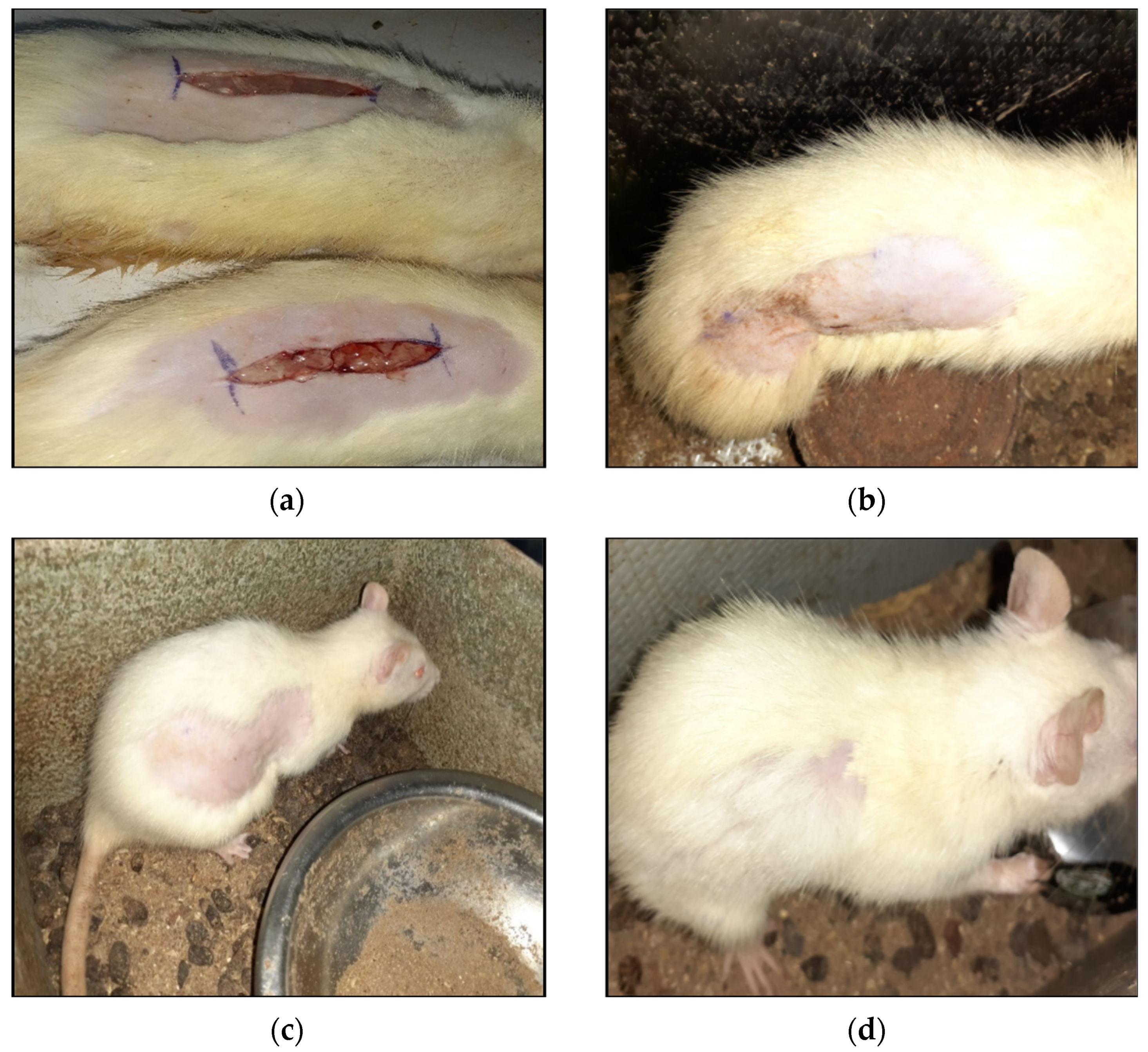
2.9. Effect of PBA-Alg Hydrogel on Wound Healing

The study was conducted according to the guidelines of the Declaration of Helsinki, and approved by the Institutional Review Board (ethics committee) of the University of Nigeria Nsukka/Faculty of Pharmaceutical Sciences. Adult Swiss albino rats (170–300 g; average body length (nose tip to tail base) = 19 cm) of both sexes were used to evaluate wound healing using an incision model. The animals were divided into five subgroups (four treatment groups and one control group) of five rats each. The animals were anesthetized with intramuscular ketamine (50 mg/kg) and xylazine (10 mg/kg). The hair on the dorsal region of the animals was shaved off, and a 4 cm paravertebral incision was made through the skin and cutaneous muscles. The incisions for each treatment group were covered with preparations containing 3% w/w PBA-Alg hydrogel, 4% PBA-Alg hydrogel, 3% PBA-Alg hydrogel/Savlon antiseptic, 3% sodium alginate hydrogel, or neomycin sulfate Vaseline gauze, while the control group received no treatment.
The wound area was measured on alternating days during a 9-day study period (days 0, 2, 4, 6, 8, and 9). Wound closure time was recorded as the number of days required for the scab to fall off with no raw wound left behind. Wound closure or size reduction was calculated from Equation (3):
W o u n d   c l o s u r e   % = i n i t i a l   w o u n d   s i z e w o u n d   s i z e   a t   t i m e   t i n i t i a l   w o u n d   s i z e × 100

3. Results and Discussion

3.1. Formulation and Relevant Characterization of Hydrogels

We have previously shown [13] that the pendant boronic acid groups of PBA-Alg can interact with the vicinal diols present on the same or different alginate pyranose rings in a basic environment, leading to the formation of pH-dependent reversible hydrogels. The phenylboronic acid–alginate conjugate was synthesized with a degree of substitution (DS) of 25% and we have already demonstrated that it forms non-cytotoxic clear hydrogels under suitable basic conditions [13]. However, the inclusion of magnesium aluminometasilicate prior to the base-catalyzed gelation produced opaque whitish nanocomposite hydrogels with modified physical properties (Figure 1). These hydrogels were stable and compatible with collagen (vide infra).

3.1.1. Interfacial Tension between Fluids and Hydrogel

The interfacial tension between the soft hydrogels and the selected fluids (PBS and water) was relatively low, likely because the hydrogels were miscible with these fluids and could dissolve in them over time. The interfacial tension between the hydrogels and PBS and distilled water was 1.89 and 1.70 dynes/cm, respectively. The slight difference in the interfacial tension of the material with PBS and water is not significant (p > 0.05), and may be attributed to the inhibitory common ion effect with PBS.
The relatively low interfacial tension suggests that these hydrogels would interact effectively with biofluids when in contact with biological surfaces, allowing continuous engagement. This attribute would be valuable in the process of wound healing via fluidization [23] and biodegradation [24] of hydrogel material because it would increase the surface area available for activity and breakdown.

3.1.2. Moisture Uptake and Loss of Films

The hydrogels did not absorb further moisture under an experimental relative humidity of 84%. However, under reduced relative humidity, they did lose moisture. The three concentrations (3%, 4%, and 3% hydrogel plus antiseptic) all exhibited 60% moisture loss in a dry environment created with anhydrous calcium chloride. This high level of moisture loss may be due to the nature of the bonds and level of moisture trapped in the network. The results indicate that this hydrogel favors moisture loss rather than uptake. In the case of nanocomposites (Figure 1), while those prepared with 20 mg and 80 mg of silicates did not show any moisture uptake, the hydrogels containing 140 mg and 200 mg of the silicate showed 2% and 2.5% moisture uptake, respectively. Moreover, about 55% moisture loss was observed for the nanocomposites.

3.1.3. FTIR Spectroscopy

Protein–polymer interaction can greatly affect healing, clotting, and other bioprocesses. Thus, we performed a series of FTIR measurements to gain insight regarding the compatibility between PBA-Alg gel and biological systems (i.e., extracted collagen). The FTIR spectrum of the PBA-Alg complex (Figure S1) showed a broad band at 3280 cm−1, which indicates the strong presence of the –OH functional groups of boronic acid and alginate, while the peak at 2929.7 cm−1 represents the C–H stretch from alginate. In the spectrum of the extracted collagen (Figure S2), the broad band at 3291 cm−1 shows the presence of –OH, while a small weak peak of amine was observed around 3675.2 cm−1. Further, C–H bond stretching vibrations were observed at 2918.5 cm−1 and 2847.7 cm−1. The band of C=O was also observed around 1606.5 cm−1; the smaller peaks around it represent C=C bond stretching vibrations. The FTIR spectrum of the hydrogel–collagen mixture (Figure S3) exhibits peaks of the components; those at 3283.8 cm−1, 2918.5 cm−1/2847.7 cm−1, and 1602.8 cm−1 represent –OH, C–H, and C=O, respectively, with that at 3675.2 cm−1 indicating the presence of amines. Observations of only minor shifts indicate that the mixtures are stable and compatible.

3.1.4. Self-Healing Properties

In agreement with our previous study [13], the 3% w/v hydrogels showed complete self-repair or closure within 7 min after the bulk soft gel was cut into two pieces and placed back in contact. A gradual and visual disappearance of the interface was observed, until the healing process concluded with no visible demarcation. A gradual reduction in the area of punched holes was also observed, with complete closure after approximately 10 min. Previous rheological studies also demonstrated the thixotropic nature of these gel networks, in good agreement with the macroscopic self-healing phenomenon [13]. It is desirable for these biomimetic materials to have the ability to repair damage and consequently restore lost or degenerated properties and functions by utilizing capabilities inherently available in the system, in this case, a reversible cyclic reaction involving the boronic acid groups and vicinal diols present.
In the case of the composite hydrogels, samples prepared with 20 mg and 80 mg silicate self-repaired completely after 20 min and 30 min, respectively, whereas it required 60 min for those prepared with 140 mg and 200 mg. Thus, the mineral seems to interfere with the dynamic covalent interactions which is the primary mechanism of self-healing in PBA-Alg conjugates.

3.1.5. Blood Clotting Time

Clotting time is the time required for a sample of blood to coagulate in vitro under standard conditions; the normal clotting time in humans is approximately 2–8 min [25]. Clotting times are presented in Table 1. Mean clotting times for the 3%, 4% hydrogels and 3% hydrogel/Savlon mixture were 9.1, 9.3, and 10.7 min, respectively. Test blood dropped on an untreated Petri dish (no hydrogel) coagulated in 4.2 min. These results indicate that the hydrogels prolonged clotting time by around 5–6 min.
Notably, the natural clotting process requires calcium ions [26]. The alginate in the hydrogel may be bonding with the calcium ions in the blood, thereby inhibiting clotting and prolonging clotting time; the cross-linking of alginate with calcium ions is well established [27]. Biomolecules such as fibrin, which are necessary in clot formation [28], might have been affected. This clotting time prolongation may be appropriate or desirable in thromboembolic conditions, where blood coagulation can result in disastrous blocking of tiny blood vessels, as well as other biomedical applications.
Nanocomposite hydrogels containing 20 mg magnesium aluminometasilicate caused blood clotting within 4 min, which is comparable to the untreated (control) blood. However, the nanocomposite hydrogel with a higher concentration of the silicate (200 mg) showed a very high anti-coagulant property with blood clotting time taking as long as 180 min (3 h) (Table 1). In this case, clotting factors may have been disrupted. Conversely, the lower silicate concentrations showed shorter clotting times compared to the non-composite PBA-Alg hydrogels, which may indicate that the low content silicates competitively bound to the PBA-Alg, reducing the effect of the latter on blood clotting. Nevertheless, as the silicate concentration and surface area increased, it caused extensive adsorption onto clotting factors which resulted in extended clotting time. The high adsorption capacity of these magnesium aluminosilicates may have influenced the result. In terms of applications, these results show that low silicate-based PBA-Alg may be favored if normal blood clotting is desired, whereas high silicate-based PBA-Alg would be preferred if an anti-coagulant effect is desired. A one-way analysis of variance (ANOVA) was used to analyze blood clotting data with statistical significance at p < 0.05.

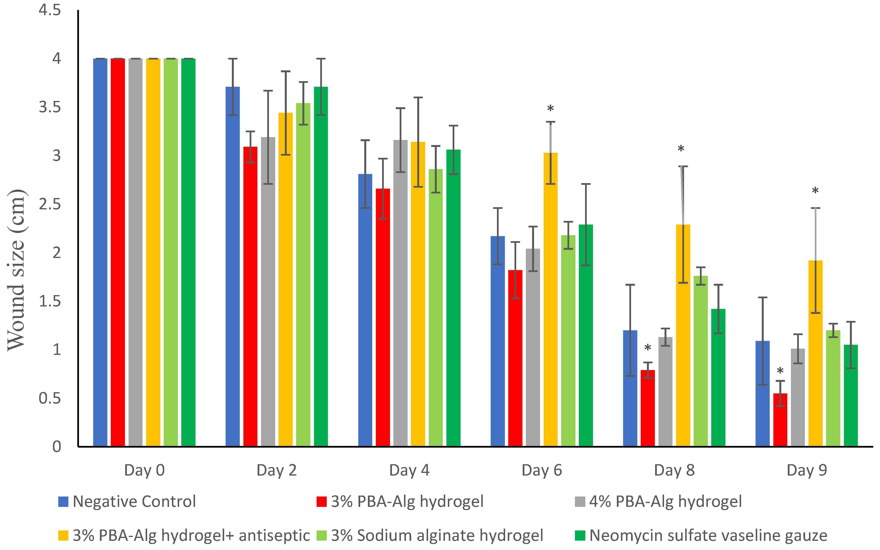
3.1.6. Wound Size Reduction

Wound size measurements on each evaluation day (Figure 2) showed significantly smaller wound size, higher wound size reduction, and faster healing in the group treated with 3% w/w hydrogel alone compared to the other groups. Generally, there was no significant difference (p > 0.05) in wound size among the other groups although the group treated with a combination of 3% hydrogel and Savlon antiseptic showed significantly lower healing at the later days (8 and 9). The wound length of groups treated with 3% hydrogel was reduced from 4 cm at day 0 to 1.82 cm at day 6, equivalent to a 54.5% reduction in wound size.
Certain factors may be responsible for the improved wound healing observed with the 3% w/w samples. Materials containing boronic acid have been reported to bind with the glycoproteins present in cell membranes that cause the proliferation of lymphocytes and other related cells, facilitating faster wound healing [29,30]. Boronic acid units are able to covalently bond to biological macromolecules with cis-diol moieties, resulting in the formation of boronate ester rings [31,32]. Indeed, prior reports have shown that boronic acid-functionalized platforms are useful for wound healing [33]. Furthermore, alginate-based hydrogels have structural similarities to the extracellular matrix, which may facilitate the process of wound healing and closure [26]. The higher wound size reduction observed in the 3% phenylboronic acid–alginate treatment compared to its corresponding sodium alginate treatment show that the attachment of phenylboronic acid to alginate is essential for its improved wound healing activity. We therefore suspect that the unique bonding characteristics of boronic acids may be contributory. The gross appearance of the wound on days 0 and 9 is presented in Figure 3. Typical wound dressings require replacement as surfaces in direct contact with the wound usually degenerate. Therefore, the capacity of the formulated hydrogels to continuously repair and self-heal may reduce the need for replacement by keeping interactive surfaces fresh. Conversely, the combination of PBA-Alg and antiseptic may have caused reduced activity because of interference in the performance of the PBA-Alg. Moreover, delayed wound healing has been observed in the use of some antiseptics or antimicrobial agents [34] and this may be more pronounced where the antiseptic is persistently present in the wound area as is the case in our study.

3.1.7. Wound Closure and Scar Formation

The treatment group that received the 3% hydrogel regrew fur faster than the other groups, which still had bare skin at day 9 (Table 2). The degree of wound closure is evident from the scar (Figure 3). A moist wound environment (maintained by the hydrogels, in this experiment) promotes epithelialization [35] better than a dry environment, as a moist environment supports the migration of epidermal cells. Previous reports showed that water vapor transmission rate values of 200–250 mg/cm2/day are more suitable to maintain moist wound zone and to create an ideal healing environment [36,37]. It seems our formulations were able to maintain a moisture equilibrium by losing and then replacing moisture within its matrix. The direction of migration of keratinocytes is controlled by the arrangement of the extracellular matrix [38], and the hydrogel network can provide a similar scaffold.

4. Conclusions

The phenylboronic acid–alginate complexes described in this work can be formulated as soft self-repairing hydrogels with wound healing properties. These hydrogels have favorable physico-chemical properties and stability, delayed blood clotting, and enabled effective wound closure depending on the composition of the materials. Silicate composites of these hydrogels provided modifications in the hematological effects with low-dose silicates (20 mg, 1.2 wt%) maintaining normal clotting, whereas high-dose silicates (200 mg, 12 wt%) prolonged blood clotting to as much as 3 h. Among the different formulations, the 3% PBA-Alg hydrogels showed the highest wound healing capacity. This preliminary study points out potential novel wound healing applications of phenylboronic acid–alginate complexes and biorelevant qualities of their magnesium aluminometasilicate composites.

Supplementary Materials

The following are available online at https://www.mdpi.com/article/10.3390/app11156945/s1, Figure S1: FTIR spectrum of phenylboronic acid–alginate (PBA-Alg) complex. Figure S2: FTIR spectrum of acid-soluble collagen. Figure S3: FTIR spectrum of PBA-Alg complex/collagen mixture.

Author Contributions

Conceptualization, C.O.A. and D.D.D.; methodology, C.O.A. and C.C.M.; formal analysis, I.T.N.; investigation, C.O.A., C.C.M. and C.S.; writing—original draft preparation, C.O.A. and C.C.M.; writing—review and editing, C.O.A., D.D.D., C.S. and I.T.N.; supervision, C.O.A.; project administration, C.O.A. All authors have read and agreed to the published version of the manuscript.

Funding

D.D.D. thanks the Spanish Ministry of Science and Innovation for the grant PID2019-105391GB-C21.

Institutional Review Board Statement

The study was conducted according to the guidelines of the Declaration of Helsinki and approved by the Institutional Review Board (ethics committee) of the University of Nigeria Nsukka/Faculty of Pharmaceutical Sciences (protocol code is FPSRE/UNN/20/00024, dated 15 September 2020).

Informed Consent Statement

Not applicable.

Data Availability Statement

Data are contained within the article and supplementary material.

Acknowledgments

D.D.D. thanks the Spanish Ministry of Science and Innovation for the Senior Beatriz Galindo Award (BEAGAL18/00166) and NANOtec, INTech, Cabildo de Tenerife, and ULL for laboratory facilities. C.S. thanks Proyecto Inserción Académica (VRA-UC).

Conflicts of Interest

The authors declare no conflict of interest. The funders had no role in the design of the study; in the collection, analyses, or interpretation of data; in the writing of the manuscript, or in the decision to publish the results.

References

  1. Rodrigues, M.; Kosaric, N.; Bonham, C.A.; Gurtner, G.C. Wound healing: A cellular perspective. Physiol. Rev. 2019, 99, 665–706. [Google Scholar] [CrossRef] [PubMed]
  2. Amendola, V.; Meneghetti, M. Self-healing at the nanoscale. Nanoscale 2009, 1, 74–88. [Google Scholar] [CrossRef] [PubMed]
  3. Fratzl, P. Biomimetic materials research: What can we really learn from nature’s structural materials? J. R. Soc. Interface 2007, 4, 637–642. [Google Scholar] [CrossRef] [PubMed] [Green Version]
  4. Davami, K.; Mohsenizadeh, M.; Mitcham, M.; Damasus, P.; Williams, Q.; Munther, M. Additively manufactured self-healing structures with embedded healing agent reservoirs. Sci. Rep. 2019, 9, 7474. [Google Scholar] [CrossRef]
  5. Wei, Z.; Yang, J.H.; Zhou, J.; Xu, F.; Zrínyi, M.; Dussault, P.H.; Osada, Y.; Chen, Y.M. Self-healing gels based on constitutional dynamic chemistry and their potential applications. Chem. Soc. Rev. 2014, 43, 8114–8131. [Google Scholar] [CrossRef]
  6. Agubata, C.O.; Ezeanya, N.I.; Nzekwe, I.T.; Uzondu, S.W.; Mbah, C.C. Characterization and sustainable anti-ulcer activity of hydrogels based on acid-diol reactions. Sustain. Chem. Pharm. 2021, 21, 100414. [Google Scholar] [CrossRef]
  7. Cambre, J.N.; Sumerlin, B.S. Biomedical applications of boronic acid polymers. Polymer 2011, 52, 4631–4643. [Google Scholar] [CrossRef] [Green Version]
  8. Marco-Dufort, B.; Tibbitt, M.W. Design of moldable hydrogels for biomedical applications using dynamic covalent boronic esters. Mater. Today Chem. 2019, 12, 16–33. [Google Scholar] [CrossRef]
  9. Liu, H.; Li, Y.; Sun, K.; Fan, J.; Zhang, P.; Meng, J.; Wang, S.; Jiang, L. Dual-responsive surfaces modified with phenylboronic acid-containing polymer brush to reversibly capture and release cancer cells. J. Am. Chem. Soc. 2013, 135, 7603–7609. [Google Scholar] [CrossRef] [PubMed]
  10. Ryu, J.H.; Lee, G.J.; Shih, Y.V.; Kim, T.; Varghese, S. Phenylboronic acid-polymers for biomedical applications. Curr. Med. Chem. 2019, 26, 6797–6816. [Google Scholar] [CrossRef]
  11. Aeridou, E.; Díaz, D.D.; Alemán, C.; Pérez-Madrigal, M.M. Advanced functional hydrogel biomaterials based on dynamic B-O bonds and polysaccharide building blocks. Biomacromolecules 2020, 21, 3984–3996. [Google Scholar] [CrossRef]
  12. Abasalizadeh, F.; Moghaddam, S.V.; Alizadeh, E.; Akbari, E.; Kashani, E.; Fazljou, S.M.B.; Torbati, M.; Akbarzadeh, A. Alginate-based hydrogels as drug delivery vehicles in cancer treatment and their applications in wound dressing and 3D bioprinting. J. Biol. Eng. 2020, 14, 8. [Google Scholar] [CrossRef]
  13. Pettignano, A.; Grijalvo, S.; Häring, M.; Eritja, R.; Tanchoux, N.; Quignard, F.; Díaz, D.D. Boronic acid-modified alginate enables direct formation of injectable, self-healing and multistimuli-responsive hydrogels. Chem. Commun. 2017, 53, 3350–3353. [Google Scholar] [CrossRef] [Green Version]
  14. Suresh, K.; Häring, M.; Kumaraswamy, G.; Díaz, D.D. On the sensitivity of alginate rheology to composition. Soft Matter 2019, 15, 159–165. [Google Scholar] [CrossRef]
  15. Mishra, D.K.; Yadav, K.S.; Prabhakar, B.; Gaud, R.S. Nanocomposite for cancer targeted drug delivery. In Applications of Nanocomposite Materials in Drug Delivery; Inamuddin Asiri, A.M., Mohammed, A., Eds.; Woodhead publishing: Sawston, UK, 2018; pp. 323–337. [Google Scholar]
  16. Hári, J.; Pukánszky, B. Nanocomposites: Preparation, structure and properties. In Applied Plastics Engineering Handbook: Processing and Materials; Kutz, M., Ed.; William Andrew Publishing: New York, NY, USA, 2011; pp. 109–142. [Google Scholar]
  17. Gaharwar, A.K.; Kishore, V.; Rivera, C.; Bullock, W.; Wu, C.J.; Akkus, O.; Schmidt, G. Physically crosslinked nanocomposites from silicate-crosslinked PEO: Mechanical properties and osteogenic differentiation of human mesenchymal stem cells. Macromol. Biosci. 2012, 12, 779–793. [Google Scholar] [CrossRef]
  18. Meng, H.; Xiao, P.; Gu, J.; Wen, X.; Xu, J.; Zhao, C.; Zhang, J.; Chen, T. Self-healable macro-/microscopic shape memory hydrogels based on supramolecular interactions. Chem. Commun. 2014, 50, 12277–12280. [Google Scholar] [CrossRef] [PubMed]
  19. Butt, H.; Graf, K.; Kappl, M. Physics and Chemistry of Interfaces, 3rd ed.; Wiley-VCH: Weinheim, Germany, 2013; pp. 1–495. [Google Scholar]
  20. Lyklema, J. Fundamentals of Interface and Colloid Science: Liquid-Fluid Interfaces; Elsevier: Amsterdam, The Netherlands, 2000; pp. 1–751. [Google Scholar]
  21. Henriksen, K.; Karsdal, M.A. Type 1 Collagen. In Biochemistry of Collagens, Laminins and Elastin. Structure, Function and Biomarkers; Karsdal, M.A., Ed.; Academic Press: Cambridge, MA, USA, 2016; pp. 1–11. [Google Scholar]
  22. Kumar, S.S.; George, J.; Mukkadan, J.K. Bleeding time and clotting time in healthy male and female college students of Karukutty village, Kerala. Health Prospect. 2013, 12, 7–9. [Google Scholar] [CrossRef] [Green Version]
  23. Tetley, R.J.; Staddon, M.F.; Heller, D.; Hoppe, A.; Banerjee, S.; Mao, Y. Tissue fluidity promotes epithelial wound healing. Nat. Phys. 2019, 15, 1195–1203. [Google Scholar] [CrossRef] [PubMed] [Green Version]
  24. Tyman, J.H.P.; Bruce, I.E. Surfactant properties and biodegradation of polyethoxylates from phenolic lipids. J. Surfactants Deterg. 2004, 7, 169–173. [Google Scholar] [CrossRef]
  25. Kang, W.; Lim, H.; Kim, B.; Lee, Y.; Hahm, K.; Kim, S. Assessment of the effects of sugammadex on coagulation profiles using thromboelastographic parameters. Sci. Rep. 2020, 10, 19860. [Google Scholar] [CrossRef]
  26. Singh, S.; Dodt, J.; Volkers, P.; Hethershaw, E.; Philippou, H.; Ivaskevicius, V.; Imhof, D.; Oldenburg, J.; Biswas, A. Structure functional insights into calcium binding during the activation of coagulation factor XIII A. Sci. Rep. 2019, 9, 11324. [Google Scholar] [CrossRef]
  27. Lee, K.Y.; Mooney, D.J. Alginate: Properties and biomedical applications. Prog. Polym. Sci. 2012, 37, 106–126. [Google Scholar] [CrossRef] [Green Version]
  28. Litvinov, R.I.; Weisel, J.W. What is the biological and clinical relevance of fibrin? Semin. Thromb. Hemost. 2016, 42, 333–343. [Google Scholar] [CrossRef] [Green Version]
  29. Manju, S.; Antonty, M.; Sreenivasan, K. Synthesis and evaluation of a hydrogel that binds glucose and releases ciprofloxacin. J. Mater. Sci. 2010, 45, 4006–4012. [Google Scholar] [CrossRef]
  30. Uçan, U.S.; Sayin, Z. Proliferation effects of phenylboronic acid and boric acid on canine peripheral blood mononuclear cells. Turk. J. Vet. Anim. Sci. 2019, 43, 229–234. [Google Scholar] [CrossRef]
  31. Brooks, W.L.A.; Deng, C.C.; Sumerlin, B.S. Structure–reactivity relationships in boronic acid–diol complexation. ACS Omega 2018, 3, 17863–17870. [Google Scholar] [CrossRef] [PubMed] [Green Version]
  32. Elshaarani, T.; Yu, H.; Wang, L.; Ullah, R.S.; Haroon, M.; Khan, R.U.; Fahad, S.; Khan, A.; Nazir, A.; Usman, M. Synthesis of hydrogel-bearing phenylboronic acid moieties and their applications in glucose sensing and insulin delivery. J. Mater. Chem. B 2018, 6, 3831–3854. [Google Scholar] [CrossRef] [PubMed]
  33. Beyranvand, S.; Pourghobadi, Z.; Sattari, S.; Soleymani, K.; Donskyi, I.; Gharabaghi, M.; Unger, W.E.S.; Farjanikish, G.; Nayebzadeh, H.; Adeli, M. Boronic acid functionalized graphene platforms for diabetic wound healing. Carbon 2020, 158, 327–336. [Google Scholar] [CrossRef]
  34. Thomas, G.W.; Rael, L.T.; Bar-Or, R.; Shimonkevitz, R.; Mains, C.W.; Slone, D.S.; Craun, M.L.; Bar-Or, D. Mechanisms of delayed wound healing by commonly used antiseptics. J. Trauma 2009, 66, 82–91. [Google Scholar] [CrossRef]
  35. Junker, J.P.E.; Kamel, R.A.; Caterson, E.J.; Eriksson, E. Clinical impact upon wound healing and inflammation in moist, wet, and dry environments. Adv. Wound Care 2013, 2, 348–356. [Google Scholar] [CrossRef] [PubMed] [Green Version]
  36. Mouro, C.; Gomes, A.P.; Ahonen, M.; Fangueiro, R.; Gouveia, I.C. Chelidonium majus L. Incorporated emulsion electrospun PCL/PVA_PEC nanofibrous meshes for antibacterial wound dressing applications. Nanomaterials 2021, 11, 1785. [Google Scholar] [CrossRef]
  37. Xu, R.; Xia, H.; He, W.; Li, Z.; Zhao, J.; Liu, B.; Wang, Y.; Lei, Q.; Kong, Y.; Bai, Y.; et al. Controlled water vapor transmission rate promotes wound-healing via wound re-epithelialization and contraction enhancement. Sci. Rep. 2016, 6, 24596. [Google Scholar] [CrossRef] [Green Version]
  38. Patel, G.K.; Wilson, C.H.; Harding, K.G.; Finlay, A.Y.; Bowden, P.E. Numerous keratinocyte subtypes involved in wound re-epithelialization. J. Investig. Dermatol. 2006, 126, 497–502. [Google Scholar] [CrossRef] [Green Version]
Figure 1. (a) Chemical structure of PBA-Alg conjugate used in this study. (b) Schematic representation of the composites. Images of formulated nanocomposite hydrogels labeled NH-20 (c), NH-80 (d), NH-140 (e), and NH-200 (f) representing composites with 20 mg, 80 mg, 140 mg, and 200 mg of magnesium aluminometasilicate, respectively, in combination with 50 mg of PBA-Alg.
Applsci 11 06945 g001
Figure 2. Wound size profile obtained in animal groups after various treatments. N = 5 groups of 5 rats per treatment and control. Treatments: 3% PBA-Alg hydrogel, 4% PBA-Alg hydrogel, 3% PBA-Alg hydrogel + antiseptic. Values are presented as mean ± standard deviation. Data were analyzed using one-way analysis of variance (ANOVA). Significant difference at * p < 0.05 with 3% PBA-Alg hydrogels showing significantly smaller wound size compared to other treatments.
Applsci 11 06945 g002
Figure 3. (a) Day 0 animal incision wound. Incision length = 4 cm, (b) day 9 scar formation, (c,d) gross appearance after wound closure and scar formation on the healed area using 3% hydrogel. Note: average rat body length (nose tip to tail base) = 19 cm.
Applsci 11 06945 g003
Table 1. In vitro blood clotting time.
TreatmentClotting Time (min)
3% PBA-Alg9.09 ± 0.04 *
4% PBA-Alg9.34 ± 0.03 *
3% PBA-Alg + Savlon10.75 ± 0.04 *
none (negative control)4.20 ± 0.02
nanocomposite (20 mg silicate)4.08 ± 0.02
nanocomposite (80 mg silicate)5.55 ± 0.03 *
nanocomposite (140 mg silicate)11.50 ± 0.05 *
nanocomposite (200 mg silicate)180.00 ± 0.12 **
*, ** indicate statistical significance at p < 0.05, p < 0.01, respectively, compared to control.
Table 2. Wound closure time.
DrugWound Closure Day
3% PBA-Alg hydrogelday 14
4% PBA-Alg hydrogel day 14
3% PBA-Alg hydrogel + Savlonday 16
3% sodium alginate hydrogelday 15
Negative controlday 15
Neomycin sulfate Vaseline gauzeday 14
Publisher’s Note: MDPI stays neutral with regard to jurisdictional claims in published maps and institutional affiliations.

