Next Article in Journal
Using Convolutional Neural Network and Candlestick Representation to Predict Sports Match Outcomes
Next Article in Special Issue
Bacterial Adhesion to Grade 4 and Grade 5 Turned and Mildly Acid-Etched Titanium Implant Surfaces: An In Vitro and Ex Vivo Study
Previous Article in Journal
Spatial Variation of Water Chemistry in Aries River Catchment, Western Romania
Previous Article in Special Issue
Assessment of Fit on Ten Screw-Retained FrameworksRealized through Digital Full-Arch Implant Impression
 
 
Retraction published on 15 November 2021, see Appl. Sci. 2021, 11(22), 10756.
Article

Accuracy Assessment of Implant Placement in a Newly Developed Dynamic Navigation System: A Pilot Study

1
Section of Fixed Prosthodontics, Division of Oral Rehabilitation, Faculty of Dental Science, Kyushu University, Fukuoka 812-8582, Japan
2
Division of Advanced Dental Devices and Therapeutics, Faculty of Dental Science, Kyushu University, Fukuoka 812-8582, Japan
*
Author to whom correspondence should be addressed.
Appl. Sci. 2021, 11(14), 6593; https://doi.org/10.3390/app11146593
Submission received: 31 May 2021 / Revised: 7 July 2021 / Accepted: 15 July 2021 / Published: 17 July 2021 / Retracted: 15 November 2021
(This article belongs to the Special Issue New Materials and Technologies for Implant Dentistry)

Abstract

:
The aim of this pilot study was to evaluate the accuracy of a newly developed dynamic navigation system and to compare the accuracy between flapless and open-flap surgery, and between surgeons. The subjects were patients who were scheduled to receive implants of the same size using the newly developed dynamic navigation system. The study’s procedures included cone beam computed tomography (CBCT) filming with fiducials, virtual planning of implant placement and the use of motion tracking technology for calibration and practical implant placement. The accuracy was evaluated using preoperative (virtual implant) and postoperative (actual implant) CBCT images based on angular, apical, coronal and vertical deviations. The differences of deviations between flapless and open-flap surgery, and between two surgeons, were statistically compared. In total, 66 implants were placed in 39 patients. The median and interquartile range of angular, apical, coronal and vertical deviations were 3.07° (2.52–3.54°), 0.96 mm (0.75–1.42 mm), 0.76 mm (0.57–1.37 mm) and 0.71 mm (0.61–0.88 mm), respectively. These deviations were similar to those found in previous studies. Flapless surgery resulted in a more accurate placement with respect to apical and coronal deviations, and the differences between the two surgeons were limited. The newly developed dynamic navigation system is considered to be eligible for clinical use.

1. Introduction

Dynamic navigation surgery for dental implant placement has been increasingly used because it has the following advantages: (1) the availability of immediate placement after planning, (2) on-site alteration of implant placement dependent upon the clinical situation and (3) minimally invasive intervention due to real-time visualization [1,2,3,4]. In general, the workflow of dynamic navigation surgery includes images of cone beam computed tomography (CBCT) with fiducials, virtual planning of implant placement and the use of motion tracking technology for calibration and practical implant placement [4,5,6]. Briefly, fiducial markers, attached to the patient’s jaw during CBCT exposure, are registered in software. These markers and markers on the surgical handpiece are captured with a tracking camera in order to display the relative position of the jaw and the drill (burr) in CBCT images. These provide guidance for freehand real-time drilling and implant placement. This means that data acquisition and registration are the most crucial procedures for accomplishing the ideal placement as planned.
There have been many reports evaluating the accuracy of implant placement using dynamic navigation [2,3,6,7,8,9,10,11,12,13]. These previous studies evaluated the placement accuracy by comparing between preoperative and postoperative CBCT images based on angle deviation and/or lateral deviation and/or depth deviation. The results of these studies concluded that dynamic navigation surgery could achieve a more accurate implant placement as compared to freehand implant surgery, and a similar accuracy to static guided surgery. The important point is to recognize the procedures required for accurate placement. According to previous studies that validated static guided surgery, the type of guide (full-guided or half-guided) [14], digital workflow [15,16], surgical and patient’s conditions [17,18,19] and the type of guide support [20] can all affect the accuracy. In dynamic navigation surgery, the steps that can cause deviations are considered to be similar. Even in such a situation, the vertical position of the implant platform (the depth of the implant position) in open-flap surgery is the only implant position we can confirm under direct vision. Flapless placement, which is a minimally invasive procedure, requires virtual visualization of the surgical site (bone morphology and mucosal thickness), optimal virtual planning and accurate surgical technique. Previous studies have suggested that computer-guided surgery, with the proper workflow, permitted the surgeon to conduct accurate flapless surgery [3,6,7,9]. Regarding surgical technique in dynamic navigation, the influence of the surgical situation on the accuracy is controversial [2,4,21]. A dynamic navigation system with a higher accuracy is beneficial, but demanding. This means that validation of the accuracy is essential.
The present pilot study aims to evaluate the accuracy of implant placement using a newly developed dynamic navigation system, by comparing between preoperative and postoperative CBCT images. In addition, the deviations of implant positions were statistically compared between flapless and open-flap surgery, and between surgeons.

2. Materials and Methods

2.1. Study Design

The protocol of this study was approved by the institutional ethical committee (approval number: 28–119) and conducted in accordance with the Helsinki Declaration.
The candidates for this study were patients who visited the Department of Prosthodontics, Kyushu University Hospital for the purpose of implant rehabilitation between April 2018 and February 2021. The inclusion criteria were as follows: (1) patients who were 20 years old or more, (2) patients who understood the purpose of this study and (3) patients without systemic disease unsuitable for the surgical procedure. To evaluate under the same analytical conditions, patients who would receive the same implants (Ø 4.1 mm × 10 mm, Straumann® Dental Implant System, Basel, Switzerland) were selected, which diminished the effect of implant size (diameter and length) on the implant positions. The exclusion criteria included: (1) patients who could not agree the use of this dynamic navigation system and (2) patients with gingival inflammation and dental plaque accumulation. The patients who met the inclusion criteria and provided informed consent were enrolled in this study.

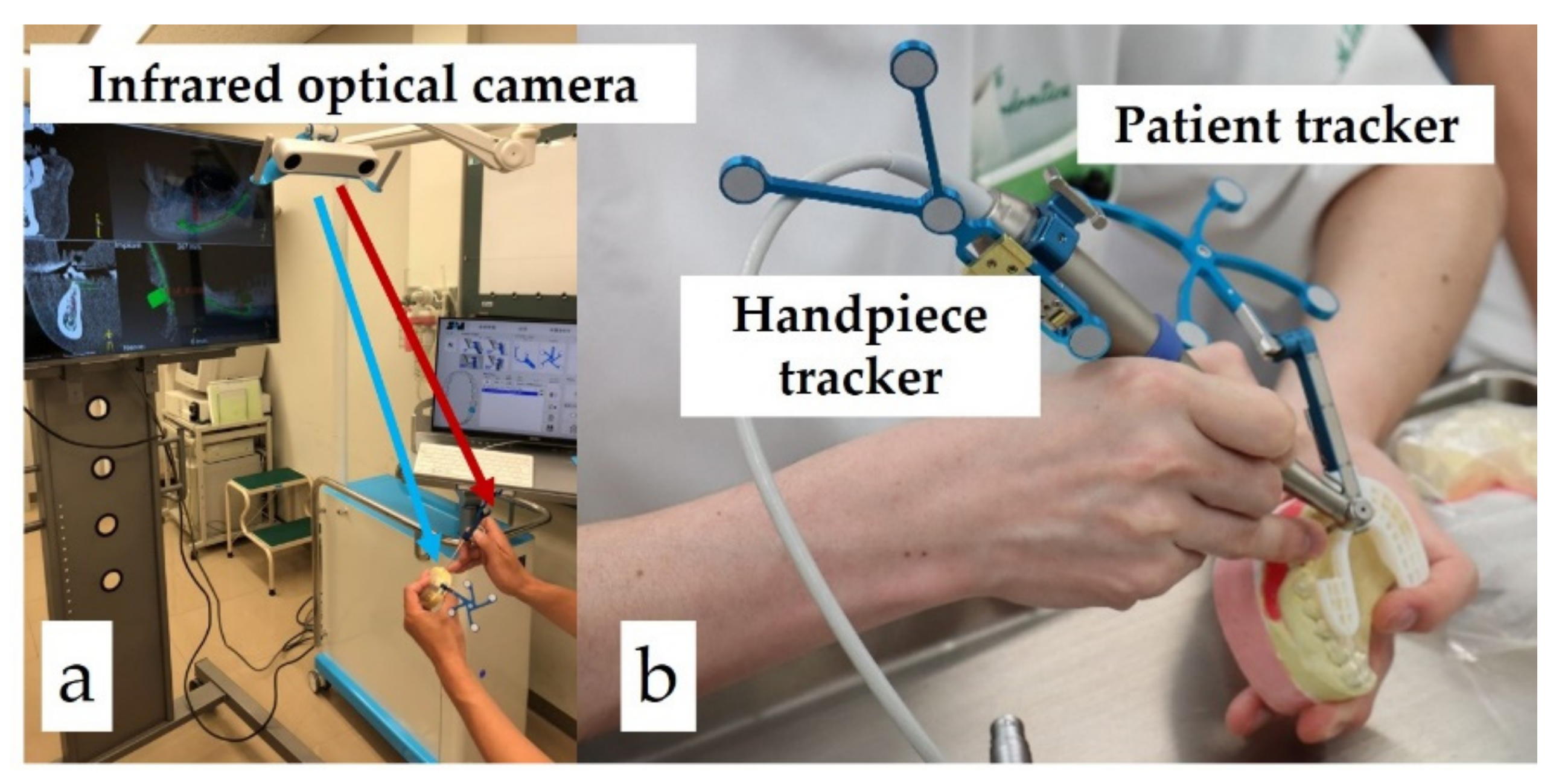
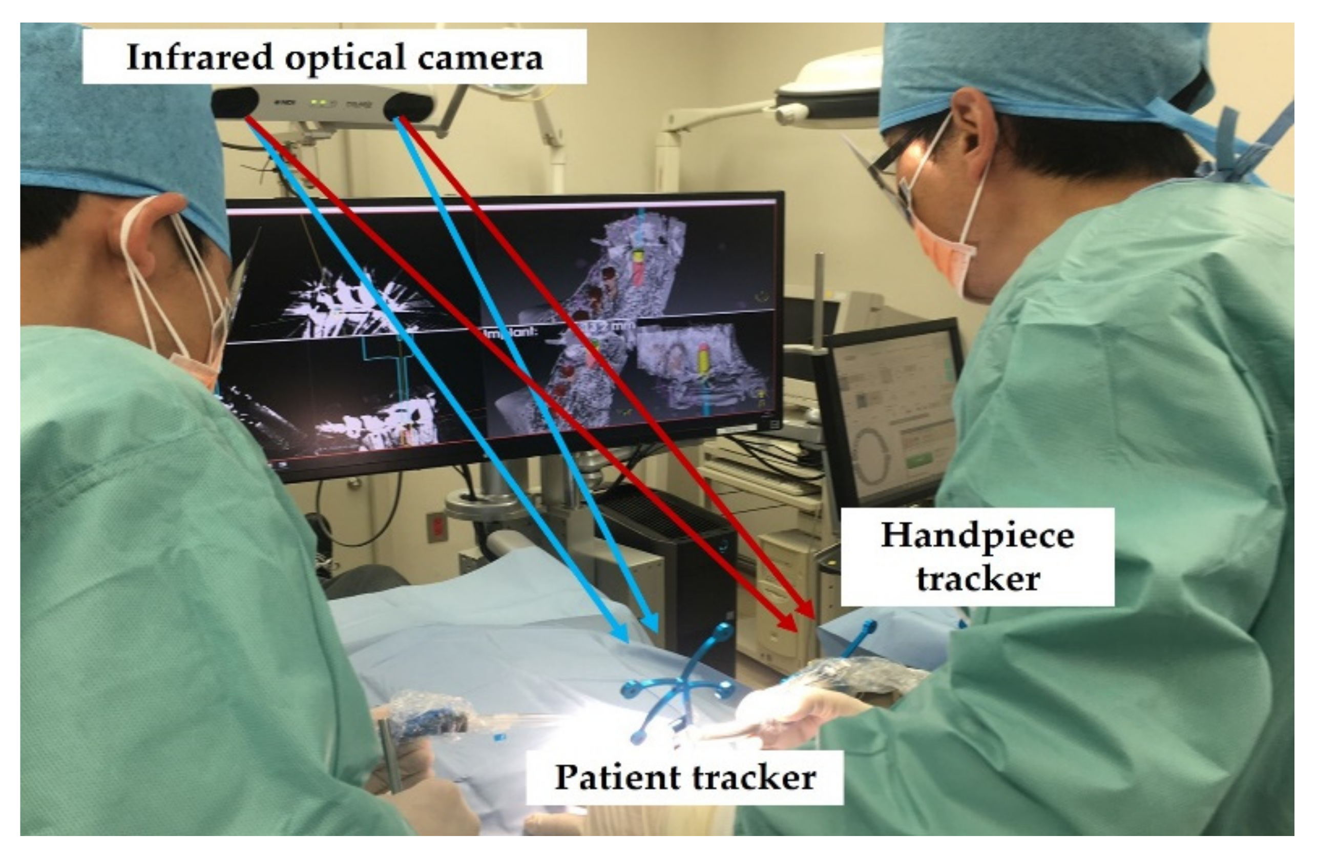
2.2. Dynamic Navigation System

In this study, a newly developed dynamic navigation system using a conventional infrared optical camera (ImVision, Safe Approach Medical Inc., Fukuoka, Japan) was tested. The preliminary study revealed that the working volume of the infrared camera was from 50 cm to 130 cm. In addition, the preliminary study also revealed that the deviation of this system was within 0.1 mm and 0.4°.

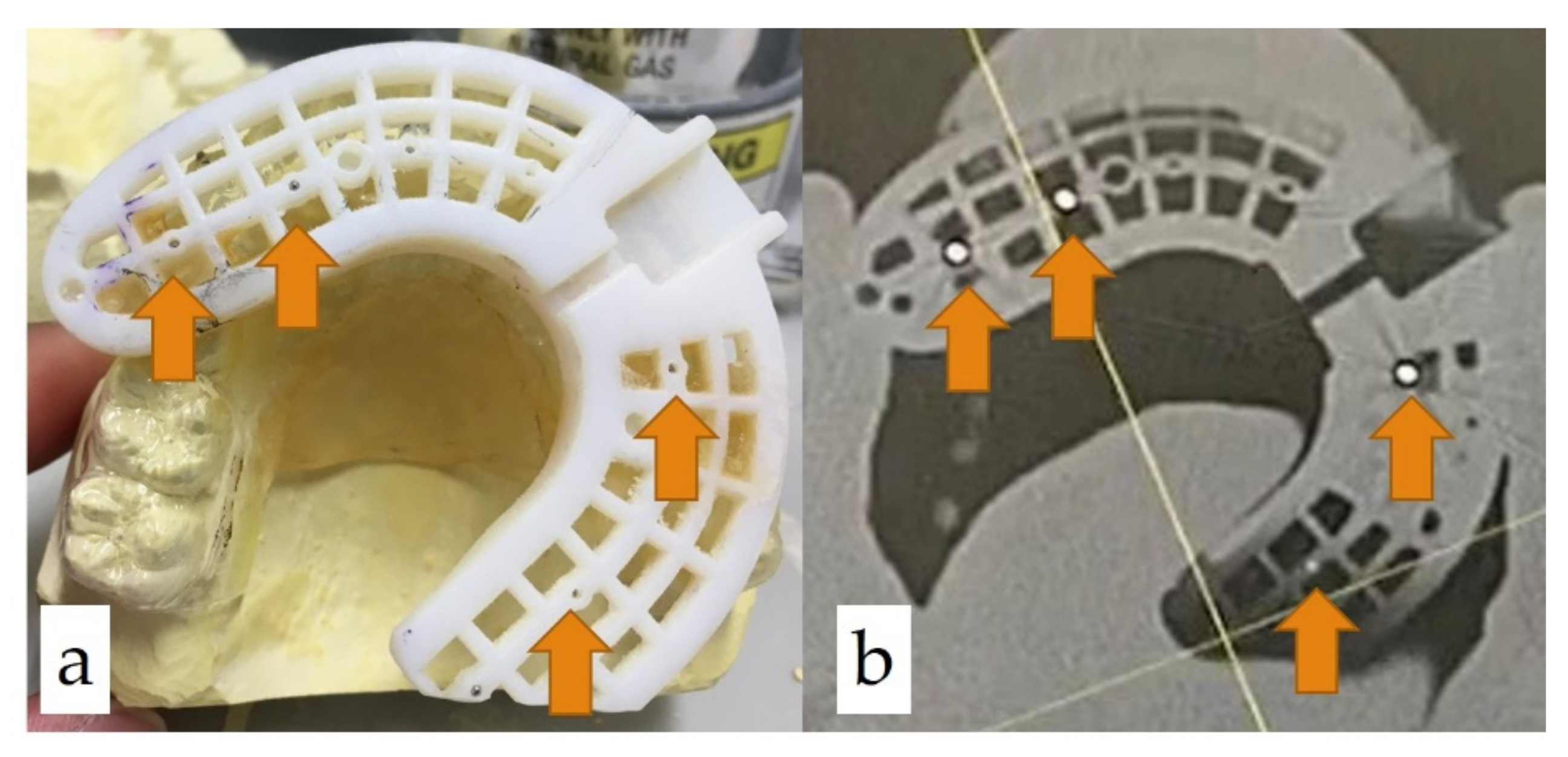
2.3. Preoperative Procedure

All patients were subjected to CBCT examination with prefabricated thermoplastic stents (Figure 1). The stents contained small titanium discs (diameter 1.0 mm and height 1.0 mm, more than 3) as fiducial markers and could be fixed onto remaining teeth rigidly. These were also used as registration devices. Three-dimensional models (3D models) were created from CBCT data (Digital Imaging and Communications in Medicine data: DICOM data) using software for image analysis and scientific visualization (3D Slicer, National Institutes of Health, Bethesda, MD, USA). These 3D models were imported into the navigation system. This procedure was effective in reducing artifacts caused by metallic prostheses. After the registration of the titanium discs incorporated in the stent, the tracking frames were attached to the stent extraorally, and to the handpiece, for virtual implant planning (Figure 2). These frames were captured with the infrared optical camera system for tracking. Prior to virtual planning, these tracking frames were recognized as tags by the infrared optical camera and were calibrated, including the length of the drills. The virtual position of the implant was planned with this optical tracking system using the model and the handpiece. This planning procedure was performed by T.S. and R.O., who also rechecked it.

2.4. Implant Placement Using Dynamic Navigation

Prior to surgery, the stent with a tracking frame was rigidly fixed at the same position of residual dentition. The tracking frame for the handpiece and the infrared optical camera system were also prepared, and the calibration of these frames were performed in the same manner as for the planning procedure (Figure 3).
All implants were placed under local anesthesia by T.S. and R.O. At a site with adequate keratinized mucosa, a flapless surgery was performed [6,22,23]. During surgery, the implant position was determined according to the virtual planning image and the real-time position, which were displayed on a monitor screen. The drilling procedure was followed according to the manufacturer’s instructions. After implant placement, antibiotics and analgesic drugs were provided to all patients.

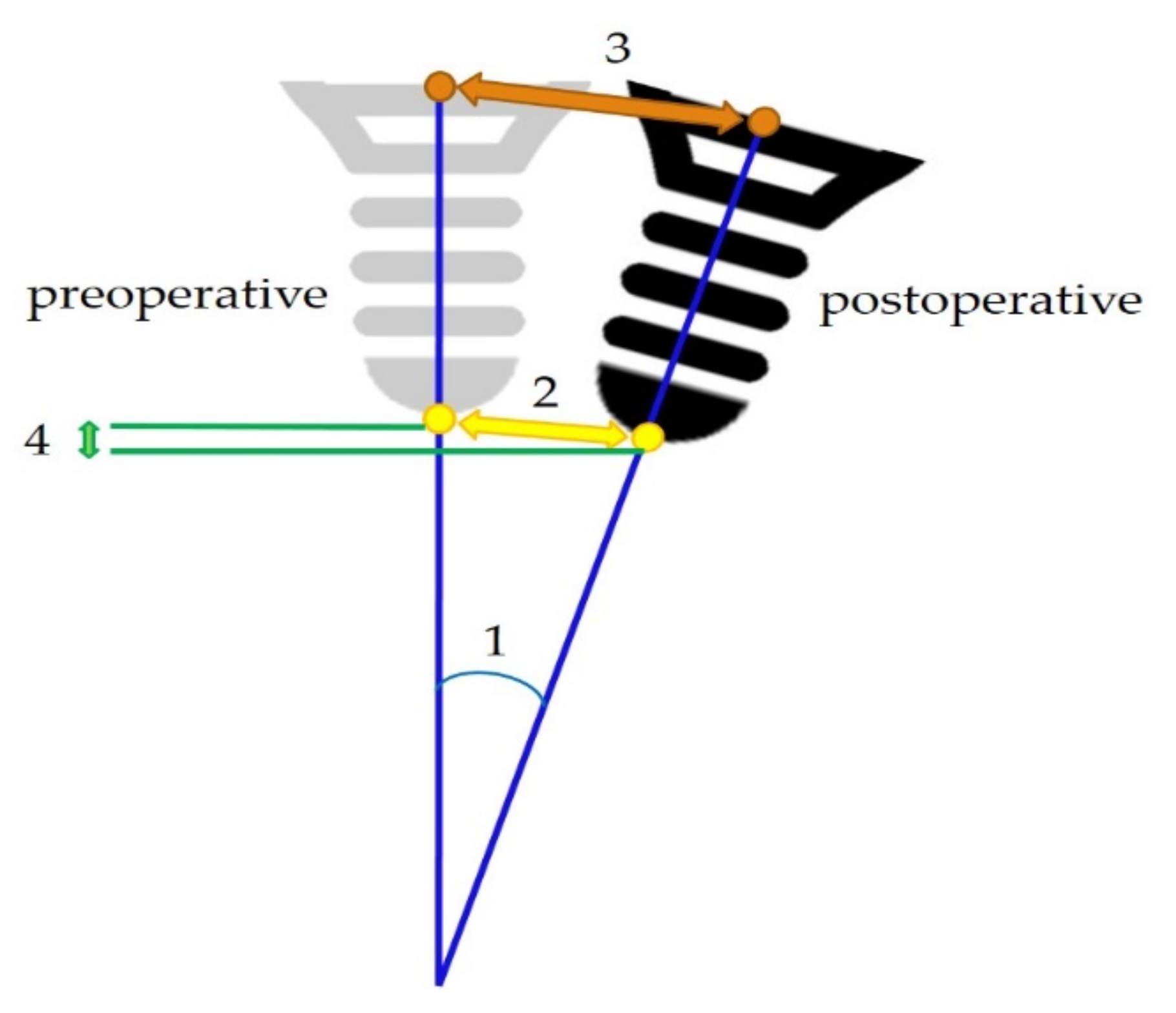
2.5. Statistical Evaluation of Implant Position Accuracy

All patients received a CT examination in the same manner within 1 month after surgery. Titanium discs in the stent in preoperative and postoperative 3D models were matched and the deviations between preoperative (planned) and postoperative (actual placed) implant position were measured. The measurement items were: (1) angular deviation between the axes of the implants from the sagittal view, (2) apical lateral deviation between the implants from the sagittal view, (3) coronal lateral deviation between implants from the sagittal view and (4) vertical (depth) deviation between the implants from the sagittal view (Figure 4). To describe each deviation, three measurements were conducted for each sample and their mean values were used as the final deviation value.
Numerical data were presented as median and interquartile ranges (IQR). The measurement values were compared according to the surgical procedures (flapless and open-flap surgery) and the surgeons (R.O.: 28 years’ experience or T.S.: 4 years’ experience). The deviations were statistically compared by using the Mann–Whitney U test and statistical significance was set at p < 0.05. Each effect size (r) was also calculated to measure the strength of the relationship between each pair of variables, because the sample size was limited in this study. The statistical analyses were performed using the JMP15 software (SAS Institute Inc., Cary, NC, USA).

3. Results

3.1. Subject Profiles and Implant Distribution

A total of 66 implants were placed in 39 patients (8 males and 31 females). The median age of the subjects was 65 years old (IQR: 49–73). All implants were included in the final analyses. The distributions of the placed implants are provided in Table 1. In this study, 46 implants (6 males and 23 females; 66 years old, IQR: 51–74) were placed using flapless surgery and 20 implants were placed using open-flap surgery (two males and eight females; 53.5 years old, IQR: 45.25–70.25). As described above, all patients were treated with the same diameter and length of implant (Ø 4.1 mm × 10 mm). All surgical procedures were completed without any adverse events.

3.2. Mean and Median Values of Each Deviation and Comparison with Previous Reviews

The median and IQR values of implant angular deviation, apical lateral deviation, coronal lateral deviation and vertical (depth) deviation were 3.07° (2.52–3.54°), 0.96 mm (0.75–1.42 mm), 0.76 mm (0.57–1.37 mm) and 0.71 mm (0.61–0.88 mm), respectively. (A Shapiro–Wilk test revealed that the deviation data did not show any normal distributions except for vertical deviations, and the present data were described as median and IQR.) To compare the present results with previous studies, the results reported in previous systematic reviews and meta-analyses are shown in Table 2 [24,25,26,27]. Most of the previous studies reported the data as means and 95% confidential intervals; the present data were also described in this way, for easy comparison. These deviations were similar to the results of previous studies, although this observation was not analyzed statistically.

3.3. Comparative Analysis between Flapless Surgery and Open-Flap Surgery

Each deviation was statistically compared between flapless surgery and open-flap surgery. The results of the data comparison are presented in Table 3. There were significant differences in apical (p < 0.01, r = 0.36; medium) and coronal (p < 0.05, r = 0.30; medium) lateral deviations, although there were no differences in angle and vertical deviations. These findings revealed that the implants were placed more accurately when using dynamic navigation in flapless surgery.

3.4. Comparative Analysis between Surgeons

The effect of surgical experience on the accuracy of implant placement was also evaluated (Table 4). This comparison only included implants placed in flapless surgery. In this study, a well-experienced surgeon (R.O.) had previously placed 31 implants, and the other (T.S.) had placed 15 implants. The present analyses identified a significant difference in coronal lateral deviation only (p < 0.01, r = 0.33; medium), and not in other deviations.

4. Discussion

Previous studies compared the accuracy of implant positions (coincidence between preoperative and postoperative positions) between dynamic navigation surgery, static guided surgery and freehand surgery. Most of them concluded that dynamic navigation surgery could enable accurate implant placement, with similar accuracy to static guided surgery and greater accuracy than freehand surgery [3,6,7,8,9,10,11,12,13]. Current systematic reviews and meta-analyses have also noted that dynamic navigation surgery achieved a clinically acceptable accuracy, although heterogeneity must be taken into account [24,25,26,27]. The mean values of the deviations shown in Table 2 fell into the intermediate values of previous reports. Similarly to these previous studies, this study compared preoperative and postoperative implant positions to evaluate the accuracy and the practicality of our system. In addition, the effects of different surgical procedures (flapless and open-flap surgeries) and surgeons’ experiences on the accuracy of implantation were also assessed. To define the number of subjects, we calculated the sample size based on the results of the present study; these were 71 (angular deviations), 49 (apical deviations), 67 (coronal deviations) and 217 (vertical deviations). Although previous clinical studies which evaluated the accuracy of a dynamic navigation system had a sample size between 20 and 231 [6,9,10,11,12,13,16], which seemed to be wide-ranging, there was, unfortunately, an insufficient number of patients for formal statistical analysis in this study. Effect sizes were calculated to compensate for this insufficient sample size and to evaluate validity; medium effect sizes were confirmed in our statistical comparisons, which showed significant differences and are displayed in Table 3 and Table 4. However, it was decided that this study was conducted as a pilot study, due to the insufficient sample size.
Prior to the discussion of our results, it is necessary to mention the difference between previous dynamic navigation systems and the newly developed one. The dynamic navigation systems in previous studies used visible wavelength light for their optical cameras. The new system in this study uses a conventional infrared optical camera, as in some previous studies [28,29,30]. Dynamic navigation systems using an infrared optical camera for tracking have been known to be stable regardless of the environment, especially with respect to brightness in the surgical room, and have been widely used because of their compact and simple system. The system with an infrared optical camera went off-patent in 2018 and the development of this new system was undertaken. This study was designed to verify the accuracy and feasibility of this newly developed system.
The mean values of the deviations in this study were intermediate values in all categories between the smallest and the largest deviations (Table 2). This finding implied that the newly developed system is clinically acceptable. However, a remarkable finding was that the implants placed in flapless surgeries were more accurate than the ones placed in open-flap surgeries, and the two surgical methods had medium strength. One proposed cause of this is that the surgeons tended to be more careful in an invisible situation, but this suggestion might be incorrect in this study. However, visual information during open-flap surgery might make the surgeon feel at ease. Actually, on-site modification of an implant position is known to be one of the specific features of dynamic navigation surgery [2,3,4,6,9,12,31]. Although all implants were intended to be placed according to preoperative planning in this study, “real” visual information in open-flap surgery might disturb accurate implant placement. The effect of the surgeons’ experiences on the accurate positioning was also examined. Previous studies demonstrated that the contribution of the surgeons’ experiences to the accuracy remains controversial [4,9,21,31,32,33]. A significant difference between the deviations of implants placed by more and less experienced surgeons was observed only in coronal lateral deviation and the calculation of effect size indicated that the strength was medium. It was suggested that the first drill tended to be unstable on crestal bone. However, the drill became stable in the implant bed, meaning that the modification of the preparation of the implant bed, according to preoperative planning, could be possible and easier. Although this is nothing more than our speculation, a well-experienced surgeon (R.O.) might manage the first drilling. However, no significant differences between the two surgeons were observed in the other deviations, and it could be concluded that the effect of the surgeons’ experiences on the accurate positioning was limited, although further evaluation with more surgeons might be favorable.
A limitation of this study was that some variables, including bone quality, implant size (length and diameter) and implant sites, were not evaluated. Regardless of these variables, the surgeons generally need to confirm what they are doing using both a monitor and the implant site itself during the surgery. This implies that posterior sites are much more difficult than anterior sites. In this study, the implant beds were prepared according to dynamic navigation without a surgical guide. However, a previous study indicated that dynamic navigation with a surgical guide enabled more accurate implant placement [10]. As mentioned above, the position of the first drill prior to drilling might influence the implant position, especially the coronal lateral position. In other words, implant placement might be modified to be more accurate from the proper coronal position (the first step of drilling). In a future study, the comparison between anterior and posterior implant positioning, with or without a surgical stent, will be required. It will use a greater number of implants and subjects, in addition to other variables. If sufficiently many subjects can be enrolled, the randomization of the patients can be possible and the quality of the analyses will be higher. However, one of the main purposes of this study was to evaluate the accuracy of this system. Although this study had some limitations, it has suggested the clinical viability of this dynamic navigation system, as judged from the statistical variability (mean and 95% CI or median and IQR). To demonstrate the clinical viability clearly, a well-designed study must be conducted in the future.

5. Conclusions

Although the sample size of this study might be insufficient, the newly developed dynamic navigation system seems to achieve a more accurate implant positioning, similar to those of previous studies. According to the present results, this system might be eligible for clinical use, especially flapless surgery, which enables a minimally invasive procedure and limits the differences between surgeons. Further studies with more subjects and more surgeons will be required to validate the effectiveness properly.

Author Contributions

Conceptualization, Y.O.; methodology, Y.O.; software, T.S. and R.O.; validation, Y.O.; formal analysis, Y.O.; investigation, T.S., R.O. and A.T.; resources, R.O.; data curation, T.S. and Y.O.; writing—original draft preparation, Y.O.; writing—review and editing, Y.A.; visualization, T.S. and R.O.; supervision, K.K.; project administration, R.O. and K.K.; funding acquisition, R.O. All authors have read and agreed to the published version of the manuscript.

Funding

This research was funded by Sugiyama Chemical and Industrial Laboratory.

Institutional Review Board Statement

The study was conducted according to the guidelines of the Declaration of Helsinki and approved by the Institutional Review Board of Kyushu University Hospital (approval number: 28–119).

Informed Consent Statement

Informed consent was obtained from all subjects involved in the study, if applicable.

Data Availability Statement

The datasets used and/or analyzed during the current study are available from the corresponding author on reasonable request.

Conflicts of Interest

One of the co-authors (R.O.) acted as a medical adviser with no stake in the company which developed this system. He did not analyze the data at all and did not receive any funds. K.K. belongs to the Division of Advanced Dental Devices and Therapeutics, Faculty of Dental Science, Kyushu University. This division is endowed by GC Corporation, Tokyo, Japan. GC Corporation had no specific roles in this study. The authors, including them (R.O. and K.K.), declare no conflict of interest. The company also had no role in the design of the study; in the collection, analyses, or interpretation of data; in the writing of the manuscript; or in the decision to publish the results.

References

  1. Block, M.S.; Emery, R.W. Static or Dynamic Navigation for Implant Placement-Choosing the Method of Guidance. J. Oral Maxillofac. Surg. 2016, 74, 269–277. [Google Scholar] [CrossRef]
  2. Block, M.S.; Emery, R.W.; Lank, K.; Ryan, J. Implant Placement Accuracy Using Dynamic Navigation. Int. J. Oral Maxillofac. Implants 2017, 32, 92–99. [Google Scholar] [CrossRef]
  3. Chen, C.K.; Yuh, D.Y.; Huang, R.Y.; Fu, E.; Tsai, C.F.; Chiang, C.Y. Accuracy of Implant Placement with a Navigation System, a Laboratory Guide, and Freehand Drilling. Int. J. Oral Maxillofac. Implants 2018, 33, 1213–1218. [Google Scholar] [CrossRef]
  4. Gargallo-Albiol, J.; Barootchi, S.; Salomó-Coll, O.; Wang, H.L. Advantages and disadvantages of implant navigation surgery. A systematic review. Ann. Anat. 2019, 225, 1–10. [Google Scholar] [CrossRef] [PubMed]
  5. Panchal, N.; Mahmood, L.; Retana, A.; Emery, R., 3rd. Dynamic Navigation for Dental Implant Surgery. Oral Maxillofac. Surg. Clin. N. Am. 2019, 31, 539–547. [Google Scholar] [CrossRef]
  6. Kaewsiri, D.; Panmekiate, S.; Subbalekha, K.; Mattheos, N.; Pimkhaokham, A. The accuracy of static vs. dynamic computer-assisted implant surgery in single tooth space: A randomized controlled trial. Clin. Oral Implants Res. 2019, 30, 505–514. [Google Scholar] [CrossRef]
  7. Emery, R.W.; Merritt, S.A.; Lank, K.; Gibbs, J.D. Accuracy of Dynamic Navigation for Dental Implant Placement-Model-Based Evaluation. J. Oral Implantol. 2016, 42, 399–405. [Google Scholar] [CrossRef]
  8. Mediavilla Guzmán, A.; Riad Deglow, E.; Zubizarreta-Macho, Á.; Agustín-Panadero, R.; Hernández Montero, S. Accuracy of Computer-Aided Dynamic Navigation Compared to Computer-Aided Static Navigation for Dental Implant Placement: An In Vitro Study. J. Clin. Med. 2019, 8, 2123. [Google Scholar] [CrossRef] [Green Version]
  9. Stefanelli, L.V.; DeGroot, B.S.; Lipton, D.I.; Mandelaris, G.A. Accuracy of a Dynamic Dental Implant Navigation System in a Private Practice. Int. J. Oral Maxillofac. Implants 2019, 34, 205–213. [Google Scholar] [CrossRef] [PubMed]
  10. Sun, T.M.; Lee, H.E.; Lan, T.H. Comparing Accuracy of Implant Installation with a Navigation System (NS), a Laboratory Guide (LG), NS with LG, and Freehand Drilling. Int. J. Environ. Res. Public Health 2020, 17, 2107. [Google Scholar] [CrossRef] [Green Version]
  11. Aydemir, C.A.; Arısan, V. Accuracy of dental implant placement via dynamic navigation or the freehand method: A split-mouth randomized controlled clinical trial. Clin. Oral Implants Res. 2020, 31, 255–263. [Google Scholar] [CrossRef]
  12. Wu, D.; Zhou, L.; Yang, J.; Zhang, B.; Lin, Y.; Chen, J.; Huang, W.; Chen, Y. Accuracy of dynamic navigation compared to static surgical guide for dental implant placement. Int. J. Implant Dent. 2020, 6, 78. [Google Scholar] [CrossRef] [PubMed]
  13. Edelmann, C.; Wetzel, M.; Knipper, A.; Luthardt, R.G.; Schnutenhaus, S. Accuracy of Computer-Assisted Dynamic Navigation in Implant Placement with a Fully Digital Approach: A Prospective Clinical Trial. J. Clin. Med. 2021, 10, 1808. [Google Scholar] [CrossRef]
  14. Gargallo-Albiol, J.; Barootchi, S.; Marqués-Guasch, J.; Wang, H.L. Fully Guided Versus Half-Guided and Freehand Implant Placement: Systematic Review and Meta-analysis. Int. J. Oral Maxillofac. Implants 2020, 35, 1159–1169. [Google Scholar] [CrossRef] [PubMed]
  15. Al Yafi, F.; Camenisch, B.; Al-Sabbagh, M. Is Digital Guided Implant Surgery Accurate and Reliable? Dent. Clin. N. Am. 2019, 63, 381–397. [Google Scholar] [CrossRef]
  16. Cristache, C.M.; Burlibasa, M.; Tudor, I.; Totu, E.E.; Di Francesco, F.; Moraru, L. Accuracy, Labor-Time and Patient-Reported Outcomes with Partially versus Fully Digital Workflow for Flapless Guided Dental Implants Insertion-A Randomized Clinical Trial with One-Year Follow-Up. J. Clin. Med. 2021, 10, 1102. [Google Scholar] [CrossRef]
  17. El Kholy, K.; Janner, S.F.M.; Schimmel, M.; Buser, D. The influence of guided sleeve height, drilling distance, and drilling key length on the accuracy of static Computer-Assisted Implant Surgery. Clin. Implant Dent. Relat. Res. 2019, 21, 101–107. [Google Scholar] [CrossRef] [Green Version]
  18. Li, J.; Chen, Z.; Chan, H.L.; Sinjab, K.; Yu, H.; Wang, H.L. Does flap opening or not influence the accuracy of semi-guided implant placement in partially edentulous sites? Clin. Implant Dent. Relat. Res. 2019, 21, 1253–1261. [Google Scholar] [CrossRef] [PubMed]
  19. Putra, R.H.; Yoda, N.; Iikubo, M.; Kataoka, Y.; Yamauchi, K.; Koyama, S.; Cooray, U.; Astuti, E.R.; Takahashi, T.; Sasaki, K. Influence of bone condition on implant placement accuracy with computer-guided surgery. Int. J. Implant Dent. 2020, 6, 62. [Google Scholar] [CrossRef]
  20. El Kholy, K.; Lazarin, R.; Janner, S.F.M.; Faerber, K.; Buser, R.; Buser, D. Influence of surgical guide support and implant site location on accuracy of static Computer-Assisted Implant Surgery. Clin. Oral Implants Res. 2019, 30, 1067–1075. [Google Scholar] [CrossRef]
  21. Sun, T.M.; Lee, H.E.; Lan, T.H. The influence of dental experience on a dental implant navigation system. BMC Oral Health 2019, 19, 222. [Google Scholar] [CrossRef] [Green Version]
  22. Kaku, T.; Ogino, Y.; Matsushita, Y.; Koyano, K. Technique for identifying keratinized gingival tissue and soft tissue morphology on 3-dimensional implant-planning software images. J. Prosthet. Dent. 2016, 116, 21–24. [Google Scholar] [CrossRef]
  23. Lin, G.H.; Chan, H.L.; Wang, H.L. The significance of keratinized mucosa on implant health: A systematic review. J. Periodontol. 2013, 84, 1755–1767. [Google Scholar] [CrossRef]
  24. Bover-Ramos, F.; Viña-Almunia, J.; Cervera-Ballester, J.; Peñarrocha-Diago, M.; García-Mira, B. Accuracy of Implant Placement with Computer-Guided Surgery: A Systematic Review and Meta-Analysis Comparing Cadaver, Clinical, and In Vitro Studies. Int. J. Oral Maxillofac. Implants 2018, 33, 101–115. [Google Scholar] [CrossRef] [PubMed]
  25. Wei, S.M.; Zhu, Y.; Wei, J.X.; Zhang, C.N.; Shi, J.Y.; Lai, H.C. Accuracy of dynamic navigation in implant surgery: A systematic review and meta-analysis. Clin. Oral Implants Res. 2021, 32, 383–393. [Google Scholar] [CrossRef]
  26. Schnutenhaus, S.; Edelmann, C.; Knipper, A.; Luthardt, R.G. Accuracy of Dynamic Computer-Assisted Implant Placement: A Systematic Review and Meta-Analysis of Clinical and In Vitro Studies. J. Clin. Med. 2021, 10, 704. [Google Scholar] [CrossRef]
  27. Jorba-García, A.; González-Barnadas, A.; Camps-Font, O.; Figueiredo, R.; Valmaseda-Castellón, E. Accuracy assessment of dynamic computer-aided implant placement: A systematic review and meta-analysis. Clin. Oral Investig. 2021, 25, 2479–2494. [Google Scholar] [CrossRef]
  28. Hong, J.; Matsumoto, N.; Ouchida, R.; Komune, S.; Hashizume, M. Medical navigation system for otologic surgery based on hybrid registration and virtual intraoperative computed tomography. IEEE Trans. Biomed. Eng. 2009, 56, 426–432. [Google Scholar] [CrossRef]
  29. Matsumoto, N.; Oka, M.; Cho, B.; Hong, J.; Jinnouchi, M.; Ouchida, R.; Hashizume, M.; Komune, S. Cochlear implantation assisted by noninvasive image guidance. Otol. Neurotol. 2012, 33, 1333–1338. [Google Scholar] [CrossRef] [PubMed]
  30. Oka, M.; Cho, B.; Matsumoto, N.; Hong, J.; Jinnouchi, M.; Ouchida, R.; Komune, S.; Hashizume, M. A preregistered STAMP method for image-guided temporal bone surgery. Int. J. Comput. Assist. Radiol. Surg. 2014, 9, 119–126. [Google Scholar] [CrossRef]
  31. Block, M.S.; Emery, R.W.; Cullum, D.R.; Sheikh, A. Implant Placement Is More Accurate Using Dynamic Navigation. J. Oral Maxillofac. Surg. 2017, 75, 1377–1386. [Google Scholar] [CrossRef] [PubMed]
  32. Golob Deeb, J.; Bencharit, S.; Carrico, C.K.; Lukic, M.; Hawkins, D.; Rener-Sitar, K.; Deeb, G.R. Exploring training dental implant placement using computer-guided implant navigation system for predoctoral students: A pilot study. Eur. J. Dent. Educ. 2019, 23, 415–423. [Google Scholar] [CrossRef] [PubMed]
  33. Pellegrino, G.; Bellini, P.; Cavallini, P.F.; Ferri, A.; Zacchino, A.; Taraschi, V.; Marchetti, C.; Consolo, U. Dynamic Navigation in Dental Implantology: The Influence of Surgical Experience on Implant Placement Accuracy and Operating Time. An in Vitro Study. Int. J. Environ. Res. Public Health 2020, 17, 2153. [Google Scholar] [CrossRef] [PubMed] [Green Version]
Figure 1. Preoperative procedure: (a) prefabricated thermoplastic stent with fiducial markers (small titanium discs; arrow heads); (b) CBCT image.
Applsci 11 06593 g001
Figure 2. Preoperative procedure (calibration, virtual planning and simulation): (a) calibration of patient and handpiece tracker; (b) virtual planning and simulation.
Applsci 11 06593 g002
Figure 3. Implant placement using the present dynamic navigation system.
Applsci 11 06593 g003
Figure 4. A schema for each deviation measurement from sagittal view: (1) angle deviation; (2) apical lateral deviation; (3) coronal lateral deviation; (4) vertical (depth) deviation.
Applsci 11 06593 g004
Table 1. Distribution of implants.
MaxillaMandible
Incisors and Canines8 (0) *2 (1) *
Premolars and Molars30 (6) *26 (13) *
* Total number of implants (the number of implants placed in open-flap surgery).
Table 2. Results of deviations in previous studies and the present study.
Angular Deviations (°)Apical Deviation (mm)Coronal Deviation (mm)Vertical Deviation (mm)
Bover-Ramos et al. [24] (mean ± SD) 3.98 ± 0.331.40 ± 0.121.10 ± 0.990.74 ± 0.10
Wei et al. [25] (mean, 95% CI) 4.22 (2.74, 5.68)1.45 (1.19, 1.72)1.11 (0.96, 1.26)Not analyzed
Schnutenhaus et al. [26] (mean, 95% CI) 4.10 (3.12, 5.10)1.33(0.98, 1.68).1.00 (0.83, 1.16)Not analyzed
Jorba-García et al. [27] (mean, 95% CI) 3.68 (3.61, 3.74)0.90 (0.83, 0.97)0.69 (0.67, 0.72)0.73 (0.70, 0.76)
Present study (mean, 95% CI)2.93 (2.75, 3.10)1.15 (1.01, 1.29)0.96 (0.84, 1.08)0.71 (0.66, 0.77)
Present study (median, IQR)3.07 (2.52–3.54)0.96 (0.75–1.42)0.76 (0.57–1.37)0.71 (0.61–0.88)
SD: standard deviation, CI: confidence interval, IQR: interquartile range.
Table 3. Statistical comparisons between flapless surgery and open-flap surgery (Mann–Whitney U test).
Angular Deviations (°)Apical Deviation (mm)Coronal Deviation (mm)Vertical Deviation (mm)
Flapless surgery2.92 (2.37–3.41)0.93 (0.72–1.11)0.68 (0.56–1.21)0.72 (0.61–0.88)
Open-flap surgery3.34 (2.64–3.65)1.37 (0.90–1.88)1.27 (0.72–1.67)0.67 (0.51–0.90)
p value0.06<0.01<0.050.70
Effect size (r)0.230.360.30−0.04
Table 4. Statistical comparisons between more experienced and less experienced surgeons (Mann–Whitney U test).
Angular Deviations (°)Apical Deviation (mm)Coronal Deviation (mm)Vertical Deviation (mm)
Well-experienced2.76 (2.38–3.22)0.93 (0.71–1.12)0.61 (0.51–0.88)0.71 (0.61–0.88)
Less-experienced3.29 (2.35–3.62)0.92 (0.76–1.05)1.15 (0.62–1.63)0.71 (0.63–0.89)
p value0.110.96<0.010.95
Effect size (r)0.19−0.0040.330.007
Publisher’s Note: MDPI stays neutral with regard to jurisdictional claims in published maps and institutional affiliations.

