Article

Transferred Cold Atmospheric Plasma Treatment on Melanoma Skin Cancer Cells with/without Catalase Enzyme In Vitro

1
Graduate Institute of Biomedical Materials and Tissue Engineering, International Ph.D. Program in Biomedical Engineering, College of Biomedical Engineering, Taipei Medical University, Taipei 110, Taiwan
2
Department of Materials Engineering, Ming Chi University of Technology, New Taipei City 243, Taiwan
3
Center for Plasma and Thin Film Technologies, Ming Chi University of Technology, New Taipei City 243, Taiwan
4
Department of Obstetrics and Gynecology, Taipei Medical University Hospital, Taipei 110, Taiwan
5
Cell Physiology and Molecular Image Research Center, Taipei Medical University, Wan Fang Hospital, Taipei 116, Taiwan
*
Authors to whom correspondence should be addressed.
These authors contributed equally to this work.
Appl. Sci. 2021, 11(13), 6181; https://doi.org/10.3390/app11136181
Submission received: 13 May 2021 / Revised: 29 June 2021 / Accepted: 30 June 2021 / Published: 2 July 2021
(This article belongs to the Special Issue The Applications of Plasma Techniques II)

Abstract

:
Cold atmospheric plasma (CAP) is a promising tool to overcome certain cancerous and precancerous conditions in dermatology. A scheme of transferred CAP was first developed to treat melanoma (B16F10) skin cancer cells as well as non-malignant (L929) cells in vitro. CAP was transferred using a silicone tube with a jet system that was developed and was assessed as to whether it could generate reactive oxygen and nitrogen species (RONS) at near-room temperature. The transferred CAP was characterized electrically and spectroscopically. Biological data showed that the transferred CAP killed cancer cells but not non-malignant (L929) cells. Plasma treatment was effective with a time duration of 30 s, whereas non-malignant (L929) cells were less damaged during plasma treatment. In addition, catalase (CAT) enzyme was applied to neutralize and detoxify the RONS generated by the transferred CAP. These findings suggest that transferred CAP can be considered a melanoma cancer therapy.

1. Introduction

Cancer is one of the major diseases and a leading cause of death worldwide. Among the various forms of cancer, melanoma skin cancer is regarded as one of the deadliest cancers. A survey revealed that melanoma skin cancer is rising faster than other types of cancers particularly in developed countries [1], which is probably due to the increasing ultraviolet (UV) radiation and holes in the ozone layer [2].
Advancements in cancer therapies are specifically due to innovative technologies from various fields of science and engineering which have improved diagnostic and treatment systems of patients. In the case of skin cancer cells, several treatment methods are suggested; however, the suggested treatments include undesired side effects. For instance, surgery as a primary treatment is able to remove skin cancer cells, including some healthy cells around cancer cells. Such treatment may leave scarring, and in some cases may even be painful. In the case of immunotherapy, several types of chemo-drugs are used for treating skin cancer, but they have various unwanted side effects as well. Chemotherapy is known as a common option for treating skin cancer cells [3]. Since this treatment is drug-based, in addition to side effects such as hair loss and so on, it is regarded as an ineffective treatment due to drug resistance [4]. As reported in a previously published article, various skin cancer cell treatments were well categorized, including the side effects of conventional treatments for skin cancer cells [5].
Radiation treatment uses high-energy radiation, such as x-rays or particles, to kill skin cancer cells. However, the side effects of radiation treatment are not usually limited to areas that receive radiation. Typical side effects may include changes in skin color, nausea, hair loss, and fatigue [6]. Suggested treatments for skin melanomas showed several advantages and disadvantages for clinical patients. Furthermore, all melanoma patients must undergo a screening process and get motivation or advice from dermatologists. They might be able to avoid experiencing possible side effects from the therapy. Determining how to prevent side effects during melanoma treatments and make melanoma patients comfortable is important and urgently needed. One of the novel melanoma treatments relies on plasma at atmospheric pressure.
In recent decades, plasma-based treatments were introduced in the field of medicine. Plasma is the fourth state of matter, including charged particles (ions and electrons), photons, and neutral atoms; it has a net neutral charge [7]. Plasma medicine has emerged as an interdisciplinary research field combining plasma physics, plasma chemistry, biology, and clinical medicine [8]. Plasma sources, in this field, are mainly focused on plasmas at atmospheric pressure that generate charged particles, radicals, excited species, and reactive oxygen (ROS) and nitrogen species (RNS). When interacting with ambient air, atmospheric-pressure plasma transfers the energy of particles of a noble gas with oxygen and nitrogen in ambient air; consequently, numerous ROS and RNS are generated such as oxygen radicals, nitrogen species, and so on. Here, we also merge ROS and RNS to call them RONS. The cytotoxicity effects of these species contribute to biomedical treatments [9,10,11,12], specifically in cancer therapies [13,14,15].
Generally, cold atmospheric plasma (CAP) sources are generated with various electrode configurations [16]. Herein, a dielectric barrier discharge (DBD)-based plasma jet configuration was employed, as it is more adaptable and safer for biomedical treatment. A specific configuration of the DBD-based plasma jet, which is called an extendable plasma jet, was utilized for melanoma cell treatment. Similar configurations were reported for endoscopic plasma applications [17], inner surface modification [18,19], and bio-targeting of human cell lines, such as A431 (skin carcinoma), HEK 293 (kidney embryonic cells), and A549 cell lines (human lung adenocarcinoma cells) [20]. In the case of biomedical treatment, the use of transferred CAPs, including helium and neon gases, were reported [17,20]; however, using a transferred CAP including argon gas, has not yet been mentioned for melanoma cancer cells. It should be noted that argon gas was just used for conventional plasma jet systems [20]. It has been found that the temperature of argon plasma with the configuration of the conventional plasma jet rose to around 40 °C. This heating problem is due to using argon gas [21] that would not be biocompatible. Whereas, using a scheme of transferred plasma, the gas temperature cools when the plasma gas is flowing from upstream to downstream in the jet system. In this case, the gas temperature is kept at a biocompatible level approaching room temperature to avoid harming patients’ healthy cells. Furthermore, using the transferred plasma-based configuration, the high-voltage side is kept away to enhance patient safety for future clinical applications. In addition, compared to the large area irradiated with the volume DBD to kill or inactivate melanoma cancer cells [22], the small area of the transferred plasma (with a diameter of around 1~2 mm) are more applicable and allow us to localize the plasma treatment on the skin cancer cells/tumors.
RONS are considered to be bioactive products generated by treatment with CAP. Previous findings show that the conventional CAPs offer an approach for treating melanoma skin cancer by RONS induction of cellular apoptosis [22,23,24]. Induction of cellular apoptosis by the conventional CAP treatment occurs through the establishment of RONS. The formation of RONS by conventional CAP can also cause damage to DNA and cancer cell death. On the other hand, catalase (CAT) is responsible for neutralizing hydrogen peroxide and for abrogating toxic RONS production [25] possibly by breaking hydrogen peroxide down into water and oxygen. Herein, it is also investigated whether the addition of CAT is able to prevent the toxic effects of RONS, generated by transferred CAP as a developed plasma source.
In this study, an argon-based transferred CAP was developed for melanoma skin cancer cells in vitro. First, the transferred CAP with argon gas was characterized electrically and spectroscopically, then it was utilized for melanoma (B16F10) cell and non-malignant (L929) cell treatments individually, as well as melanoma (B16F10) cell treatment with the addition of CAT.

2. Materials and Methods

2.1. Transfer-CAP System

A scheme of transferring CAP using a silicone tube was utilized to produce a flexible plasma for the cell treatment. The experimental setup of the transferred CAP is illustrated in Figure 1. In this configuration, a 10-cm-long silicone tube was responsible for transferring the plasma jet. Argon gas was utilized in this investigation as it is known to be an affordable gas compared to other noble gases, such as helium and neon. Pure argon gas was adjusted by a mass flow controller to a flow rate of 8 standard liters per minute (SLM).

2.2. Transfer-CAP Generation

2.2.1. Electrical Measurement

A positive microsecond pulsed discharge with an on-time of 27 µs, an off-time of 47 µs (at a frequency of 12.5 kHz and duty cycle of 36%), and a peak voltage of 8.5 kV provided electrical discharges. A typical voltage-current characteristic corresponding to the transfer-CAP system is shown in Figure 2.

2.2.2. Emission and Spectroscopic Measurements

An intensified charge-coupled device (ICCD) and optical emission spectroscopy (OES) were utilized to characterize the reactive species generated by the transferred CAP when it interacted with ambient air. The measurement region was adjusted to be downstream of the jet system as shown in Figure 1a.

2.3. Cell Experiments

L929 (a non-malignant mouse fibroblast cell line, accession number: ATCC® CCL-1™) and B16F10 (a mouse melanoma skin cancer cell line, accession number: ATCC® CRL-6475™) cells used for this study were cultured in a cell incubator under standard culture conditions: humidified 5% CO2, at atmospheric pressure, at 37 °C in Dulbecco’s modified Eagle medium with 1% (v/v) penicillin/streptomycin, 10% (v/v) fetal bovine serum (FBS), 1% (v/v) 1-glutamine, and 1% (v/v) non-essential amino acids (NEAAs).

2.3.1. Cell Viability Analysis

We individually seeded L929 and B16F10 cells into 96-well plates (at 104 cells/well, with 5% CO2, at atmospheric pressure, at 37 °C) then cultured them for 2 days until they reached confluence. Before plasma treatment, we removed all culture medium from all wells and treated cells with transferred CAP for 30 s (with and without the addition of CAT from bovine liver with the concentration of 1 mg/50 mL). Subsequently, to test the cell viability after transfer-CAP treatment, we added fresh culture medium (200 μL/well) and a 3-(4,5-dimethylthiazol-2-yl)-2,5-diphenyltetrazolium bromide (MTT) solution (1 mg/mL, 20 μL/well) for a 1.5-h incubation. The supernatant medium was then removed, and dimethyl sulfoxide (DMSO) solution (200 μL/well) was added to dissolve the cellular formazan crystals which had formed. Formazan was quantified with a plate reader (SpactraMax 190) at a wavelength of 570 nm. Data are expressed as an average absorbance (optical density; OD) of triplicate experimental samples + standard deviation (SD) of the average.

2.3.2. Live/Dead Experiment

We seeded B16F10 cells (2 × 10⁵ cells/dish into 35-mm confocal dishes) and cultured them for 2 days in a 5% CO2 atmosphere at 37 °C. After transfer-CAP treatment, morphological changes in melanoma cells were assessed following staining protocols of a LIVE/DEAD® Viability (Calcein AM)/Cytotoxicity (EthD-III) Assay Kit (ThermoFisher). Fluorescent signals of live/dead cells were then observed with an IX81 optical microscope (Olympus, Tokyo, Japan). Morphological changes of cells treated with transferred CAP were also microscopically analyzed with the IX81 optical microscope (Olympus).

2.3.3. Fluorescence RONS and Catalase Level Analysis in Cells

In this study, B16F10 cells were seeded on confocal dishes incubated under 5% CO2 at 37 °C until adherence and confluence were achieved. Before CAP treatment, the medium was removed. Then, transferred CAP was applied for 30 s. Fluorescent 4′,6-diamidino-2-phenylindole (DAPI) was used to stain cell nuclei. Cells were stained with 2′,7′-dichlorodihydrofluorescein diacetate (H2DCF-DA) [26] to detect the amount of intracellular RONS generated using an IX81 fluorescent microscope (Olympus) after treatment. The immunofluorescence was employed to examine the expression of catalase in L929 cells and B16F10 cells. The bioactivity of catalase was assessed with fluorescence. Cells were seeded into 96-well (104 cells/wells) until attachment and blocked by 1% of serum. For immunofluorescence staining, catalase primary antibodies conjugating fluorescein isothiocyanate diluted 1:39 in PBS were incubated with the cells for 1 h. The unbound antibodies were washed with PBS. Fluorescence emission measurements were determined through a fluorescence microplate reader (n = 13).

2.3.4. Statistical Analysis

Experimental data are presented as the average (AVE) ± SD. Test results were statistically evaluated using Student’s t-test. The obtained values were considered to be statistically significant at p < 0.05.

3. Results and Discussion

3.1. Transfer-CAP Characterization

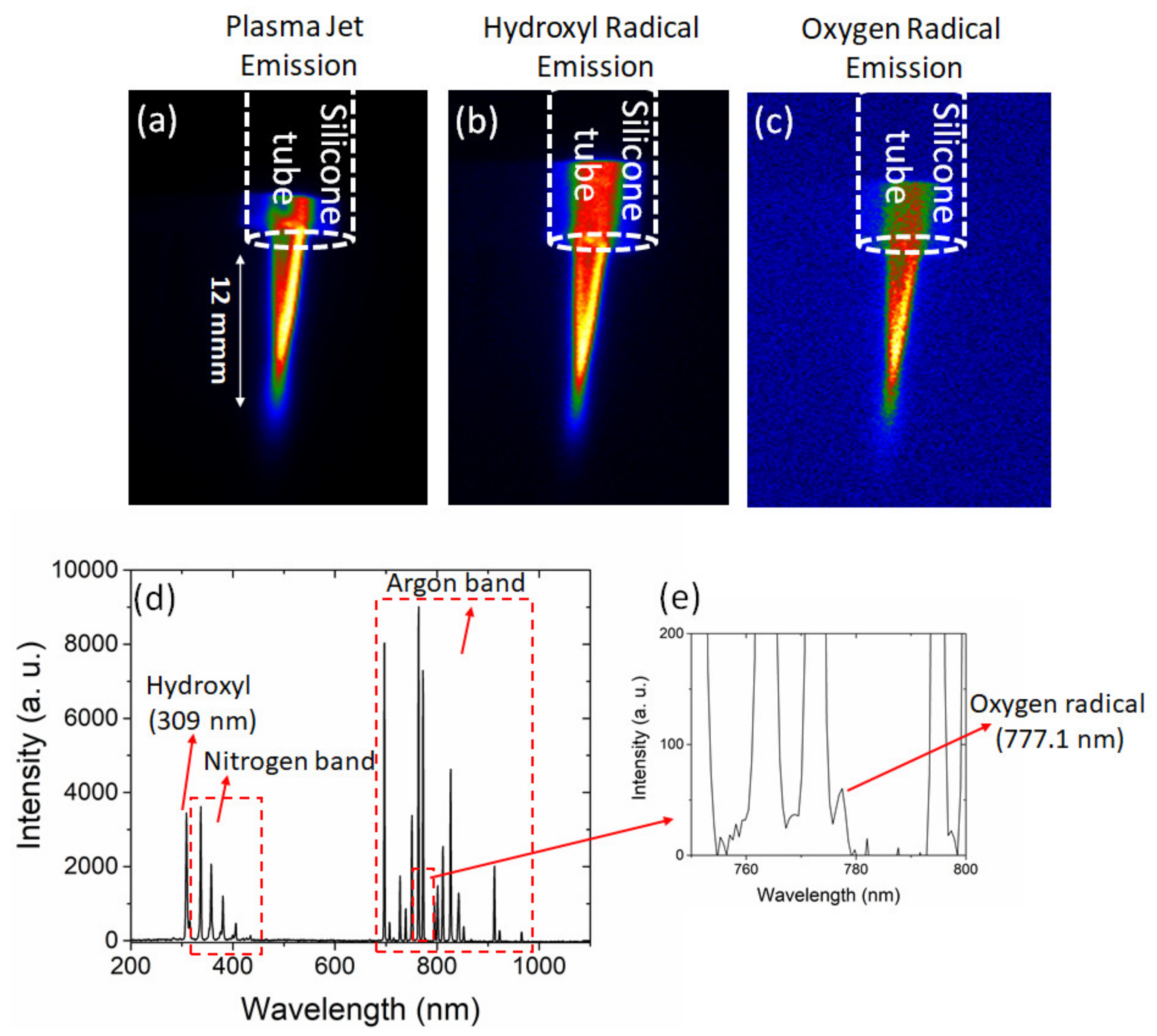
Figure 3a shows the emission of the transferred CAP. Using optical filters with 50% transmission, which were mounted in front of the ICCD camera, hydroxyl, and atomic oxygen radicals were observed as shown in Figure 3b,c, respectively. The dominant species of the spectra measured by OES were a hydroxyl peak (309 nm) and a nitrogen band (300~450 nm) as well as an argon band, as shown in Figure 3d. A similar spectrum related to the conventional argon-based plasma jet was reported for melanoma tumor treatment [27]. Moreover, a small peak of atomic oxygen radical (777.1 nm) was observed in the spectra (Figure 3e). The small peak of atomic oxygen radical was also reported in a radio-frequency plasma jet with argon [28] and argon/oxygen gas feeds [29]. Although the gas feed used in our system was argon, the generated RONS such as hydroxyl and atomic oxygen radicals as well as nitrogen species observed by OES were mainly respectively generated via energetic electron collisions with water molecules, oxygen, and nitrogen of the ambient air [30]. As a result, the presence of RONS generated by the transferred CAP was confirmed by emission and spectroscopic characterizations.

Gas Temperature

Biomedical applications of CAP first utilized plasma at thermal equilibrium, which was based on thermal energy for tissue removal, disinfection, and cauterization of thermally stable biomedical instruments. However, it is not suitable for heat-sensitive substances such as living tissues, as cells cannot tolerate such high temperatures. Recent advances in novel plasma sources, which can generate transferred CAP under atmospheric pressure in an open space at nearly room temperature, have allowed direct contact between live human cells and plasma yet avoid thermal damage. Implementation of cold plasma is essential for biological or medical treatment (Figure 4a), as only plasma with a temperature slightly higher than the body temperature which can be utilized to avoid harm and distress to patients’ cells. As shown in Figure 4b,c, an infrared (IR) camera was used to confirm that the gas temperature remained near room temperature after passing through an extended silicon tube. The thermal image data (Figure 4b,c) show that the temperature in the area of the transferred CAP remained under 30 °C during plasma treatment. For instance, a conventional argon-based CAP with a reported temperature of 35 °C to 40 °C was utilized for melanoma cells treatment [23]. A helium-based CAP with a temperature-controlled environment was also presented for melanoma treatment [24] although using argon is more affordable than helium gas. On the other hand, as mentioned in the introduction, using argon gas leads to a heating problem that causes a relatively high gas temperature compared to the room temperature. The treated biological cells and tissue could be damaged by heating with a temperature around 40 °C although there is no specific threshold temperature for heating damage [31]. In our case, during the plasma treatment, as shown in Figure 4b, the thermal image presents a gas temperature less than 30 °C, which is much safer for avoiding possible heating damage to biological cells and tissue.

3.2. Cell Viability

Cell lines of non-malignant murine (L929) fibroblasts and malignant murine melanoma (B16F10) cells were employed in this study. We examined whether the transferred CAP could kill B16F10 cancer cells in an in vitro model as well as explore the potential mechanisms that allow for the specific ablation of cancer cells without affecting non-malignant (L929) cells. We cultured cells in cell medium as shown in Figure 5a. Non-malignant (L929) cells and cancerous B16F10 cells were next collected by trypsinization and centrifugation (at 1200 rpm for 5 min). Collected cells were sub-cultured until used further. For plasma treatment, we removed the culture medium and applied the transfer-CAP system to cells. As a result, after transfer-CAP treatment, using an MTT assay and visible microscopy, we investigated cell viability and morphology as shown in Figure 5b,c, respectively. Cell viability results showed that the number of viable non-malignant (L929) cells was ca. 2~3-fold higher than the number of cancer B16F10 cells after the transferred CAP treatment. Therapeutic effect is one of the most imperative considerations of cancer treatment. Spectroscopic (Figure 3d,e) and MTT (Figure 5b) data suggest that the generation of RONS by transferred CAP preferentially killed malignant melanoma (B16F10) cells without exhibiting significant cytotoxicity toward non-malignant (L929) cells. Similar published findings also supported our experimental data [23,24]. In addition, the optical morphological data showed that the transferred CAP resulted in the separation of cancer cells compared to the before transfer-CAP treatment (Figure 5c). As shown in Figure 5c, optical image data suggests that the transferred CAP also could cause cell shrinkage and damage cancer cells. Transfer-CAP treatment thus led to morphological changes of melanoma cancer cells.

3.3. Live/Dead Experiment (Control, 30 s, and 30 s with CAT)

The transferred CAP generates RONS, including hydroxyl, atomic oxygen radicals, and nitrogen species, as shown in Figure 3. RONS-induced death of cells is a strategy for cancer treatment [32]. At low levels, RONS play crucial roles during redox homeostasis and in biological signals. However, high levels of RONS are able to break the balance, causing irreversible oxidative damage to lipids, carbohydrates, proteins, and DNA. RONS can be catalytically decomposed into water and oxygen by CAT, which defends cells from RONS oxidative damage [32]. CAT is a biological enzyme that plays key roles in cellular antioxidant defense mechanisms precipitated by the accumulation of RONS. To further examine the CAT and CAP relationship on B16F10 cancer cells, CAT was added to melanoma cancer (B16F10) cells before transfer-CAP treatment in the following cell viability test. In the live/dead study, 1 mg CAT/50 mL was pre-added to the cell culture medium for the co-culture of B16F10 cancer cells, and then cells were treated with transferred CAP. As shown in Figure 6a, the fluorescent-green represents live cells and red represents dead cells, which differ in the two treated groups. In the control group (B16F10 cells plus fluorescent dyes), 30 s of CAP treatment, leads to cell death. In the group after co-culture with CAT, it can clearly be observed that CAT might preserve cells alive even after treatment with transferred CAP. In addition, the MTT assay was used for further quantification (Figure 6b). The MTT data suggest that CAT adds potential protection to the transferred CAP resulting in a higher cell viability, compared to cells treated by the transferred CAP without CAT (Figure 6b). Furthermore, Image J software was used to quantitatively calculate green live cell fluorescent signals (Figure 6c). As shown in Figure 6b,c, the transferred CAP-treated group given CAT had higher cell viability than that of the transferred CAP-treated group without CAT. The addition of biological CAT is recognized to be a mechanism of cellular defense against RONS. Its function helps to balance the amount of cell RONS, whereas an imbalance between RONS and RONS-scavenging enzymes can lead to an event called oxidative stress [33].

3.4. Cell RONS Analysis (Control, 30 s, and 30 s with CAT)

Cellular RONS were seen to increase under CAP treatment, as shown in Figure 7a. The production of RONS in cells exists in equilibrium with antioxidant defenses. At moderate levels, RONS are believed to be important for regulating physiological and biological functions involved in development, including cell-cycle progression and differentiation, proliferation, and migration. RONS play vital roles in the immune system, maintaining a redox balance, and are associated with the bioactivation of various cell signaling pathways. Excess RONS in cells causes damage to cellular proteins, lipids, nucleic acids, organelles, and membranes that can lead to activation of cell death processes, including cell apoptosis. Apoptosis is a greatly modulated process that is critical for the survival and development of multicellular organisms. These multicellular organisms usually must discard cells that are possibly harmful or superfluous or that possess accumulated mutations. Apoptosis features a representative set of biochemical, pathological, and morphological aspects whereby cells undergo a sequence of self-destruction [34].
We used a cellular RONS analytical method to confirm the CAT and CAP roles on B16F10 cancer cells. Cell RONS levels can be determined in live cells by converting the non-fluorescent 2′,7′-dichlorofluorescein diacetate (DCFDA) that is oxidized into the fluorescent tracer, 2′, 7′-dichlorofluorescein (DCF). The generated fluorescent signal is directly proportional to the amount of DCFDA chemically oxidized into DCF. As the fluorescent emission is at the wavelength of ca. 529 nm, it can be determined by fluorescence microscopy, thereby measuring hydroxyl, peroxyl, and other RONS bioactivity in tested cells. Thus, melanoma cells were treated with the transferred CAP in the same way as the aforementioned cell culture method, then cells were stained with DAPI, and DCFDA RONS fluorescence was detected. The fluorescence intensity of RONS was much higher in CAP-treated cells (Figure 7a). The generated cellular RONS signals potentially came from the contributions of applied CAP or cell death after CAP treatment. However, the detailed mechanisms of RONS generation inside cells need to be investigated in the future. At the same time, the group that received CAT, after quantitative plasma treatment and analysis with Image J software (Figure 7b), possessed lower RONS fluorescent signals than the transfer-CAP-treated group without CAT, which confirms the original hypothesis. Furthermore, an interesting research [35], reported that CAP can help in wound healing (non-malignant L929 cells). According to published literature, a moderate ROS level is vital for promoting cell proliferation [36]. A moderate level of ROS plays an essential role in the cell signaling that controls cell survival and cell proliferation. However, a rise in ROS level could damage cell components for example DNA, lipids, proteins, triggering an imbalance among cellular reduction-oxidation (redox) situations and causing homeostasis disruption (in the case of cold-plasma treated cancer cells). By comparing the intensities of the immune-stained CAT level, the staining protein level detected by fluorescence microplate reader in non-malignant L929 cells group was much higher than that in the malignant B16F10 cells cell lines (p < 0.05), which suggests possible higher expression of CAT in L929 cells. The higher CAT expression from L929 cells group might possess an augmented ROS scavenging efficacy, compared to that of the B16F10 cells group, thus causing an anticancer effect when CAP is applied on B16F10 cells (Figure 7c). At present, thus we conclude that CAP can kill B16F10 melanoma cells effectively.

4. Conclusions

A transferred argon-driven plasma jet using a silicone tube was employed to make a flexible plasma jet system for melanoma skin cancer cell treatment. Results showed that the transferred plasma jet was able to effectively kill (inactivate) melanoma skin cancer cells. In addition, the presence of the CAT enzyme confirmed that RONS play key roles in killing melanoma cancer cells. The transferred CAP treatment was effective with a short time duration of 30 s, whereas non-malignant (L929) cells were less damaged during plasma treatment. This means that the transferred plasma jet can be considered for melanoma skin cancer therapy. Even though our and other groups proved CAP has anticancer outcomes, detailed investigation is still needed to understand the mechanisms underlying this result.

Author Contributions

Investigation, data analysis, Y.-H.C.; supervision, J.-H.H.; investigation, data analysis, I.-T.W.; investigation, P.-R.J., Y.-Y.Y.; supervision, J.-W.L.; writing—original draft preparation, writing—review and editing, N.B., E.-Y.C. All authors have read and agreed to the published version of the manuscript.

Funding

This research work was financially supported by the Ministry of Science and Technology, Taiwan (MOST 108-2320-B-038-061-MY3 and 108-2221-E-038-017-MY3).

Institutional Review Board Statement

Not applicable.

Informed Consent Statement

Not applicable.

Data Availability Statement

Not applicable.

Acknowledgments

Not applicable.

Conflicts of Interest

The authors declare no conflict of interest.

References

  1. Erdei, E.; Torres, S.M. A new understanding in the epidemiology of melanoma. Expert Rev. Anticancer. Ther. 2010, 10, 1811–1823. [Google Scholar] [CrossRef] [PubMed] [Green Version]
  2. D’Orazio, J.; Jarrett, S.; Amaro-Ortiz, A.; Scott, T. UV radiation and the skin. Int. J. Mol. Sci. 2013, 1, 12222–12248. [Google Scholar] [CrossRef] [PubMed] [Green Version]
  3. Ikegawa, S.; Saida, T.; Obayashi, H.; Sasaki, A.; Esumi, H.; Ikeda, S.; Kiyohara, Y.; Hayasaka, K.; Ishihara, K. Cisplatin combination chemotherapy in squamous cell carcinoma and adenoid cystic carcinoma of the skin. J. Dermatol. 1989, 16, 227–230. [Google Scholar] [CrossRef] [PubMed]
  4. Kalal, B.S.; Upadhya, D.; Pai, V.R. Chemotherapy resistance mechanisms in advanced skin cancer. Oncol. Rev. 2017, 11, 326. [Google Scholar] [CrossRef]
  5. Abdullah, Z.; Mohtar, J.A.; Zaaba, S.K. Review on Melanoma Skin Cancer Treatment by Cold Atmospheric Plasma. J. Telecommun. Electron. Comput. Eng. JTEC 2018, 10, 97–100. [Google Scholar]
  6. Baskar, R.; Lee, K.A.; Yeo, R.; Yeoh, K.-W. Cancer and radiation therapy: Current advances and future directions. Int. J. Med. Sci. 2012, 9, 193. [Google Scholar] [CrossRef] [Green Version]
  7. Burm, K. Plasma: The fourth state of matter. Plasma Chem. Plasma Process. 2012, 32, 401–407. [Google Scholar] [CrossRef]
  8. Laroussi, M. Cold plasma in medicine and healthcare: The new frontier in low temperature plasma applications. Front. Phys. 2020, 8, 74. [Google Scholar] [CrossRef]
  9. Duarte, S.; Panariello, B.H. Comprehensive biomedical applications of low temperature plasmas. Arch. Biochem. Biophys. 2020, 693, 108560. [Google Scholar] [CrossRef] [PubMed]
  10. Martines, E. Special issue “Plasma Technology for Biomedical Applications”; Multidisciplinary Digital Publishing Institute: Basel, Switzerland, 2020. [Google Scholar]
  11. Privat-Maldonado, A.; Schmidt, A.; Lin, A.; Weltmann, K.-D.; Wende, K.; Bogaerts, A.; Bekeschus, S. Ros from physical plasmas: Redox chemistry for biomedical therapy. Oxidative Med. Cell. Longev. 2019, 2019. [Google Scholar] [CrossRef] [Green Version]
  12. Borges, A.C.; Kostov, K.G.; Pessoa, R.S.; de Abreu, G.M.A.; Lima, G.d.G.; Figueira, L.W.; Koga-Ito, C.Y. Applications of Cold Atmospheric Pressure Plasma in Dentistry. Appl. Sci. 2021, 11, 1975. [Google Scholar] [CrossRef]
  13. Privat-Maldonado, A.; Bogaerts, A. Plasma in Cancer Treatment; Multidisciplinary Digital Publishing Institute: Basel, Switzerland, 2020. [Google Scholar]
  14. Stope, M.B. Plasma oncology-Physical plasma as innovative tumor therapy. J. Cancer 2020, 53, 56. [Google Scholar]
  15. Keidar, M.; Shashurin, A.; Volotskova, O.; Stepp, M.A.; Srinivasan, P.; Sandler, A.; Trinket, B. Cold atmospheric plasma in cancer therapy. Phys. Plasmas 2013, 20, 057101. [Google Scholar] [CrossRef]
  16. Brandenburg, R. Dielectric barrier discharges: Progress on plasma sources and on the understanding of regimes and single filaments. Plasma Sources Sci. Technol. 2017, 26, 053001. [Google Scholar] [CrossRef]
  17. Robert, E.; Vandamme, M.; Brullé, L.; Lerondel, S.; le Papebe, A.; Sarrona, V.; Rièsa, D.; Darnya, T.; Doziasa, S.; Colletaf, G.; et al. Perspectives of endoscopic plasma applications. Clin. Plasma Med. 2013, 1, 8–16. [Google Scholar] [CrossRef]
  18. Onyshchenko, I.; de Geyter, N.; Nikiforov, A.Y.; Morent, R. Atmospheric pressure plasma penetration inside flexible polymeric tubes. Plasma Process. Polym. 2015, 12, 271–284. [Google Scholar] [CrossRef]
  19. Prysiazhnyi, V.; Saturnino, V.F.; Kostov, K.G. Transferred plasma jet as a tool to improve the wettability of inner surfaces of polymer tubes. Int. J. Polym. Anal. Charact. 2017, 22, 215–221. [Google Scholar] [CrossRef] [Green Version]
  20. Schweigert, I.; Zakrevsky, D.; Gugin, P.; Yelak, E.; Golubitskaya, E.; Troitskaya, O.; Koval, O. Interaction of cold atmospheric argon and helium plasma jets with bio-target with grounded substrate beneath. Appl. Sci. 2019, 9, 4528. [Google Scholar] [CrossRef] [Green Version]
  21. Wang, S.; der Gathen, V.S.; Döbele, H. Discharge comparison of nonequilibrium atmospheric pressure Ar/O2 and He/O2 plasma jets. Appl. Phys. Lett. 2003, 83, 3272–3274. [Google Scholar] [CrossRef]
  22. Fridman, G.; Shereshevsky, A.; Jost, M.M.; Brooks, A.D.; Fridman, A.; Gutsol, A.; Vasilets, V.; Friedman, G. Floating electrode dielectric barrier discharge plasma in air promoting apoptotic behavior in melanoma skin cancer cell lines. Plasma Chem. Plasma Process. 2007, 27, 163–176. [Google Scholar] [CrossRef]
  23. Alimohammadi, M.; Golpour, M.; Sohbatzadeh, F.; Hadavi, S.; Bekeschus, S.; Niaki, H.A.; Valadan, R.; Rafiei, A. Cold atmospheric plasma is a potent tool to improve chemotherapy in melanoma in vitro and in vivo. Biomolecules 2020, 10, 1011. [Google Scholar] [CrossRef]
  24. Zucker, S.N.; Zirnheld, J.; Bagati, A.; DiSanto, T.M.; des Soye, B.; Wawrzyniak, J.A.; Etemadi, K.; Nikiforov, M.; Berezney, R. Preferential induction of apoptotic cell death in melanoma cells as compared with normal keratinocytes using a non-thermal plasma torch. Cancer Biol. Ther. 2012, 13, 1299–1306. [Google Scholar] [CrossRef] [PubMed]
  25. Cloux, A.-J.; Aubry, D.; Heulot, M.; Widmann, C.; ElMokh, O.; Piacente, F.; Cea, M.; Nencioni, A.; Bellotti, A.; Bouzourène, K.; et al. Reactive oxygen/nitrogen species contribute substantially to the antileukemia effect of APO866, a NAD lowering agent. Oncotarget 2019, 10, 6723. [Google Scholar] [CrossRef]
  26. Chwa, M.; Atilano, S.R.; Reddy, V.; Jordan, N.; Kim, D.W.; Kenney, M.C. Increased stress-induced generation of reactive oxygen species and apoptosis in human keratoconus fibroblasts. Investig. Ophthalmol. Vis. Sci. 2006, 47, 1902–1910. [Google Scholar] [CrossRef] [Green Version]
  27. Rafiei, A.; Sohbatzadeh, F.; Hadavi, S.; Bekeschus, S.; Alimohammadi, M.; Valadan, R. Inhibition of murine melanoma tumor growth in vitro and in vivo using an argon-based plasma jet. Clin. Plasma Med. 2020, 19, 100102. [Google Scholar] [CrossRef]
  28. Cullen, P.; Milosavljević, V. Spectroscopic characterization of a radio-frequency argon plasma jet discharge in ambient air. Prog. Theor. Exp. Phys. 2015, 2015. [Google Scholar] [CrossRef] [Green Version]
  29. Lim, J.-P.; Uhm, H.S.; Li, S.-Z. Influence of oxygen in atmospheric-pressure argon plasma jet on sterilization of Bacillus atrophaeous spores. Phys. Plasmas 2007, 14, 093504. [Google Scholar] [CrossRef]
  30. Lietz, M.A.; Kushner, M.J. Molecular admixtures and impurities in atmospheric pressure plasma jets. J. Appl. Phys. 2018, 124, 153303. [Google Scholar] [CrossRef] [Green Version]
  31. Yarmolenko, P.S.; Moon, E.J.; Landon, C.; Manzoor, A.; Hochman, D.W.; Viglianti, B.L.; Dewhirst, M.W. Thresholds for thermal damage to normal tissues: An update. Int. J. Hyperth. 2011, 27, 320–343. [Google Scholar] [CrossRef] [PubMed]
  32. Sun, K.; Gao, Z.; Zhang, Y.; Wu, H.; You, C.; Wang, S.; An, P.; Suna, C.; Sun, B. Enhanced highly toxic reactive oxygen species levels from iron oxide core–shell mesoporous silica nanocarrier-mediated Fenton reactions for cancer therapy. J. Mater. Chem. B 2018, 6, 5876–5887. [Google Scholar] [CrossRef]
  33. Nantapong, N.; Murata, R.; Trakulnaleamsai, S.; Kataoka, N.; Yakushi, T.; Matsushita, K. The effect of reactive oxygen species (ROS) and ROS-scavenging enzymes, superoxide dismutase and catalase, on the thermotolerant ability of Corynebacterium glutamicum. Appl. Microbiol. Biotechnol. 2019, 103, 5355–5366. [Google Scholar] [CrossRef] [PubMed]
  34. Redza-Dutordoir, M.A.; Averill-Bates, D. Activation of apoptosis signalling pathways by reactive oxygen species. Biochim. Biophys. Acta BBA Mol. Cell Res. 2016, 1863, 2977–2992. [Google Scholar] [CrossRef] [PubMed]
  35. Liu, J.-R.; Xu, G.-M.; Shi, X.-M.; Zhang, G.-J. Low temperature plasma promoting fibroblast proliferation by activating the NF-κB pathway and increasing cyclinD1 expression. Sci. Rep. 2017, 7, 11698. [Google Scholar] [CrossRef] [PubMed] [Green Version]
  36. Kim, S.J.; Kim, H.S.; Seo, Y.R. Understanding of ROS-Inducing Strategy in Anticancer Therapy. Oxidative Med. Cell. Longev. 2019, 2019, 5381692. [Google Scholar] [CrossRef] [PubMed]
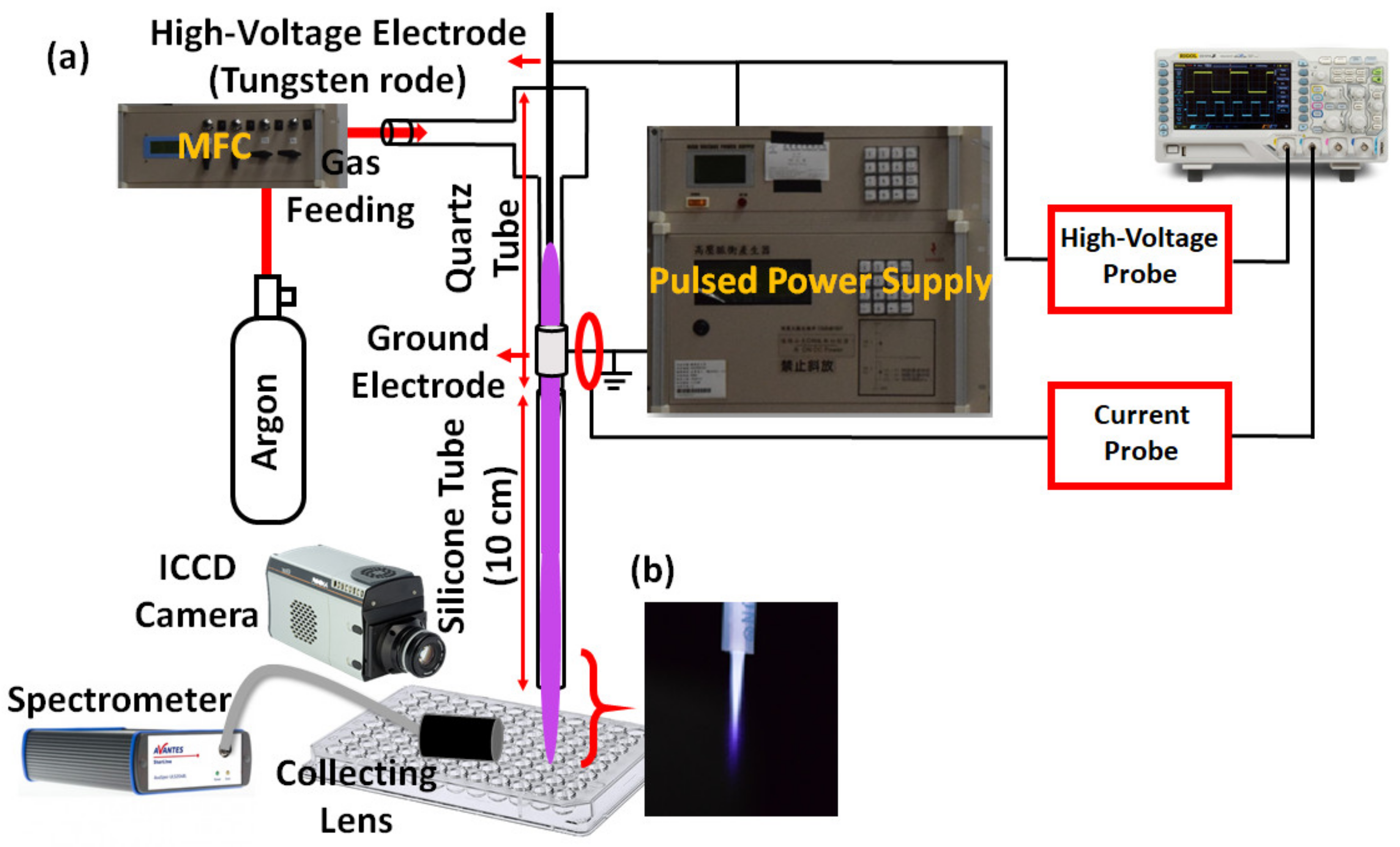
Figure 1. (a) Experimental setup of the transferred argon plasma jet system including an ICCD camera for emission characterization and spectrometer for spectroscopic characterization. (b) Image of the transferred cold atmospheric plasma (CAP) including argon gas.
Applsci 11 06181 g001
Figure 2. A typical waveform of applied voltage and plasma current.
Applsci 11 06181 g002
Figure 3. Transferred cold atmospheric plasma (CAP) emission captured by an ICCD camera with an exposure time of 0.5 s. (a) Transferred CAP emission without an optical filter. (b) Hydroxyl radical emission using an optical filter with 50% transmission. (c) Atomic oxygen radical emission using an optical filter with 50% transmission. (d) A typical spectrum obtained with a spectrometer during transferred CAP treatment including hydroxyl and (e) atomic oxygen radicals.
Applsci 11 06181 g003
Figure 4. (a) The image of transferred cold atmospheric plasma, (b,c) thermal images captured by the IR camera.
Applsci 11 06181 g004
Figure 5. (a) Cell culture and collection of L929 and B16F10 cells. (b) Cell viability of L929 and B16F10 cells before and after plasma treatment. (c) B16F10 cell morphology was imaged by optical microscopy at different magnifications before and after transferred CAP treatment. (* p < 0.05).
Applsci 11 06181 g005
Figure 6. (a) Fluorescence image of B16F10 cells treated with transferred cold atmospheric plasma (CAP) after 30 s without and with CAT. Green fluorescence represents live cells, while red fluorescence represents dead cells. (b) Cell viability (MTT) after 30 s of treatment without and with CAT. Cell viability was calculated with normalizing the survival of the treated cells group (transferred CAP 30 s) as 100%. (c) Fluorescence intensity after 30 s of treatment without and with CAT (green alive cell fluorescent signals). (* p < 0.05) Control: B16F10 cells plus fluorescent dyes.
Applsci 11 06181 g006
Figure 7. Cellular (B16F10 cells) reactive oxygen and nitrogen species (RONS) generation. (a) Qualitative and (b) quantitative RONS data indicating that cells that received catalase (CAT) could inhibit RONS generation after cold atmospheric plasma (CAP) treatment compared to the CAP-treated group (without CAT). (c) Cellular CAT levels were determined by immunofluorescence. (* p < 0.05).
Applsci 11 06181 g007
Publisher’s Note: MDPI stays neutral with regard to jurisdictional claims in published maps and institutional affiliations.