Article

Processed Scutellaria baicalensis Georgi Extract Alleviates LPS-Induced Inflammatory and Oxidative Stress through a Crosstalk between NF-κB and KEAP1/NRF2 Signaling in Macrophage Cells

1
Cnh Center for Cancer Research, Cnh Corporation, Gangnam-gu, Seoul 06154, Korea
2
Department of Dermatology, Bundang Jesaeng General Hospital, Seongnam 13590, Gyeonggi, Korea
3
Department of Natural Medicine, College of Pharmacy, Dongduk Women’s University, Seongbuk-gu, Seoul 02748, Korea
4
Division of Pharmacology, College of Korean Medicine, Semyung University, Jecheon 27136, Korea
5
Soram Korean Medicine Hospital, Gangnam-gu, Seoul 06154, Korea
*
Author to whom correspondence should be addressed.
These authors contributed equally.
Appl. Sci. 2021, 11(13), 6055; https://doi.org/10.3390/app11136055
Submission received: 14 May 2021 / Revised: 22 June 2021 / Accepted: 25 June 2021 / Published: 29 June 2021
(This article belongs to the Special Issue Advances in Anti-inflammatory Plants)

Abstract

:
Prolonged inflammation results in chronic diseases that can be associated with a range of factors. Medicinal plants and herbs provide synergistic benefits based on the interaction of multiple phytochemicals. The dried root of Scutellaria baicalensis Georgi and its compounds possess anti-inflammatory, anti-oxidative, and anticancer effects. Processing is a traditional method to achieve clinical benefits by improving therapeutic efficacy and lowering toxicity. In this study, we investigated the anti-inflammatory and anti-oxidant effect of processed Scutellaria baicalensis Georgi extract (PSGE) against lipopolysaccharide (LPS) stimulated RAW 264.7 cells. Data using Griess assay and ELISA showed that PSGE decreased nitric oxide and prostaglandin E2 (PGE2) levels against LPS. PSGE treatment up-regulated 15-hydroxyprostaglandin dehydrogenase (PGDH), while cyclooxygenase (COX)-2 and microsomal prostaglandin E synthase (mPGES)-1 expression did not change. Interestingly, PGE2 inhibition was regulated by prostaglandin catabolic enzyme 15-PGDH rather than COX-2/mPGES-1, enzymes essential for PGE2 synthesis. Additionally, PSGE-suppressed LPS-induced IL-6 and TNF-α production through NF-κB signaling. NF-κB release from an inactive complex was inhibited by HO-1 which blocked IκBα phosphorylation. The ROS levels lowered by PSGE were measured with the H2DCFDA probe. PSGE activated NRF2 signaling and increased antioxidant Hmox1, Nqo1, and Txn1 gene expression, while reducing KEAP1 expression. In addition, pharmacological inhibition of HO-1 confirmed that the antioxidant enzyme induction by PSGE was responsible for ROS reduction. In conclusion, PSGE demonstrated anti-inflammatory and anti-oxidant effects due to NRF2/HO-1-mediated NF-κB and ROS inhibition.

1. Introduction

Inflammation is the body’s nonspecific response to infectious or non-infectious etiologies, which accompanies pain, heat, redness and swelling [1,2]. The triggered acute inflammatory responses are resolved by the innate immune cells, such as macrophages and neutrophils [3]. However, prolonged inflammation, also referred to as chronic inflammation, causes additional damage to organs including the brain, lungs, heart, digestive tract, kidneys, intestinal tract, and reproductive system. Failure to prevent chronic inflammation results in chronic diseases such as type 2 diabetes, cancer, rheumatoid arthritis, neurodegenerative, cardiovascular and bowel diseases [3]. Dysregulation of nuclear factor kappa-B (NF-κB), Janus kinase (JAK)-signal transducer and activator of transcription (STAT), and mitogen-activated protein kinase (MAPK) pathways were involved in causing the inflammatory diseases [4]. Glucocorticoids (GCs) and non-steroidal anti-inflammatory drugs (NSAIDs) are the major classes of anti-inflammatory drugs. GCs inhibit the production of prostaglandins and other inflammatory proteins. On the other hand, NSAIDs target the cyclooxygenase (COX) regulation of prostaglandins and thromboxanes synthesis [5,6]. However, GCs potentiate the risk of musculoskeletal, metabolic, cardiovascular, central nervous system, and gastrointestinal diseases [7]. NSAIDs are also associated with side effects such as gastric ulcers and bleeding, renal injury, and cardiovascular diseases [8]. Therefore, it is necessary to discover new therapeutic modalities for inflammatory conditions.
Medicinal plants have provided therapeutic benefits for different cultures around the world over a long period of time. Asian countries for centuries have been using traditional herbal medicine with its unique treatment methods [9]. Scientific studies have explored the chemical profiles of traditional herbs and screened their pharmacological activity to discover lead molecules for modern medicine [10].
The dried root of Scutellaria baicalensis Georgi (SG) has a long history as a traditional herbal medicine for treating gastrointestinal and respiratory infections, bleeding, inflammation, and insomnia [11]. Pharmacological studies have reported the anti-inflammatory, anti-oxidative, anticancer, anticonvulsant, and antiviral effects of SG extracts [12,13,14,15,16]. The major active constituents responsible for SG’s function are baicalin, baicalein, wogonin, and wogonoside [17]. More than 180 drugs in the Chinese Pharmacopoeia contain SG, which is used in two ways: non-processed and processed forms [18]. Processing (Paozhi in Chinese and Poje in Korean) is a series of techniques such as cutting, frying with or without liquid excipients, roasting, and steaming, which are used to achieve different clinical needs from the same herb [19]. Previous study has reported that processed Scutellaria baicalensis Georgi (PSG) inhibited the expression level of chymase, tumor necrosis factor-α (TNF-α), COX-2, NF-κB, and the activity of myeloperoxidase in dextran sulfate sodium-induced colitis mouse model [20]. PSG treatment decreased NF-κB-mediated pro-inflammatory cytokines, serum inflammatory and oxidative stress biomarker levels in acute lung injury induced by lipopolysaccharide (LPS) [21]. However, the potential molecular basis behind the anti-inflammatory effects of PSG remains unclear. Therefore, in this article, we aim to unravel the mechanism of PSG against inflammatory and oxidative stress.

2. Materials and Methods

2.1. Preparation of SGE and PSGE

SG was purchased from CK PHARM (Boeun-gun, Korea). PSG preparation was performed following a reported method with modification [22]. SG was soaked in 30% EtOH for 30 min. A convection oven (JSOF-150, JS Research Inc., Gongju, Korea) was used to roast SG for 1 h 20 min in 200 °C. SG was reversed every 5 min while roasting to prevent burning of herb. SG and PSG were separately boiled in 2 L of 30% EtOH for 2 h in 100 °C followed by filtration and evaporation. The extraction yield (w/w) of lyophilized SG extract (SGE) and PSG extract (PSGE) was 33.9% and 29.7% respectively. The voucher specimens (SGE: BON190510.39, PSGE: BON190515.JC39) were deposited at the herbarium of Korean Medicine at Semyung University.

2.2. Liquid Chromatography–Mass Spectrometry (LC–MS) Analysis

SGE and PSGE were analyzed using LC–MS, on a Waters 2695 LC system interfaced with DAD and micro mass ZQ. The LC-MS conditions for qualitative and quantitative analysis of baicalin, baicalein, and wogonin in the samples were set as shown in Table 1 and analyzed. The standard compounds of baicalin, baicalein, and wogonin were diluted in methanol for the calibration curve. The calibration curve was generated by plotting the peak intensities against the standard concentrations of baicalin, baicalein, and wogonin. The regression equations were obtained using the formula y = ax + b, where y, x, a, and b correspond to peak area, compound concentration, slope of calibration curve, and y-intercept respectively. The limit of detection (LOD) and limit of quantitation (LOQ) of baicalin, baicalein, and wogonin were calculated by the following formula in Table 2.
LOD = 3.3 × σ/S
LOQ = 10 × σ/S
σ: standard deviation, S: slope of calibration curve.

2.3. Materials and Chemicals

Dulbecco’s modified eagle medium (DMEM) and fetal bovine serum (FBS) from Gibco (Carlsbad, CA, USA). Dimethyl sulfoxide (DMSO), lipopolysaccharide (LPS), 3-(4,5-dimethylthiazol-2-yl)-2,5-diphenyltetrazoliumbromide (MTT) and dexamethasone (DXM) from Sigma-Aldrich (St. Louis, MO, USA). Penicillin-streptomycin from Welgene (Gyeongsangbuk-do, Korea). Tin protoporphyrin IX (SnPPIX) from Cayman Chemicals (San Diego, CA, USA). NF-κB p65 (ab7970) antibody from Abcam (Cambridge, UK). Antibodies against iNOS (#13120), COX-2 (#12282), IκBα (#4812), p-IκBα (#2859), HO-1 (#70081), Lamin A/C (#4777), NRF2 (#12721), HRP linked anti-mouse IgG (7076), and HRP linked anti-rabbit IgG from Cell Signaling Technology (Bervely, MA, USA). Antibodies against β-actin (A5411) from Sigma Aldrich (St. Louis, MO, USA), KEAP1 (sc-514914) from Santa Cruz Biotechnology (Dallas, TX, USA), and HSP90 (13171-1-1AP) from Proteintech (Rosemont, IL, USA). 15-PGDH (160140) and mPGES-1 (9160615) antibodies from Cayman Chemicals (San Diego, CA, USA).

2.4. Cell Culture

Mouse macrophage RAW 264.7 (KCLB No. 40071) cell line from Korean Cell Line Bank (Seoul, Korea) were cultured with DMEM supplemented with 10% FBS, 1% antibiotics (100 IU/mL penicillin and 100 μg/mL streptomycin) in an incubator at 37 °C and 5% CO2.

2.5. Cell Viability Assay

Cell cytotoxicity after PSGE treatment was examined by the MTT assay. A density of 1.4 × 10⁴ cells/well were seeded and incubated for 24 h. Cells were treated with several concentrations of PSGE for 24 h. Media was removed and media containing MTT solution (0.5 mg/mL) was added per well, and the cells were incubated for another 4 h. Next, the MTT reagent and media were discarded and 100 µL DMSO was added to dissolve the formed formazan crystals. Finally, the plate was gently shaken on a shaker, and absorbance was measured at 570 nm with a microplate reader (Biotek, VT, USA).

2.6. Nitric Oxide (NO) Assay

The amount of NO was determined by the levels of nitrite with the cultured RAW 264.7 cell supernatant using Griess reagent containing 0.1% N-(1-Naphthyl) ethylenediamine dihydrochloride in double-distilled water and 1% sulfanilamide in 5% phosphoric acid. Briefly, RAW 264.7 cells (5 × 10⁵ cells/well) were seeded and incubated for 24 h. Following pre-treatment with different concentrations of PSGE and 20 µM DXM for 2 h, the cells were stimulated with LPS (1 µg/mL) for an additional 24 h. Culture media was collected, and the amount of nitrite was measured using Griess reagent. Griess reagent was added to an equal volume and the plate was gently agitated for 10 min on a shaker at room temperature (RT). 540 nm absorbance was monitored by a microplate reader and the concentration of nitrite was determined from a standard curve generated from known sodium nitrite concentrations.

2.7. IL-6, TNF-α and PGE2 Assay

RAW 264.7 cells (1 × 10⁵ cells/well) were pretreated with different concentrations of PSGE and DXM 20 µM for 2 h and then exposed to LPS 1µg/mL for 24 h. The supernatant of each well was collected and centrifuged at 1500 rpm at 4 °C for 10 min. IL-6 and TNF-α concentrations were measured using mouse IL-6 and TNF-α ELISA kit (Invitrogen, Carlsbad, CA, USA) and PGE2 using mouse PGE2 ELISA kit (R&D Systems, Minneapolis, MN, USA), following the manufacturer’s instructions.

2.8. Western Blotting

Cells were rinsed with chilled PBS and lysed in RIPA containing 0.1% SDS, 1% Triton X-100, 0.5% sodium deoxycholate, 150 mM NaCl, 50 mM Tris HCL (pH 7.4), 2 mM EDTA and I X protease inhibitor cocktail. The samples were incubated on ice for 10 min and vortexed for 10 s after an interval time for lysis. The cell debris was cleared by centrifugation at 14,000× g for 15 min. Nuclear and cytoplasmic proteins were separated by NE-PER nuclear and cytoplasmic extraction kit (Thermo Fisher Scientific, Rockford, IL, USA) following the manufacturer’s instructions. The protein concentration was determined by Coomassie dye-binding (Bradford) assay using the colorimetric protein assay kit (Biorad, Hercules, CA, USA). Cell proteins were separated by SDS-PAGE and transblotted to nitrocellulose membranes. The membranes were then blocked for 1 h at RT in 5% skim milk, followed by an overnight incubation at 4 °C with a specific primary antibody. The next day, the membranes were washed and incubated for an additional 1 h with HRP-conjugated secondary antibody at RT after washing three times with TBST. Bands were detected by ECL (LPS Solution, Daejeon, Korea), and band intensities were analyzed with Image J.

2.9. Cellular Reactive Oxygen Species (ROS) Assay

The Cellular ROS Assay Kit (Abcam, Cambridge, UK) is a DCFDA/H2DCFDA dye-based assay to detect intracellular hydroxyl radical, peroxy radical, and other ROS activity. RAW 264.7 cells (2.5 × 10⁴ cells/well) were seeded in 96 well clear bottom, black walled plates and incubated for 24 h. After 24 h, cells were washed with 1X buffer and treated with PSGE, DXM or SnPPIX for the recommended time, followed by LPS for another 24 h. Cells were then stained with diluted DCFDA solution 100 μL/well (final concentration, 20 μM) for 45 min at 37 °C in the dark, and fluorescence was measured at (Ex/Em = 485/535 nm).

2.10. Real-Time qPCR (RT-qPCR) Analysis

Total RNA was isolated from cells with RNeasy Mini Kit (Qiagen, Hilden, Germany). Total RNA (1 μg) was reversely transcribed with high capacity cDNA reverse transcription kit (Applied Biosystems, Foster City, CA, USA). RT-qPCR was performed using TaqMan probes with the universal PCR Master Mix (Applied Biosystems, Foster City, CA, USA) using StepOnePlus real time PCR system (Applied Biosystems, Foster City, CA, USA). The TaqMan probes used were Hemeoxygenase 1 (Hmox1, Mm00516005_m1), NAD(P)H dehydrogenase quinone 1 (Nqo1, Mm01253561_m1); Thioredoxin 1 (Txn1, Mm00726847_s1); β-actin (Acbt, Mm00607939_s1). Based on the reference Acbt gene, the relative gene expression analysis was performed using the delta-delta Ct formula.

2.11. Statistical Analysis

Data is shown as means ± SEM and are pooled from three independent experiments. Statistical analysis was performed by ANOVA with Tukey’s Multiple Comparison Test using software GraphPad Prism 5.1. (GraphPad Software, San Diego, CA, USA). p-values < 0.05 were considered statistically significant.

3. Results

3.1. LC-MS Analysis

LC-MS was used to analyze the retention time and mass pattern of SGE or PSGE compared with standards. The standards baicalin, baicalein, and wogonin were detected at 7.56 min, 12.74 min, and 16.38 min respectively (Figure 1A–E). According to the LC-MS quantitative analysis results, the contents of baicalin, baicalein, and wogonin were 219.89 ± 1.24 mg/g, 10.21 ± 0.14 mg/g, and 2.55 ± 0.05 mg/g in SGE, and 212.79 ± 0.12 mg/g, 15.41 ± 0.12 mg/g, and 4.59 ± 0.04 mg/g in PSGE respectively.

3.2. PSGE Modulated the Release of NO and PGE2

In order to examine the cytotoxicity of PSGE, we performed a MTT assay in RAW 264.7 cells. Evaluation on cells treated with different doses (50 to 1000 µg/mL) of PSGE were evaluated. PSGE did not cause cell toxicity up to 400 µg/mL (Figure 2A). Experiments with PSGE concentration ranging from 50 to 200 µg/mL and DXM 20 µM, as positive control, were performed in the study.
We investigated whether PSGE regulated the formation of inflammatory mediators such as nitric oxide (NO) and prostaglandin E2 (PGE2) in LPS-challenged RAW 264.7 cells. PSGE treatment considerably inhibited NO and PGE2 production, in a dose dependent manner, compared to LPS-treated cells (Figure 2B,C). PSGE (200 µg/mL) suppressed NO production by 0.28-fold and almost completely diminished PGE2 production when compared with LPS-treated cells. Moreover, it is notable that PSGE caused more extensive inhibition compared to the positive control DXM. We next examined the effect of PSGE on the inducible nitric oxide synthase (iNOS) and the cyclooxygenase (COX)-2 expression levels. The expression of iNOS was significantly increased by LPS but decreased with PSGE treatment dose-dependently (Figure 2D,E). Despite the significant PGE2 inhibition, COX-2 levels were unaffected suggesting that PSGE might target the downstream of COX-2 in the PGE2 synthesis cascade (Figure 2D,F). The results of microsomal prostaglandin E synthase (mPGES)-1 expression, a terminal synthase of PGE2, showed that mPGES-1 also did not influence PGE2 inhibition by PSGE (Figure 2D,G). We next examined the expression levels of 15-hydroxyprostaglandin dehydrogenase (PGDH), a PGE2 degrading enzyme. LPS-treated cells showed 0.5-fold decrease in 15-PGDH levels compared to control. PSGE inverted the LPS-mediated 15-PGDH suppression by 2-fold (Figure 2D,H). These results indicate that 15-PGDH induced the PGE2 inhibition by PSGE.

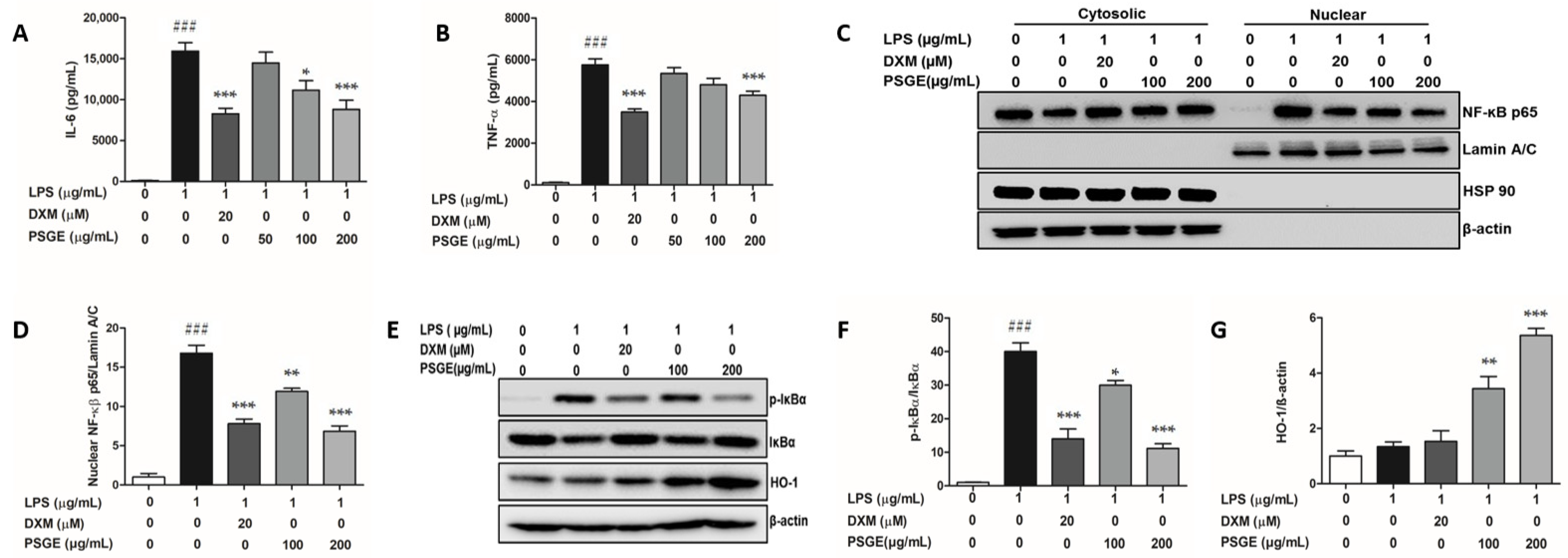
3.3. PSGE Regulated the NF-κB-Mediated Inflammatory Responses

The effect of PSGE on pro-inflammatory cytokines such as TNF-α and interleukin 6 (IL-6) was investigated in LPS stimulated RAW 264.7 cells. PSGE significantly suppressed IL-6 levels at 100 and 200 µg/mL and TNF-α levels at 200 µg/mL (Figure 3A,B). Notably, at 200 µg/mL, PSGE inhibited IL-6 more strongly (0.55-fold), similar to DXM, compared to TNF-α (0.75-fold). Based on these anti-inflammatory effects of PSGE, we determined whether PSGE could modulate nuclear translocation of NF-κB, a transcription factor associated with pro-inflammatory responses. PSGE significantly down-regulated the nuclear expression of NF-κB p65 by 0.70-fold (at 100 µg/mL) and 0.57-fold (at 200 µg/mL) respectively compared to LPS-treated cells (Figure 3C,D). Inhibitor kappa B-alpha (IκBα) phosphorylation involved in the regulatory steps for LPS-mediated NF-κB activation was determined by Western blot analysis. IκBα phosphorylation increased by 40-fold in LPS-treated cells but PSGE treatment significantly attenuated this effect dose-dependently (Figure 3E,F). Next, we examined the role of Heme Oxygenase-1 (HO-1) on NF-κB p65 nuclear translocation. PSGE treatment augmented HO-1 expression by 1.55-fold (at 100 μg/mL) and 3-fold (at 200 μg/mL) compared to LPS-treated cells (Figure 3E,G). Our data suggested that PSGE upregulated HO-1 expression, maintained the retention of IκBα/NF-κB complex, and prevented the release of NF-κB into the nucleus.

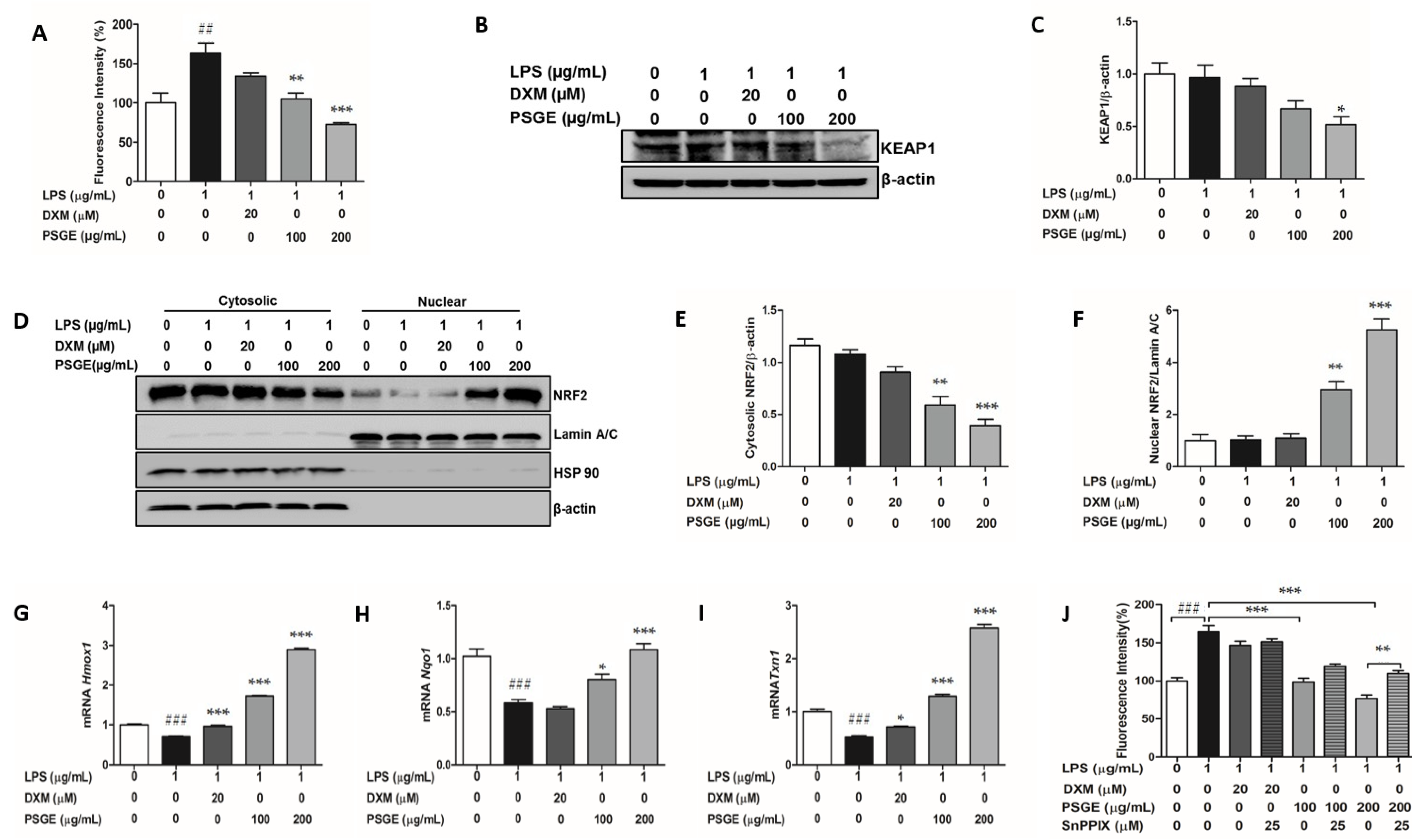
3.4. PSGE Activated the NRF2-Mediated Antioxidant Enzymes

PSGE significantly protected RAW 264.7 cells against intracellular accumulation of reactive oxygen species (ROS) caused by LPS. Pretreatment with 100 and 200 µg/mL PSGE before 24 h incubation with LPS, inhibited ROS generation by 0.64 and 0.44-fold (Figure 4A). The interaction between nuclear factor E2-related factor 2 (NRF2) and Kelch-like ECH-associated protein 1 (KEAP1) regulates antioxidant gene expression [23]. PSGE 200 µg/mL treatment decreased KEAP1 expression significantly by 0.46-fold compared to only LPS-treated cells (Figure 4B,C). Consequently, we examined whether PSGE could induce NRF2 nuclear translocation when treated with LPS. PSGE exhibited a 1.9-fold (at 100 µg/mL) and 4-fold (at 200 µg/mL) increase in nuclear NRF2 levels (Figure 4D,F). However, KEAP1 and nuclear NRF2 expression levels did not show any significant change with LPS stimulation (Figure 4B–F). Further results showed NRF2-dependent induction of antioxidant Txn1, Hmox1, and Nqo1 genes with PSGE treatment. PSGE ameliorated the significantly decreased Txn1 (0.53-fold), Hmox1 (0.71-fold), and Nqo1 (0.58-fold) gene expression caused by LPS. At 200 µg/mL, PSGE upregulated Txn1, Hmox1, and Nqo1 mRNA levels by 3.6, 3.0, and 2.1-fold (Figure 4G–I). In order to determine whether PSGE modulated antioxidant enzymes lead to reduction of ROS levels, RAW 264.7 cells were pretreated with SnPPIX (a HO-1 inhibitor) followed by PSGE and LPS. The SnPPIX reversed the inhibitory effects of PSGE on ROS generation after LPS stimulation (Figure 4J). These data suggest that PSGE elevated antioxidant enzymes to attenuate LPS-induced ROS production via NRF2-KEAP1 pathway.

4. Discussion

The pathogenesis of chronic inflammatory diseases such as type 2 diabetes, cancer, rheumatoid arthritis, neurological, and cardiovascular and inflammatory bowel diseases are linked with inflammation and oxidative stress [24]. Interaction of multiple phytochemicals in medicinal plants and herbs provide synergistic effects on anti-inflammatory and antioxidant activity [25]. A large number of studies have determined the pharmacological activities of SGE and its chemical constituents, such as anti-viral, anti-tumor, anti-bacterial, antioxidant, anti-inflammatory, hepatoprotective, and neuroprotective activities [26]. Processing is a traditional herbal method to achieve clinical benefits by improving therapeutic efficacy and also by lowering toxicity [27]. The alteration in therapeutic and toxic effects induced by processing is suggested to be related with structural transformation such as hydrolysis, oxidation, decomposition, isomerization, and other chemical reactions [28]. Comparative analysis of the major flavonoids, baicalin, baicalein, and wogonin contained in SGE and PSGE suggested that baicalin content was higher in SGE than PSGE. Interestingly, baicalein and wogonin contents were higher in PSGE than SGE. A report on the change in chemical components of SG after processing showed that stir-frying with alcohol and stir-frying till charred decreased baicalin but increased baicalein and wogonin contents [29]. Earlier studies suggested that the high temperature inhibited glucuronidase activity and prevented hydrolysis of baicalin or wogonin in SG [30,31]. However, understanding the diverse factors influencing glucuronidase activity remains unclear, since it has been reported that baicalin, baicalein, and wogonin contents are increased by heat processing [21,32].
NO is considered as an inflammatory mediator and regulator. In response to inflammatory stimuli, iNOS synthesize NO, inducing proinflammatory and destructive effects that lead to pathological conditions [33]. PSGE pretreatment suppressed NO increase after LPS treatment, which was considerably more potent than DXM treatment (Figure 2B). PGE2 is recognized as a bioactive lipid that mediates proinflammatory and immunomodulatory effects. In response to inflammatory stimuli, COX-2 and mPGES-1 mediate concomitant generation of PGE2. The elevated PGE2 levels can also be inactivated by the catabolic enzyme, 15-PGDH [34]. Our data showed that PSGE strongly suppressed PGE2 production to a greater extent than DXM, while COX-2 and mPGES-1 expression remained unchanged (Figure 2C,D,F,G). Interestingly, PSGE promoted 15-PGDH expression, which in turn impaired PGE2 synthesis (Figure 2D,H). However, previous studies reported that the regulatory effects of SG and PSG on PGE2 production are dependent on COX-2 downregulation, which remains controversial [35,36]. Other studies have shown that thiazolidinediones pioglitazone and rosiglitazone inhibited PGE2 production in A427 and A549 lung cancer cells by the catabolic 15-PGDH rather than the synthetic COX-2/mPGES-1 [37]. Further study is needed to fully understand how 15-PGDH expression, altered by PSGE, leads to PGE2 inhibition without significant change in COX-2 expression. Additionally, the ability of PSGE to increase 15-PGDH may offer therapeutic benefits without resulting in cardiotoxicity known in selective COX-2 inhibitors. PSGE also demonstrated anti-inflammatory effects by decreasing IL-6 and TNF-α release (Figure 3A,B).
NF-κB is a transcription factor that plays a central role in regulating a large array of genes during immune response and inflammation [38]. NF-κB activity is tightly regulated by IκBα binding in the cytoplasm. Inflammatory stimuli induce IκBα phosphorylation and degradation result in NF-κB nuclear translocation [39]. Our data demonstrated that PSGE inhibition on IL-6 and TNF-α levels involved the prevention of NF-κB release from an inactive complex due to dephosphorylation of IκBα (Figure 3A–F). Studies have suggested that HO-1 acts on IκB phosphorylation and inhibits activation of NF-κB dependent gene expression [40,41,42]. PSGE treatment increased HO-1 expression, which correlated with decreased IκBα phosphorylation (Figure 3E,F). These data support the idea that the anti-inflammatory effect of PSGE is mediated by NF-κB regulation via HO-1. The NF-κB and NRF2 pathways play an integral role to maintain redox balance and inflammatory homeostasis upon disruption by cellular stressors. The NRF2 activation is negatively regulated by KEAP1, which promotes ubiquitination and degradation of NRF2. Under stress, KEAP1 is modified and allows NRF2 to translocate to the nucleus where the transcription levels of antioxidant enzymes such as HO-1, NQO1, and TRX1 are activated. The KEAP1/NRF2 signaling pathway negatively regulates the NF-κB-induced cytokine expression by inhibiting intracellular ROS levels [23,43]. Many recent studies provide compelling evidence for the antioxidant and anti-inflammatory role of HO-1 [44]. Our data showed that PSGE pretreatment prevented LPS stimulated increase in ROS production. This was achieved through NRF2 dissociation from KEAP1, leading to nuclear translocation and accumulation of NRF2 (Figure 4A–F). Furthermore, considerable enrichment of nuclear NFR2 facilitated antioxidant enzyme upregulation and provided robust protection against oxidative stress. We used SnPPIX to confirm whether the antioxidant effect of PSGE against LPS triggered ROS generation was obtained by enhancement of HO-1. HO-1 inhibition restored intracellular ROS levels suppressed by PSGE (Figure 4G–J). Previous studies on LPS-induced experimental models have shown that SG components such as baicalein, apigetrin, and baicalin alleviate elevated inflammatory cytokines by NF-κB regulation and oxidative damage by HO-1 induction [45,46,47]. Here, we identified that HO-1 may play dual roles in anti-inflammatory and anti-oxidative effects of PSGE mediated by crosstalk between NF-κB and the KEAP1/NRF2 signaling pathway.

5. Conclusions

PSGE shows synergistic effect by targeting both inflammatory and oxidative stress conditions. The pharmacological functions of PSGE were due to inhibiting NF-κB activation and increasing nuclear NRF2 accumulation. NRF2 activated by PSGE mediated HO-1 upregulation, which resulted in preventing IκBα phosphorylation and ROS production. In addition, PGE2 production by PSGE involved catabolic 15-PGDH expression rather than synthetic COX-2/mPGES-1 expression. The dual function of PSGE may serve as a potential drug target for chronic inflammatory diseases.

Author Contributions

Conceptualization, E.H.K. and W.K.; investigation, A.A., E.H.K., J.-H.L. and K.-H.L.; resources, K.-H.L. and S.S.; writing-preparation and editing, A.A., E.H.K. and W.K.; visualization, E.H.K.; supervision, S.S. and W.K.; funding acquisition, W.K. All authors have read and agreed to the published version of the manuscript.

Funding

This work was carried out by a grant from Korea Health Technology R&D Project through the Korea Health Industry Development Institute (KHIDI), funded by the Ministry of Health & Welfare, Republic of Korea (grant number: HF20C0073).

Institutional Review Board Statement

Not applicable.

Informed Consent Statement

Not applicable.

Data Availability Statement

The data are available from the corresponding author upon request.

Conflicts of Interest

The authors declare no conflict of interest.

References

  1. Jabbour, H.N.; Sales, K.J.; Catalano, R.D.; Norman, J.E. Inflammatory pathways in female reproductive health and disease. Reproduction 2009, 138, 903–919. [Google Scholar] [CrossRef]
  2. Takeuchi, O.; Akira, S. Pattern recognition receptors and inflammation. Cell 2010, 140, 805–820. [Google Scholar] [CrossRef] [Green Version]
  3. Ahmed, A.U. An overview of inflammation: Mechanism and consequences. Front. Biol. 2011, 6, 274. [Google Scholar] [CrossRef]
  4. Reuter, S.; Gupta, S.C.; Chaturvedi, M.M.; Aggarwal, B.B. Oxidative stress, inflammation, and cancer: How are they linked? Free. Radic. Biol. Med. 2010, 49, 1603–1616. [Google Scholar] [CrossRef] [Green Version]
  5. Lima, A.; Alvim, H. Review on non-steroid antiinflammatory: Acetylsalicylic acid. Rev. Inic. Ciente. Ext. 2018, 1, 169–174. [Google Scholar]
  6. Meek, I.L.; Van de Laar, M.A.; E Vonkeman, H. Non-steroidal anti-inflammatory drugs: An overview of cardiovascular risks. Pharmaceuticals 2010, 3, 2146–2162. [Google Scholar] [CrossRef] [Green Version]
  7. Moghadam-Kia, S.; Werth, V.P. Prevention and treatment of systemic glucocorticoid side effects. Int. J. Dermatol. 2010, 49, 239–248. [Google Scholar] [CrossRef] [PubMed] [Green Version]
  8. Wongrakpanich, S.; Wongrakpanich, A.; Melhado, K.; Rangaswami, J. A comprehensive review of non-steroidal anti-inflammatory drug use in the elderly. Aging Dis. 2018, 9, 143–150. [Google Scholar] [CrossRef] [PubMed] [Green Version]
  9. Khan, H. Medicinal plants in light of history: Recognized therapeutic modality. J. Evid. Based Complement. Altern. Med. 2014, 19, 216–219. [Google Scholar] [CrossRef] [PubMed]
  10. Koparde, A.; Doijad, R.; Magdum, C. Natural products in drug discovery. In Pharmacognosy-Medicinal Plants; IntechOpen: London, UK, 2019. [Google Scholar]
  11. Zhao, T.; Tang, H.; Xie, L.; Zheng, Y.; Ma, Z.; Sun, Q.; Li, X. Scutellaria baicalensis georgi. (lamiaceae): A review of its traditional uses, botany, phytochemistry, pharmacology and toxicology. J. Pharm. Pharmacol. 2019, 71, 1353–1369. [Google Scholar] [CrossRef] [Green Version]
  12. Shao, Z.H.; Li, C.Q.; Vanden Hoek, T.L.; Becker, L.B.; Schumacker, P.T.; Wu, J.A.; Attele, A.S.; Yuan, C.S. Extract from scutellaria baicalensis georgi attenuates oxidant stress in cardiomyocytes. J. Mol. Cell. Cardiol. 1999, 31, 1885–1895. [Google Scholar] [CrossRef] [PubMed]
  13. Ikemoto, S.; Sugimura, K.; Yoshida, N.; Yasumoto, R.; Wada, S.; Yamamoto, K.; Kishimoto, T. Antitumor effects of scutellariae radix and its components baicalein, baicalin, and wogonin on bladder cancer cell lines. Urology 2000, 55, 951–955. [Google Scholar] [CrossRef]
  14. Wang, H.H.; Liao, J.F.; Chen, C.F. Anticonvulsant effect of water extract of scutellariae radix in mice. J. Ethnopharmacol. 2000, 73, 185–190. [Google Scholar] [CrossRef]
  15. Chu, M.; Chu, Z.Y.; Wang, D.D. The extract of compound radix scutellariae on mrna replication and ifn expression of influenza virus in mice. Zhong Yao Cai = Zhongyaocai = J. Chin. Med. Mater. 2007, 30, 63–65. [Google Scholar]
  16. Yoon, S.B.; Lee, Y.J.; Park, S.K.; Kim, H.C.; Bae, H.; Kim, H.M.; Ko, S.G.; Choi, H.Y.; Oh, M.S.; Park, W. Anti-inflammatory effects of scutellaria baicalensis water extract on lps-activated raw 264.7 macrophages. J. Ethnopharmacol. 2009, 125, 286–290. [Google Scholar] [CrossRef]
  17. Li, C.; Lin, G.; Zuo, Z. Pharmacological effects and pharmacokinetics properties of radix scutellariae and its bioactive flavones. Biopharm. Drug Dispos. 2011, 32, 427–445. [Google Scholar] [CrossRef]
  18. Commission, C.P. Chinese Pharmacopoeia; China Medical Science Press: Beijing, China, 2015; Volume 1, pp. 191–193. [Google Scholar]
  19. Sheridan, H.; Kopp, B.; Krenn, L.; Guo, D.; Sendker, J. Traditional chinese herbal medicine preparation: Invoking the butterfly effect. Science 2015, 350, S64–S66. [Google Scholar]
  20. Choi, Y.A.; Kang, O.H.; Park, H.J.; Tae, J.; Kim, D.K.; Kang, C.S.; Choi, S.C.; Yun, K.J.; Choi, S.J.; Nah, Y.H.; et al. Effect of processed scutellaria baicalensis on dextran sulfate sodium-induced colitis in mice. Int. J. Mol. Med. 2005, 16, 667–672. [Google Scholar]
  21. Shin, Y.O.; Park, C.H.; Lee, G.H.; Yokozawa, T.; Roh, S.S.; Rhee, M.H. Heat-processed scutellariae radix enhances anti-inflammatory effect against lipopolysaccharide-induced acute lung injury in mice via nf- kappa b signaling. Evid. Based Complement. Altern. Med. eCAM 2015, 2015, 456846. [Google Scholar] [CrossRef] [Green Version]
  22. Shin, S.; Shin, Y.; Lee, J.; Lee, A.; Kim, M.; Park, C.; Seo, B.; Roh, S. Ethanol-heated processed scutellariae radix improve inflammatory response through an inhibitory effect against oxidative stress in mice with the lipopolysaccharide-induced intestine injury of mice. Korea J. Herbol. 2015, 30, 81–88. [Google Scholar] [CrossRef]
  23. Yamamoto, M.; Kensler, T.W.; Motohashi, H. The keap1-nrf2 system: A thiol-based sensor-effector apparatus for maintaining redox homeostasis. Physiol. Rev. 2018, 98, 1169–1203. [Google Scholar] [CrossRef] [PubMed] [Green Version]
  24. Hussain, T.; Tan, B.; Yin, Y.; Blachier, F.; Tossou, M.C.B.; Rahu, N. Oxidative stress and inflammation: What polyphenols can do for us? Oxidative Med. Cell. Longev. 2016, 2016, 7432797. [Google Scholar] [CrossRef] [PubMed] [Green Version]
  25. Phan, M.A.T.; Paterson, J.; Bucknall, M.; Arcot, J. Interactions between phytochemicals from fruits and vegetables: Effects on bioactivities and bioavailability. Crit. Rev. Food Sci. Nutr. 2018, 58, 1310–1329. [Google Scholar] [CrossRef] [PubMed]
  26. Wang, Z.L.; Wang, S.; Kuang, Y.; Hu, Z.M.; Qiao, X.; Ye, M. A comprehensive review on phytochemistry, pharmacology, and flavonoid biosynthesis of scutellaria baicalensis. Pharm. Biol. 2018, 56, 465–484. [Google Scholar] [CrossRef] [Green Version]
  27. Wu, X.; Wang, S.; Lu, J.; Jing, Y.; Li, M.; Cao, J.; Bian, B.; Hu, C. Seeing the unseen of chinese herbal medicine processing (paozhi): Advances in new perspectives. Chin. Med. 2018, 13, 4. [Google Scholar] [CrossRef]
  28. Cai, B.; Qin, K.; Wu, H.; Cai, H.; Lu, T.; Zhang, X. Chemical mechanism during chinese medicine processing. Prog. Chem. 2012, 24, 637. [Google Scholar] [CrossRef]
  29. Ham, I.H.; Maeng, W.M.; Yang, G.S.; Kim, D.H.; Kim, D.H.; Cho, J.H.; Choi, H.Y. Study on the variation of components from scutellariae radix by processing and storage condition. Korea J. Herbol. 2007, 22, 189–199. [Google Scholar]
  30. Levvy, G.A. Baicalinase, a plant beta-glucuronidase. Biochem. J. 1954, 58, 462–469. [Google Scholar] [CrossRef]
  31. Ku, S.; Zheng, H.; Park, M.S.; Ji, G.E. Optimization of β-glucuronidase activity from lactobacillus delbrueckii rh2 and and its use for biotransformation of baicalin and wogonoside. J. Korean Soc. Appl. Biol. Chem. 2011, 54, 275–280. [Google Scholar] [CrossRef]
  32. Park, C.H.; Shin, M.-R.; An, B.K.; Joh, H.W.; Lee, J.C.; Roh, S.-S.; Yokozawa, T. Heat-processed scutellariae radix protects hepatic inflammation through the amelioration of oxidative stress in lipopolysaccharide-induced mice. Am. J. Chin. Med. 2017, 45, 1233–1252. [Google Scholar] [CrossRef]
  33. Korhonen, R.; Lahti, A.; Kankaanranta, H.; Moilanen, E. Nitric oxide production and signaling in inflammation. Curr. Drug Targets Inflamm. Allergy 2005, 4, 471–479. [Google Scholar] [CrossRef]
  34. Finetti, F.; Travelli, C.; Ercoli, J.; Colombo, G.; Buoso, E.; Trabalzini, L. Prostaglandin e2 and cancer: Insight into tumor progression and immunity. Biology 2020, 9, 424. [Google Scholar] [CrossRef]
  35. Kim, E.H.; Shim, B.; Kang, S.; Jeong, G.; Lee, J.S.; Yu, Y.B.; Chun, M. Anti-inflammatory effects of scutellaria baicalensis extract via suppression of immune modulators and map kinase signaling molecules. J. Ethnopharmacol. 2009, 126, 320–331. [Google Scholar] [CrossRef]
  36. Jeong, K.; Shin, Y.C.; Park, S.; Park, J.S.; Kim, N.; Um, J.Y.; Go, H.; Sun, S.; Lee, S.; Park, W.; et al. Ethanol extract of scutellaria baicalensis georgi prevents oxidative damage and neuroinflammation and memorial impairments in artificial senescense mice. J. Biomed. Sci. 2011, 18, 14. [Google Scholar] [CrossRef] [Green Version]
  37. Hazra, S.; Batra, R.K.; Tai, H.H.; Sharma, S.; Cui, X.; Dubinett, S.M. Pioglitazone and rosiglitazone decrease prostaglandin e2 in non-small-cell lung cancer cells by up-regulating 15-hydroxyprostaglandin dehydrogenase. Mol. Pharmacol. 2007, 71, 1715–1720. [Google Scholar] [CrossRef] [PubMed] [Green Version]
  38. Oeckinghaus, A.; Ghosh, S. The nf-kappab family of transcription factors and its regulation. Cold Spring Harb. Perspect. Biol. 2009, 1, a000034. [Google Scholar] [CrossRef] [PubMed]
  39. Juvekar, A.; Manna, S.; Ramaswami, S.; Chang, T.P.; Vu, H.Y.; Ghosh, C.C.; Celiker, M.Y.; Vancurova, I. Bortezomib induces nuclear translocation of ikappabalpha resulting in gene-specific suppression of nf-kappab--dependent transcription and induction of apoptosis in ctcl. Mol. Cancer Res. MCR 2011, 9, 183–194. [Google Scholar] [CrossRef] [PubMed] [Green Version]
  40. Liu, G.H.; Qu, J.; Shen, X. Nf-kappab/p65 antagonizes nrf2-are pathway by depriving cbp from nrf2 and facilitating recruitment of hdac3 to mafk. Biochim. Biophys. Acta 2008, 1783, 713–727. [Google Scholar] [CrossRef] [PubMed] [Green Version]
  41. Lee, D.S.; Kim, K.S.; Ko, W.; Li, B.; Keo, S.; Jeong, G.S.; Oh, H.; Kim, Y.C. The neoflavonoid latifolin isolated from meoh extract of dalbergia odorifera attenuates inflammatory responses by inhibiting nf-kappab activation via nrf2-mediated heme oxygenase-1 expression. Phytother. Res. PTR 2014, 28, 1216–1223. [Google Scholar] [CrossRef]
  42. Jiang, L.; Jiang, Q.; Yang, S.; Huang, S.; Han, X.; Duan, J.; Pan, S.; Zhao, M.; Guo, S. Gyy4137 attenuates lps-induced acute lung injury via heme oxygenase-1 modulation. Pulm. Pharmacol. Ther. 2019, 54, 77–86. [Google Scholar] [CrossRef]
  43. Saha, S.; Buttari, B.; Panieri, E.; Profumo, E.; Saso, L. An overview of nrf2 signaling pathway and its role in inflammation. Molecules 2020, 25, 5474. [Google Scholar] [CrossRef] [PubMed]
  44. Campbell, N.K.; Fitzgerald, H.K.; Dunne, A. Regulation of inflammation by the antioxidant haem oxygenase 1. Nat. Rev. Immunol. 2021, 21, 411–425. [Google Scholar]
  45. Lee, Y.M.; Cheng, P.Y.; Chim, L.S.; Kung, C.W.; Ka, S.M.; Chung, M.T.; Sheu, J.R. Baicalein, an active component of scutellaria baicalensis georgi, improves cardiac contractile function in endotoxaemic rats via induction of heme oxygenase-1 and suppression of inflammatory responses. J. Ethnopharmacol. 2011, 135, 179–185. [Google Scholar] [CrossRef] [PubMed]
  46. Lim, H.S.; Kim, O.S.; Kim, B.Y.; Jeong, S.J. Apigetrin from scutellaria baicalensis georgi inhibits neuroinflammation in bv-2 microglia and exerts neuroprotective effect in ht22 hippocampal cells. J. Med. Food 2016, 19, 1032–1040. [Google Scholar] [CrossRef] [PubMed]
  47. Meng, X.; Hu, L.; Li, W. Baicalin ameliorates lipopolysaccharide-induced acute lung injury in mice by suppressing oxidative stress and inflammation via the activation of the nrf2-mediated ho-1 signaling pathway. Naunyn-Schmiedeberg’s Arch. Pharmacol. 2019, 392, 1421–1433. [Google Scholar] [CrossRef]
Figure 1. Liquid chromatography–mass spectrometry chromatogram and cell viability. (A) Baicalin, (B) Baicalein, (C) Wogonin, (D) Scutellaria baicalensis Georgi extract (SGE), (E) Processed Scutellaria baicalensis Georgi extract (PSGE).
Applsci 11 06055 g001
Figure 2. Cell viability and effects of PSGE against LPS-induced NO and PGE2 production and related iNOS, COX-2, mPGES-1 and 15-PGDH expression in RAW 264.7 cells. (A) Cell viability of PSGE in RAW 264.7 cells. RAW 264.7 cells were treated with 50−1000 µg/mL of PSGE for 24 h and cell viability was determined by MTT assay. (B) NO levels were measured by Griess assay, (C) PGE2 levels were measured by ELISA assay, (D) Protein expression and quantitative analysis of (E) iNOS, (F) COX-2, (G) mPGES-1 and (H) 15-PGDH expression relative to β-actin. RAW 264.7 cells were pre-treated with indicated concentration of PSGE or DXM for 2 h followed by LPS for 24 h. DXM was used as positive control. Data shown represent mean ± SEM (n = 3). (### p < 0.001 vs. control; * p < 0.05, ** p < 0.01, and *** p < 0.001 vs. LPS).
Applsci 11 06055 g002
Figure 3. Effects of PSGE against LPS-induced IL-6, TNF-α cytokine expression, and NF-κB signaling in RAW 294.7 cells. Production of (A) IL-6 and (B) TNF-α levels were measured by ELISA. RAW 264.7 cells were pre-treated with indicated concentration of PSGE or DXM for 2 h followed by LPS for 24 h. (C) Western blot analysis of nuclear and cytosolic fractions using NF-κB p65 antibody, (D) quantitative analysis of nuclear NF-κB p65 relative to Lamin A/C, (E) Protein expression and quantitative analysis of (F) p-IκBα relative to IκBα and (G) HO-1 relative to β-actin. RAW 264.7 cells were pretreated with the indicated concentrations of PSGE and DXM for 2 h followed by LPS for 2 h. Lamin A/C and HSP90 were used as cytosolic and nuclear markers. DXM as positive control. Data shown represent mean ± SEM (n = 3). (### p < 0.001 vs. control; * p < 0.05, ** p < 0.01, and *** p < 0.001 vs. LPS).
Applsci 11 06055 g003
Figure 4. Effect of PSGE against LPS-induced intracellular ROS levels, KEAP1/NRF2 signaling and antioxidant gene expression in RAW 264.7 cells. (A) Intracellular ROS levels were assayed using oxidation of H2DCFDA fluorescence probe. RAW 264.7 cells were pre-treated with indicated concentration of PSGE or DXM for 2 h followed by LPS for 24 h.(B) Protein expression and quantitative analysis of (C) KEAP1 relative to β-actin, (D) Western blot analysis of nuclear and cytosolic fractions using NRF2 antibody, quantitative analysis of (E) cytosolic NRF2 relative to β-actin, (F) nuclear NRF2 relative to Lamin A/C. RAW 264.7 cells were pre-treated with indicated concentration of the PSGE or DXM for 2 h followed by LPS for 30 min. The levels of (G) Hmox1, (H) Nqo1 and (I) Txn1 mRNA expression were analyzed by RT-qPCR. RAW 264.7 cells were pre-treated with indicated concentration of the PSGE or DXM for 2 h followed by LPS for 2 h. (J) Changes in ROS levels by SnPPIX. RAW 264.7 cells were pre-treated with indicated concentration of SnPPIX (HO-1 inhibitor) for 1 h and then DXM or PSGE for 2 h followed by LPS for 24 h. Lamin A/C and HSP90 were used as cytosolic and nuclear markers. DXM as positive control. Data shown represent mean ± SEM (n = 3). (## p < 0.01 and ### p < 0.001 vs. control; * p < 0.05, ** p < 0.01, and *** p < 0.001 vs. LPS).
Applsci 11 06055 g004
Table 1. The analysis conditions of liquid chromatography–mass spectrometry.
ParametersConditions
Analytical columnTSK-gell ODS-80Ts (4.6 mm × 150 mm)
Column temperature35 ℃
Injection volume10 µL
UV Wavelength277 nm
Mobile phaseFinal time (min)Water in 0.1% formic acidAcetonitrile
0.08020
30.02080
30.10100
40.00100
40.18020
Flow rate1.0 mL/min
VoltagesCapillary3.00 kV
Cone40 V
Extractor2 V
RF Lens0.2 V
Source Temperature120 ℃
Desolvation Temperature400 ℃
Gas flowDesolvation600 L/h
Cone30 L/h
Table 2. Calibration curves, correlation coefficients, limit of detection (LOD) and limit of quantitation (LOQ) of baicalin, baicalein, and wogonin.
CompoundsLinear Range (µg/mL)Regression EquationR2LOD (µg/mL)LOQ (µg/mL)
baicalin62.5–1000y = 428.15x + 4238.5113.0939.67
baicalein12.5–200y = 529.06x − 8488.30.988347.94145.27
wogonin6.25–100y = 983.39x − 328.620.99991.665.02
Publisher’s Note: MDPI stays neutral with regard to jurisdictional claims in published maps and institutional affiliations.

