Article

The Role of Prediabetes as a Predictive Factor for the Outcomes in Patients with STEMI. Which Is the Right Range of Glycated Hemoglobin to Adopt in This Setting?

by
Kyriazoula Chatzianagnostou
1,
Letizia Guiducci
2,
Umberto Paradossi
1,
Alberto Ranieri De Caterina
1,
Annamaria Mazzone
1,
Sergio Berti
1 and
Cristina Vassalle
1,3,*
1
Fondazione CNR-Regione Toscana G. Monasterio, 54100 Pisa, Italy
2
Clinical Physiology Institute, CNR, 56124 Pisa, Italy
3
Cristina Vassalle, Fondazione CNR-Regione Toscana G. Monasterio, via Moruzzi 1, 56100 Pisa, Italy
*
Author to whom correspondence should be addressed.
Appl. Sci. 2021, 11(12), 5518; https://doi.org/10.3390/app11125518
Submission received: 26 April 2021 / Revised: 7 June 2021 / Accepted: 10 June 2021 / Published: 15 June 2021
(This article belongs to the Special Issue Trends and Prospects in Pathophysiology of Diet-Related Diseases)

Abstract

:

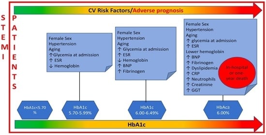
Featured Application

HbA1c represents a continuum in terms of CV risk in STEMI patients. Moreover, HbA1c measurement methods are standardized, quick, easy, cheap. These reasons justify routine HbA1c testing and HbA1 5.70–5.99% inclusion in the preT2D category for early identification of STEMI patients who may benefit from interventional strategies and lifestyle changes, to prevent progression of preT2D status into overt T2D.

Abstract

Background: Prediabetes (preT2D) is considered a subtle adverse cardiovascular (CV) risk factor after acute myocardial infarction. Glycated hemoglobin (HbA1c) ranges to identify preT2D are different between ADA and WHO guidelines (5.7–6.4 vs. 6.0–6.4%, respectively). Aim: To evaluate the prognostic value of HbA1c different preT2D-ranges and their correlation with demographic, instrumental, and laboratory parameters in STEMI. Methods: A total of 1681 patients (mean age 67 ± 13 years; 1217 males) were enrolled. Admission HbA1c was used to identify patients with no-T2D (<5.7%), HbA1c 5.70–5.99%, and WHO-preT2D with HbA1c 6–6.49%, and T2D (HbA1c ≥ 6.5). Results: HbA1c 5.7–5.99, WHO-preT2D, and T2D progressively correlated with an increasing number of CV risk factors. However, only T2D, but not preT2D, was significantly associated with adverse prognosis (in-hospital and one-year death). Conclusions: PreT2D is correlated with CV risk factors, but not with adverse prognosis as compared to no-T2D. Nonetheless, routine HbA1c testing in the STEMI population and HbA1c-5.7–5.99 patient inclusion in the preT2D category may help to identify those who may benefit from intervention and lifestyle strategies to early prevent preT2D progression.

Graphical Abstract

1. Introduction

Prediabetes (preT2D) is considered a subtle adverse CV risk factor for prognosis after acute ST-elevation myocardial infarction (STEMI) [1]. This fact because the complications of T2D begin early in the progression from normal glucose tolerance to overt Type 2 diabetes (T2D).
Glycated hemoglobin (HbAc1) represents the biomarker most recently introduced as a diagnostic measure of diabetes by the American Diabetes Society (ADA) in 2010, in addition to fasting blood glucose (FGP), and blood glucose-two hours from the glycemic load (OGTT) [2]. The fact that the test for HbAc1 can be performed at any time of the day because it can be performed without the need for fasting, and therefore is not affected by acute or individual variations, makes it a reproducible tool and an ideal candidate for screening subjects with a high risk of T2D. In acute clinical settings, as in acute myocardial infarction (AMI), hyperglycemia occurring at admission generally mirrors the so-called “stress hyperglycemia” [3]. Thus, alterations of blood glucose in patients with AMI may reflect a temporary stress-induced phenomenon rather than a stationary pre-existing abnormal glucose tolerance. In this case, the use of HbA1c may be particularly useful to distinguish between stress hyperglycemia and hyperglycemia caused by a dysglycemic status.
However, the advice on ranges of HbA1c to identify preT2D are different between Scientific Societies. In fact, ADA indicated the range between 5.70–6.40% (corresponding to 38–48 mmol/mol), whereas the World Health Organization (WHO) guidelines defined the preT2D status in presence of HbA1c value between 6.00% and 6.49% (42–48 mmol/mol) [4].
At now, it is not clearly determined if the use of WHO-preT2D defined ranges is appropriate in STEMI setting, or the inclusion of HbA1c between 5.70–5.99% in the preT2D category, as in the ADA guidelines, is better because already representative of a dysmetabolic condition that could correlate with adverse prognosis and increase CV risk.
Our study aimed to evaluate the prognostic value of HbA1c different preT2D-ranges and their correlation with demographic, instrumental, and laboratory parameters related to CV risk, in a large population of STEMI patients.

2. Materials and Methods

2.1. Population Characteristics and Data Acquisition

One-thousand six-hundred and eighty-one STEMI patients (mean age 67 ± 13 years; 1217 males), admitted from April 2006 to December 2016, were enrolled at the Ospedale del Cuore G. Pasquinucci-Clinical Cardiology Department (Massa, Italy). STEMI definition follows published SC/ACCF/AHA/WHF guidelines for STEMI criteria and management [5]. Within 90 min of admission, all patients underwent coronary angiography with subsequent PCI.
Data concerning all the information of the subject, including demographic and laboratory parameters, concomitant diseases, characteristics of STEMI, angiographic findings, outcomes of revascularization procedure, in-hospital complications, and mortality, were extracted by a computerized clinical database (MATRIX) of our Hospital. We recorded demographic information, cardiovascular history, cardiovascular risk factors. Hypertension was defined in case of blood pressure higher than 140/90 mmHg or current use of antihypertensive medications. Dyslipidemia was defined by the use of lipid-lowering treatments or by fasting low-density lipoprotein levels >150 mg/dL. Smoking history was ascertained by the clinical anamnesis.
Patients were considered eligible to be enrolled in the study based on inclusion criteria that were: (1) male and female adult patients, admitted to the coronary care unit for chest pain and subsequently proven STEMI; (2) patients subject to percutaneous coronary revascularization and stenting of the culprit lesion within 24 h from the onset of symptoms. Exclusion criteria were the following: (1) severe systemic diseases; (2) systemic inflammatory disease; (3) patients refusing or unable to supply written Informed Consent; (4) patients who did not undergo HbA1c; (5) diabetic patients with HbA1c < 6.5% treated with diet therapy, anti-diabetic drugs, or insulin therapy (n = 109).
Standard therapy (e.g., aspirin, beta-blockers, ACE-inhibitors, diuretics, statins) was administered in all eligible patients.
Informed consent was obtained from each patient (or from their relatives where necessary) before the angiogram, and the study was approved by the local ethics committee.

2.2. Measurements and Follow-Up

Routinely performed admission HbA1c was used to identify patients with no-T2D (<5.70%), HbA1c 5.70–5.99%, and WHO-preT2D with HbA1c 6.00–6.49%, T2D (HbA1c ≥ 6.50%).
The diagnosis of myocardial infarction was based on the documentation of chest pain persisting >20 min, persistent electrocardiographic ST-segment changes, or Q wave development, associated with biomarker increase [5].
Complete revascularization was defined when no total occlusion and no residual stenosis >70% (for left main >50%) was found in any major coronary artery or their major branches at discharge.
Echocardiography, performed within 24 h from hospital admission, was used to assess LV ejection fraction (EF) through modified Simpson’s rule with biplane planimetry [6]. Follow-up data were obtained at 12 months from at least one of the following four sources: a review of the patient’s record, telephone interview conducted by trained personnel, personal communication with the patient’s physician, or medical control at the outpatient clinical units.

2.3. Statistical Analysis

Continuous variables were reported as mean ± SD. Owing to skewness, Log transformations of fibrinogen, C reactive protein (CRP), erythrocyte sedimentation rate (ESR), brain natriuretic peptide (BNP), glycemia, gamma-glutamyl transferase (GGT), neutrophils, lymphocytes, HbA1c were used to perform statistical analyses. Then, log-transformed values were then back-transformed for data presentation. Comparisons between continuous variables were evaluated by Student’s t-test, while differences between categorical variables were made by the Chi-square analysis. Comparisons among three or more independent groups were analyzed using the ANOVA test and p for trend reported. Regression analysis was performed to assess the relationship between continuous variables. A p-value < 0.05 was chosen as the level of significance.

3. Results

3.1. Patient Characteristics

Patients (n = 1681, mean age 67 ± 13 years; 1217 males), admitted to the Heart Hospital Pasquinucci of Fondazione Gabriele Monasterio (Italy, April 2006–December 2016), were included. Prevalence of preT2D resulted 29% by ADA criteria and 12% by WHO criteria (p < 0.001; Figure 1). The prevalence of dysglycemia (cumulative combination of diabetes and prediabetes) was found to be 48% and 31% according to ADA and WHO criteria, respectively.

3.2. Correlates of HbA1c as Qualitative Variable

Routinely performed admission HbA1c was used to identify patients with no history of T2D (N-HbA1c < 5.70%, n = 875), with T2D (T2D, HbA1c ≥ 6.50%, n = 320), and patients with HbA1c between 5.70% to 5.99% (5.70–5.99 group; n = 290) and WHO-preT2D group with HbA1c between 6.00–6.49% (n = 196). The distribution of risk factors among the study subjects in the HbA1c categories are shown in Table 1.
Patients in the group of HbA1c 5.7–5.99 were more frequently females (<0.001), hypertensive (<0.001) and older (<0.001), and had significantly higher glycemia at admission (<0.001), ESR (<0.001), and lower hemoglobin (<0.001) than those in the N-HbA1c group (Table 1). Patients in the WHO-preT2D group were more frequently females (<0.001), hypertensive (<0.001), and older (<0.001), and had significantly higher glycemia (<0.001), BNP (<0.001), fibrinogen (<0.05), and ESR (<0.001) at admission and lower hemoglobin (<0.001) than those in the N-HbA1c group (Table 1). Patients in the T2D group were more frequently females (<0.001), hypertensive (<0.001), dyslipidemic (<0.001) and older (<0.001), and had significantly higher CRP (<0.001), neutrophils (<0.001), glycemia (<0.001), BNP (<0.001), fibrinogen (<0.001), creatinine (<0.001), GGT (<0.001), and ESR (<0.001) at admission and lower hemoglobin (<0.001) than those in the N-HbA1c group (Table 1)
Prevalence of preT2D and T2D according to age intervals is reported in Figure 2.

3.3. Correlates of HbA1c as Continuous Variable

When HbA1c was considered as a continuous variable, significant association was found with female sex (<0.001), aging (<0.001), hypertension (<0.001), dyslipidemia (<0.001), smoking habit (<0.001), EF (≤0.01), BMI (<0.001), creatinine (<0.001), glycemia at admission (<0.001), BNP (<0.001), fibrinogen (<0.001), hemoglobin (<0.01), CRP, (<0.001), GGT (<0.001), neutrophils (<0.001), ESR (<0.001) (Table 2).

3.4. Follow-Up

The occurrence of in-hospital and one-year deaths was reported in Figure 3. Only T2D, but not the preT2D groups, were significantly associated with adverse prognosis in terms of in-hospital or one-year death (Figure 3). Specifically, in-hospital death totaled for 4 (1.4%), 4 (2%), and 18 (5.6%) in HbA1c 5.70–5.99%, WHO-preT2D, and T2D group versus 15 (1.7%) for the N-HbA1c, respectively (trend for ANOVA; p < 0.001). Moreover, one-year death occurred in 22 (7.6%), 17 (8.7%) and 44 (13.8%) for HbA1c 5.70–5.99%, WHO-preT2D, and T2D group versus 54 (6.2%) for the N-HbA1c, respectively (trend for ANOVA; p < 0.001). In-hospital and one-year death were also both associated with higher HbA1C value, when evaluated as a categorical rather than a continuous variable (upper panels reporting % of deaths, <0.001, <0.001 T2D versus N-HbA1c, respectively; lower panels showing values of HbA1c in patients with compared to those without events <0.001, <0.001, respectively) (Figure 3).

4. Discussion

The main findings of the present study are (1) the prevalence of preT2D is high among AMI patients, especially when ADA criteria are applied (2) preT2D and T2D are correlated with a progressively growing number of CV risk factors according to increased HbA1c values; (3) T2D patients, but not the preT2D, were significantly associated with adverse prognosis in terms of in-hospital or one-year death.
For many patients, the AMI hospitalization represents a period in which previously unrecognized glucidic abnormalities may be diagnosed. Thanks to improvements in laboratory standardization, the utilization of HbA1c as a diagnostic test for dysglycemia in addition to the traditional criteria based on fasting plasma glucose or oral glucose tolerance testing has been suggested [2,7]. However, there were differences in the criteria of defining preT2D between ADA and WHO. These differences may be particularly critical in specific CV settings, as in AMI, where the HbA1c test may be more practical because its measurement does not require a fasting status, and as a long-term glycemic biomarker HbA1c is less affected than glucose by acute stress and illness [8]. Moreover, HbA1c may represent a better tool in predicting CV risk, as this biomarker is more strongly associated with CV risk and death when compared with fasting glucose [9]. In the current study, we observed that majority of AMI patients had dysglycemia, with a prevalence of preT2D of 29 and 12% according to HbA1c ADA and WHO criteria, respectively. A comparable preT2D prevalence in AMI patients of 11% was previously found in agreement with our WHO definition finding, utilizing the criteria of fasting glucose ≥110 to <126 mg/dL for preT2D definition [10]. In any case, this high prevalence of pre-T2D in AMI patients gives the theoretical chance to identify patients who may benefit more from lifestyle change or therapeutical interventions (e.g., metformin) to prevent progression to overt T2D, and reduce the risk of adverse CV events [11]. This is important also in view of evidence suggesting that preT2D may develop in overt T2D with an approximately 5–10% annual risk, whereas lifestyle modification in preT2D patients is associated with 40–70% relative-risk reduction [12]. Moreover, this fact may be relevant, as generally subjects with preT2D, even those admitted with AMI, receive less aggressive treatment than those with T2D, often due to physician unawareness of their potentially high CV risk, representing a high-risk population under-recognized and under-treated. Interestingly, there was a higher prevalence of preT2D among females (37% versus 26% in males, <0.001: unshown data), associated with a higher prevalence of hypertension (68 versus 53% in males, <0.001: unshown data) in our population. Previous studies have reported hypertension to be associated with preT2D further increasing the risk of CV disease [13,14,15], which may have particular significance in female patients, giving an early opportunity to intervene with lifestyle and therapeutic measures for health maintenance and T2D and CV disease prevention, especially in this specific subgroup of patients.
In the present study, the risk for in-hospital and one-year mortality was approximately triple and double, respectively for patients with compared with those without T2D. This result confirms previous data suggesting that the risk for death and other adverse events is approximately double for acute and stable coronary artery disease coronary patients with T2D versus without T2D [16,17]. However, the risk for patients with preT2D was similar to that of patients with noT2D. Also, this result is in agreement with previous findings suggesting that patients with preT2D have a better CV prognosis compared with those with T2D [16,17]. In this context, it is clear that T2D duration and cumulative preT2D hyperglycemic insults among patients with long-standing T2D acquire critical importance for the risk of adverse outcomes [18,19,20,21]. Thus, preT2D may represent a stage at which fewer adverse repercussions of hyperglycemia occurred, including those on arterial vessels. Nonetheless, glucotoxicity may precede overt hyperglycemia and beta-cell dysfunction, and as an early event is reversible, opening the possibility of timely interventions, that may modify the course of hyperglycemia and prevent or delay long-term complications [22]. Accordingly, the correlation of HbA1c 5.7–5.99 with hypertension and ESR, that we found in our population, denotes a possible relationship with endothelial dysfunction and inflammation, as well as the association of WHO-preT2D group with hypertension, aging, BNP, fibrinogen, and ESR, indicates a growing CV risk with the progression of glucidic abnormalities, giving the chance of prompt interventions already in these early disease phases. In fact, the relationship between HbA1C when evaluated as a continuous variable and CV risk factor suggests that the risk associated with HbA1c is rather a continuum process, and the increased CV risk may begin at a range well below the threshold for T2D diagnosis [23]. Accordingly, baseline HbA1c represented a stronger predictor of subsequent T2D and cardiovascular events than fasting glucose in no-T2D subjects [9].

5. Conclusions

Given the high number of preT2D and T2D observed in this study, patients who present with AMI (especially female patients) should be carefully screened for dysglycemia, taking into account that HbA1c rather represents a continuum in terms of increasing CV risk. Moreover, if preT2D correlates with CV risk factors, but not with adverse prognosis as compared to N-HbA1c, the lack of a clear association between preT2D and mortality should nor discourage clinicians to apply strategies to prevent progression towards overt T2D, and reduce CV risk. Thus, lifestyle modification (predominantly exercise and weight loss) and stop smoking and lipid and hypertension control must be strongly encouraged also in the preT2D phase. In AMI settings, HbA1c evaluation may be particularly useful as this biomarker is not affected by the stress or fasting status, such as OGTT. However, one important limitation for HbA1c may be lower sensitivity for identifying subjects with early impairment in the beta-cell function who are at increased future risk for T2D, whereas an OGTT is preferable. Nonetheless, the combination of HbA1c evaluation, followed by a later OGTT could be one possible efficacious strategy to catch as many patients with glucose abnormalities as possible.

Author Contributions

Conceptualization, K.C., L.G. and C.V.; methodology, K.C.; data patient collection K.C., A.R.D.C., U.P., A.M. and S.B.; statistical analysis, writing—original draft preparation, C.V.; review and editing, K.C., L.G., U.P., A.M., S.B. and C.V. All authors have read and agreed to the published version of the manuscript.

Funding

This research received no external funding.

Institutional Review Board Statement

All the data have been acquired in the context of institutional assistance within clinical care purposes in a retrospectively collected modality by our Institute electronic databank which saves demographic, clinical, laboratory, instrumental data of all consecutive patients admitted to our department (IMA registry).

Informed Consent Statement

All the data were acquired through the informed consent given to each patient and signed, as normally carried out when accessing our Hospital as part of the institutional activity.

Conflicts of Interest

The authors declare no conflict of interest.

Abbreviations

preT2DPrediabetes
T2DType 2 diabetes
CVCardiovascular
STEMIAcute ST-elevation Myocardial Infarction
HbA1c Glycated Hemoglobin
ADAAmerican Diabetes Association
WHOWorld Health Organization
FPBFasting Blood Glucose
OGTTOral glucose Test Tolerance
EFEjection Fraction
LVLeft Ventricle
LVEFLeft Ventricular ejection Fraction
CRPC Reactive Protein
ESRErythrocyte Sedimentation Rate
BNPBrain Natriuretic Peptide
GGTGamma Glutamyl Transferase
BMIBody Mass Index
AMIAcute myocardial infarction

References

  1. Otten, R.; Kline-Rogers, E. Impact of pre-diabetic state on clinical outcomes in patients with acute coronary syndrome. Heart 2005, 91, 1466–1468. [Google Scholar] [CrossRef] [Green Version]
  2. American Diabetes Association. Diagnosis and classification of diabetes mellitus. Diabetes Care 2014, 37 (Suppl. 1), S81–S90. [Google Scholar] [CrossRef] [PubMed] [Green Version]
  3. Angeli, F.; Reboldi, G. Hyperglycemia in acute coronary syndromes: From mechanisms to prognostic implications. Ther. Adv. Cardiovasc. Dis. 2015, 9, 412–424. [Google Scholar] [CrossRef] [PubMed]
  4. Bergman, M.; Abdul-Ghani, M. Review of methods for detecting glycemic disorders. Diabetes Res. Clin. Pract. 2020, 165, 108233. [Google Scholar] [CrossRef]
  5. Third universal definition of myocardial infarction. J. Am. Coll. Cardiol. 2012, 60, 1581–1598. [CrossRef] [PubMed] [Green Version]
  6. Lang, R.M.; Bierig, M.; Devereux, R.B.; Flachskampf, F.A.; Foster, E.; Pellikka, P.A.; Picard, M.H.; Roman, M.J.; Seward, J.; Shanewise, J.; et al. Recommendations for chamber quantification. Eur. J. Echocardiogr. 2006, 7, 79–108. [Google Scholar] [CrossRef]
  7. World Health Organization; IDF. Definition and Diagnosis of Diabetes Mellitus and Intermediate Hyperglycaemia: Report of a WHO/IDF Consultation; World Health Organization: Geneva, Switzerland, 2006. [Google Scholar]
  8. Soler, N.G.; Frank, S. Value of glycosylated hemoglobin measurements after acute myocardial infarction. JAMA 1981, 246, 1690–1693. [Google Scholar] [CrossRef]
  9. Selvin, E.; Steffes, M.W.; Zhu, H.; Matsushita, K.; Wagenknecht, L.; Pankow, J.; Coresh, J.; Brancati, F.L. Glycated Hemoglobin, Diabetes, and Cardiovascular Risk in Nondiabetic Adults. N. Engl. J. Med. 2010, 362, 800–811. [Google Scholar] [CrossRef] [PubMed]
  10. Giraldez, R.R.; Clare, R.M. Prevalence and clinical outcomes of undiagnosed diabetes mellitus and prediabetes among patients with high-risk non-ST-segment elevation acute coronary syndrome. Am. Heart J. 2013, 165, 918–925. [Google Scholar] [CrossRef] [PubMed]
  11. The Diabetes Prevention Program. Long-term effects of lifestyle intervention or metformin on diabetes development and microvascular complications over 15-year follow-up: The Diabetes Prevention Program Outcomes Study. Lancet Diabetes Endocrinol. 2015, 3, 866–875. [Google Scholar] [CrossRef] [Green Version]
  12. Tabák, A.G.; Herder, C. Prediabetes: A high-risk state for diabetes development. Lancet 2012, 379, 2279–2290. [Google Scholar] [CrossRef] [Green Version]
  13. Cheung, B.M.; Li, C. Diabetes and hypertension: Is there a common metabolic pathway? Curr. Atheroscler. Rep. 2012, 14, 160–166. [Google Scholar] [CrossRef] [PubMed] [Green Version]
  14. DeFronzo, R.A.; Ferrannini, E. Insulin resistance: A multifaceted syndrome responsible for NIDDM, obesity, hypertension, dyslipidemia, and atherosclerotic cardiovascular disease. Diabetes Care 1991, 14, 173–194. [Google Scholar] [CrossRef]
  15. Shahim, B.; Gyberg, V. Undetected dysglycaemia common in primary care patients treated for hypertension and/or dyslipidaemia: On the need for a screening strategy in clinical practice. A report from EUROASPIRE IV a registry from the EuroObservational Research Programme of the European Society of Cardiology. Cardiovasc. Diabetol. 2018, 17, 21. [Google Scholar] [PubMed] [Green Version]
  16. Farhan, S.; Redfors, B. Impact of Pre-Diabetes on Coronary Plaque Composition and Clinical Outcome in Patients with Acute Coronary Syndromes: An Analysis from the PROSPECT Study. JACC Cardiovasc. Imaging 2019, 12, 733–741. [Google Scholar] [CrossRef] [PubMed]
  17. Haffner, S.M.; Lehto, S. Mortality from coronary heart disease in subjects with type 2 diabetes and in nondiabetic subjects with and without prior myocardial infarction. N. Engl. J. Med. 1998, 339, 229–234. [Google Scholar] [CrossRef]
  18. Lenzen, M.; Ryden, L.; Öhrvik, J.; Bartnik, M.; Malmberg, K.; Reimer, W.S.O.; Simoons, M.L.; on Behalf of the Euro Heart Survey Investigators. Diabetes known or newly detected, but not impaired glucose regulation, has a negative influence on 1-year outcome in patients with coronary artery disease: A report from the Euro Heart Survey on diabetes and the heart. Eur. Heart J. 2006, 27, 2969–2974. [Google Scholar] [CrossRef] [PubMed] [Green Version]
  19. Kuusisto, J.; Mykkanen, L. NIDDM and its metabolic control predict coronary heart disease in elderly subjects. Diabetes 1994, 43, 960–967. [Google Scholar] [CrossRef]
  20. Lindsey, J.B.; House, J.A.; Kennedy, K.F.; Marso, S.P. Diabetes Duration Is Associated with Increased Thin-Cap Fibroatheroma Detected by Intravascular Ultrasound With Virtual Histology. Circ. Cardiovasc. Interv. 2009, 2, 543–548. [Google Scholar] [CrossRef] [Green Version]
  21. Natarajan, S.; Liao, Y.; Sinha, D.; Cao, G.; McGee, D.L.; Lipsitz, S.R. Sex Differences in the Effect of Diabetes Duration on Coronary Heart Disease Mortality. Arch. Intern. Med. 2005, 165, 430–435. [Google Scholar] [CrossRef] [Green Version]
  22. Stolar, M. Glycemic control and complications in type 2 diabetes mellitus. Am. J. Med. 2010, 123, S3–S11. [Google Scholar] [CrossRef] [PubMed]
  23. Dankner, R.; Danoff, A. Can ‘personalized diagnostics’ promote earlier intervention for dysglycaemia? Hypothesis ready for testing. Diabetes Metab. Res. Rev. 2010, 26, 7–9. [Google Scholar] [CrossRef] [PubMed]
Figure 1. Prevalence of no-T2D, preT2D, and T2D according to ADA and WHO criteria.
Applsci 11 05518 g001
Figure 2. Interval age-specific prevalence of diabetes and prediabetes.
Applsci 11 05518 g002
Figure 3. In-hospital and one-year death according to HbA1C, when evaluated as categorical rather than continuous variable (upper and lower panels, respectively).
Applsci 11 05518 g003
Table 1. Demographic, clinical, and laboratory parameters according to HbA1c intervals.
HbA1c
<5.7%
HbA1c
5.7–5.99%
HbA1c
6–6.49%
HbA1c
>6.49%
Total population 875 (52)290 (17)196 (12)320 (19)
Females183 (21)99 (34)71 (36)111 (35)
Males692 (79)191 (66)120 (64)209 (65)
Age
<66 years (50th percentile)485 (55)120 (41)68 (35)117 (37)
66–77 years (75th percentile)221 (25)76 (26)57 (29)97 (30)
>77 years167 (20)94 (32)71 (36)106 (33)
CV risk factors
Hypertension431 (50)177 (61)129 (66)220 (69)
Dyslipidemia320 (37)121 (42)79 (40)156 (49)
Current/ex smoking habit419 (48)111 (38)78 (40)108 (34)
Ejection fraction (%)46 ± 944 ± 944 ± 944 ± 9
Body mass index (kg/m2)26 ± 427 ± 427 ± 529 ± 5
Laboratory parameters
Creatinine (mg/dL)1 ± 0.61.1 ± 0.61.2 ± 0.71.4 ± 1.1
Glycemia (mg/dL)111 ± 29125 ± 37141 ± 48195 ± 85
Brain natriuretic peptide (pg/mL)211 ± 353270 ± 447353 ± 665350 ± 514
Fibrinogen (mg/dL)327 ± 110333 ± 110354 ± 111358 ± 115
Hemoglobin (g/dL)14 ± 213 ± 213 ± 213 ± 2
C reactive protein (mg/dL)2 ± 42.1 ± 42.5 ± 52.7 ± 4
Gamma glutamyltransferase (UI/L)31 ± 3631 ± 2733 ± 3636 ± 32
Monocytes (109/L)0.7 ± 0.40.7 ± 0.40.7 ± 0.50.7 ± 0.4
Neutrophils (109/L)8.6 ± 3.68.8 ± 3.79.2 ± 3.79.6 ± 4.2
Erythrocyte sedimentation rate (mm/h)19 ± 1825 ± 2125 ± 2027 ± 24
Table 2. Demographic, clinical, and laboratory parameters according to HbA1c values.
HbA1c%p
Total population 5.9 ± 1.1
Females6.1 ± 1.2
Males5.9 ± 1.1<0.001
Age
<66 years (50th percentile)5.9 ± 1.1
66–77 years (75th percentile)6 ± 1.1
>77 years6.1 ± 0.9<0.001
CV risk factors
No-hypertension5.8 ± 1
Hypertension6.1 ± 1.2<0.001
No-dyslipidemia5.9 ± 1.1
Dyslipidemia6.1 ± 1.1≤0.01
No-smoking habit6 ± 1.1
Current/ex smoking habit5.8 ± 1<0.001
Ejection fraction (%)r = −0.1≤0.01
Body mass index (kg/m2)r = 0.2<0.001
Laboratory parameters
Creatinine (mg/dL)r = 0.1<0.001
Glycemia (mg/dL)r = 0.6<0.001
Brain natriuretic peptide (pg/mL)r = 0.1<0.001
Fibrinogen (mg/dL)r = 0.1<0.001
Hemoglobin (g/dL)r = −0.1<0.01
C reactive protein (mg/dL)r = 0.2<0.001
Gamma glutamyltransferase (UI/L)r = 0.1<0.001
Monocytes (109/L)r = 0.02ns
Neutrophils (109/L)r = 0.1<0.001
Erythrocyte sedimentation rate (mm/h)r = 0.1<0.001
Publisher’s Note: MDPI stays neutral with regard to jurisdictional claims in published maps and institutional affiliations.

