Article

The Structural Characteristics of Collagen in Swim Bladders with 25-Year Sequence Aging: The Impact of Age

1
College of Science, Shantou University, Shantou 515063, China
2
Analysis and Testing Center, Shantou University, Shantou 515063, China
3
Guangdong Provincial Key Laboratory of Marine Biotechnology, Shantou University, Shantou 515063, China
4
College of Marine Sciences, South China Agricultural University, Guangzhou 510642, China
*
Author to whom correspondence should be addressed.
Xiaofeng He and Liling Xie are co-first author.
Appl. Sci. 2021, 11(10), 4578; https://doi.org/10.3390/app11104578
Submission received: 3 April 2021 / Revised: 11 May 2021 / Accepted: 11 May 2021 / Published: 17 May 2021
(This article belongs to the Special Issue Nutraceuticals: Food and Nutritional Applications)

Abstract

:
Aged swim bladders from the yellow drum (Protonibea diacanthus) are considered collagen-based functional food with extremely high market value. The structural integrity of collagen may be crucial for its biological functions. In the current study, swim bladders with 25-year-old sequences were collected and found to be basically composed of collagen. Then, thermogravimetry (TG), differential scanning calorimetry (DSC), X-ray diffraction (XRD), and attenuated total reflectance–Fourier transform infrared spectroscopy (ATR–FTIR) were conducted to evaluate the integrity of the peptide chain and triple helix in the collagen. The structures of microfibers and fiber bundles were revealed with atomic force microscopy (AFM), scanning electrical microscopy (SEM), and optical spectroscopy. The collagens in the aged swim bladders were found to have similar thermal properties to those of fresh ones, but the relative content of the triple helixes was found to be negatively correlated with aging. The secondary structure of the remaining triple helix showed highly retained characteristics as in fresh swim bladders, and the microfibrils also showed a similar D-period to that of the fresh one. However, the fiber bundles displayed more compact and thick characteristics after years of storage. These results indicate that despite 25 years of aging, the collagen in the swim bladders was still partially retained with structures.

1. Introduction

The swim bladder is a gas-filled regulating organ that can be found in most bony fishes. It is responsible for balance, floating, sinking, and protecting internal organs in fish [1]. In Asian countries, there is a long history of using the swim bladder as a luxury food with nutritional and medicinal value [2]. In the field of traditional Chinese medicine, it is believed that swim bladders can relieve gastrointestinal bleeding and postpartum hemorrhage. In addition, aged swim bladders in particular were believed to possess these functions. It has been reported that the declared annual import value of swim bladders has reached USD 264–394 million in Hongkong, similar to that of sea cucumbers, shark fins, and abalone [2].
Recent research defined collagens (type I) as a major ingredient in swim bladders [3], and the biological functions of collagen in regard to benefiting tissue regeneration, tissue structuring [4], and especially, wound healing, have been identified [5]. These findings are in agreement with the efficacy of swim bladders as stated by the theory of traditional Chinese medicine.
Type I collagens are composed of peptide chains with a repeating (Gly–X–Y) sequence as their primary structure; the peptide chains are observed to form a left-handed triple helix structure that is flanked by helical telopeptides [6]. The triple helix structures are further covalently bound to form microfibers with a staggered mode and then assembled into fiber bundles of a larger size and longer length (Figure 1).
Due to its highly organized character, collagen’s structure is sensitive to temperature, pH, ions, solvents, enzymes, and microorganisms. Extracted collagens can be easily denatured and lose their triple helix structures once the temperature increases to 40 °C [7]. Changes in pH are able to cause a structural transformation via changing the valence state of amino acids on side chains [8]. The free ions in the solution could lead to triple helix dissociation and reformation, thereby affecting the collagen structures [9]. Collagen structures highly rely on hydrogen bonds and hydrophobic forces, which can easily be degraded by solvents [10]. The covalent bonds inside peptide chains have been observed to be vulnerable and could be breached by enzymes; therefore, many microorganisms can also degrade collagen [11].
The multiple biological functions of collagens are generally attributed to their highly organized molecular and supramolecular structures [12]. Notably, the wound healing ability, described by experts in the field of traditional medicine as an ability to relieve bleeding, is basically due to the inherent self-reassembling nature of the triple helix structure [4]. However, the self-reassembly ability can be irreversibly impacted once collagen loses its structure following certain denaturation processes [4,13,14].
Notably, a large group of traditional Chinese medicinal practitioners claimed that swim bladders retain high medicinal value after years of storage. Therefore, aged swim bladders are still being traded on the market as functional food and have extremely high market value. However, the related evidence and scientific information are limited. For instance, does the swim bladder remain an integrated structure? This question is believed to be related to the biological and medicinal functions of this luxury food due to the importance of the structural integrity of collagens in the expression of biological functions.
Considering the importance of the structural integrity of collagen in expressing medicinal functions, swim bladders with 25-year-old sequences were collected. Their structures were evaluated on the basis of the following aspects: amino acid composition, integrity of the peptide chain, characteristics of the triple helix, and morphologies of the microfibril and fiber bundles. The results provide more background knowledge about aged swim bladders and may also provide support to determining the commercial value of aged swim bladders.

2. Materials and Methods

2.1. Materials

Yellow drum fish (Protonibea diacanthus) were acquired from an aquafarm at Shantou (23°23′00.00″ N, 116°41′00.00″ E, Shantou, China). The swim bladder was taken from the yellow drum, and the lipid layer was removed by hand and then air dried (25 °C for 48 h) to obtain the fresh swim bladder (FSB). The 1, 5, and 25-year-old swim bladders (1-YSB, 5-YSB, and 25-YSB, respectively) were prepared in the same manner as the FSB and were provided by the Chaocai Research Institute (Shantou, China). The FSB was heated in a water bath at 100 °C for 25 min to obtain the thermally denatured swim bladder (TDSB). Chloramine-T, p-dimethylaminobenzaldehyde, phenyl isothiocyanate, and L-amino acid standards were purchased from Aladdin (Shanghai, China). Hydrochloric acid, sodium hydroxide, n-propanol, triethylamine, n-hexane, and acetonitrile (chromatographic grade) were purchased from Xilong science (Shantou, China). All other chemicals were analytical grade.

2.2. Moisture and Collagen Content

Moisture content analysis was performed by direct drying [15]. Briefly, samples were cut into small pieces (1 × 1 cm) and weighed. Afterwards, samples were transferred to a DZF-6020 vacuum oven (Boxun, Suzhou, China) and heated at 105 °C for 4 h to remove the moisture; then, the weights of the samples were recorded. Moisture contents were calculated using Equation (1).
Moisture (%) = (M1 − M2)/M1 × 100%
where M1 and M2 represent the weight of the sample before and after drying, respectively.
The collagen contents were analyzed using a previously published protocol [16] and modified as follows: A 0.1000 g sample was mixed with 4 mL HCl (6 mol/L) and hydrolyzed at 120 °C for 6 h. The pH of the hydrolysis fluid was adjusted with 1 mL NaOH (0.15 mol/L), and the volume was topped up to 50 mL with distilled water. Then, 60 μL neutralized hydrolysate was taken and mixed with a 60 μL chloramine-T solution (0.065 mol/L, dissolved in a n-propanol–water solution, 1:1, v:v). Following this, we left the solution for 20 min at room temperature. Afterwards, 60 μL of Ehrlich’s reagent (1 mol/L) was added to the mixture, and then it was heated in a water bath (60 °C) for 20 min. Finally, the absorbance of the sample was measured at 560 nm, and hydroxyproline concentrations were obtained with reference to a standard curve from the hydroxyproline standard sample (Aladdin, Shanghai, China); collagen contents were calculated using Equation (2).
Collagen (%) = 50 × CHyp/0.1000 × 11.1
where CHyp is the hydroxyproline concentration. The standard curve is provided on Figure S1.

2.3. Composition of Amino Acids

The amino acid compositions were analyzed in accordance with a report of Heinrikson et al. [17]. Briefly, samples were ground into fine powder with an A800 mini pulverizer (Jinxi Machinery Factory, Shanghai, China). A 0.1000 g amount of powdered sample was dissolved into a 4 mL HCl (6 mol/L) solution and heated at 150 °C for 5 h to obtain the hydrolysate; then, the hydrolysate was neutralized with 1 mL of sodium hydroxide (5 mol/L). Then, 1 mL of neutralized hydrolysate was taken and transferred into a centrifuge tube and mixed with 0.5 mL of phenyl isothiocyanate (0.1 mol/L, dissolved in acetonitrile) and 0.5 mL of triethylamine (1 mol/L, dissolved in acetonitrile). Afterwards, the mixture was incubated in the dark for 1 h and then extracted with 2 mL of n-hexane. After extraction, the lower phase of the mixture was taken and filtered with a 0.45 μm filter and then subjected to high-performance liquid chromatography (HPLC) analysis.
The HPLC analysis was conducted according to the method of Heinrikson et al., which we sum up briefly as follows [17]: The sample (20 μL) was injected onto an Eclipse XDB-C18 reverse phase column (5 μm, 4.6 × 250 mm, Agilent, Santa Clara, CA, USA) on an LC-16 HPLC (Shimadzu, Tokyo, Japan) at a flow rate of 1.0 mL/min and a column temperature of 30 °C. The mobile phase A was prepared by mixing acetonitrile with a 0.1 mol/L sodium acetate–water solution (3:97, v:v), and mobile phase B was an acetonitrile–water solution (4:1, v:v). The gradient elution method was carried out as follows: 0–5 min: 0–9% B; 10–13 min: 9–10% B; 13–15 min: 13–16% B; 15–17 min: 16–20% B; 17–25 min: 20–30% B; 30 min: 70% B; 35–40 min: 100% B; 50 min: 0% B. The detection wavelength was 254 nm. The amino acids were identified, and concentrations were calculated by peak area information referencing with L-amino acid standards (Aladdin, China). Standard curves for individual amino acids are provided in Figure S2.

2.4. Thermogravimetric and Differential Scanning Calorimetry (TG-DSC) Analysis

TG analysis was performed in accordance with a report by Yu et al. [18] and modified as follows: A 5 mg powdered sample was weighed and then transferred into a Q600 synchronous thermal analyzer (TA instruments, New Castle, DE, USA). The sample was balanced at 30 °C for 5 min; then the gravimetrical changes were recorded across a range of 30 to 800 °C with a 10 °C/min heating rate. Nitrogen gas with a flow rate of 50 mL/min was chosen as the atmosphere.
DSC analysis was carried out with reference to a report by Safandowska et al. [19]. In short, a 6 mg powdered sample was sealed into an aluminum cell and then transferred into a DSC-2000 analyzer (TA instruments, New Castle, DE, USA). The thermal behavior of the sample was recorded within a range of from 20 to 180 °C and a 5 °C/min heating rate, and nitrogen gas with a flow rate of 50 mL/min was chosen as the atmosphere and an empty cell was used as the reference.

2.5. Attenuated Total Reflectance–Fourier Transform Infrared Spectroscopy (ATR–FTIR)

Samples were cut into small pieces (0.5 × 0.5 cm) and lyophilized to remove moisture. The ATR–FTIR spectra of swim bladders were obtained in attenuated total reflectance (ATR) mode using a Nicolet is50 FTIR spectrometer (Thermo Fisher Scientific, Carlsbad, CA, USA) equipped with a diamond reflector accessory. Samples were tightly placed onto the diamond reflector accessory. ATR–FTIR spectra were acquired at 25 °C with a resolution of 2 cm−1, and the measurement range was 4000–525 cm−1. An air infrared spectrum of the blank sample was collected to eliminate the influence from the background peak before detection. Analysis of the spectral data was carried out using OMNIC 8.0 (Thermo Nicolet, Carlsbad, CA, USA). The ATR–FTIR spectra were subjected to fitting with Gaussian/Lorentz functions [20] using the PeakFit v4.12 (Systat, San Jose, CA, USA) software; a D2 baseline, 10% smoothing spectroscopy, and Gaussian area peak type were chosen during spectrum fitting.

2.6. X-Ray Diffraction (XRD) Analysis

Samples were cut into small pieces (1 × 1 cm) to fit the sample plate of an ADVANCE D8 X-ray diffractometer (Bruker, Berlin, Germany), which was equipped with a Cu Kα radiation source and a Lynxeye array detector. Based on a previous report [8], A 2.2 kW X-ray tube power, 40 kV pipe pressure, and a 40 mA pipe flow were taken for X-ray emission. The scan type and sola slit were set as coupled two theta and 2.0 mm, respectively. Data were collected within a 2θ range of from 4 to 50° with a 0.2° step size and a 4°/min scanning speed. The diameter of the triple helix was calculated with Bragg’s equation [8] (Equation (3)):
d (nm) = λ/2sinθ
where d is the diameter of the triple helix structure, λ is the wavelength of the radiation source (λ = 0.15418 nm), and θ is the angle of the diffraction peak.

2.7. Atomic Force Microscope (AFM) Analysis

The sliced samples (20 μm thickness) were placed on a freshly cleaved mica to observe the transverse striation period (D-period) of collagens with an NT-MDT AFM (NTEGRA, Russia) instrument. The AFM was equipped with a Si cantilever (NT-MDT, HA_NC, Moscow, Russia) which had a 10 nm tip curvature radius and a 235 kHz resonant frequency and an aluminum reflex coated silicon tip (force constant 40 N m−1). All of the AFM images were recorded in semi-contact mode at room temperature with a relative humidity of 30%. Both 2D and 3D images were captured simultaneously at imaging speeds of 1.2 Hz, scanning 512 lines. Measurements were taken from 2D images using the Image Analysis 3.5.0 (NT-MDT, Moscow, Russia).

2.8. Optical Microscopy Analysis

The sample was cut into pieces (1 × 1 cm) and then transferred into a centrifuge tube; a swelling process was carried out with the addition of 40 mL of distilled water, followed by 6 h of standing still. Afterwards, the sample was cut into slices (8 μm thickness) with a NX50 freezing microtome (Thermo, Carlsbad, CA, USA). Based on Zhu’s report [21], the G1006 Masson staining suit (Servicebio, Nanjin, China) was chosen for staining. Specifically, a slice was saturated with 20 mL Masson A solution for 12 h and then washed with distilled water. Afterwards, the slice was saturated with a 20 mL coloring agent (Masson B–Masson C = 1:1, v:v), dyed for 1 min, and then washed with distilled water 3 times. The slice was then sequentially rinsed with 5 mL of an acidified ethanol solution (HCl–ethanol, 1:99, v:v), 5 mL of Masson D solution, 5 mL of Masson E solution, and 5 mL of Masson F solution. Afterwards, the slice was washed with 5 mL of a 1% acetic acid water solution to remove organic solvents, and it was dehydrated with ethanol and xylene sequentially followed by sealing with rhamsan gum. The images of samples were taken on a positive optical microscope (Nikon Eclipse E100, Tokyo, Japan) equipped with an imaging system (Nikon DS-U3, Tokyo, Japan).

2.9. Scanning Electron Microscope (SEM) Analysis

Sliced samples (20 μm thickness) were prepared as described above and loaded on a stub using copper double-tape and then analyzed with a JSM-6360 LA SEM instrument (JEOL, Tokyo, Japan) for surface micromorphological observation. The signals were taken with an acceleration voltage of 10 kV and a beam current of 1 μA in a vacuum chamber [22].

2.10. Statistical Analysis

Data are presented as means ± standard deviations (SDs). One-way analysis of variance (ANOVA) was performed to determine the differences between samples. The differences between the means were compared using Tukey’s post hoc test (p < 0.05).

3. Results

3.1. Moisture, Collagen, and Amino Acid Compositions of Swim Bladders

The composition of the swim bladder was analyzed in terms of moisture, collagen, and amino acids, as shown in Table 1. The collagen contents of swim bladders were found to range from 35 to 90%, and moisture content ranged from 10 to 57%. The total combined content of collagen and moisture in swim bladders was found to be higher than 90%.
A total of 16 kinds of amino acids were detected in swim bladders. Glycine, hydroxyproline, and proline were found to be dominant amino acids. The relative content of glycine was found to range from 33 to 35%, the relative content of hydroxyproline ranged from 8 to 9%, and the relative content of proline ranged from 12 to 18%. The content of total amino acids (including proline and hydroxyproline) was found to range from 21 to 28%.

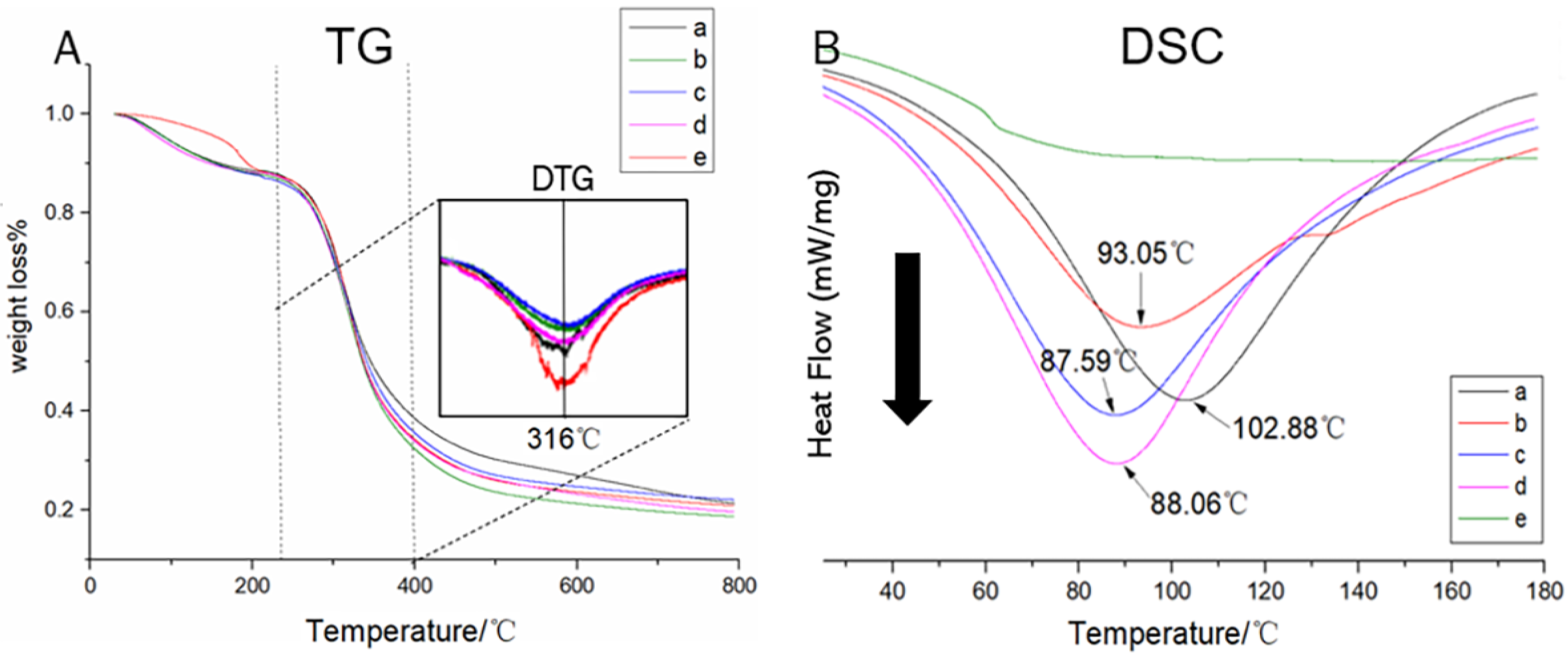
3.2. Thermal Decomposition Profiles of the Swim Bladder

The decomposition processes of the peptide chains were observed using TG–DTG (Figure 2A). Distinct weight losses were observed from 235 to 400 °C, which was mainly attributed to the cleavage of covalent bonds inside the peptide chains [23]. After the derivative calculation of weight loses, the DTG curves accompanied with decomposition temperature (Td) were obtained. The results showed that all samples provided a similar Td (316 °C), indicating that the covalent bonds inside the peptide chains were not destroyed after years of storage.
The triple helix is composed of three peptide chains but is sensitive to thermal treatment. Based on previous reports [19,24,25], the triple helix could be destroyed and showed thermal denaturation in DSC. Therefore, DSC analysis was conducted, and the results are shown in Figure 2B. The FSB, 1-YSB, 5-YSB, and 25-YSB showed Tm values higher than 87 °C. These Tm values were similar to those of dry collagen mentioned in a previous report [25]; therefore, the existence of the triple helix in aged swim bladders was preliminarily indicated. However, the TDSB showed a flattened thermal curve in the DSC results, which reflected that the triple helix was not observed. Interestingly, the Tm values of the swim bladders appeared to be negatively related to the age of the swim bladders, which could be attributed to the differences in the triple helix contents in these samples.

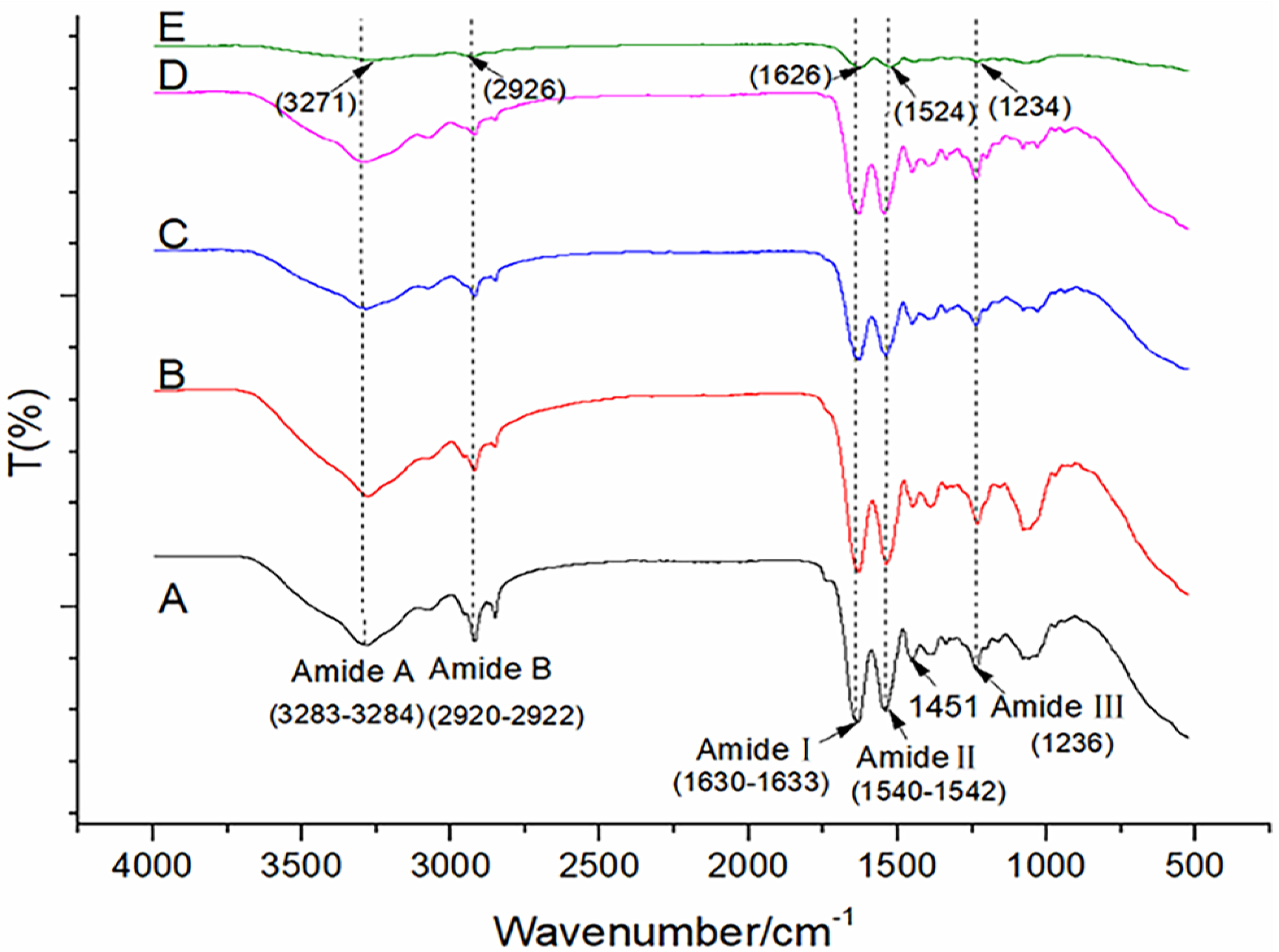
3.3. ATR–FTIR Spectrum of the Triple Helix in the Swim Bladder

The ATR–FTIR is a useful tool for obtaining information about triple helix structures. The spectra of the swim bladders are shown in Figure 3. It can be clearly observed that the FSB, 1-YSB, 5-YSB, and 25-YSB presented similar bands and peaks in the spectrum.
Constant bands at amide A (3283–3284 cm−1, attributed to N–H and O–H stretching), amide B (2920–2922 cm−1, -CH2 asymmetric stretching), amide I (1630–1633 cm−1, C=O stretching), amide II (1540–1542 cm−1, N–H bending and C–N stretching), and amide III (1236 cm−1, C–H stretching) were observed in the FSB, 1-YSB, 5-YSB, and 25-YSB. These observed bands and peaks represent the typical characteristics of collagens, which is in agreement with previous reports [26,27]. However, for the TDSB, significantly lower transmittances were observed. The present bands and peaks were found to drift to different wavenumbers (3271, 2926, 1626, and 1524 cm−1). In addition, the peaks from –CH2 vibration (1451 cm−1) and amide II (1540 cm−1) also disappeared in the TDSB.
Based on Şulea’s report [28], a triple helix should have (1) an AIII/A1450 (absorbance ratio) value higher than 1.0, and (2) a νI–νII (wavenumber) value lower than 100. The FSB, 1-YSB, 5-YSB, and 25-YSB showed properties that were in agreement with the abovementioned requirements (Table 2), indicating the existence of triple helixes in these samples.
The characteristics of the triple helix structure were acquired by fitting the curve of amide I with Gaussian/Lorentz functions [20] (Figure S3), and the results are shown in Table 2. The relative contents of the α-helixes, β-sheets, and random coils in the 1-YSB, 5-YSB, and 25-YSB were statistically the same as those in the FSB (p < 0.05).
As for the triple helix in the TDSB, the relative contents of α-helixes (different to that of the 25-YSB), β-sheets (different to those of the 1-YSB, 5-YSB, and 25-YSB), β-turns (different to those of the 1-YSB, 5-YSB, and 25-YSB), and random coils (different to those of the 5-YSB and 25-YSB) showed different values in comparison with other samples, which implies that the structure of the retained triple helix in the TDSB possessed different characteristics.
The characteristics of the triple helix structure were observed by fitting the curve of amide I with the Gaussian equation (Figure S3), and the results are shown in Table 2. The relative contents of α-helixes, β-sheets, and random coils in the 1-YSB, 5-YSB, and 25-YSB were statistically the same as those in the FSB (p < 0.05).
As for the triple helix in the TDSB, the relative contents of the α-helixes (different to that of the 25-YSB), β-sheets (different to those of the 1-YSB, 5-YSB, and 25-YSB), β-turns (different to those of the 1-YSB, 5-YSB, and 25-YSB), and random coils (different to those of the 5-YSB and 25-YSB) showed different values in comparison with other samples, which implies that the structure of the retained triple helix in the TDSB showed different characteristics.

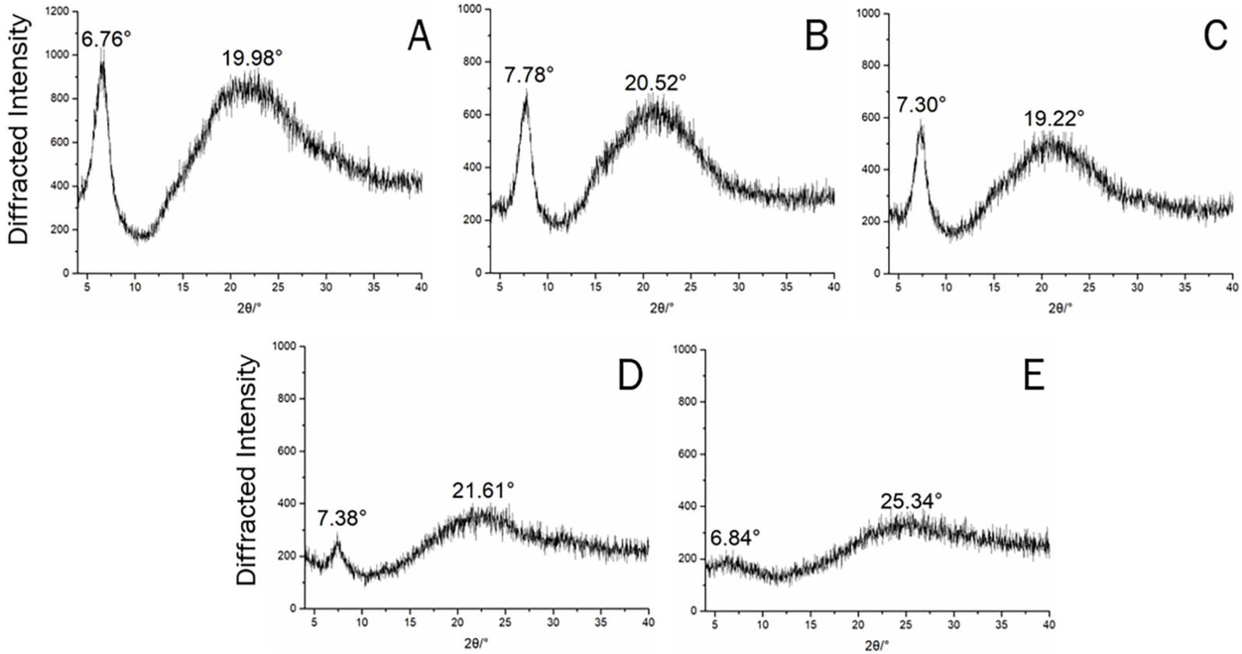
3.4. XRD Spectrum of Triple Helix Structures in the Swim Bladder

XRD provides detailed information about triple helix structures. Two diffraction peaks were detected in the FSB, 1-YSB, 5-YSB, and 25-YSB, which showed sharp peaks at around 6° to 8° and other broad peaks at 19° to 22°, respectively (Figure 4). The sharp peaks at 6° to 8° were found to be associated with the diameter of the triple helix, and the peaks that were at around 19° to 22° were normally attributed to the diffraction from the adjacent repeating units [23]. More importantly, the intensities of these diffraction peaks were found to correspond to the relative contents of the triple helix. Therefore, diffraction peaks were integrated to obtain the peak areas (Table 3).
Table 3 shows that the FSB, 1-YSB, 5-YSB, and 25-YSB showed peak areas higher than those of the TDSB. This trend can be observed either from areas of the peak that increased from 6° to 8° or areas of peaks that increased from 19° to 22°. More importantly, these peak areas were found to be negatively related to swim bladder age; the older the swim bladder, the smaller the peak areas.
Based on Bragg’s equation [8], the diameter of the triple helix was calculated with reference to the angle of the diffusion peak that appeared at from 6° to 8° (Table 3). The results imply that triple helixes in the FSB, 1-YSB, 5-YSB, and 25-YSB had diameters ranging from 1.14 to 1.31, which are similar results to those of previous reports on observations of natural collagens.

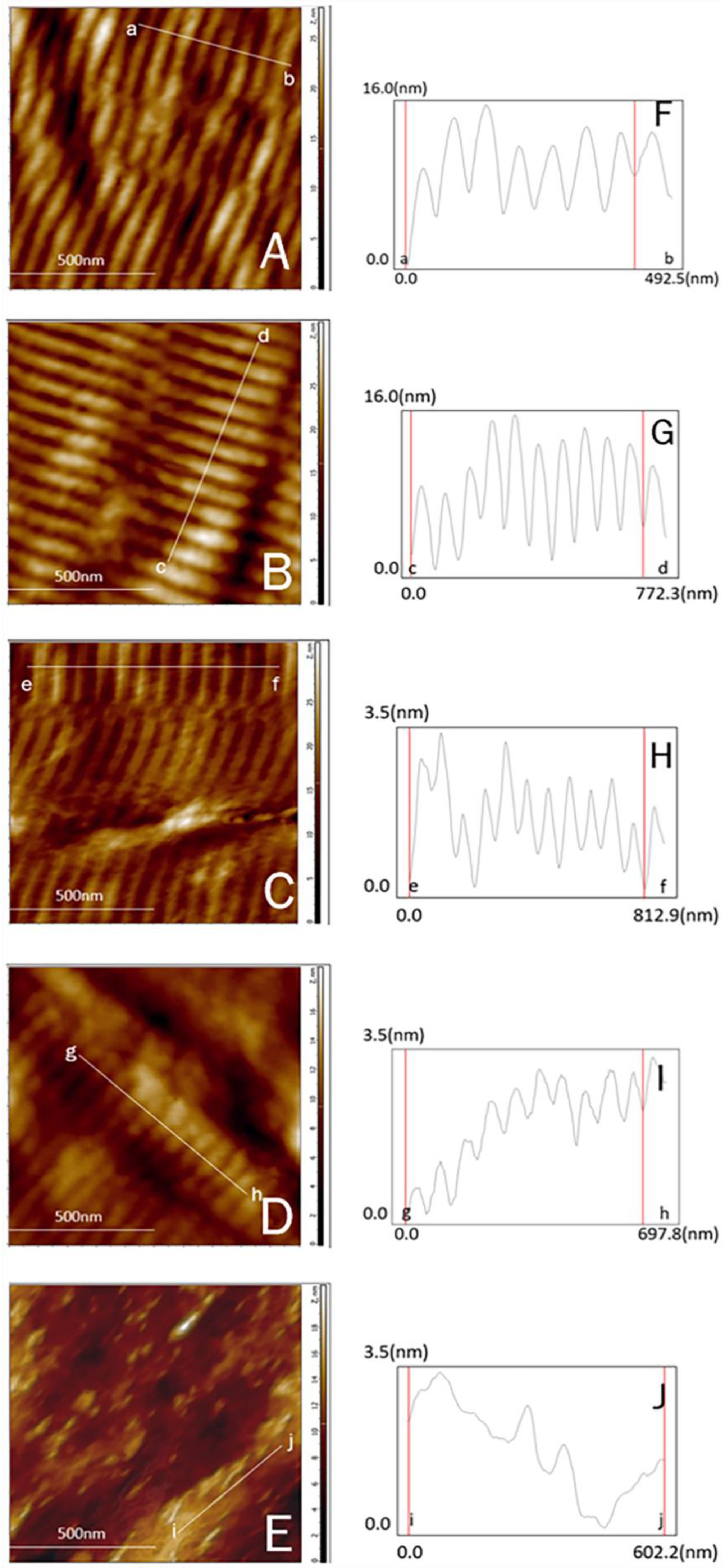
3.5. Characteristics of Microfibrils in Swim Bladders

The swim bladders were probed with AFM to observe the microfibril structures, and those are shown in Figure 5. The significate periodic cross-striated characters were observed in the FSB, 1-YSB, 5-YSB, and 25-YSB, but not in the TDSB. These cross-striated characteristics, attributed to the well-organized gap and overlap of the triple helix [29], were used to identify the integrity of the collagen structures but on a microfibril level. As for the sample without triple helix structures (TDSB), the cross-striated characteristics disappeared.
The surfaces of the collagens were measured along the axial direction of the microfibrils. Accordingly, the average dimension of a single cross-stripe was calculated to obtain the D-period of the microfibrils (Table 4). The FSB, 1-YSB, 5-YSB, and 25-YSB showed a D-period ranging from 70 to 77 nm, which is in agreement with that observed in native collagens [29].

3.6. Optical Microscopy and SEM Observation of Collagen Fiber Bundles

The swim bladders were treated with Masson trichrome staining to observe the histological characteristics of the collagen fiber bundles (Figure 6A–E). The fiber bundles were found to be regularly arranged in the FSB, 1-YSB, 5-YSB and 25-YSB. However, the TDSB showed a transparency, which could be attributed to the poor uptake of Masson trichrome staining as a result of the denatured collagen structure. Furthermore, the fiber bundles in the 1-YSB, 5-YSB, and 25-YSB were observed to be compact and thick, whereas the fiber bundles in the FSB showed unconsolidated and tenuous features.
The cross-sections of swim bladders were further observed with SEM. Fiber bundles that were filiform in character were observed in the FSB. On the contrary, the compact bundles were found in the cross-sections of the 1-YSB, and the bundles were barely seen in the 5-YSB and 25-YSB. As for the TDSB, a smooth surface was observed without any bundle-like characteristics.

4. Discussion

Collagens are reported to have a self-assembly ability [21], and thus are considered an important natural resource and serve as functional ingredients in foods [30,31,32]. The composition analysis showed that the combined moisture and collagen contents of the analyzed swim bladders were higher than 90%. Therefore, besides moisture, swim bladders were basically composed of collagens. These results are in agreement with other reports about the composition of swim bladders from other fishes [33,34,35].
The swim bladder with a 25-year sequence age was investigated regarding (1) the amino acid composition, (2) peptide chains, (3) triple helix structures, (4) microfibrils, and (5) fiber bundles.
Peptide chains are basic structural units in collagen fibers; they are mainly composed of amino acids with a (Gly–X–Y)n repeating sequence, and hydroxyproline is the typical amino acid and frequently appears on X or Y positions [6]. According to the amino acid compositions, glycine contents were found to range from 33 to 35%; the ranges of hydroxyproline and proline were from 8 to 9% and 12 to 18%, respectively. The glycine content in the swim bladders was in agreement with the theoretical value of model collagen molecules (1/3 of total amino acids); the amino acid contents were also similar to those of pure collagen [6,34]. It was further proved that swim bladders are basically composed of collagens.
Similar Td values were observed in different swim bladders from TG-DTG analysis, indicating that the covalent linkages inside peptide chains were not affected by aging. The triple helix is considered a typical structure that is rarely found in other proteins and only appears in collagen [36,37]. The existence of the triple helix in swim bladders was primarily revealed by DSC. The result indicated that swim bladders were found to have thermal denaturation temperatures similar to those of dry collagen [3] but different to that of the thermally denatured sample (TDSB). It was implied that the triple helix may exist in swim bladders. Furthermore, their presented Tm values were slightly different when comparisons were made within samples aged for different lengths of time. This indicates that the triple helix structures in swim bladders may be impacted by aging.
Therefore, the characteristics of the triple helix were further observed by XRD and ATR–FTIR. Based on the XRD result, typical diffusion signals of the triple helix were observed. The ATR–FTIR spectra of aged swim bladders also showed similar wavenumbers to those of the FSB, indicating that the triple helixes in aged swim bladders are similar to those in native collagen [34]. The results from the secondary calculation of bands and peaks further proved the deduction that the collagens in swim bladders retain triple helix structures after years of storage.
Based on the peak areas that appeared on the XRD spectrum, it can be concluded that the retained quantities of triple helix were negatively related to age. This result is in agreement with the observation from DSC, which further emphasized that the loss of the triple helix structure happened over time.
The results obtained by Gaussian/Lorentz functions implied that in the aged swim bladders, the relative contents of α-helixes, β-sheets, and random coils were found to be statistically the same as those in the FSB. The Bragg equation calculation results implied that the triple helix in aged swim bladders had a similar value to those observed in natural collagens [19,34]. These observations implied that, after years of aging, the retained triple helix still presents natural structural characteristics.
Regarding the microfibrils, despite the swim bladders being aged for years and the triple helix structures being partially lost, the cross-striated characteristics still appeared in the swim bladders. As for the swim bladder without the triple helix structure (TDSB), the cross-striated characteristics of the microfibrils were not detected. The D-period of the microfibrils was similar to those of native collagens [29]. This phenomenon indicates that the structure of the microfibrils did not change after years aging, which could be attributed to the preserved structures of the retained triple helix observed from ATR–FTIR and XRD analysis.
The quality of the fiber bundles was observed by optical microscopy and SEM. Compact and thick fiber bundles were observed in aged swim bladders—significantly different to the histological characteristics of the FSB. This could be attributed to the loss of moisture during aging. As for the TDSB, bundle-like characteristics were not observed.

5. Conclusions

Swim bladders with a 25-year aging sequence were collected, and the integrities of the collagen structure were evaluated. The swim bladders were basically composed of collagen, and, after years of aging, the integrities of the peptide chains were retained, but the collagen structures were affected. The triple helix content in the swim bladders was found to be negatively correlated with aging. However, the remaining triple helix was well preserved, and cross-sections of microfibrils can still be observed after 25 years. The fiber bundles, formed by microfibrils, showed compact and thick histological properties. These results imply that after years of aging (up to 25 years), structures of collagen in swim bladders are partially retained, and that the biological and medicinal functions of swim bladders might be retained after years of aging.

Supplementary Materials

The following are available online at https://www.mdpi.com/article/10.3390/app11104578/s1. Figure S1: The standard curve for collagen content calculation; Figure S2: The standard curves for individual amino acid content calculations. Figure S3: The Gaussian/Lorentz function-fitted amid I band (1600–1700 cm−1) on the ART-FTIR spectrum.

Author Contributions

Conceptualization, B.T. and X.W.; methodology, B.T.; validation, F.L. and X.W.; formal analysis, X.H.; investigation, F.L.; resources, F.L. and X.Z.; data curation, X.Z. and L.X.; writing—original draft preparation, X.H.; writing—review and editing, B.T.; supervision, L.X.; project administration, X.W. and F.L.; funding acquisition, X.W. All authors have read and agreed to the published version of the manuscript.

Funding

This research was financially supported by Natural Science Foundation of Guangdong Province, China (2020A1515011357); the Department of Education of Guangdong Province, China, (2018KQNCX081) and (2018KCXTD012); the Key Program of Marine Economy Development (Six Marine Industries) Special Foundation of Department of Natural Resources of Guangdong Province (GDNRC [2020]076); and the Research and Development Start-up Foundation (NTF18031).

Institutional Review Board Statement

Not applicable. This study did not involve humans or animals.

Acknowledgments

The authors wish to thank the Chaocai research institute for providing swim bladders.

Conflicts of Interest

The authors declare no conflict of interest.

References

  1. Fänge, R. Gas exchange in fish swim bladder. Rev. Physiol. Biochem. Pharmacol. 1983, 97, 111–158. [Google Scholar] [CrossRef] [PubMed]
  2. De Mitcheson, Y.S.; To, A.W.-L.; Wong, N.W.; Kwan, H.Y.; Bud, W.S. Emerging from the murk: Threats, challenges and opportunities for the global swim bladder trade. Rev. Fish Biol. Fisher. 2019, 29, 809–835. [Google Scholar] [CrossRef]
  3. Liu, D.; Liang, L.; Regenstein, J.M.; Zhou, P. Extraction and characterisation of pepsin-solubilised collagen from fins, scales, skins, bones and swim bladders of bighead carp (Hypophthalmichthys nobilis). Food Chem. 2012, 133, 1441–1448. [Google Scholar] [CrossRef]
  4. Zhu, S.; Yuan, Q.; Yin, T.; You, J.; Gu, Z.; Xiong, S.; Hu, Y. Self-assembly of collagen-based biomaterials: Preparation, characterizations and biomedical applications. J. Mater. Chem. B 2018, 6, 2650–2676. [Google Scholar] [CrossRef]
  5. Palmieri, B. Heterologous collagen in wound healing: A clinical study. Int. J. Tissue React. 1992, 14, 21–25. [Google Scholar] [CrossRef]
  6. Brown, E.M. Development and utilization of a bovine type I collagen microfibril model. Int. J. Biol. Macromol. 2013, 53, 20–25. [Google Scholar] [CrossRef]
  7. Sousa, R.O.; Alves, A.L.; Carvalho, D.N.; Martins, E.; Oliveira, C.; Silva, T.H.; Reis, R.L. Acid and enzymatic extraction of collagen from Atlantic cod (Gadus Morhua) swim bladders envisaging health-related applications. J. Biomater. Sci. Polym. Ed. 2020, 31, 20–37. [Google Scholar] [CrossRef]
  8. Maxwell, C.A.; Wess, T.J.; Kennedy, C.J. X-ray Diffraction Study into the Effects of Liming on the Structure of Collagen. Biomacromolecules 2006, 7, 2321–2326. [Google Scholar] [CrossRef]
  9. Meng, Z.; Zheng, X.; Tang, K.; Liu, J.; Ma, Z.; Zhao, Q. Dissolution and regeneration of collagen fibers using ionic liquid. Int. J. Biol. Macromol. 2012, 51, 440–448. [Google Scholar] [CrossRef]
  10. Zhao, X.; Qian, Y.; Li, G.-J.; Tan, J. Preventive effects of the polysaccharide of Larimichthys crocea swim bladder on carbon tetrachloride (CCl4)-induced hepatic damage. Chin. J. Nat. Med. 2015, 13, 521–528. [Google Scholar] [CrossRef]
  11. Okada, T.; Hayashi, T.; Ikada, Y. Degradation of collagen suture in vitro and in vivo. Biomaterials 1992, 13, 448–454. [Google Scholar] [CrossRef]
  12. Bretaud, S.; Nauroy, P.; Malbouyres, M.; Ruggiero, F. Fishing for collagen function: About development, regeneration and disease. Sem. Cell Dev. Biol. 2019, 89, 100–108. [Google Scholar] [CrossRef] [PubMed]
  13. Hickman, D.; Sims, T.J.; Miles, C.A.; Bailey, A.J.; de Mari, M.; Koopmans, M. Isinglass/collagen: Denaturation and functionality. J. Biotechnol. 2000, 79, 245–257. [Google Scholar] [CrossRef]
  14. Teng, B.; Petrie, P.R.; Smith, P.A.; Bindon, K.A. Comparison of water addition and early-harvest strategies to decrease alcohol concentration in Vitis vinifera cv. Shiraz wine: Impact on wine phenolics, tannin composition and colour properties. Aust. J. Grape Wine Res. 2020, 26, 158–171. [Google Scholar] [CrossRef]
  15. Sun, J.; Shi, X.; Zhang, H.; Xia, L.; Guo, Y.; Sun, X. Detection of moisture content in peanut kernels using hyperspectral imaging technology coupled with chemometrics. J. Food Process Eng. 2019, 42, e13263. [Google Scholar] [CrossRef]
  16. Kesava Reddy, G.; Enwemeka, C.S. A simplified method for the analysis of hydroxyproline in biological tissues. Clin. Biochem. 1996, 29, 225–229. [Google Scholar] [CrossRef]
  17. Heinrikson, R.L.; Meredith, S.C. Amino acid analysis by reverse-phase high-performance liquid chromatography: Precolumn derivatization with phenylisothiocyanate. Anal. Biochem. 1984, 136, 65–74. [Google Scholar] [CrossRef]
  18. Yu, X.; Guo, L.; Liu, M.; Cao, X.; Shang, S.; Liu, Z.; Huang, D.; Cao, Y.; Cui, F.; Tian, L. Callicarpa nudiflora loaded on chitosan-collagen/organomontmorillonite composite membrane for antibacterial activity of wound dressing. Int. J. Biol. Macromol. 2018, 120, 2279–2284. [Google Scholar] [CrossRef]
  19. Safandowska, M.; Pietrucha, K. Effect of fish collagen modification on its thermal and rheological properties. Int. J. Biol. Macromol. 2013, 53, 32–37. [Google Scholar] [CrossRef]
  20. Belbachir, K.; Noreen, R.; Gouspillou, G.; Petibois, C. Collagen types analysis and differentiation by FTIR spectroscopy. Anal. Bioanal. Chem. 2009, 395, 829–837. [Google Scholar] [CrossRef]
  21. Zhu, L.; Li, J.; Wang, Y.; Sun, X.; Li, B.; Poungchawanwong, S.; Hou, H. Structural feature and self-assembly properties of type II collagens from the cartilages of skate and sturgeon. Food Chem. 2020, 331, 127340. [Google Scholar] [CrossRef]
  22. Susanto, A.; Satari, M.H.; Abbas, B.; Koesoemowidodo, R.S.A.; Cahyanto, A. Fabrication and characterization of chitosan-collagen membrane from barramundi (Lates calcarifer) scales for guided tissue regeneration. Eur. J. Dent. 2019, 13, 370–375. [Google Scholar] [CrossRef] [PubMed] [Green Version]
  23. Badii, F.; MacNaughtan, W.; Mitchell, J.R.; Farhat, I.A. The Effect of drying temperature on physical properties of thin gelatin films. Dry. Technol. 2014, 32, 30–38. [Google Scholar] [CrossRef]
  24. Gaurav Kumar, P.; Nidheesh, T.; Govindaraju, K.; Jyoti; Suresh, P.V. Enzymatic extraction and characterisation of a thermostable collagen from swim bladder of rohu (Labeo rohita). J. Sci. Food Agric. 2017, 97, 1451–1458. [Google Scholar] [CrossRef] [PubMed]
  25. Liu, W.; Tian, Z.; Li, C.; Li, G. Thermal denaturation of fish collagen in solution: A calorimetric and kinetic analysis. Thermochim. Acta. 2014, 581, 32–40. [Google Scholar] [CrossRef]
  26. Chadefaux, C.; Le Hô, A.-S.; Bellot-Gurlet, L.; Reiche, I. Curve-fitting micro-ATR-FTIR studies of the amide I and II bands of type I collagen in archaeological bone materials. E-Preser. Sci. 2009, 6, 129–137. [Google Scholar] [CrossRef]
  27. Petibois, C.; Gouspillou, G.; Wehbe, K.; Delage, J.P.; Déléris, G. Analysis of type I and IV collagens by FT-IR spectroscopy and imaging for a molecular investigation of skeletal muscle connective tissue. Anal. Bioanal. Chem. 2006, 386, 1961–1966. [Google Scholar] [CrossRef]
  28. Şulea, D.; Micutz, M.; Albu, M.; Staicu, T.; Leca, M. Collagen-thuja tincture biomaterials for wound treatment. 2. Hydrogels and porous matrices. Rev. Roum. Chim. 2011, 56, 129–136. [Google Scholar] [CrossRef]
  29. Strasser, S.; Zink, A.; Janko, M.; Heckl, W.M.; Thalhammer, S. Structural investigations on native collagen type I fibrils using AFM. Biochem. Bioph. Res. Co. 2007, 354, 27–32. [Google Scholar] [CrossRef]
  30. Hashim, P.; Mohd Ridzwan, M.S.; Bakar, J.; Mat Hashim, D. Collagen in food and beverage industries. Int. Food Res. J. 2015, 22, 1–8. [Google Scholar] [CrossRef]
  31. Subhan, F.; Hussain, Z.; Tauseef, I.; Shehzad, A.; Wahid, F. A review on recent advances and applications of fish collagen. Crit. Rev. Food Sci. Nutr. 2021, 61, 1027–1037. [Google Scholar] [CrossRef] [PubMed]
  32. Lin, F.; Zhang, H.; Yu, J.; Yu, C.; Chen, C.; Sun, Z.; Wang, S.; Wen, X. Effects of dietary selenium on growth performance, antioxidative status and tissue selenium deposition of juvenile Chu’s croaker (Nibea coibor). Aquaculture 2021, 536, 736439. [Google Scholar] [CrossRef]
  33. Gauza-Włodarczyk, M.; Kubisz, L.; Włodarczyk, D. Amino acid composition in determination of collagen origin and assessment of physical factors effects. Int. J. Biol. Macromol. 2017, 104, 987–991. [Google Scholar] [CrossRef] [PubMed]
  34. Sinthusamran, S.; Benjakul, S.; Kishimura, H. Comparative study on molecular characteristics of acid soluble collagens from skin and swim bladder of seabass (Lates calcarifer). Food Chem. 2013, 138, 2435–2441. [Google Scholar] [CrossRef]
  35. Lin, F.; Rong, H.; Lin, J.; Yuan, Y.; Yu, J.; Yu, C.; You, C.; Wang, S.; Sun, Z.; Wen, X. Enhancement of collagen deposition in swim bladder of Chu’s croaker (Nibea coibor) by proline: View from in-vitro and in-vivo study. Aquaculture 2020, 523, 735175. [Google Scholar] [CrossRef]
  36. Shoulders, M.D.; Raines, R.T. Collagen structure and stability. Annu. Rev. Biochem. 2009, 78, 929–958. [Google Scholar] [CrossRef] [Green Version]
  37. Teng, B.; Jian, X.; Gao, Y.; Chen, W. Comparison of polyflavonoids in bayberry tanning effluent and commercial bayberry tannin: Prerequisite information for vegetable tanning effluent recycling. J. Clean. Prod. 2016, 112, 972–979. [Google Scholar] [CrossRef]
Figure 1. Structural characteristics of collagen.
Applsci 11 04578 g001
Figure 2. TG-DTG (A) and DSC (B) observations of the decompositions of peptide chains and triple helix structures in FSB (a), 1-YSB (b), 5-YSB(c), 25-YSB (d), and TDSB (e).
Applsci 11 04578 g002
Figure 3. The ATR–FTIR spectra of (A) FSB, (B) 1-YSB, (C) 5-YSB, (D) 25-YSB, and (E) TDSB.
Applsci 11 04578 g003
Figure 4. The diffraction of the triple helix in FSB (A), 1-YSB (B), 5-YSB (C), 25-YSB (D), and TDSB (E) observed from XRD analysis.
Applsci 11 04578 g004
Figure 5. The AFM image of the D-periodic cross-striated microfibrils in FSB (A), 1-YSB (B), 5-YSB (C), 25-YSB (D), and TDSB (E); line measurements on the microdissected collagen fibril between points a and b (F), points c and d (G), points e and f (H), points g and h (I), and points i and j (J) on surface of each sample.
Applsci 11 04578 g005
Figure 6. The optical microscopy image (×100 times) of fiber bundles in FSB (A), 1-YSB (B), 5-YSB (C), 25-YSB (D), and TDSB (E) after Masson’s trichrome staining; SEM images (×1000 times) of fiber bundles in FSB (F), 1-YSB (G), 5-YSB (H), 25-YSB (I), and TDSB (J).
Applsci 11 04578 g006
Table 1. Basic compositions of FSB, 1-YSB, 5-YSB, and 25-YSB 1.
FSB1-YSB5-YSB25-YSB
Collagen (%)35.41 ± 0.70 b75.36 ± 0.47 a77.03 ± 13.38 a90.38 ± 3.02 a
Moisture (%)56.98 ± 1.04 a16.00 ± 0.46 b14.63 ± 0.57 b10.31 ± 0.20 c
Moisture and collagen in total (%)92.21 ± 0.43 a91.12 ± 0.16 a91.34 ± 13.19 a100.58 ± 2.95 a
Amino acid composition (mol %)
Glycine33.50 ± 0.23 a35.34 ± 2.34 a33.48 ± 0.62 a33.54 ± 0.44 a
Hydroxyproline9.57 ± 1.54 a8.85 ± 2.46 a8.80 ± 0.21 a9.42 ± 0.25 a
Proline18.81 ± 0.53 a12.31 ± 0.57 b13.15 ± 0.28 b12.43 ± 0.02 b
Arginine1.26 ± 0.02 c2.11 ± 0.13 a1.81 ± 0.03 a b1.72 ± 0.08 b
Valine2.13 ± 0.03 b2.65 ± 0.08 a1.25 ± 0.02 c1.38 ± 0.002 c
Methionine1.74 ± 0.04 b2.09 ± 0.07 a1.45 ± 0.02 c0.54 ± 0.003 d
Isoleucine0.67 ± 0.03 b3.10 ± 0.08 a0.46 ± 0.01 c0.44 ± 0.01 c
Leucine2.55 ± 0.02 a0.94 ± 0.04 c2.46 ± 0.05 a2.26 ± 0.02 b
Phenylalanine1.59 ± 0.05 b1.79 ± 0.05 a1.31 ± 0.02 c1.27 ± 0.001 c
Lysine3.36 ± 0.08 a3.35 ± 0.09 a2.77 ± 0.06 b2.96 ± 0.01 b
Alanine4.74 ± 0.22 d11.80 ± 0.12 a10.06 ± 0.16 c10.86 ± 0.12 b
Tyrosine0.79 ± 0.03 b1.01 ± 0.03 a0.71 ± 0.02 b0.42 ± 0.02 c
Aspartic acid7.87 ± 0.99 a5.10 ± 0.08 b5.74 ± 0.69 a b5.95 ± 0.04 a b
Glutamate6.04 ± 0.28 b6.42 ± 1.23 b11.54 ± 0.23 a11.37 ± 0.002 a
Serine5.38 ± 0.13 a3.13 ± 0.17 b4.09 ± 0.03 b5.44 ± 0.50 a
Total imino acids 228.38 ± 1.01 a21.16 ± 1.90 b21.94 ± 0.49 b21.85 ± 0.27 b
1 Values are means ± SDs (n = 3). Values with different superscript letters within the same row are significantly different (p < 0.05). 2 Total amino acids were composed of proline and hydroxyproline.
Table 2. Secondary calculation of wavenumbers extracted from ATR–FTIR spectra 1.
FSB1-YSB5-YSB25-YSBTDSB
From maximum absorbance/wavenumber calculation
AIII/A14501.06 ± 0.01 ab1.14 ± 0.06 a1.01 ± 0.01 b1.10 ± 0.02 a0.95 ± 0.01 c
νI–νII92.41 ± 0.35 b92.30 ± 3.43 b88.66 ± 0.25 b88.97 ± 1.64 b102.50 ± 1.01 a
From fitting by Gaussian equation
α-helix (%)24.35 ± 0.56 ab21.31 ± 1.94 b23.34 ± 0.71 ab25.50 ± 0.96 a20.57 ± 2.12 b
β-sheet (%)37.55 ± 1.30 a42.24 ± 2.90 a38.49 ± 0.95 a37.57 ± 2.03 a30.42 ± 1.71 b
β-turn (%)15.65 ± 0.56 ab12.82 ± 0.94 c14.01 ± 0.41 bc13.79 ± 1.12 bc17.69 ± 0.52 a
Random coil (%)15.22 ± 0.22 b17.30 ± 2.73 ab14.45 ± 0.77 b14.19 ± 0.24 b21.29 ± 1.44 a
1 Values are means ± SDs (n = 3). Values with the different superscript letters within the same column are significantly different (p < 0.05).
Table 3. Structural characteristics of collagens in the swim bladder obtained from XRD analysis.
FSB1-YSB5-YSB25-YSBTDSB
Peak area (6° to 8°)1182.8753.4603.5180.7165.9
Peak area (19° to 22°)5481.43567.52806.91713.4927.6
Diameter of triple helix1.311.141.221.20--
Table 4. Length of D-period (nm) in FSB, 1-YSB, 5-YSB, and 25-YSB 1.
FSB1-YSB5-YSB25-YSB
D-Period (nm)70.82 ± 0.66 b76.93 ± 0.24 a75.30 ± 2.40 a70.70 ± 0.73 b
1 Values are means ± SDs (n = 3). Values with different superscript letters within the same row are significantly different (p < 0.05).
Publisher’s Note: MDPI stays neutral with regard to jurisdictional claims in published maps and institutional affiliations.

