
Early Treatment with a Slow Maxillary Ni–Ti Leaf Springs Expander

Abstract
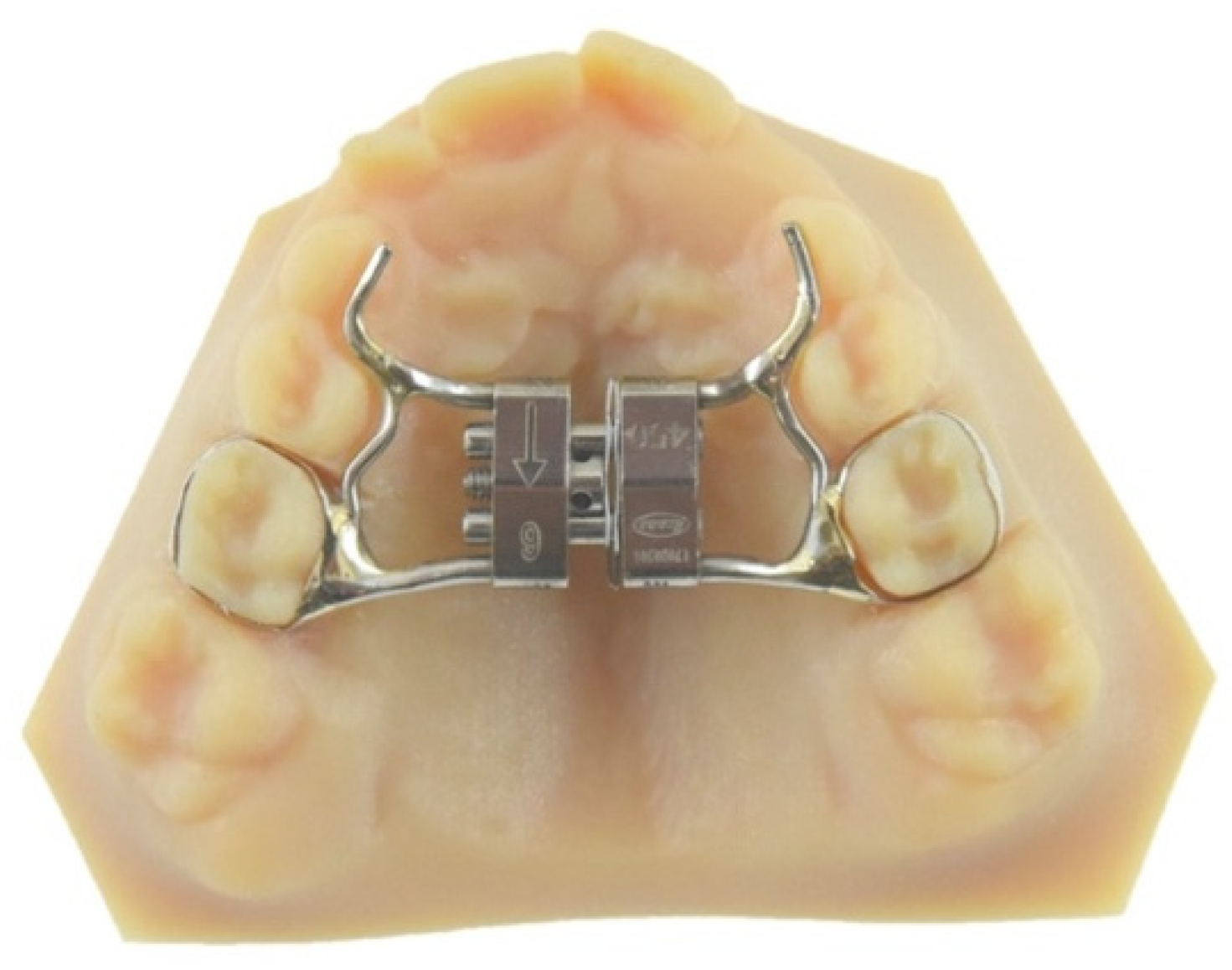
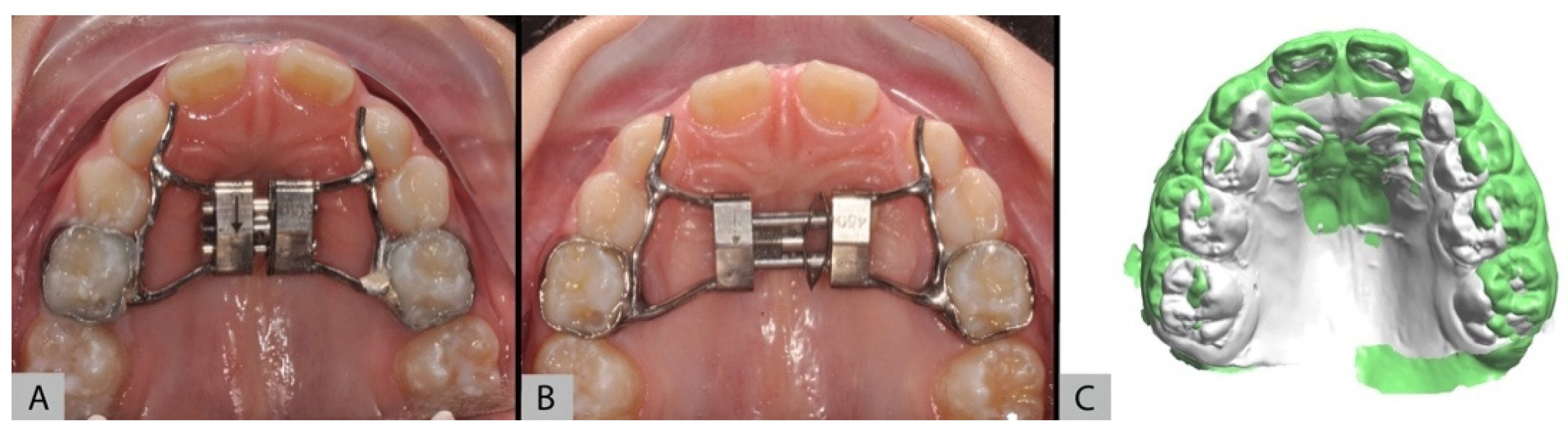
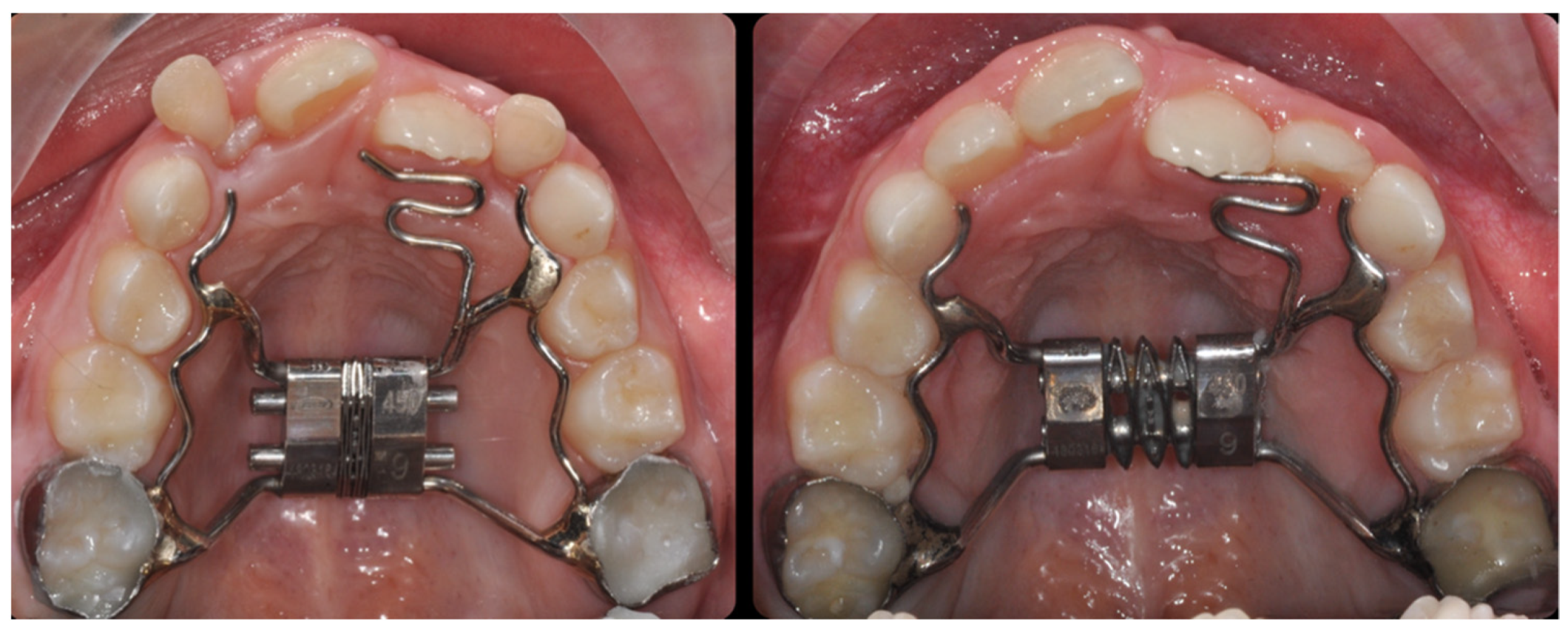
1. Introduction
- (a)
- Reactivable;
- (b)
- Preactivated.
- (a)
- Active component;
- (b)
- Action modalities.
- (1)
- 6 mm screw—450 gr;
- (2)
- 6 mm screw—900 gr;
- (3)
- 9 mm screw—450 gr;
- (4)
- 9 mm screw—900 gr.
- -
- No compliance needed;
- -
- Reduced need for clinical checks;
- -
- Painless;
- -
- Controlled, light and graded force production;
- -
- No risks of overexpansion;
- -
- Easiness of activation;
- -
- Vestibular tooth inclination control.
- (1)
- 6 mm—450 gr;
- (2)
- 6 mm—900 gr;
- (3)
- 9 mm—450 gr;
- (4)
- 9 mm—900 gr.
2. Materials and Methods
- -
- Publication date from 1993 to 2020;
- -
- Articles in English, Spanish or Italian;
- -
- Full text available;
- -
- Scientific validity;
- -
- Articles published before 1993;
- -
- No full text available;
- -
- Articles without L.E. or analogue appliances references;
- -
- Articles without statistical data.
3. Results
4. Discussion
5. Conclusions
- -
- Absence of collaboration from patients and parents;
- -
- Visual control of activation, reducing the risk of operator-dependent error;
- -
- Safety in use;
- -
- Mainly body movement of the teeth;
- -
- Predetermined, light and constant forces;
- -
- Predictability of the result;
- -
- Ease of activation;
- -
- Simplification of clinical procedures, reducing the number of visits.
Author Contributions
Funding
Conflicts of Interest
References
- De Sousa, R.V.; Ribeiro, G.L.A.; Firmino, R.T.; Martins, C.C.; Granville-Garcia, A.F.; Paiva, S.M. Prevalence and Associated Factors for the Development of Anterior Open Bite and Posterior Crossbite in the Primary Dentition. Braz. Dent. J. 2014, 25, 336–342. [Google Scholar] [CrossRef] [PubMed]
- Beretta, M.; Lanteri, C.; Lanteri, V.; Cherchi, C.; Franchi, L.; Gianolio, A. Evolution of the Leaf Expander: A Maxillary Self Expander. J. Clin. Orthod. 2019, 53, 260–266. [Google Scholar] [PubMed]
- Maspero, C.; Prevedello, C.; Giannini, L.; Galbiati, G.; Farronato, G. Atypical swallowing: A review. Minerva Stomatol. 2014, 63, 217–227. [Google Scholar] [PubMed]
- Maspero, C.; Cavagnetto, D.; Abate, A.; Cressoni, P.; Farronato, M. Effects on the Facial Growth of Rapid Palatal Expansion in Growing Patients Affected by Juvenile Idiopathic Arthritis with Monolateral Involvement of the Temporomandibular Joints: A Case-Control Study on Posteroanterior and Lateral Cephalograms. J. Clin. Med. 2020, 9, 1159. [Google Scholar] [CrossRef]
- Maspero, C.; Farronato, M.; Bellincioni, F.; Cavagnetto, D.; Abate, A. Assessing mandibular body changes in growing subjects: A comparison of CBCT and reconstructed lateral cephalogram measurements. Sci. Rep. 2020, 10, 11722. [Google Scholar] [CrossRef]
- Prado, G.P.R.; Furtado, F.; Aloise, A.C.; Biló, J.P.R.; Ferreira, L.M.; Pereira, M.D. Stability of surgically assisted rapid palatal expansion with and without retention analyzed by 3-dimensional imaging. Am. J. Orthod. Dentofac. Orthop. 2014, 145, 610–616. [Google Scholar] [CrossRef]
- Abate, A.; Cavagnetto, D.; Rusconi, F.; Cressoni, P.; Esposito, L. Safety and Effects of the Rapid Maxillary Expander on Temporomandibular Joint in Subjects Affected by Juvenile Idiopathic Arthritis: A Retrospective Study. Children 2021, 8, 33. [Google Scholar] [CrossRef] [PubMed]
- Abate, A.; Cavagnetto, D.; Fama, A.; Maspero, C.; Farronato, G. Relationship between Breastfeeding and Malocclusion: A Systematic Review of the Literature. Nutrients 2020, 12, 3688. [Google Scholar] [CrossRef]
- Zhou, Y.; Long, H.; Ye, N.; Xue, J.; Yang, X.; Liao, L.; Lai, W. The effectiveness of non-surgical maxillary expansion: A meta-analysis. Eur. J. Orthod. 2014, 36, 233–242. [Google Scholar] [CrossRef]
- Farronato, M.; Cavagnetto, D.; Abate, A.; Cressoni, P.; Fama, A.; Maspero, C. Assessment of condylar volume and ramus height in JIA patients with unilateral and bilateral TMJ involvement: Retrospective case-control study. Clin. Oral Investig. 2019. [Google Scholar] [CrossRef]
- Cossellu, G.; Ugolini, A.; Beretta, M.; Farronato, M.; Gianolio, A.; Maspero, C.; Lanteri, V. Three-Dimensional Evaluation of Slow Maxillary Expansion with Leaf Expander vs. Rapid Maxillary Expansion in a Sample of Growing Patients: Direct Effects on Maxillary Arch and Spontaneous Mandibular Response. Appl. Sci. 2020, 10, 4512. [Google Scholar] [CrossRef]
- Isaacson, R.J.; Ingram, A.H. Forces Produced By Rapid Maxillary Expansion. Angle Orthod. 1964, 34, 261–270. [Google Scholar]
- Liu, S.; Xu, T.; Zou, W. Effects of rapid maxillary expansion on the midpalatal suture: A systematic review. Eur. J. Orthod. 2015, 37, 651–655. [Google Scholar] [CrossRef] [PubMed]
- Giudice, A.L.; Nucera, R.; Perillo, L.; Paiusco, A.; Caccianiga, G. Is Low-Level Laser Therapy an Effective Method to Alleviate Pain Induced by Active Orthodontic Alignment Archwire? A Randomized Clinical Trial. J. Évid. Based Dent. Pract. 2019, 19, 71–78. [Google Scholar] [CrossRef] [PubMed]
- Maspero, C.; Farronato, M.; Bellincioni, F.; Annibale, A.; Machetti, J.; Abate, A.; Cavagnetto, D. Three-Dimensional Evaluation of Maxillary Sinus Changes in Growing Subjects: A Retrospective Cross-Sectional Study. Materials 2020, 13, 1007. [Google Scholar] [CrossRef]
- Farronato, G.; Giannini, L.; Galbiati, G.; Cannalire, P.; Martinelli, G.; Tubertini, I.; Maspero, C. Oral tissues and orthodontic treatment: Common side effects. Minerva Stomatol. 2013, 62, 431–446. [Google Scholar]
- Maspero, C.; Abate, A.; Cavagnetto, D.; Fama, A.; Stabilini, A.; Farronato, G.; Farronato, M. Operculectomy and spontane-ous eruption of impacted second molars: A retrospective study. J. Biol. Regul. Homeost. Agents 2019, 33, 1909–1912. [Google Scholar] [CrossRef]
- Abate, A.; Cavagnetto, D.; Fama, A.; Matarese, M.; Bellincioni, F.; Assandri, F. Efficacy of Operculectomy in the Treatment of 145 Cases with Unerupted Second Molars: A Retrospective Case–Control Study. Dent. J. 2020, 8, 65. [Google Scholar] [CrossRef]
- Silvestrini-Biavati, A.; Angiero, F.; Gambino, A.; Ugolini, A. Do changes in spheno-occipital synchondrosis after rapid maxillary expansion affect the maxillomandibular complex? Eur. J. Paediatr. Dent. 2013, 14, 63–67. [Google Scholar]
- Lanteri, C.; Beretta, M.; Lanteri, V.; Gianolio, A.; Cherchi, C.; Franchi, L. The Leaf Expander for Non-Compliance Treatment in the Mixed Dentition. J. Clin. Orthod. 2016, 50, 552–560. [Google Scholar]
- Lanteri, V.; Gianolio, A.; Gualandi, G.; Beretta, M. Maxillary Tridimensional Changes after Slow Expansion with Leaf Ex-pander in a Sample of Growing Patients: A Pilot Study. Eur. J. Paediatr. Dent. 2018. [Google Scholar] [CrossRef]
- Donohue, V.E.; Marshman, L.A.G.; Winchester, L.J. A clinical comparison of the quadhelix appliance and the nickel titanium (tandem loop) palatal expander: A preliminary, prospective investigation. Eur. J. Orthod. 2004, 26, 411–420. [Google Scholar] [CrossRef]
- Caniklioglu, M.C. Use of a nickel titanium palatal expander in cleft-palate cases. J. Clin. Orthod. 2004, 38, 374–377. [Google Scholar] [PubMed]
- Ciambotti, C.; Ngan, P.; Durkee, M.; Kohli, K.; Kim, H. A comparison of dental and dentoalveolar changes between rapid palatal expansion and nickel-titanium palatal expansion appliances. Am. J. Orthod. Dentofac. Orthop. 2001, 119, 11–20. [Google Scholar] [CrossRef] [PubMed]
- Didier, H.; Assandri, F.; Gaffuri, F.; Cavagnetto, D.; Abate, A.; Villanova, M.; Maiorana, C. The Role of Dental Occlusion and Neuromuscular Behavior in Professional Ballet Dancers’ Performance: A Pilot Study. Healthcare 2021, 9, 251. [Google Scholar] [CrossRef] [PubMed]
- Arndt, W.V. Nickel titanium palatal expander. J. Clin. Orthod. 1993, 27, 129–137. [Google Scholar] [PubMed]
- Maspero, C.; Fama, A.; Cavagnetto, D.; Abate, A.; Farronato, M. Treatment of dental dilacerations. J. Biol. Regul. Homeost. Agents 2019, 33, 1623–1628. [Google Scholar] [PubMed]
- Cossellu, G.; Lanteri, V.; Butera, A.; Sarcina, M.; Farronato, G. Effects of six different preventive treatments on the shear bond strength of orthodontic brackets:in vitrostudy. Acta Biomater. Odontol. Scand. 2015, 1, 13–17. [Google Scholar] [CrossRef] [PubMed]
- Cossellu, G.; Lanteri, V.; Butera, A.; Laffi, N.; Merlini, A.; Farronato, G. Timing considerations on the shear bond strength of orthodontic brackets after topical fluoride varnish applications. J. Orthod. Sci. 2017, 6, 11–15. [Google Scholar] [CrossRef] [PubMed]
- Scribante, A.; Farahani, M.R.D.; Marino, G.; Matera, C.; Baena, R.R.Y.; Lanteri, V.; Butera, A. Biomimetic Effect of Nano-Hydroxyapatite in Demineralized Enamel before Orthodontic Bonding of Brackets and Attachments: Visual, Adhesion Strength, and Hardness in In Vitro Tests. BioMed Res. Int. 2020, 2020, 6747498. [Google Scholar] [CrossRef]
- Lanteri, V.; Cossellu, G.; Farronato, M.; Ugolini, A.; Leonardi, R.; Rusconi, F.; De Luca, S.; Biagi, R.; Maspero, C. Assessment of the Stability of the Palatal Rugae in a 3D-3D Superimposition Technique Following Slow Maxillary Expansion (SME). Sci. Rep. 2020, 10, 2676. [Google Scholar] [CrossRef]
- Cossellu, G.; Lanteri, V.; Lione, R.; Ugolini, A.; Gaffuri, F.; Cozza, P.; Farronato, M. Efficacy of ketoprofen lysine salt and paracetamol/acetaminophen to reduce pain during rapid maxillary expansion: A randomized controlled clinical trial. Int. J. Paediatr. Dent. 2018, 29, 58–65. [Google Scholar] [CrossRef] [PubMed]
- Lanteri, V.; Cavagnetto, D.; Abate, A.; Mainardi, E.; Gaffuri, F.; Ugolini, A.; Maspero, C. Buccal Bone Changes Around First Permanent Molars and Second Primary Molars after Maxillary Expansion with a Low Compliance Ni–Ti Leaf Spring Expander. Int. J. Environ. Res. Public Health 2020, 17, 9104. [Google Scholar] [CrossRef] [PubMed]
- Lanteri, V.; Cossellu, G.; Gianolio, A.; Beretta, M.; Lanteri, C.; Cherchi, C.; Farronato, G. Comparison between RME, SME and Leaf Expander in growing patients: A retrospective postero-anterior cephalometric study. Eur. J. Paediatr. Dent. 2018, 19, 199–204. [Google Scholar] [CrossRef] [PubMed]
- Lanteri, V.; Farronato, M.; Ugolini, A.; Cossellu, G.; Gaffuri, F.; Parisi, F.M.R.; Cavagnetto, D.; Abate, A.; Maspero, C. Vol-umetric Changes in the Upper Airways after Rapid and Slow Maxillary Expansion in Growing Patients: A Case-Control Study. Materials 2020, 13, 2239. [Google Scholar] [CrossRef] [PubMed]
- Grassia, V.; D’Apuzzo, F.; Jamilian, A.; Femiano, F.; Favero, L.; Perillo, L. Comparison between rapid and mixed maxillary expansion through an assessment of arch changes on dental casts. Prog. Orthod. 2015, 16, 20. [Google Scholar] [CrossRef] [PubMed]
- Christie, K.F.; Boucher, N.; Chung, C.-H. Effects of bonded rapid palatal expansion on the transverse dimensions of the maxilla: A cone-beam computed tomography study. Am. J. Orthod. Dentofac. Orthop. 2010, 137, S79–S85. [Google Scholar] [CrossRef]
- Garrett, B.J.; Caruso, J.M.; Rungcharassaeng, K.; Farrage, J.R.; Kim, J.S.; Taylor, G.D. Skeletal effects to the maxilla after rapid maxillary expansion assessed with cone-beam computed tomography. Am. J. Orthod. Dentofac. Orthop. 2008, 134, 8.e1–8.e11. [Google Scholar] [CrossRef]
- Martina, R.; Cioffi, I.; Farella, M.; Leone, P.; Manzo, P.; Matarese, G.; Portelli, M.; Nucera, R.; Cordasco, G. Transverse changes determined by rapid and slow maxillary expansion—A low-dose CT-based randomized controlled trial. Orthod. Craniofac. Res. 2012, 15, 159–168. [Google Scholar] [CrossRef]
- Lagravère, M.O.; Major, P.W.; Flores-Mir, C. Skeletal and dental changes with fixed slow maxillary expansion treatment: A Systematic Review. J. Am. Dent. Assoc. 2005, 136, 194–199. [Google Scholar] [CrossRef]
- Farronato, G.; Giannini, L.; Galbiati, G.; Maspero, C. Comparison of the dental and skeletal effects of two different rapid palatal expansion appliances for the correction of the maxillary asymmetric transverse discrepancies. Minerva Stomatol. 2012, 61, 45–55. [Google Scholar] [PubMed]
- Fama, A.; Cavagnetto, D.; Abate, A.; Mainardi, E.; De Filippis, A.; Esposito, L. Treatment of Dental Dilacerations. Narrative review. Dent. Cadmos 2021, 89, 174–178. [Google Scholar]
- Garib, D.G.; Henriques, J.F.C.; Janson, G.; Freitas, M.R.; Coelho, R.A. Rapid maxillary expansion--tooth tissue-borne versus tooth-borne expanders: A computed tomography evaluation of dentoskeletal effects. Angle Orthod. 2005, 75, 548–557. [Google Scholar] [PubMed]
- Manzella, K.; Franchi, L.; Al-Jewair, T. Correction of maxillary transverse deficiency in growing patients with permanent dentitions. J. Clin. Orthod. 2018, 52, 148–156. [Google Scholar]
- Maspero, C.; Abate, A.; Bellincioni, F.; Cavagnetto, D.; Lanteri, V.; Costa, A.; Farronato, M. Comparison of a tridimensional cephalometric analysis performed on 3T-MRI compared with CBCT: A pilot study in adults. Prog. Orthod. 2019, 20, 1–10. [Google Scholar] [CrossRef]


| Author | Date | Desing | Journal | Sample | Statistics | Conclusion |
|---|---|---|---|---|---|---|
| Cossellu et al. | 2020 | Retrospective cohort study | Appl. Sci | Leaf 69 RME 21 |
| Leaf Expander anchored on primary molars is an effective treatment option to correct maxillary transverse deficiencies and cross-bites. The data also demonstrated a statistically significant mandibular spontaneous response. |
| Lanteri | 2018 | Single arm retrospective study | Eur J Paediatr Dent | Leaf 10 |
| The data suggest that slow maxillary expansion using Leaf Expander appliance could be a reasonable alternative to conventional maxillary expansion therapy in the early mixed dentition. |
| Lanteri | 2020 | Retrospective cohort study | Sci Rep. | Leaf 27 NC 27 |
| It demonstrated that the 3D anatomical morphology of the palatal rugae area is not affected by specific dental treatments, thus indicating that it can maintain its morphological patterns of individuality even after undergoing SME. |
| Cossellu | 2019 | Prospective randomised controlled clinical trial | Int J Paediatr Dent | Leaf 40 RME 40 |
| Subjects that took ketoprofen lysine salt experienced lower levels of pain during the fourth, fifth and sixth days of screw activation showing to be more effective than paracetamol/acetaminophen, probably due to the anti-inflammatory properties of the analgesic drug. The use of the analgesic during the first three days seems to be even more effective reducing pain until the first day of activation with almost no pain for the whole activation period. |
| Lanteri | 2020 | Single arm retrospective study | Int. J. Environ. Res. Public Health | Leaf 20 |
| It appears that buccal bone thickness vestibular to first molars was not significantly reduced after maxillary expansion with the Leaf Expander. The clinical use of a slow maxillary expander with NiTi springs appears efficient and safe in in the correction of maxillary hypoplasia during mixed dentition. |
| Lanteri | 2018 | Retrospective cohort study | Eur J Paediatr Dent | Leaf 10 RME 10 SME 10 |
| The results of our research confirm the effectiveness of the Leaf Expander in the correction of transversal deficiency in growing patients. The advantages of this device are that it is extremely easy to use, requires no compliance from the patient and their parents, provides the possibility of obtaining a predominant bodily tooth movement and facilitates a slow midpalatal suture opening with the use of predetermined and constant forces. The effects are similar to those reached with the RME and the SME appliances both clinically and radiographically. |
| Lanteri | 2020 | Retrospective cohort study | Materials (Basel) | RME 22 SME 22 |
| The results of this research confirm the effectiveness of SME in treating maxillary hypoplasia in the mixed dentition. This treatment appeared effective in increasing pharyngeal airway and MSV in patients with maxillary hypoplasia. No statistically significant difference was noted when comparing its results to those obtained using a conventional Hyrax-RME. |
Publisher’s Note: MDPI stays neutral with regard to jurisdictional claims in published maps and institutional affiliations. |
© 2021 by the authors. Licensee MDPI, Basel, Switzerland. This article is an open access article distributed under the terms and conditions of the Creative Commons Attribution (CC BY) license (https://creativecommons.org/licenses/by/4.0/).
Share and Cite
Vella, M.; Cressoni, P.; Tripicchio, C.; Mainardi, E.; Esposito, L. Early Treatment with a Slow Maxillary Ni–Ti Leaf Springs Expander. Appl. Sci. 2021, 11, 4541. https://doi.org/10.3390/app11104541
Vella M, Cressoni P, Tripicchio C, Mainardi E, Esposito L. Early Treatment with a Slow Maxillary Ni–Ti Leaf Springs Expander. Applied Sciences. 2021; 11(10):4541. https://doi.org/10.3390/app11104541
Chicago/Turabian StyleVella, Massimiliano, Paolo Cressoni, Cinzia Tripicchio, Eleonora Mainardi, and Luca Esposito. 2021. "Early Treatment with a Slow Maxillary Ni–Ti Leaf Springs Expander" Applied Sciences 11, no. 10: 4541. https://doi.org/10.3390/app11104541
APA StyleVella, M., Cressoni, P., Tripicchio, C., Mainardi, E., & Esposito, L. (2021). Early Treatment with a Slow Maxillary Ni–Ti Leaf Springs Expander. Applied Sciences, 11(10), 4541. https://doi.org/10.3390/app11104541