Article

An In Vitro Evaluation of the Biocidal Effect of Oregano and Cloves’ Volatile Compounds against Microorganisms Colonizing an Oil Painting—A Pioneer Study

by
Lucrezia Gatti
1,
Federica Troiano
1,
Violetta Vacchini
2,
Francesca Cappitelli
1 and
Annalisa Balloi
2,3,*
1
Department of Food, Environmental and Nutritional Sciences, Università degli Studi di Milano, Via Celoria 2, 20133 Milano, Italy
2
Micro4yoU s.r.l., Via Morozzo della Rocca 6, 20123 Milano, Italy
3
Accademia delle Belle Arti di Brera, Via Brera 28, 20121 Milano, Italy
*
Author to whom correspondence should be addressed.
Appl. Sci. 2021, 11(1), 78; https://doi.org/10.3390/app11010078
Submission received: 9 November 2020 / Revised: 17 December 2020 / Accepted: 21 December 2020 / Published: 24 December 2020
(This article belongs to the Special Issue Microbial Communities in Cultural Heritage and Their Control)

Abstract

:

Featured Application

We hypothesize an effective and potentially color respectful method of essential oils (EOs) application in a cleaning procedure for biodeteriorated oil paintings. The procedure consists of flowing a thin film with the EOs onto an evaporating surface and then placing it parallel to the painting by using some supports, so that the vapors of the EOs may homogeneously reach the painting surface, thereby avoiding a direct contact of the EOs with pigments. Future work should be conducted to verify the feasibility of this methodology in a real case study.

Abstract

In this study, the biocidal activity of two plant derivatives (oregano and cloves’ essential oils—EOs) was evaluated, as a potential innovative and eco-friendly cleaning method for canvas paintings. The object of the study was the oil painting on canvas entitled “Studio di nudo” (Giovanni Maria Mossa, 1921), showing stains caused by microorganisms. The research focused on: (1) isolation and identification of microorganisms associated with discolorations on the obverse and reverse sides of the canvas; (2) evaluation of biocidal activity of selected EOs against fungal and bacterial collections. The phylogenetic identification was conducted with both cultivation and molecular methods. The canvas was mainly colonized by Penicillium, Aspergillus, and Cephaloteca fungal genera and by bacteria of the Bacillus genus. To evaluate the biocidal effect of the EOs’ volatile components only, an antibiogram assay (agar disc diffusion method) and a customized assay (named the contactless test) were conducted. Tested EOs showed antimicrobial activity on fungi and bacteria. However, compared to cloves, oregano EO exhibited a better inhibition activity both in contact and contactless tests. The work is pioneering for the use of EOs’ volatile compounds against oil painting biodeteriogens, and gives insights into possible extended, innovative and eco-friendly cleaning methods for painting control procedures.

1. Introduction

In current conservation practices, the use of biocides is still the most popular method in cleaning procedures for different biodeteriorated artefacts. However, the strong negative impact of these chemicals on human health, object surfaces and ecosystems are pushing researchers and conservators to find alternative solutions that are selective against biodeteriogens and at the same time environmentally friendly and safe for humans. Moreover, the choice of ecofriendly biocides is led by the EU regulation U Directive 98/8/EC, which recommends the withdrawal from the market of biocides harmful to humans and non-target organisms. Alternative products involve the use of natural molecules, such as, for example, plant derivates [1,2].
In particular the essential oils (EOs) obtained by distillation or pressing of aromatic plants are well known for their bactericidal and fungicidal properties [3]. They are mixtures of volatile compounds, insoluble in water but soluble in organic substances, characterized by low molecular weights and strong odors [4]. Their antimicrobial activity is due to their bioactive compounds such as phenols, quinines, and tannins, acting through various mechanisms, such as modification of the membrane structure or alteration of the enzymatic activity [5].
Today EOs are widely used in the food processing, phyto-sanitary, pharmaceutical, and cosmetic sectors [6,7,8]. Moreover, in recent years, a large number of studies have investigated the use of EOs for the control of microbial colonization on cultural heritage surfaces [3].
Oregano (Origanum vulgare) proved to be effective against fungi isolated on wood and stone artefacts [9,10]. Clove (Syzygium aromaticum) and garlic (Allium sativum) oils proved potent antimicrobials against different fungal species including Aspergillus niger [11].
Methodologies of application of EOs to the surfaces vary, and in almost all cases they imply a direct contact between oils and artefact surfaces, such as the application by brush, with packaging or padding methods or in a thickening solution (e.g., cellulose and/or sepiolite) [12].
EOs’ roles as antimicrobial agents have been tested on paper documents [13,14,15], historical textiles [16], wooden and stone artefacts [17,18], and also on objects of large dimensions [19]. Elsayed and Shabana [20] evaluated the effect of some EOs on fungal infestation on simulated painting models. However, despite the increasing interest, EOs have never been applied on real oil paintings. This is not surprising, as by coming into contact with pigments, EOs could act as a solvent and cause irreversible damage on painted surfaces. Citrus essential oils are in use in some “green” cleaning formulations and are known to have powerful solubilization abilities [21,22]. Moreover, paintings possess a multi-material nature (sometimes with unknown composition due to the artists having prepared their own blends), and their reaction with EOs might be unpredictable.
We hypothesize the possibility to use the volatile organic compounds (VOCs) of the essential oils for the cleaning procedure used on oil paintings, in order to avoid direct contact between the EOs and the painting surface. In the present work, the isolation and identification of the fungal and bacterial taxa from stained areas on the obverse and reverse surfaces of an oil painting on canvas were conducted, and the antimicrobial activity of oregano and cloves EOs were assessed against the isolates. Moreover, oregano EO was also tested using its volatile components only. Even if this is a preliminary Proof of Concept work, aimed to verify if indeed the VOCs of the EOs could have a significant inhibiting action on biodeteriogens of oil paintings, it can open up new ways to preserve oil paintings.

2. Materials and Methods

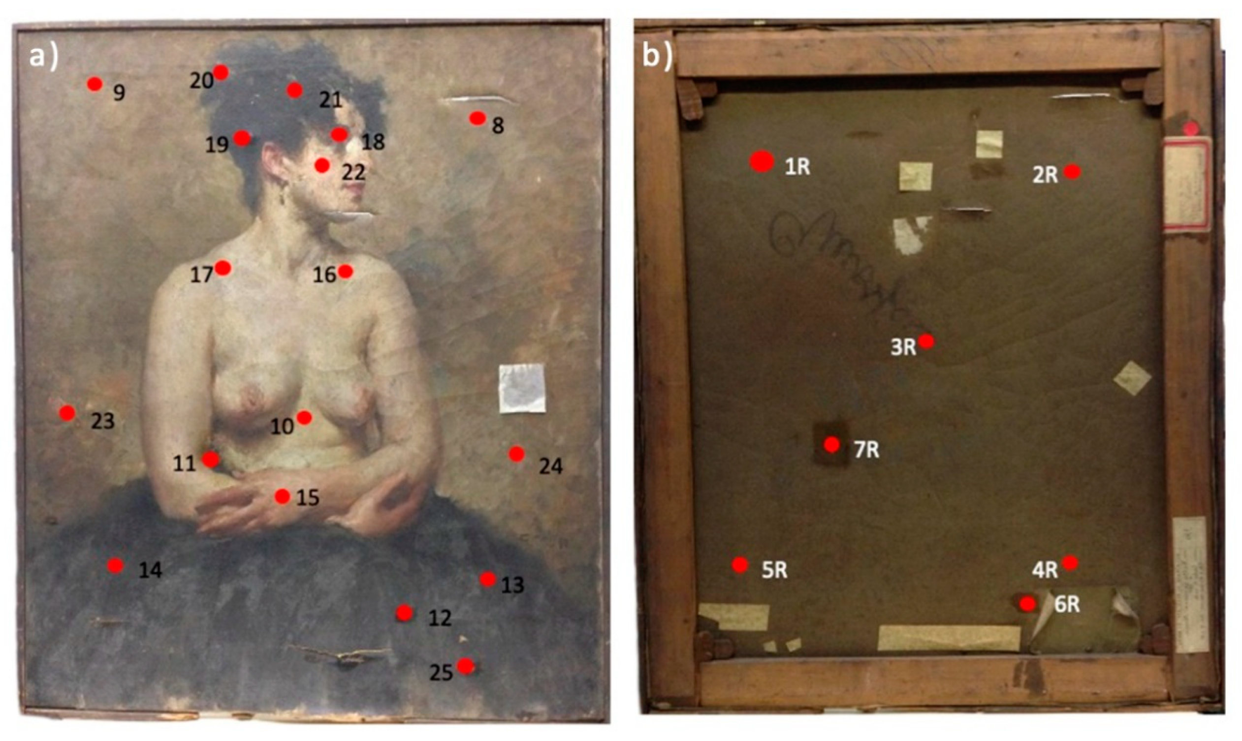
2.1. Artwork Description and Conservation State

The painting which is the object of the study is an oil on canvas, entitled “Studio di Nudo”, painted in 1921 by the Italian painter Giovanni Maria Mossa (1896, Tempio Pausania, Sardinia—1982, Pianico, Lombardy). With this portrait, the young artist won the Hayez Prize that allowed him to attend the “Accademia delle Belle Arti” di Brera in Milan. The painting represents a dancer, sitting frontally but with her face turned in profile. The woman is wearing a dark tutu that covers the lower part of her body. The figure stands out against a dark background, without a spatial perspective. Stylistically, even if the work belongs to the author’s youthful period, the characteristics of the painter are recognizable, but it is possible to find uncertainties in the drawing of the body and in the disproportion of the neck and arms compared to the rest of the body.
According to the claims by the conservators working with this painting, the canvas showed a microbial attack resulting in numerous discolorations both on the reverse, where brown dot-like areas are clearly visible, and on the obverse sides, visible especially in the areas of the body skin, due to the chromatic contrast with the pigment. The canvas surface also shows signs of laceration, dabbed during previous restorations with glue.

2.2. Sampling Method

Samples were collected by using a sterile nitrocellulose membrane (Sartorius AG, Gottingen, Germany—17.34 cm2): 18 sampling points were identified from the obverse side of the canvas, in correspondence with visible biological attack (Figure 1a): four on the background (sampling points 8F, 9F, 23F, 24F), 2 on hair (20F, 21F), 2 on the face (18F, 22F), 4 on rosy parts (10F, 15F, 16F, 17F), 2 on drapery (13F, 14F), and 4 on glue (11F, 12F, 19F, 25F). Seven sampling points were collected from the reverse side as well (Figure 1b): four samples were taken in correspondence with the lower-external edges of the canvas (1R, 2R, 4R, 5R) a central one (3R), and two in correspondence with the glue used to dab the tears in the canvas (6R, 7R).
The membranes were placed on the surface to be sampled, giving a homogeneous pressure for thirty seconds, with a sterile cotton swab [23]. Each area was sampled twice, and each nitrocellulose membrane was transferred into petri dishes with either of two different media: Potato Dextrose Agar (PDA; 4 g/L potato extract, 20 g/L glucose, 15 g/L agar) for fungi cultivation; Nutrient Broth Agar (NB; 10 g/L peptone, 5 g/L sodium chloride, 10 g/L beef extract, 15 g/L agar) for bacteria cultivation, with the addition of cycloheximide (50 µg mL−1) to prevent fungal growth.

2.3. Isolation and Identification of Microbial Community (Fungi and Bacteria)

Petri dishes inoculated with nitrocellulose membranes were incubated under sterile conditions at 30 °C: bacterial and fungal growth was followed up to 7 and 20 days, respectively. For each sampling point, results are expressed as CFU/dm2. On the basis of macroscopic features, bacterial and fungal colonies were selected and transferred into new single petri dishes with a specific growth medium and incubated at 30 °C.
After a visual examination, bacterial and fungal isolates were clustered according to their morphological characteristics and an evaluation of the relative abundance of each morphological group on the painting was possible. DNA was extracted from all isolates of each cluster according to Troiano et al. [24]. Once extracted, DNA was amplified through the Polymerase Chain Reaction. 16S region was amplified for bacteria, by using 16F (5′-AGAGTTTGATCCTGGCTCAG-3′) and 16R (5′-CTACGGCTACCTTGTTACGA-3′) primers, and a chemical protocol according to Rizzi et al. [25]. The thermal protocol provides for one cycle at 94 °C for 4 min, 35 cycles at 94 °C for 45 s, at 55 °C for 1 min and at 72 °C for 2 min, a second cycle at 72 °C for 10 min and a last cycle at 12 °C constant until samples removal. The end volume of the reaction was 50 μL, 48 μL of mix solution and 2 μL of DNA from each sample. ITS fungal region was amplified using ITS1F (5′-CTTGGTCATTTAGAGGAAGTAA-3′) and ITS4 (5′-TCCTCCGCTTATTGATATGC-3′) primers and chemical protocol as follows: buffer 1X, magnesium chloride (MgCl2) 1.5 mM, normal deoxynucleotides triphosphate (dNTP) mix 0.12 mM, 16F 0.30 μM, 16R 0.30 μM, thermostable DNA polymerase (Taq) 1 U. The thermic protocol was conducted according to Manter et al. [26]. The end volume of reaction was 1 μL of DNA in 49 μL of Master Mix buffer. PCR products were sequenced by Macrogen Inc. (Seoul, Korea) and then analyzed through BLASTn software [27] and Classifier Ribosomal Database Project [28] for bacteria, while for fungi Mycobank software [29] was used (accessed in May 2019). The 16S rRNA genes and fungal ITS sequences were deposited in the European Nucleotide Archive (ENA) under the unique accession numbers ERZ1667197 and ERZ1667410, respectively, and were registered under the study PRJEB40902.

2.4. Essential Oils

The essential oils used in this study were oregano (Origanum vulgare L., 1753, Fitomedical) and clove (Syzygium aromaticum (L.) Merr. et L.M. Perry, Fitomedical, Binasco, Italy). EOs were used diluted with ethanol 70% in the ratio 2:1 and ethanol 70% (EtOH) was used as a negative control as suggested in the research by Borrego et al. 2012 [15].

2.4.1. Contact Test with Essential Oils

One bacterial or fungal strain for each morphological cluster was inoculated in Petri dishes containing NB and PDA respectively. Three filter paper discs (47 mm) were placed on each plate after being soaked with 10 μL of oregano EO or clove EO or 70% ethanol, the latter used as a negative control. The test was performed in duplicate. Plates were incubated at 30 °C until the growth was visible. After growth, the inhibition halo around each bacterial colony was measured and the standard deviation was calculated. Since for fungi an inhibition halo radius was difficult to measure, it was observed by naked eye and recorded using the following scale: [+] for medium inhibition (approximately 0.2–1 cm), [++] for high inhibition (approximately 1–2 cm), [+++] for total inhibition and [–] for no inhibition.

2.4.2. Contactless Test with Essential Oils

In order to evaluate the inhibition properties exclusively of the EOs volatile components, a contactless experiment was conducted. One fungal or bacterial strain for each morphological cluster was plated on PDA and NBA respectively. The test was performed in duplicate. Plates were overturned and 100 μL of oregano, 100 μL of cloves, and 100 μL of 70% ethanol, used as a negative control, were placed on the lids of petri dishes separately. The plates were incubated in reverse at 30 °C. After growth an inhibition halo was measured for bacterial strains, whereas for fungal strains the inhibited area was evaluated by naked eye using the following scale: [+] for medium inhibition (approximately one third of the petri dish), [++] for high inhibition (approximately the half of the petri dish), and [+++] for total inhibition. Regarding the inhibition scale degrees, see Table S1.

3. Results and Discussion

Modern oil paintings have a complex composition, with a mixture of inorganic and organic materials [30]. For these reasons, the choice of the best method for controlling painting biodeterioration is critical for successfully treating contaminated artworks. Biocidal treatments are usually employed for controlling microbial growth and their efficacy as well as their drawbacks have been widely discussed in literature [3]. One of the well-known drawbacks is the difficulty in applying antimicrobial compounds without damaging the pictorial layers [31,32]. However, natural products, such as essential oils and plant derivatives, due to their biocidal activity, represent a useful tool in the control of biodeterioration of cultural heritage, without negative environmental and human impacts.
Previous studies reported that the major components of oregano EO are the terpenes thymol and carvacrol [33], whereas the principal component of clove EO is eugenol [34]. The antimicrobial activity of oregano is mainly due to the presence of thymol and carvacrol. Actually, this activity is based on the molecular hydrophobicity of terpenes which promotes the partition of the EOs in the lipids of the cell membrane, leading to membrane permeabilization and leakage of cytoplasmic content [35]. Furthermore, thymol acts on fungi, modifying the morphology of the hyphae and causing their aggregation, with consequences on the diameter of the hyphae themselves and on the breakdown of the cell barrier [36]. Eugenol is an amphipathic hydroxyphenyl propene, active against fungi and a wide range of Gram-negative and Gram-positive bacteria [37]. The mechanisms of action are different and include changes in the morphology and disruption of the cytoplasmatic membrane, the production of Reactive Oxygen Species (ROS) and the inhibition of some essential enzymes, such as proteases, histidine carboxylases, amylases, and ATPases [37].
In this framework, the aim of the study was to determine whether oregano and clove EOs could be applied in cleaning procedures for oil canvas paintings attacked by microorganisms. For the study, an oil canvas with a visible microbial attack was selected. Once the fungi and bacteria colonizing the canvas were identified, the isolates were exposed directly and indirectly to the selected EOs.
The results showed that the biocidal effect of EOs’ volatile components were effective on the isolates, suggesting their potential for possible application in a real cleaning procedure.

3.1. Isolation and Morphological Characterization of Microbial Community (Fungi and Bacteria)

By using nitrocellulose membranes as a sampling method, coupled with cultural analysis, 62 fungi and 20 bacteria were isolated from the stained areas of the oil painting. A quantitative microbial risk assessment was evaluated via a direct count of colonies on Petri dishes. The results of the bacterial count were of 0.7 CFU/dm2 on the reverse side and 1.7 CFU/dm2 on the obverse side. The results showed a higher concentration of colonies on the obverse side of the painting, especially on sample points 15F, 17F, 19F, and 25F. As regards fungal counts, 0.14 CFU/dm2 on the reverse and 2.9 CFU/dm2 on the obverse sides were recorded. In general the microbial count on both obverse and reverse sides of the canvas case study was not particularly high [23,38]. This can be explained by the use of a non-invasive sampling technique using the nitrocellulose membrane and the possibility that not all the microbial strains might have been cultured due to the limits of the cultivation methods [39].
According to the results obtained by the cultural analysis, a greater level of bacterial and fungal contamination was observed on the obverse side of the canvas painting. This is in contrast with what is reported in the literature, where a greater microbial contamination is usually found on the reverse of the canvas [38,40]. The more consistent colonization of the obverse rather than the reverse sides could be due to the way in which the canvas has been stored over the years, probably stacked with other paintings.
In order to get an insight into the biodiversity corresponding to the stained areas of the painting, bacterial and fungal isolates were clustered according to their morphology (Table S2). Among the 13 different bacterial clusters, Clusters I and VII were the most numerous (Figure S1a). Twenty-four different fungal clusters were identified as well; among these, 12 were represented by only one isolate, while the most abundant was Cluster IV, counting 17 colonies collected on the obverse side of the painting (Figure S1b).

3.2. Molecular Characterization of Microbial Community (Fungi and Bacteria)

Molecular characterization was conducted via a culture-dependent approach in order to isolate and carry out further investigation on the microbial community present on the artwork and in particular the potential biodeteriorative microorganisms harbored.
DNA was extracted from all bacterial and fungal strains of all clusters isolated from the oil on canvas painting; they were identified by 16S rRNA gene and ITS sequencing respectively. Results are shown in Tables S3 and S4.
Bacillus was the predominant bacterial genus (clusters I, VII, and XIII). Cluster I was the most abundant, with 20% of bacterial isolates. One member of cluster I, isolated from two sampling points on the reverse side of the painting, showed 96% similarity with Bacillus genus. Another member of cluster I, isolated in correspondence with sample 6R from the reverse side of the canvas was similar to Bacillus subtilis with 100%. Bacillus spp. are Gram-positive spore-forming bacteria, ubiquitous in the environment (soil and water) and dominant over artefact surfaces [31], such as deteriorated historical paper [41,42]. Bacillus is also responsible for structural changes of carboxymethyl cellulose during biodeterioration processes [43] and it is able to produce amylase, cellulase, protease, and acids, which are known to contribute to archaeological manuscript biodeterioration [44]. Three sampling points (21F, 18F, and 22F) chosen from the obverse side of the canvas allowed 98–99% identification of isolates with Bacillus thuringiensis (cluster I), B. simplex (cluster XIII), and B. luteus (cluster VII). Cluster VII is the second most abundant cluster, with 13.80% of bacterial isolates. Bacterial stains belonging to cluster VII were isolated from sampling points 11F, 13F, 16F, 22F, and 24F. Other identified bacteria on the obverse side belong to Cellulosimicrobium, Paenibacillus, Pseudomonas, Stenotrophomonas, and Micrococcus genera, while isolates affiliated to the Xanthomonadaceae family and Streptomyces were collected from the reverse side of the canvas. Streptomyces, isolated from stained areas of the paint layer of oil paintings, have been responsible of bio-pigment production [45]. Pseudomonas sp. can cause different types of surface deterioration, such as pigmentation, efflorescence and patinas [46]. Paenibacillus genus was isolated from human and environmental samples [47]. Xanthomonas shows cellulose structure degradation activity [48]. Stenotrophomonas was isolated and identified from an oil painting on canvas, which showed visible signs of biodeterioration [49]. Gram-positive Micrococcus luteus is known to attack cellulosic materials by lytic enzymes and pigmented components [50]. Pichia occidentalis can be used for biological detoxification of lignocellulosic hydrolysate, because it can degrade volatile fatty acids [51]. A Phaeosphaeriaceae species was isolated and identified on aged oil sludge-contaminated soil [52].
Among the fungi, sequence analysis for ITS fragments revealed that the isolates exhibited 99–100% similarity to three main genera, among which the Penicillium genus was the most abundant.
Sequences retrieved from sampling points on the obverse side of the canvas showed 99–100% similarity with Penicillium sp. According to the literature, Penicillium is associated with poorly ventilated, moist environments, representing a risk for human health and cultural heritage [39]. Several studies reported the enzymatic activity in biodeterioration of archaeological documents, and cultural heritage in general [53]. Penicillium chrysogenum, a member of the most abundant cluster II, was identified from sampling points on both obverse and reverse sides and it is known for its ability to deteriorate cellulose and lignin [54]. The majority of the isolates showed affiliation to Cephaloteca foveolata and to Aspergillus versicolor, Aspergillus insuetus, and other Aspergillus spp. and to Cladosporium sp., Alternaria sp., Pichia occidentalis, and Phaeosphaeriaceae sp. Cephalotheca foveolata is known as a human pathogen, responsible for skin infection [55]. According to literature, Aspergillus is accounted as one of the most commonly occurring fungal genera recorded on canvas paintings [39]. Together with Alternaria alternata, A. niger is reported as the most common fungal species detected on oil paintings and artworks, often the two species are isolated on the same objects [15,39,56,57]. Cladosporium, Chaetomium and Alternaria are cellulose degraders, commonly present on biodeteriorated oil paintings and cellulosic materials, as are some members of the Trichocomaceae family [23,46,56], while Pichia is a yeast genus able to ferment sugar and assimilate nitrates [58]. Sequencing did not give reliable results for some bacterial and fungal clusters: although they were not identified, one member of each non-identified cluster was tested in contact and contactless tests.

3.3. Assessment for Antifungal and Antibacterial Activity of EOs in Contact Tests

The results of the antimicrobial activity of oregano and clove extracts in contact tests revealed that the two oils had an inhibitory effect against the growth of all bacteria and almost all fungal clusters (23 out of 24) (Table 1).
In particular, fungi belonging to Penicillium chrysogenum, Cephaloteca foveolata, Cephaloteca sp., Aspergillus versicolor, and Chaetomiaceae were highly inhibited in the presence of oregano EO (indicated with “++”), while a mild effect (indicated with “+”) was observed for Aspergillus sp., Cladosporium parahalotolerans, Penicillium sp., and strains of the families Phaeosphaeriaceae and Trichocomaceae. Clove EO exhibited a high inhibitory activity on Penicillium chrysogenum, Cephaloteca foveolata, Cephaloteca sp., Aspergillus versicolor, and Chaetomiaceae and a medium inhibition on the other clusters (Table 1). A previous work reported the antifungal activity exhibited by clove and garlic oils against different fungal species including A. niger [59]. Camphor and clove EO showed antifungal activity against A. niger and A. alternata in agar plate tests as well as in simulated canvas painting models [20]. Previous studies showed the antifungal properties of different EOs, among which O. vulgare was active against several fungal species including A. niger and A. ochraceus by means of micro-, macro-dilution, and micro-atmosphere methods [18,55]. All the bacterial strains were inhibited by both EOs (Table 1). The minimum inhibition halo (0.3 ± 0.1 cm and 0.2 cm for oregano and clove, respectively) was recorded for bacteria identified as Pseudomonas psychrotolerans (Cluster VII). On the other hand, the maximum inhibition halo in the presence of oregano was measured for isolates grouped in Cluster XIII (2 cm), which showed similarity with B. simplex and in Cluster XII, affiliated with Paenibacillus sp. in the presence of clove (0.9 ± 0.2 cm). These data are in line with previous works which highlighted the inhibitory effect of EOs, and the concentration of EOs required for growth inhibition [60].

3.4. Assessment for Antifungal and Antibacterial Activity in the Contactless Test

In order to develop an application method able to prove the remote effect of EOs on microorganisms, avoiding direct contact with the pigments of paintings, a preliminary in vitro contactless test was designed in this work. Since the oregano EO gave the most promising results with the contact test, it was chosen for the contactless test. In this test microorganisms, grown on agar plates, come in contact solely with the volatile components of the EO. In detail, once the microorganisms were plated on the growth medium, a drop of the essence was placed on the lid of the plate, and the plate was turned over and incubated under sterile conditions. The result of the test is shown in Table 2.
In the test, a total inhibition effect was exhibited by oregano EO on bacterial clusters II (Xanthomonadaceae), IX (Cellulosimicrobium cellulans), and XII (Paenibacillus sp.). Moreover, if compared with the inhibition halo obtained with the contact test, oregano showed a higher effect in contactless tests both on fungi and bacteria (Table 1 and Table 2). Even if a comparison between the two tests cannot be done because of the use of different EOs’ quantities (10 μL in the contact test and 100 μL in the contactless test), the results obtained provide clues for the sole utilization of the volatile components in control practices. Previous work confirmed the antimicrobial efficacy of EOs’ vapor phase for disinfection of textiles, reporting no changes in terms of structural parameters of the object [12,16]. However, although these data provided support to the plethora of promising uses and properties exhibited by EOs, they did not avoid the procedure of direct contact of the oils with the surface of the object under study.

4. Conclusions

The research gave insight into the possibility of extending the use of EOs in the conservation of oil paintings. Indeed, the trial presented in this work was planned not with the purpose of the proper selection of suitable EOs related to the microorganisms isolated from an oil painting, but to lay the groundwork for the development of new control practices which would be suited for this kind of artwork.
In conservation, the main limit in the use of plant derivates is the methodology of application, which usually implies the direct contact of EOs with the artwork surfaces. This may cause problems due to the unpredictable reaction with pigments (and other substances used in making paintings) and EOs, such as the potential solvent effect. For this reason, here we tested oregano and cloves EOs, already reported in the literature for their inhibitory activities, focusing the attention solely on the volatile components of EOs, avoiding direct contact with pigments.
The results obtained in the present work proved that the oregano and clove EOs, and in particular the volatile components of oregano EO, are able to inhibit the growth of potential oil paintings’ biodeteriogens. We hypothesize that an effective method for EOs application could be to flow a thin film of EOs onto an evaporating surface and to place it close to the painting using some supports, so that the vapors of the EOs may homogeneously reach the painting surface, therefore avoiding direct contact of EOs with the pigments. With the aim of maximizing the antimicrobial effect of the EOs, we suggest treating the painting in a confined area, such as a display case, in order to have a saturating effect. Future studies will be devoted to verifying if this method is effectively color respectful and to establish the role of other parameters such as the minimum inhibitory concentration, exposure to light, temperature, the treatment period and modality in a real case study.

Supplementary Materials

The following are available online at https://www.mdpi.com/2076-3417/11/1/78/s1, Table S1: Inhibition scale degree; Table S2: Clusterization of bacterial isolates according to their morphology; Table S3: Clusterization of fungi isolates according to their morphology; Table S4: Clustering of bacterial strains isolated from canvas painting and their phylogenetic identification by 16S rRNA gene sequencing; Table S5: Clusterization of fungal strains isolated from canvas painting and their phylogenetic identification by ITS gene sequencing; Figure S1: Clusterization and relative abundance of bacterial (a) and fungal (b) isolates.

Author Contributions

Conceptualization: A.B.; funding acquisition: A.B.; investigation: L.G.; methodology: V.V.; project administration: A.B.; supervision: A.B. and V.V.; validation: F.C.; visualization: F.T.; writing—original draft: L.G. and V.V.; writing—review and editing: F.T. All authors have read and agreed to the published version of the manuscript.

Funding

The APC was funded by Università degli Studi di Milano, PIANO DI SOSTEGNO ALLA RICERCA 2015–2017—TRANSITION GRANT LINEA 1A PROGETTO “UNIMI PARTENARIATI H2020”, coordinator Francesca Cappitelli.

Institutional Review Board Statement

Not applicable.

Informed Consent Statement

Not applicable.

Data Availability Statement

Data available in a publicly accessible repository.

Acknowledgments

We thank Francesca Mapelli for the assistance in the submission of phylogenetic sequences. The authors also thank Chiara Nenci for her help during the sampling campaigns and for making available the painting history report.

Conflicts of Interest

The authors declare no conflict of interest.

References

  1. Casiglia, S.; Bruno, M.; Senatore, F. Activity against Microorganisms Affecting Cellulosic Objects of the Volatile Constituents of Leonotis nepetaefolia from Nicaragua. Nat. Prod. Commun. 2014, 9, 1637–1639. [Google Scholar] [CrossRef] [Green Version]
  2. Barresi, G.; Di Carlo, E.; Trapani, M.R.; Parisi, M.G.; Chille, C.; Mule, M.F.; Cammarata, M.; Palla, F. Marine organisms as source of bioactive molecules applied in restoration projects. Herit. Sci. 2015, 3, 15–18. [Google Scholar] [CrossRef] [Green Version]
  3. Fidanza, M.R.; Caneva, G. Natural biocides for the conservation of stone cultural heritage: A review. J. Cult. Herit. 2019, 38, 271–286. [Google Scholar] [CrossRef]
  4. Raut, J.S.; Karuppayil, S.M. A status review on the medicinal properties of essential oils. Ind. Crop. Prod. 2014, 62, 250–264. [Google Scholar] [CrossRef]
  5. Zoubi, Y.E.; Farah, A.; Zaroual, H.; El Ouali, A. Antimicrobial activity of Lavandula stoechas phenolic extracts against pathogenic bacteria isolated from a hospital in Morocco. Vegetos Int. J. Plant Res. 2020, 33, 703–711. [Google Scholar] [CrossRef]
  6. Ríos, J.L.; Recio, M.C. Medicinal plants and antimicrobial activity. J. Ethnopharmacol. 2005, 100, 80–84. [Google Scholar] [CrossRef]
  7. Chouhan, S.; Sharma, K.; Guleria, S. Antimicrobial Activity of Some Essential Oils-Present Status and Future Perspectives. Medicines 2017, 4, 58. [Google Scholar] [CrossRef] [Green Version]
  8. Predoi, D.; Iconaru, S.L.; Buton, N.; Badea, M.L.; Măruţescu, L. Antimicrobial Activity of New Materials Based on Lavender and Basil Essential Oils and Hydroxyapatite. Nanomaterials 2018, 8, 291. [Google Scholar] [CrossRef] [Green Version]
  9. Xie, Y.; Wang, Z.; Huang, Q.; Zhang, D. Antifungal activity of several essential oils and major components against wood-rot fungi. Ind. Crop. Prod. 2017, 108, 278–285. [Google Scholar] [CrossRef]
  10. Romano, I.; Granata, G.; Poli, A.; Finore, I.; Napoli, E.; Geraci, C. Inhibition of bacterial growth on marble stone of 18th century by treatment of nanoencapsulated essential oils. Int. Biodeterior. Biodegrad. 2020, 148, 104909. [Google Scholar] [CrossRef]
  11. Rotolo, V.; Barresi, G.; Di Carlo, E.; Giordano, A.; Lombardo, G.; Crimi, E.; Costa, E.; Bruno, M.; Palla, F. Plant extracts as green potential strategies to control the biodeterioration of cultural heritage. Int. J. Conserv. Sci. 2016, 7, 839–846. [Google Scholar]
  12. Jugreet, B.S.; Suroowan, S.; Rengasamy, R.K.; Mahomoodally, M.F. Chemistry, bioactivities, mode of action and industrial applications of essential oils. Trends Food Sci. Technol. 2020, 101, 89–105. [Google Scholar] [CrossRef]
  13. Campanella, L.; Angeloni, R.; Cibin, F.; Dell’Aglio, E.; Grimaldi, F.; Reale, R.; Vitali, M. Capsulated essential oil in gel spheres for the protection of cellulosic cultural heritage. Nat. Prod. Res. 2019, 1–8. [Google Scholar] [CrossRef]
  14. Čabalová, I.; Češek, B.; Mikala, O.; Gojný, J.; Kačík, F.; Tribulová, T. The influence of selected efficient compounds of essential oils for paper protection. J. Cult. Herit. 2019, 37, 148–154. [Google Scholar] [CrossRef]
  15. Borrego, S.; Valdés, O.; Vivar, I.; Lavin, P.; Guiamet, P.; Battistoni, P.; De Saravia, S.G.; Borges, P. Essential Oils of Plants as Biocides against Microorganisms Isolated from Cuban and Argentine Documentary Heritage. ISRN Microbiol. 2012, 2012, 1–7. [Google Scholar] [CrossRef] [PubMed] [Green Version]
  16. Matusiak, K.; MacHnowski, W.; Wrzosek, H.; Polak, J.; Rajkowska, K.; Śmigielski, K.; Kunicka-Styczyńska, A.; Gutarowska, B. Application of Cinnamomum zeylanicum essential oil in vapour phase for heritage textiles disinfection. Int. Biodeterior. Biodegrad. 2018, 131, 88–96. [Google Scholar] [CrossRef]
  17. Geweely, N.; Afifi, H.; Ibrahim, D.; Soliman, M. Inhibitory Effect of Essential Oils on Growth and Physiological Activity of Deteriorated Fungal Species Isolated from Three Archeological Objects, Saqqara excavation, Egypt. Geomicrobiol. J. 2020, 37, 520–533. [Google Scholar] [CrossRef]
  18. Stupar, M.; Grbić, M.L.; Džamić, A.; Unković, N.; Ristić, M.; Jelikić, A.; Vukojević, J. Antifungal activity of selected essential oils and biocide benzalkonium chloride against the fungi isolated from cultural heritage objects. S. Afr. J. Bot. 2014, 93, 118–124. [Google Scholar] [CrossRef]
  19. Di Vito, M.; Bellardi, M.G.; Violi, M. Gli oli essenziali entrano nei musei. Natural 2018, 1, 69–73. [Google Scholar]
  20. Elsayed, Y.; Shabana, Y. The effect of some essential oils on Aspergillus niger and Alternaria alternata infestation in archaeological oil paintings. Mediterr. Archaeol. Archaeom. 2018, 18, 71–87. [Google Scholar]
  21. Garti, N.; Yaghmur, A.; Leser, M.E.; Clement, V.; Watzke, H.J. Improved Oil Solubilization in Oil/Water Food Grade Microemulsions in the Presence of Polyols and Ethanol. J. Agric. Food Chem. 2001, 49, 2552–2562. [Google Scholar] [CrossRef]
  22. Tokuoka, Y.; Uchiyama, H.; Abe, M.; Christian, S.D. Solubilization of Some Synthetic Perfumes by Anionic-Nonionic Mixed Surfactant Systems. 1. Langmuir 1995, 11, 725–729. [Google Scholar] [CrossRef]
  23. Okpalanozie, O.E.; Adebusoye, S.A.; Troiano, F.; Polo, A.; Cappitelli, F.; Ilori, M. O Evaluating the microbiological risk to a contemporary Nigerian painting: Molecular and biodegradative studies. Int. Biodeter. Biodegr. 2016, 114, 184–192. [Google Scholar] [CrossRef]
  24. Troiano, F.; Polo, A.; Villa, F.; Cappitelli, F. Assessing the microbiological risk to stored sixteenth century parchment manuscripts: A holistic approach based on molecular and environmental studies. Biofouling 2014, 30, 299–311. [Google Scholar] [CrossRef] [PubMed]
  25. Rizzi, E.; Lari, M.; Gigli, E.; De Bellis, G.; Caramelli, D. Ancient DNA studies: New perspectives on old samples. Genet. Sel. Evol. 2012, 44, 21. [Google Scholar] [CrossRef] [Green Version]
  26. Manter, D.K.; Vivanco, J.M. Use of the ITS primers, ITS1F and ITS4, to characterize fungal abundance and diversity in mixed-template samples by qPCR and length heterogeneity analysis. J. Microbiol. Methods 2007, 71, 7–14. [Google Scholar] [CrossRef]
  27. Available online: https://blast.ncbi.nlm.nih.gov/Blast.cgi (accessed on 9 November 2020).
  28. Available online: http://rdp.cme.msu.edu/classifier/classifier.jsp (accessed on 9 November 2020).
  29. MycoBank. Available online: http://www.mycobank.org (accessed on 9 November 2020).
  30. Bonaduce, I.; Duce, C.; Lluveras-Tenorio, A.; Lee, J.; Ormsby, B.; Burnstock, A.; Berg, K.J.V.D. Conservation Issues of Modern Oil Paintings: A Molecular Model on Paint Curing. Accounts Chem. Res. 2019, 52, 3397–3406. [Google Scholar] [CrossRef]
  31. Poyatos, F.; Morales, F.; Nicholson, A.W.; Giordano, A. Physiology of biodeterioration on canvas paintings. J. Cell. Physiol. 2018, 233, 2741–2751. [Google Scholar] [CrossRef]
  32. Kakakhel, M.A.; Wu, F.; Gu, J.-D.; Feng, H.; Shah, K.; Wang, W. Controlling biodeterioration of cultural heritage objects with biocides: A review. Int. Biodeterior. Biodegrad. 2019, 143, 104721. [Google Scholar] [CrossRef]
  33. Leyva-López, N.; Gutiérrez-Grijalva, E.P.; Vazquez-Olivo, G.; Heredia, J.B. Essential Oils of Oregano: Biological Activity beyond Their Antimicrobial Properties. Molecules 2017, 22, 989. [Google Scholar] [CrossRef] [Green Version]
  34. Pramod, K.; Ansari, S.H.; Ali, J. Eugenol: A Natural Compound with Versatile Pharmacological Actions. Nat. Prod. Commun. 2010, 5, 1999–2006. [Google Scholar] [CrossRef] [PubMed] [Green Version]
  35. Mauriello, E.; Ferrari, G.; Donsì, F. Effect of formulation on properties, stability, carvacrol release and antimicrobial activity of carvacrol emulsions. Colloids Surf. B Biointerfaces 2021, 197, 111424. [Google Scholar] [CrossRef] [PubMed]
  36. Klarić, M. Šegvić; Kosalec, I.; Mastelić, J.; Piecková, E.; Pepeljnak, S. Antifungal activity of thyme (Thymus vulgaris L.) essential oil and thymol against moulds from damp dwellings. Lett. Appl. Microbiol. 2007, 44, 36–42. [Google Scholar] [CrossRef]
  37. Marchese, A.; Barbieri, R.; Coppo, E.; Orhan, I.E.; Daglia, M.; Nabavi, S.M.; Izadi, M.; Abdollahi, M.; Ajami, M. Antimicrobial activity of eugenol and essential oils containing eugenol: A mechanistic viewpoint. Crit. Rev. Microbiol. 2017, 43, 668–689. [Google Scholar] [CrossRef] [PubMed]
  38. Capodicasa, S.; Fedi, S.; Porcelli, A.M.; Zannoni, D. The microbial community dwelling on a biodeteriorated 16th century painting. Int. Biodeterior. Biodegrad. 2010, 64, 727–733. [Google Scholar] [CrossRef]
  39. Pasquarella, C.; Balocco, C.; Saccani, E.; Capobianco, E.; Viani, I.; Veronesi, L.; Pavani, F.; Pasquariello, G.; Rotolo, V.; Palla, F.; et al. Biological and microclimatic monitoring for conservation of cultural heritage: A case study at the De Rossi room of the Palatina library in Parma. Aerobiologia 2019, 36, 105–111. [Google Scholar] [CrossRef]
  40. Montanari, M.; Iotti, M.; Innocenti, G. Isolamento, identificazione e attività cellulosolitica di microrganismi fungini associati al biodeterioramento di un dipinto su tela del XIXesimo secolo. Micol. Ital. 2009, 3, 19–24. [Google Scholar]
  41. Lech, T. Evaluation of a Parchment Document, the 13th Century Incorporation Charter for the City of Krakow, Poland, for Microbial Hazards. Appl. Environ. Microbiol. 2016, 82, 2620–2631. [Google Scholar] [CrossRef] [Green Version]
  42. Kumar, S.; Priyanka; Kumar, U. Microbial Biotechnology Approaches to Monuments of Cultural Heritage; Springer Science and Business Media LLC: Singapore, 2020; pp. 1–12. [Google Scholar]
  43. Orehek, J.; Dogsa, I.; Tomšič, M.; Jamnik, A.; Kočar, D.; Stopar, D. Structural investigation of carboxymethyl cellulose biodeterioration by Bacillus subtilis subsp. subtilis NCIB 3610. Int. Biodeterior. Biodegrad. 2013, 77, 10–17. [Google Scholar] [CrossRef]
  44. Fouda, A.; Abdel-Maksoud, G.; Abdel-Rahman, M.A.; Salem, S.S.; Hassan, S.E.-D.; El-Sadany, M.A.-H. Eco-friendly approach utilizing green synthesized nanoparticles for paper conservation against microbes involved in biodeterioration of archaeological manuscript. Int. Biodeterior. Biodegrad. 2019, 142, 160–169. [Google Scholar] [CrossRef]
  45. Sakr, A.A.; Ghaly, M.F.; Edwards, H.G.M.; Ali, M.F.; Abdel-Haliem, M.E.F. Involvement of Streptomyces in the Deterioration of Cultural Heritage Materials Through Biomineralization and Bio-Pigment Production Pathways: A Review. Geomicrobiol. J. 2020, 37, 653–662. [Google Scholar] [CrossRef]
  46. Mazzoli, R.; Giuffrida, M.G.; Pessione, E. Back to the past: Find the guilty bug—Microorganisms involved in the biodeterioration of archeological and historical artifacts. Appl. Microbiol. Biotechnol. 2018, 102, 6393–6407. [Google Scholar] [CrossRef] [PubMed]
  47. Sáez-Nieto, J.; Medina-Pascual, M.; Carrasco, G.; Garrido, N.; Fernandez-Torres, M.; Villalón, P.; Valdezate, S. Paenibacillus spp. isolated from human and environmental samples in Spain: Detection of 11 new species. New Microbes New Infect. 2017, 19, 19–27. [Google Scholar] [CrossRef] [PubMed]
  48. Cao, J.; Chu, C.; Zhang, M.; He, L.; Qin, L.; Li, X.; Yuan, M. Different Cell Wall-Degradation Ability Leads to Tissue-Specificity between Xanthomonas oryzae pv. oryzae and Xanthomonas oryzae pv. oryzicola. Pathogens 2020, 9, 187. [Google Scholar] [CrossRef] [PubMed] [Green Version]
  49. Zhgun, A.A.; Avdanina, D.; Shumikhin, K.; Simonenko, N.; Lyubavskaya, E.; Volkov, I.; Ivanov, V. Detection of potential biodeterioration risks for tempera painting in 16th century exhibits from State Tretyakov Gallery. PLoS ONE 2020, 15, e0230591. [Google Scholar] [CrossRef] [PubMed]
  50. Su, X.; Zhang, S.; Mei, R.; Zhang, Y.; Hashmi, M.Z.; Liu, J.; Lin, H.; Ding, L.; Sun, F. Resuscitation of viable but non-culturable bacteria to enhance the cellulose-degrading capability of bacterial community in composting. Microb. Biotechnol. 2018, 11, 527–536. [Google Scholar] [CrossRef] [PubMed]
  51. Marjakangas, J.M.; Lakaniemi, A.-M.; Koskinen, P.E.; Chang, J.-S.; Puhakka, J.A. Lipid production by eukaryotic microorganisms isolated from palm oil mill effluent. Biochem. Eng. J. 2015, 99, 48–54. [Google Scholar] [CrossRef]
  52. Wang, H.; Kuang, S.; Lang, Q.; Yu, W. Effects of Aged Oil Sludge on Soil Physicochemical Properties and Fungal Diversity Revealed by High-Throughput Sequencing Analysis. Archaea 2018, 2018, 1–8. [Google Scholar] [CrossRef] [Green Version]
  53. Polo, A.; Cappitelli, F.; Villa, F.; Pinzari, F. Biological invasion in the indoor environment: The spread of Eurotium halophilicum on library materials. Int. Biodeterior. Biodegrad. 2017, 118, 34–44. [Google Scholar] [CrossRef]
  54. Yang, Y.; Yang, J.; Liu, J.; Wang, R.; Liu, L.; Wang, F.; Yuan, H. The composition of accessory enzymes of Penicillium chrysogenum P33 revealed by secretome and synergistic effects with commercial cellulase on lignocellulose hydrolysis. Bioresour. Technol. 2018, 257, 54–61. [Google Scholar] [CrossRef]
  55. Tsang, C.-C.; Tang, J.Y.M.; Chan, K.-F.; Lee, C.-Y.; Chan, J.; Ngan, A.H.Y.; Cheung, M.; Lau, E.C.L.; Li, X.; Ng, R.H.Y.; et al. Diversity of phenotypically non-dermatophyte, non-Aspergillus filamentous fungi causing nail infections: Importance of accurate identification and antifungal susceptibility testing. Emerg. Microbes Infect. 2019, 8, 531–541. [Google Scholar] [CrossRef] [PubMed]
  56. Kurowski, G.; Vogt, O.; Ogonowski, J. Paint-degrading microorganisms. Czas. Tech. 2017, 2017, 81–92. [Google Scholar] [CrossRef] [Green Version]
  57. Veneranda, M.; Blanco-Zubiaguirre, L.; Roselli, G.; Di Girolami, G.; Castro, K.; Madariaga, J.M. Evaluating the exploitability of several essential oils constituents as a novel biological treatment against cultural heritage biocolonization. Microchem. J. 2018, 138, 1–6. [Google Scholar] [CrossRef]
  58. Navarrete, C.; Martínez, J.L. Non-conventional yeasts as superior production platforms for sustainable fermentation based bio-manufacturing processes. AIMS Environ. Sci. 2020, 7, 289–305. [Google Scholar] [CrossRef]
  59. Borrego, S.; Gómez de Saravia, S.; Valdés, O.; Vivar, I.; Battistoni, P.; Guiamet, P. Biocidal activity of two essential oils on fungi that cause biodeterioration of paper documents. Int. J. Conserv. Sci. 2016, 7, 369–380. [Google Scholar]
  60. Paster, N.; Juven, B.J.; Shaaya, E.; Menasherov, M.; Nitzan, R.; Weisslowicz, H.; Ravid, U. Inhibitory effect of oregano and thyme essential oils on moulds and foodborne bacteria. Lett. Appl. Microbiol. 1990, 11, 33–37. [Google Scholar] [CrossRef]
Figure 1. The artwork “Studio di nudo” and position of sampling points from observe (a) and reverse (b) sides of the canvas.
Applsci 11 00078 g001
Table 1. Inhibition halo of oregano and clove essential oils on bacteria and fungi in contact tests. Tests were performed in duplicate and standard deviation was calculated. NI = not identified strain. Ethanol 70% was used as a negative control as it did not show any inhibitory effect against the isolates.
CONTACT TEST
BACTERIAISOLATESTAXAOREGANOCLOVE
IcBacillus subtilis subsp. subtilis1.0 ± 0.2 cm0.4 ± 0.1 cm
IIaXanthomonadaceae1.1 ± 0.2 cm0.4 ± 0.1 cm
IIIaStreptomyces sp.1.0 ± 0.1 cm0.5 ± 0.1 cm
IVaNI1 ± 0 cm0.3 ± 0.2 cm
VaNI1.3 ± 0.2 cm0.4 ± 0.1 cm
VIaStenotrophomonas0.9 ± 0.1 cm0.4 ± 0.1 cm
VIIaPseudomonas psychrotolerans0.3 ± 0.1 cm0.2 ± 0 cm
VIIIaXanthomonadaceae1 ± 0 cm0.3 ± 0.2cm
IXaCellulosimicrobium cellulans0.9 ± 0.1 cm0.5 ± 0 cm
XaPenibacillaceae1.4 ± 0.1 cm0.4 ± 0.2 cm
XIaNI1.3 ± 0.2 cm0.7 ± 0.1 cm
XIIaPaenibacillus sp.1.2 ± 0 cm0.9 ± 0.2 cm
XIIIaBacillus simplex2 ± 0 cm0.6 ± 0.2 cm
FUNGIIaPenicillium chrysogenum+++
IIcNI++
IIIaPenicillium chrysogenum++++
IVbCephalotheca foveolata++++
VaAspergillus sp.++
VIaCephalotheca foveolata++++
VIIaCladosporium parahalotolerans++
VIIIaNI+++
IXaCephalotheca foveolata++++
XaAspergillus versicolor++++
XIaNI++++
XIIaNI--
XIIIaPenicillium chrysogenum++++
XIVaTrichocomaceae++
XVaChaetomiaceae++++
XVIaPenicillium chrysogenum++++
XVIIaCephalotheca foveolata++++
XVIIIaNI++
XIXaPhaeosphaeriaceae++
XXaPenicillium sp.++
XXIbNI++
XXIIbNI++
XXIIIbNI+++
XXIVaNI+++
Table 2. Inhibition halo of oregano and cloves essential oils on bacteria and fungi in contactless tests. The experiment was performed in duplicate. NI = not identified strain. Ethanol 70% was used as a negative control as it did not show any inhibitory effect against the isolates.
CONTACTLESS TEST
BACTERIAISOLATESTAXAOREGANO
IcBacillus subtilis subsp. subtilis1.7 ± 0.3 cm
IIaXanthomonadaceaeTotal inhibition
IIIaStreptomyces1.6 ± 0.4 cm
IVaNI1.6 ± 0.4 cm
VaNI1.8 ± 0.2 cm
VIaStenotrophomonas1.6 ± 0.2 cm
VIIaPseudomonas psychrotolerans1.5 ± 0.5 cm
VIIIaXanthomonadaceae1.1 ± 0.4 cm
IXaCellulosimicrobium cellulansTotal inhibition
XaPenibacillaceae1.9 ± 0.1 cm
XIaNI1.6 ± 0.2 cm
XIIaPaenibacillus sp.Total inhibition
XIIIaBacillus simplex2 ± 0 cm
FUNGIIaPenicillium chrysogenum++
IIcNI+
IIIaPenicillium chrysogenum+++
IVbCephalotheca foveolata++
VaAspergillus sp.+++
VIaCephalotheca foveolata++
VIIaCladosporium parahalotolerans+
VIIIaNI+++
IXaCephalotheca foveolata+++
XaAspergillus versicolor+++
XIaNI++
XIIaNI+
XIIIaPenicillium chrysogenum+++
XIVaTrichocomaceae+
XVaChaetomiaceae++
XVIaPenicillium chrysogenum++
XVIIaCephalotheca foveolata+++
XVIIIaNI++
XIXaPhaeosphaeriaceae sp.+
XXaPenicillium sp.+
XXIbNI+++
XXIIbNI+
XXIIIbNI+
XXIVaNI+++
Publisher’s Note: MDPI stays neutral with regard to jurisdictional claims in published maps and institutional affiliations.

