Trichoderma Biomass as an Alternative for Removal of Congo Red and Malachite Green Industrial Dyes


Abstract
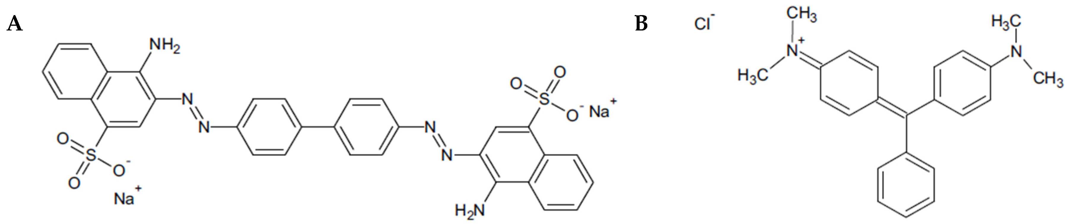
1. Introduction
2. Materials and Methods
2.1. Fresh Biomass of T. virens and T. viride
2.2. Activated Carbon and Dry Biomass of T. virens and T. viride
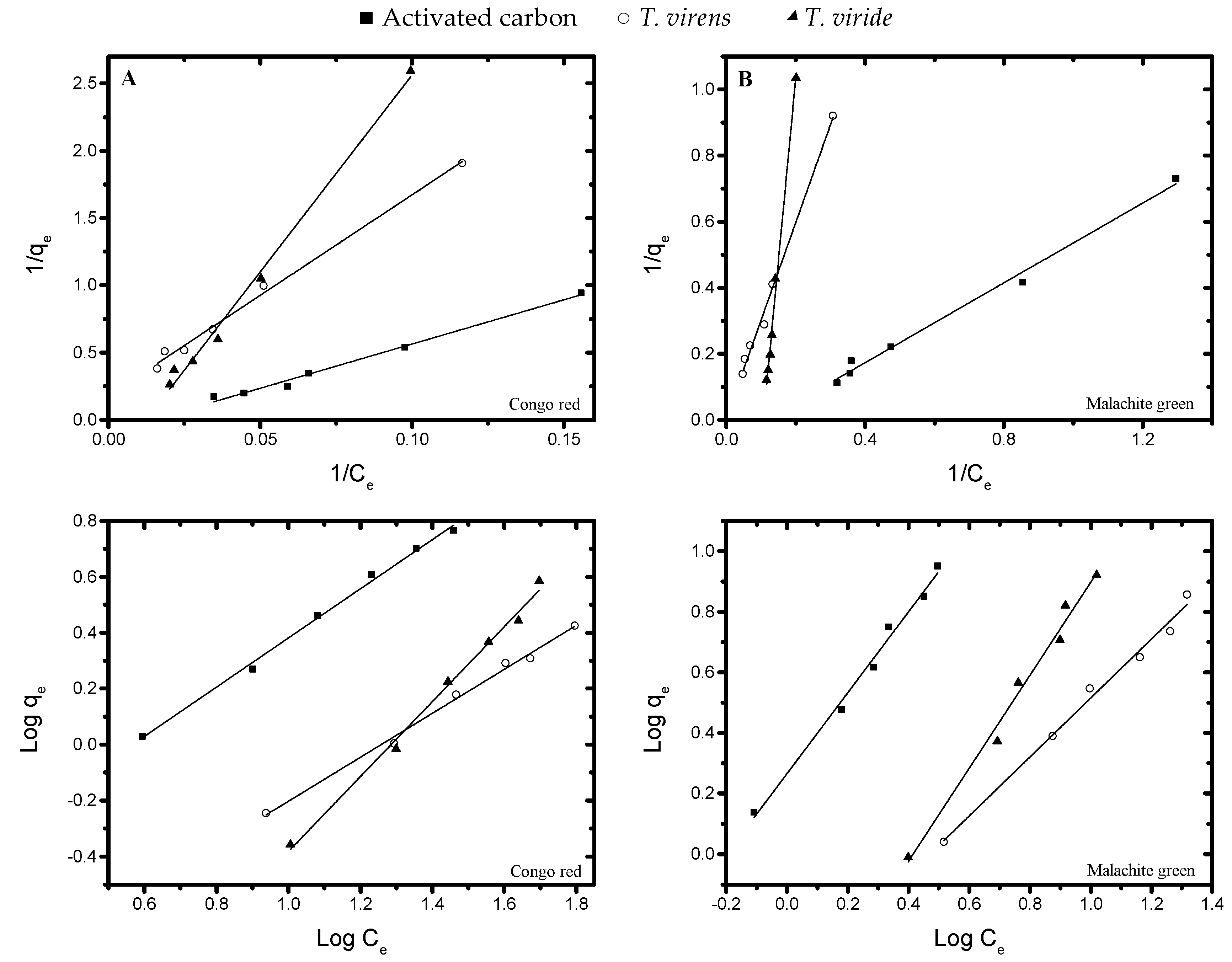
2.2.1. Equilibrium Studies
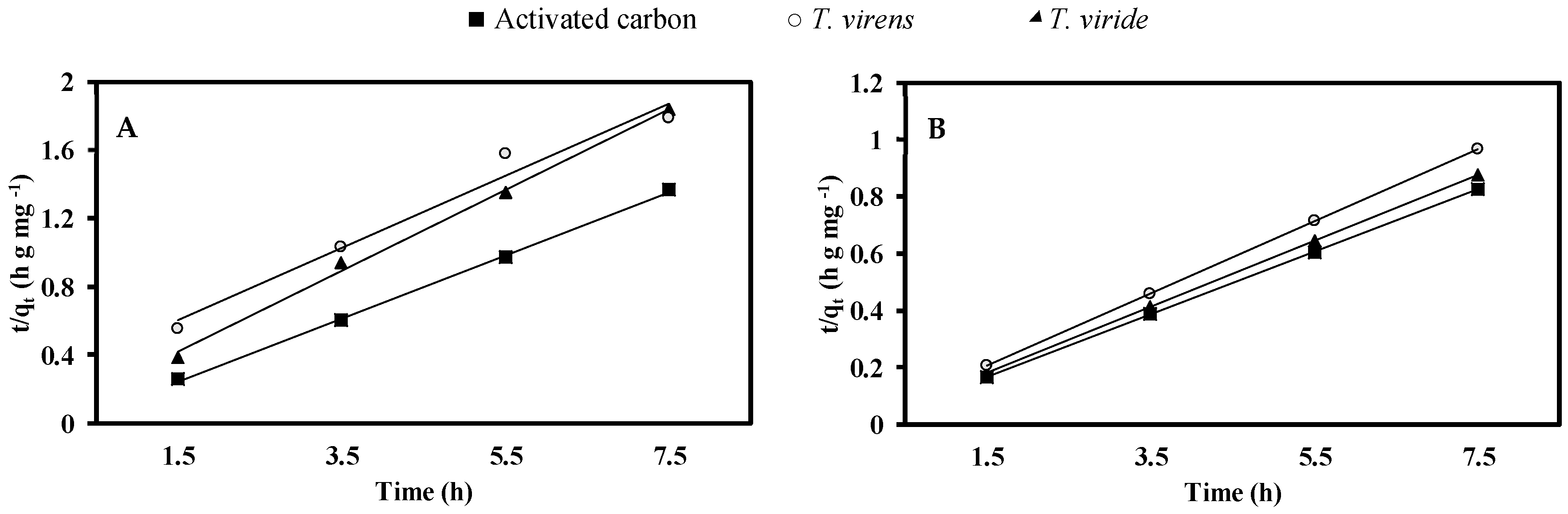
2.2.2. Kinetic Studies
2.3. Statistic Analysis
3. Results and Discussion
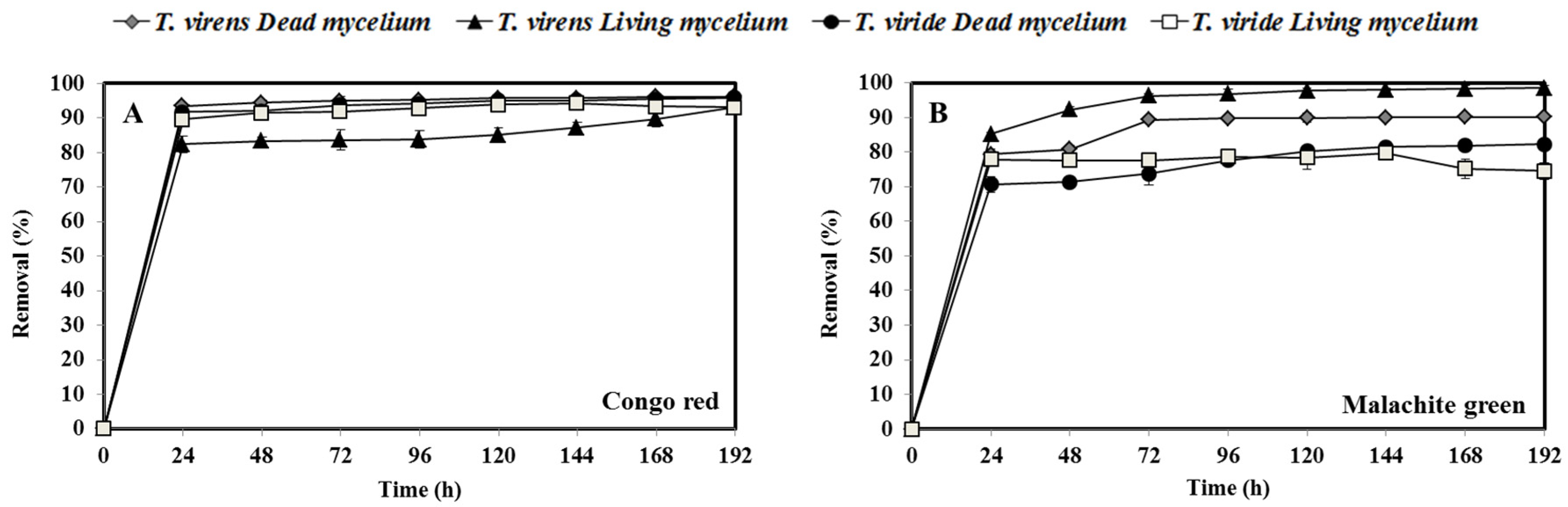
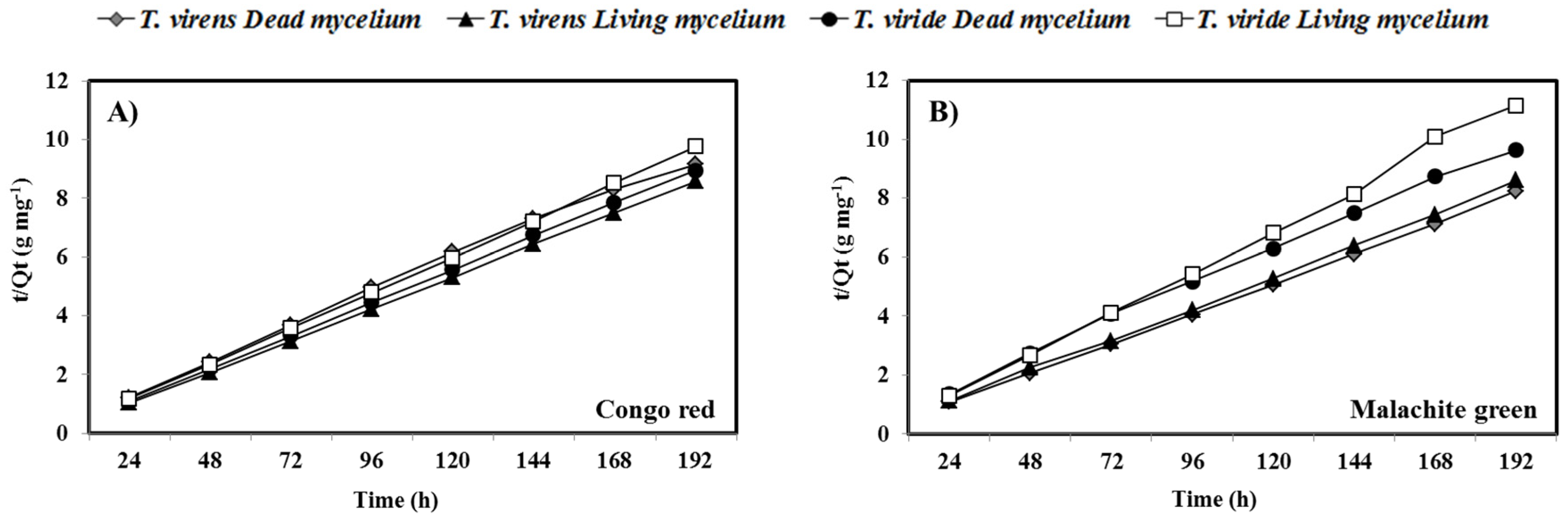
3.1. Removal of CR and MG by Fresh Biomass of T. virens and T. viride
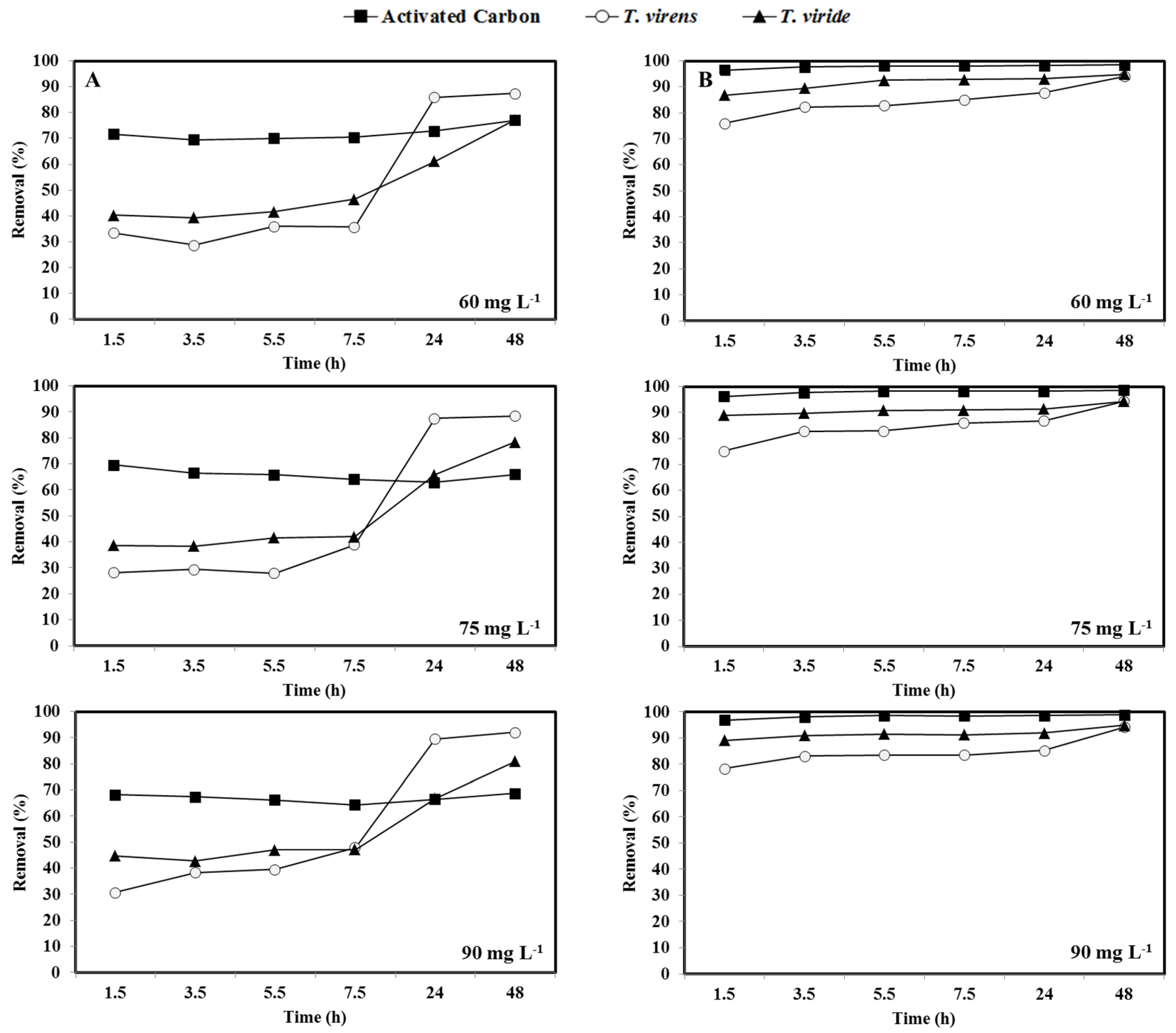
3.2. Removal of CR and MG by Activated Carbon and Dry Biomass of T. virens and T. viride
4. Conclusions
Author Contributions
Funding
Institutional Review Board Statement
Informed Consent Statement
Data Availability Statement
Acknowledgments
Conflicts of Interest
References
- Mathur, N.; Bhatnagar, P.; Sharma, P. Review of the Mutagenicity of Textile Dye Products. Univ. J. Environ. 2012, 2, 1–18. [Google Scholar]
- Shaban, M.; Abukhadra, M.R.; Shahien, M.G.; Ibrahim, S.S. Novel bentonite/zeolite-NaP composite efficiently removes methylene blue and Congo red dyes. Environ. Chem. Lett. 2018, 16, 275–280. [Google Scholar] [CrossRef]
- Kaushik, P.; Malik, A. Fungal dye decolourization: Recent advances and future potential. Environ. Int. 2009, 35, 127–141. [Google Scholar] [CrossRef] [PubMed]
- Siddiqui, S.I.; Rathi, G.; Chaudhry, S.A. Acid washed black cumin seed powder preparation for adsorption of methylene blue dye from aqueous solution: Thermodynamic, kinetic and isotherm studies. J. Mol. Liq. 2018, 264, 275–284. [Google Scholar] [CrossRef]
- Ansari, R.; Mosayebzadeh, Z. Application of polyaniline as an efficient and novel adsorbent for azo dyes removal from textile wastewaters. Chem. Pap. 2011, 65, 1–8. [Google Scholar] [CrossRef]
- Hunger, K. (Ed.) Industrial Dyes: Chemistry, Properties, Applications, 1st ed.; WILEY-VCH: Weinheim, Germany, 2003; pp. 1–12. [Google Scholar]
- Steensma, D.P. “Congo” Red Out of Africa? Arch. Pathol. Lab. Med. 2001, 125, 250–252. [Google Scholar] [PubMed]
- Jalandoni-Buan, A.C.; Lynn, A.; Decena-Soliven, A.; Cao, E.P.; Barraquio, V.L.; Barraquio, W.L. Congo Red Decolorizing Activity Under Microcosm and Decolorization of other Dyes by Congo Red Decolorizing Bacteria. Philipp. J. Sci. 2009, 138, 125–132. [Google Scholar]
- Alderman, D.J. Malachite green: A review. J. Fish. Dis. 1985, 8, 289–298. [Google Scholar] [CrossRef]
- Raval, N.P.; Shah, P.U.; Shah, N.K. Malachite green “A cationic dye” and its removal from aqueous solution by adsorption. Appl. Water Sci. 2017, 7, 3407–3445. [Google Scholar] [CrossRef]
- Knapp, J.S.; Bromley-Challenor, K.C.A. Recalcitrant organic compounds. In Handbook of Water and Wastewater Microbiology; Mara, D., Horan, N.J., Eds.; Academic: San Diego, CA, USA, 2003; pp. 559–596. [Google Scholar]
- Madsen, E.L. Biodegradability of recalcitrant aromatic compounds. In Comprehensive Biotechnology, 2nd ed.; Murray, M.Y., Ed.; Academic Press: Burlington, NJ, USA, 2011; pp. 95–103. [Google Scholar]
- Fewson, C.A. Biodegradation of xenobiotic and other persistent compounds: The causes of recalcitrance. Trends Biotechnol. 1988, 6, 148–153. [Google Scholar] [CrossRef]
- DeVito, S.C. Predicting Azo Dye Toxicity. Crit. Rev. Environ. Sci. Technol. 1993, 23, 249–324. [Google Scholar]
- Srivastava, S.; Sinha, R.; Roy, D. Toxicological effects of malachite green. Aquat. Toxicol. 2004, 66, 319–329. [Google Scholar] [CrossRef] [PubMed]
- Tara, N.; Siddiqui, S.I.; Rathi, G.; Chaudhry, S.A.; Inamuddin; Asiri, A.M. Nano-engineered Adsorbent for the Removal of Dyes from Water: A Review. Curr. Anal. Chem. 2019, 16, 14–40. [Google Scholar] [CrossRef]
- Salleh, M.A.M.; Mahmoud, D.K.; Karim, W.A.; Idris, A. Cationic and anionic dye adsorption by agricultural solid wastes: A comprehensive review. Desalination 2011, 280, 1–13. [Google Scholar] [CrossRef]
- Chanzu, H.A.; Onyari, J.M.; Shiundu, P.M. Brewers’ spent grain in adsorption of aqueous Congo Red and malachite Green dyes: Batch and continuous flow systems. J. Hazard. Mater. 2019, 380, 1–8. [Google Scholar] [CrossRef] [PubMed]
- Bhatia, D.; Sharma, N.R.; Singh, J.; Kanwar, R.S. Biological methods for textile dye removal from wastewater: A review. Crit. Rev. Environ. Sci. Technol. 2017, 47, 1836–1876. [Google Scholar] [CrossRef]
- Waghmode, T.R.; Kurade, M.B.; Sapkal, R.T.; Bhosale, C.H.; Jeon, B.H.; Govindwar, S.P. Sequential photocatalysis and biological treatment for the enhanced degradation of the persistent azo dye methyl red. J. Hazard. Mater. 2019, 371, 115–122. [Google Scholar] [CrossRef]
- Jafari, N.; Soudi, M.R.; Kasra-Kermanshahi, R. Biodegradation perspectives of azo dyes by yeasts. Microbiology 2014, 83, 484–497. [Google Scholar] [CrossRef]
- Yagub, M.T.; Sen, T.K.; Afroze, S.; Ang, H.M. Dye and its removal from aqueous solution by adsorption: A review. Adv. Colloid Interface Sci. 2014, 209, 172–184. [Google Scholar] [CrossRef]
- Katheresan, V.; Kansedo, J.; Lau, S.Y. Efficiency of various recent wastewater dye removal methods: A review. J. Environ. Chem. Eng. 2018, 6, 4676–4697. [Google Scholar] [CrossRef]
- Fomina, M.; Gadd, G.M. Biosorption: Current perspectives on concept, definition and application. Bioresour. Technol. 2014, 160, 3–14. [Google Scholar] [CrossRef] [PubMed]
- Crini, G. Non-conventional low-cost adsorbents for dye removal: A review. Bioresour. Technol. 2006, 97, 1061–1085. [Google Scholar] [CrossRef] [PubMed]
- Aksu, Z. Application of biosorption for the removal of organic pollutants: A review. Process. Biochem. 2005, 40, 997–1026. [Google Scholar] [CrossRef]
- Gadd, G.M. Biosorption: Critical review of scientific rationale, environmental importance and significance for pollution treatment. J. Chem. Technol. Biotechnol. 2009, 84, 13–28. [Google Scholar] [CrossRef]
- Park, D.; Yun, Y.S.; Park, J.M. The past, present, and future trends of biosorption. Biotechnol. Bioprocess. Eng. 2010, 15, 86–102. [Google Scholar] [CrossRef]
- Gupta, V.K.; Suhas. Application of low-cost adsorbents for dye removal—A review. J. Environ. Manag. 2009, 90, 2313–2342. [Google Scholar] [CrossRef]
- Tigini, V.; Prigione, V.; Donelli, I.; Freddi, G.; Varese, G.C. Influence of culture medium on fungal biomass composition and biosorption effectiveness. Curr. Microbiol. 2012, 64, 50–59. [Google Scholar] [CrossRef]
- Chew, S.Y.; Ting, A.S.Y. Common filamentous Trichoderma asperellum for effective removal of triphenylmethane dyes. Desalin. Water Treat. 2016, 57, 13534–13539. [Google Scholar] [CrossRef]
- Aksu, Z.; Dönmez, G. A comparative study on the biosorption characteristics of some yeasts for remazol blue reactive dye. Chemosphere 2003, 50, 1075–1083. [Google Scholar] [CrossRef]
- Kargi, F.; Ozmihci, S. Biosorption performance of powdered activated sludge for removal of different dyestuffs. Enzym. Microb. Technol. 2004, 35, 267–271. [Google Scholar] [CrossRef]
- Kapdan, I.K.; Kargia, F.; McMullan, G.; Marchant, R. Effect of environmental conditions on biological decolorization of textile dyestuff by C. versicolor. Enzym. Microb. Technol. 2000, 26, 381–387. [Google Scholar] [CrossRef]
- Cha, C.J.; Doerge, D.R.; Cerniglia, C.E. Biotransformation of Malachite Green by the Fungus Cunninghamella elegans. Appl. Environ. Microbiol. 2001, 67, 4358–4360. [Google Scholar] [CrossRef] [PubMed]
- Aksu, Z. Reactive dye bioaccumulation by Saccharomyces cerevisiae. Process. Biochem. 2003, 38, 1437–1444. [Google Scholar] [CrossRef]
- Pazarlioglu, N.K.; Urek, R.O.; Ergun, F. Biodecolourization of Direct Blue 15 by immobilized Phanerochaete chrysosporium. Process. Biochem. 2005, 40, 1923–1929. [Google Scholar] [CrossRef]
- Aksu, Z.; Çaǧatay, Ş.Ş. Investigation of biosorption of Gemazol Turquise Blue-G reactive dye by dried Rhizopus arrhizus in batch and continuous systems. Sep. Purif. Technol. 2006, 48, 24–35. [Google Scholar] [CrossRef]
- Aksu, Z.; Kiliç, N.K.; Ertuǧrul, S.; Dönmez, G. Inhibitory effects of chromium(VI) and Remazol Black B on chromium(VI) and dyestuff removals by Trametes versicolor. Enzym. Microb. Technol. 2007, 40, 1167–1174. [Google Scholar] [CrossRef]
- Matos, A.J.F.S.; Bezerra, R.M.F.; Dias, A.A. Screening of fungal isolates and properties of Ganoderma applanatum intended for olive mill wastewater decolourization and dephenolization. Lett. Appl. Microbiol. 2007, 45, 270–275. [Google Scholar] [CrossRef]
- Zhao, X.; Hardin, I.R. HPLC and spectrophotometric analysis of biodegradation of azo dyes by Pleurotus ostreatus. Dye Pigments 2007, 73, 322–325. [Google Scholar] [CrossRef]
- Sadhasivam, S.; Savitha, S.; Swaminathan, K. Redox-mediated decolorization of recalcitrant textile dyes by Trichoderma harzianum WL1 laccase. World J. Microbiol. Biotechnol. 2009, 25, 1733–1741. [Google Scholar] [CrossRef]
- Sadhasivam, S.; Savitha, S.; Swaminathan, K. Feasibility of using Trichoderma harzianum biomass for the removal of erioglaucine from aqueous solution. World J. Microbiol. Biotechnol. 2007, 23, 1075–1081. [Google Scholar] [CrossRef]
- Sadhasivam, S.; Saritha, E.; Savitha, S.; Swaminathan, K. Comparison of the efficacy of live and autoclaved mycelium of Trichoderma harzianum on the removal of trypan blue. Bull. Environ. Contam. Toxicol. 2005, 75, 1046–1053. [Google Scholar] [CrossRef] [PubMed]
- Sadhasivam, S.; Savitha, S.; Swaminathan, K. Exploitation of Trichoderma harzianum mycelial waste for the removal of rhodamine 6G from aqueous solution. J. Environ. Manag. 2007, 85, 155–161. [Google Scholar] [CrossRef]
- Sivasamy, A.; Sundarabal, N. Biosorption of an azo dye by Aspergillus niger and Trichoderma sp. fungal biomasses. Curr. Microbiol. 2011, 62, 351–357. [Google Scholar] [CrossRef] [PubMed]
- Saeed, A.; Iqbal, M.; Zafar, S.I. Immobilization of Trichoderma viride for enhanced methylene blue biosorption: Batch and column studies. J. Hazard. Mater. 2009, 168, 406–415. [Google Scholar] [CrossRef] [PubMed]
- Walsh, G.A.; Power, R.F.; Headon, D.R. Enzymes in the animal-feed industry. Trends Biotechnol. 1993, 11, 424–430. [Google Scholar] [CrossRef]
- Buchert, J.; Ranua, M.; Siika-aho, M.; Pere, J.; Viikari, L. Trichoderma reesei cellulases in the bleaching of kraft pulp. Appl. Microbiol. Biotechnol. 1994, 40, 941–945. [Google Scholar] [CrossRef]
- Rojo, F.G.; Reynoso, M.M.; Ferez, M.; Chulze, S.N.; Torres, A.M. Biological control by Trichoderma species of Fusarium solani causing peanut brown root rot under field conditions. Crop. Prot. 2007, 26, 549–555. [Google Scholar] [CrossRef]
- Zhang, C.L.; Druzhinina, I.S.; Kubicek, C.P.; Xu, T. Trichoderma biodiversity in China: Evidence for a North to South distribution of species in East Asia. FEMS Microbiol. Lett. 2005, 251, 251–257. [Google Scholar] [CrossRef]
- Karthik, V.; Saravanan, K.; Patra, C.; Ushadevi, B.; Vairam, S.; Selvaraju, N. Biosorption of Acid Yellow 12 from simulated wastewater by non-viable T. harzianum: Kinetics, isotherm and thermodynamic studies. Int. J. Environ. Sci. Technol. 2019, 16, 6895–6906. [Google Scholar] [CrossRef]
- Argumedo-Delira, R.; Alarcón, A.; Ferrera-Cerrato, R.; Almaraz, J.J.; Peña-Cabriales, J.J. Tolerance and growth of 11 Trichoderma strains to crude oil, naphthalene, phenanthrene and benzo[a]pyrene. J. Environ. Manag. 2012, 95, S291–S299. [Google Scholar] [CrossRef]
- Ibarra-Medina, V.A.; Ferrera-Cerrato, R.; Alarcón, A.; Lara-Hernández, M.E.; Valdez-Carrasco, J.M. Isolation and screening of Trichoderma strains antagonistic to Sclerotinia sclerotiorum and Sclerotinia minor. Revista Mexicana de Micología 2010, 31, 53–63. [Google Scholar]
- Wunch, K.G.; Feibelman, T.; Bennett, J.W. Screening for fungi capable of removing benzo[a]pyrene in culture. Appl. Microbiol. Biotechnol. 1997, 47, 620–624. [Google Scholar] [CrossRef]
- Ali, N.; Hameed, A.; Ahmed, S.; Khan, A.G. Decolorization of structurally different textile dyes by Aspergillus niger SA1. World J. Microbiol. Biotechnol. 2008, 24, 1067–1072. [Google Scholar] [CrossRef]
- Ramsay, J.A.; Nguyen, T. Decoloration of textile dyes by Trametes versicolor and its effect on dye toxicity. Biotechnol. Lett. 2002, 24, 1757–1761. [Google Scholar] [CrossRef]
- Binupriya, A.R.; Sathishkumar, M.; Swaminathan, K.; Kuz, C.S.; Yun, S.E. Comparative studies on removal of Congo red by native and modified mycelial pellets of Trametes versicolor in various reactor modes. Bioresour. Technol. 2008, 99, 1080–1088. [Google Scholar] [CrossRef]
- Bhattacharya, S.; Das, A.; Mangai, G.; Vignesh, K.; Sangeetha, J. Mycoremediation of Congo red dye by filamentous fungi. Braz. J. Microbiol. 2011, 42, 1526–1536. [Google Scholar] [CrossRef]
- Moturi, B.; Singara Charya, M. Decolourisation of Crystal Violet and Malachite Green By fungi. Sci. World J. 2010, 4, 28–33. [Google Scholar] [CrossRef]
- Chen, S.H.; Cheow, Y.L.; Ng, S.L.; Ting, A.S.Y. Removal of triphenylmethane dyes in single-dye and dye-metal mixtures by live and dead cells of metal-tolerant Penicillium simplicissimum. Sep. Sci. Technol. 2020, 55, 2410–2420. [Google Scholar] [CrossRef]
- Kumhar, K.C.; Babu, A.; Bordoloi, M.; Ali, A. Evaluation of Culture Media for Biomass Production of Trichoderma viride (KBN 24) and Their Production Economics. AJAF 2015, 2, 317–320. [Google Scholar] [CrossRef][Green Version]
- Singh, A.; Shahid, M.; Srivastava, M.; Pandey, S.; Sharma, A.; Kumar, V. Optimal physical parameters for growth of Trichoderma species at varying pH, temperature and agitation. Virol. Mycol. 2014, 3, 1. [Google Scholar]
- Irshad, M.; Anwar, Z.; Mahmood, Z.; Aqil, T.; Mehmmod, S.; Nawaz, H. Bio-processing of agro-industrial waste orange peel for induced production of pectinase by Trichoderma viridi; its purification and characterization. Turk. J. Biochem. 2014, 39, 9–18. [Google Scholar] [CrossRef]
- Abdel-Ghany, T.M.; Alawlaqi, M.M.; Shater, A.R.; Al Abboud, M.A. Congo red biosorption with live and dead biomass of thermophilic Aspergillus fumigates. Egypt. J. Exp. Biol. 2019, 15, 1–6. [Google Scholar] [CrossRef]
- Lu, T.; Zhang, Q.; Yao, S. Efficient decolorization of dye-containing wastewater using mycelial pellets formed of marine-derived Aspergillus niger. Chin. J. Chem. Eng. 2017, 25, 330–337. [Google Scholar] [CrossRef]
- Tatarko, M.; Bumpus, J.A. Biodegradation of Congo Red by Phanerochaete chrysosporium. Water Res. 1998, 32, 1713–1717. [Google Scholar] [CrossRef]
- Chen, Z.; Deng, H.; Chen, C.; Yang, Y.; Xu, H. Biosorption of malachite green from aqueous solutions by Pleurotus ostreatus using Taguchi method. J. Environ. Health Sci. Eng. 2014, 12, 63. [Google Scholar] [CrossRef]
- Marcharchand, S.; Ting, A.S.Y. Trichoderma asperellum cultured in reduced concentrations of synthetic medium retained dye decolourization efficacy. J. Environ. Manag. 2017, 203, 542–554. [Google Scholar] [CrossRef]
- Chen, S.H.; Ting, A.S.Y. Biosorption and biodegradation potential of triphenylmethane dyes by newly discovered Penicillium simplicissimum isolated from indoor wastewater sample. Int. Biodeterior. Biodegrad. 2015, 103, 1–7. [Google Scholar] [CrossRef]
- Chen, S.H.; Ting, A.S.Y. Biodecolorization and biodegradation potential of recalcitrant triphenylmethane dyes by Coriolopsis sp. isolated from compost. J. Environ. Manag. 2015, 150, 274–280. [Google Scholar] [CrossRef]
- Shedbalkar, U.; Jadhav, J.P. Detoxification of malachite green and textile industrial effluent by Penicillium ochrochloron. Biotechnol. Bioprocess. Eng. 2011, 16, 196–204. [Google Scholar] [CrossRef]
- Fu, Y.; Viraraghavan, T. Removal of Congo Red from an aqueous solution by fungus Aspergillus niger. Adv. Environ. Res. 2002, 7, 239–247. [Google Scholar] [CrossRef]
- Bayramoglu, G.; Arica, M.Y. Adsorption of Congo Red dye by native amine and carboxyl modified biomass of Funalia trogii: Isotherms, kinetics and thermodynamics mechanisms. Korean J. Chem. Eng. 2018, 35, 1303–1311. [Google Scholar] [CrossRef]
- Namasivayam, C.; Kanchana, N. Waste banana pith as adsorbent for color removal from wastewaters. Chemosphere 1992, 25, 1691–1705. [Google Scholar] [CrossRef]
- Namasivayam, C.; Muniasamy, N.; Gayatri, K.; Rani, M.; Ranganathan, K. Removal of dyes from aqueous solutions by cellulosic waste orange peel. Bioresour. Technol. 1996, 57, 37–43. [Google Scholar] [CrossRef]
- Mall, I.D.; Srivastava, V.C.; Agarwal, N.K.; Mishra, I.M. Removal of congo red from aqueous solution by bagasse fly ash and activated carbon: Kinetic study and equilibrium isotherm analyses. Chemosphere 2005, 61, 492–501. [Google Scholar] [CrossRef] [PubMed]
- Annadurai, G.; Juang, R.S.; Lee, D.J. Use of cellulose-based wastes for adsorption of dyes from aqueous solutions. J. Hazard. Mater. 2002, 92, 263–274. [Google Scholar] [CrossRef]
- Tor, A.; Cengeloglu, Y. Removal of congo red from aqueous solution by adsorption onto acid activated red mud. J. Hazard. Mater. 2006, 138, 409–415. [Google Scholar] [CrossRef] [PubMed]
- Wang, L.; Wang, A. Adsorption characteristics of Congo Red onto the chitosan/montmorillonite nanocomposite. J. Hazard. Mater. 2007, 147, 979–985. [Google Scholar] [CrossRef]
- Sun, G.; Xu, X. Sunflower stalk as adsorbents for color removal from textile wastewater. Ind. Eng. Chem. Res. 1997, 36, 808–812. [Google Scholar] [CrossRef]
- Namasivayam, C.; Kavitha, D. Removal of Congo red from water by adsorption on to activated carbon prepared from coir pith, an agricultural solid waste. Dyes Pigments 2002, 54, 47–58. [Google Scholar] [CrossRef]
- Lian, L.; Guo, L.; Guo, C. Adsorption of Congo red from aqueous solutions onto Ca-bentonite. J. Hazard. Mater. 2009, 161, 126–131. [Google Scholar] [CrossRef]
- Kannan, N.; Meenakshisundaram, M. Adsorption of Congo red on various activated carbons. a comparative study. Water Air Soil Pollut. 2002, 138, 289–305. [Google Scholar] [CrossRef]
- Huang, H.Y.; Luo, L.D.; Zhang, H.; Lu, Y.Q.; Zhang, D. Adsorption of Congo red from aqueous solutions by the activated carbons prepared from grapefruit peel. Appl. Mech. Mater. 2014, 529, 3–7. [Google Scholar] [CrossRef]
- Kaur, H.; Thakur, A. Adsorption of Congo red dye from aqueous solution onto ash of Cassia fistula seeds: Kinetic and thermodynamic studies. Chem. Sci. Rev. Lett. 2014, 3, 159–169. [Google Scholar]
- Mittal, A.; Thakur, V.; Mittal, J.; Vardhan, H. Process development for the removal of hazardous anionic azo dye Congo red from wastewater by using hen feather as potential adsorbent. Desalin. Water Treat. 2014, 52, 227–237. [Google Scholar] [CrossRef]
- Li, H.X.; Zhang, R.J.; Tang, L.; Zhang, J.H.; Mao, Z.G. Use of cassava residue for the removal of Congo red from aqueous solution by a novel process incorporating adsorption and in vivo decolorization. BioResources 2014, 9, 6682–6698. [Google Scholar] [CrossRef]
- Hu, Z.; Chen, H.; Ji, F.; Yuan, S. Removal of Congo Red from aqueous solution by cattail root. J. Hazard. Mater. 2010, 173, 292–297. [Google Scholar] [CrossRef]
- Adebayo, M.A.; Adebomi, J.I.; Abe, T.O.; Areo, F.I. Removal of aqueous Congo red and malachite green using ackee apple seed–bentonite composite. Colloids Interface Sci. Commun. 2020, 38, 100311. [Google Scholar] [CrossRef]
- Hameed, B.H.; El-Khaiary, M.I. Batch removal of malachite green from aqueous solutions by adsorption on oil palm trunk fibre: Equilibrium isotherms and kinetic studies. J. Hazard. Mater. 2008, 154, 237–244. [Google Scholar] [CrossRef]
- Mendez, A.; Fernandez, F.; Gasco, G. Removal of malachite green using carbon-based adsorbents. Desalination 2007, 206, 147–153. [Google Scholar] [CrossRef]
- Gupta, V.K.; Srivastava, S.K.; Mohan, D. Equilibrium uptake, sorption dynamics, process optimization, and column operations for the removal and recovery of malachite green from wastewater using activated carbon and activated slag. Ind. Eng. Chem. Res. 1997, 36, 2207–2218. [Google Scholar] [CrossRef]
- Khattri, S.D.; Singh, M.K. Colour removal from dye wastewater using sugar cane dust as an adsorbent. Adsorpt. Sci. Technol. 1999, 17, 269–282. [Google Scholar] [CrossRef]
- Khattria, S.D.; Singh, M.K. Removal of malachite green from dye wastewater using neem sawdust by adsorption. J. Hazard. Mater. 2009, 167, 1089–1094. [Google Scholar] [CrossRef] [PubMed]
- Abbas, M. Experimental investigation of activated carbon prepared from apricot stones material (ASM) adsorbent for removal of malachite green (MG) from aqueous solution. Adsorpt. Sci. Technol. 2020, 38, 24–45. [Google Scholar] [CrossRef]
- Hameed, B.H.; El-Khaiary, M.I. Malachite green adsorption by rattan sawdust: Isotherm, kinetic and mechanism modeling. J. Hazard. Mater. 2008, 159, 574–579. [Google Scholar] [CrossRef] [PubMed]
- Bulut, E.; Ozacar, M. Adsorption of malachite green onto bentonite: Equilibrium and kinetic study and process design. Microporous Mesoporous Mater. 2008, 115, 234–246. [Google Scholar] [CrossRef]
- Arabkhani, P.; Asfaram, A. Development of a novel three-dimensional magnetic polymer aerogel as an efficient adsorbent for malachite green removal. J. Hazard. Mater. 2020, 384, 121394. [Google Scholar] [CrossRef]
- Gündüz, F.; Bayrak, B. Biosorption of malachite green from an aqueous solution using pomegranate peel: Equilibrium modelling, kinetic and thermodynamic studies. J. Mol. Liq. 2017, 243, 790–798. [Google Scholar] [CrossRef]
- Vijayaraghavan, K.; Premkumar, Y.; Jegan, J. Malachite green and crystal violet biosorption onto coco-peat: Characterization and removal studies. Desalin. Water Treat. 2016, 57, 6423–6431. [Google Scholar] [CrossRef]
- Panahi, M.; Behnam, S. Biosorption of Malachite Green dye by the brown alga Dictyota cervicornis: Kinetics and isotherm study. Color. Technol. 2018, 134, 292. [Google Scholar] [CrossRef]
- Escudero, L.B.; Agostini, E.; Dotto, G.L. Application of tobacco hairy roots for the removal of malachite green from aqueous solutions: Experimental design, kinetic, equilibrium, and thermodynamic studies. Chem. Eng. Commun. 2018, 205, 122–133. [Google Scholar] [CrossRef]
- Hamitouche, A.; Haffas, M.; Boudjemaa, A.; Benammar, S.; Sehailia, M.; Bachari, K. Efficient biosorption of methylene blue, malachite green and methyl violet organic pollutants on biomass derived from Anethum graveolens: An eco-benign approach for wastewater treatment. Desalin. Water Treat. 2017, 5, 225–236. [Google Scholar] [CrossRef]


| Fungal Strain | Adsorbate | Operational Conditions | Adsorbate Concentration (mg L−1) | Adsorption Capacity (mg g−1) | Removal (%) | Ref. |
|---|---|---|---|---|---|---|
| Aspergillus fumigatus | CR | Dead fungus biomass, pH: 6, 180 min, 25 °C. | 100 | Nm | 78 | [65] |
| Aspergillus niger | CR | 0.2 g of dry fungal biomass, pH: 6.0, 42 h. | 50 | 14.7 | 89.6 | [56] |
| Aspergillus niger ZJUBE-1 | CR | pH: 2–5; 120 rpm, 28 °C, mycelial pellets (4 g L−1), 12 h. | 25–300 | 263.2 | 99 | [66] |
| Phanerochaete chrysosporium | CR | Mycelial pellets, 48 h. | 500 | Nm | 90 | [67] |
| Trametes versicolor | CR | pH: 2, sterilized biomass. | 50 | 51.8 | 90 | [58] |
| Pleurotus ostreatus | MG | pH: 2 to 10, 180 min, 25 °C. | 40 | 32.3 | 89.6 | [68] |
| Trichoderma asperellum | MG | pH: 5, immobilized biomass, 30 °C, 200 rpm. | 100 | 50.3 | Nm | [31] |
| Trichoderma asperellum | MG | 2 g of the freshly prepared fungal biomass, 14 days, 150 rpm, 30 °C. | 100 | Nm | 62 | [69] |
| Penicillium simplicissimum | MG | 2 g of the freshly prepared fungal biomass, 14 days, 150 rpm, 30 °C. | 100 | Nm | 54 | [70] |
| Coriolopsis sp. | MG | 2 g of the freshly prepared fungal biomass, 14 days, 150 rpm, 30 °C. | 100 | Nm | 52 | [71] |
| Penicillium ochrochloron | MG | 0.3 g of the freshly prepared fungal biomass, 10 days, pH: 7, 30 °C. | 50 | Nm | 93 | [72] |
| Penicillium simplicissimum | MG | 0.8 g of the freshly prepared fungal biomass, 480 min, pH: 5, 2 °C. | 50 | Nm | 88.5 | [61] |
| Penicillium simplicissimum | MG | 0.1 g of dry fungal biomass, 480 min, pH5, 2 °C. | 50 | Nm | 56.9 | [61] |
| Adsorbent | Dye | K2 (g mg−1 h−1) | qe (mg g−1) | qexp (mg g−1) | R2 |
|---|---|---|---|---|---|
| T. virens dead mycelium | CR | −0.0234 | 22.15 | 23.39 | 0.999 |
| T. virens living mycelium | CR | 0.0116 | 20.74 | 21.97 | 0.998 |
| T. viride dead mycelium | CR | −0.0422 | 21.28 | 22.44 | 0.999 |
| T. viride living mycelium | CR | −0.0275 | 19.56 | 20.65 | 0.999 |
| T. virens dead mycelium | MG | 0.0543 | 22.66 | 23.36 | 0.999 |
| T. virens living mycelium | MG | 0.1062 | 23.60 | 22.36 | 0.999 |
| T. viride dead mycelium | MG | 0.0067 | 20.29 | 18.64 | 0.998 |
| T. viride living mycelium | MG | −0.0181 | 16.87 | 17.62 | 0.998 |
| Dye | Adsorbent | Freundlich KF ((mg g−1)/ (mg L−1)1/n) | n | R2 | Langmuir qm (mg g−1) | KL (L mg−1) | R2 | RL |
|---|---|---|---|---|---|---|---|---|
| CR | Activated carbon | 0.317 | 1.136 | 0.991 | 10.684 | 0.014 | 0.996 | 0.44 |
| T.virens | 0.102 | 1.270 | 0.990 | 5.577 | 0.012 | 0.995 | 0.48 | |
| T. viride | 0.019 | 0.746 | 0.989 | 2.764 | 0.012 | 0.993 | 0.48 | |
| MG | Activated carbon | 1.843 | 0.749 | 0.991 | 14.514 | 0.114 | 0.993 | 0.88 |
| T.virens | 0.348 | 1.027 | 0.991 | 81.818 | 0.002 | 0.994 | 0.84 | |
| T. viride | 0.231 | 0.652 | 0.986 | 0.848 | 0.107 | 0.995 | 0.09 |
| Material | Adsorbate | Adsorption Capacity (mg g−1) | Concentration Range (mg L−1) | Contact Time | pH | Ref. |
|---|---|---|---|---|---|---|
| Banana peel | CR | 18.2 | 10–120 | 24 h | Nm | [78] |
| Orange peel | CR | 14.0 | 10–120 | 24 h | Nm | [78] |
| Activated red mud | CR | 7.0 | 10–90 | 90 min | Nm | [79] |
| Chitosan | CR | 81.2 | NP | 12 h | 7 | [80] |
| Sunflower stalk | CR | 37.8 | 50–1000 | 5 d | Nm | [81] |
| Coir pith | CR | 6.7 | 20–80 | Nm | 7.7 | [82] |
| Ca-bentonite | CR | 85.3 | 50–200 | 600 min | 5–10 | [83] |
| Straw carbon | CR | 403.7 | 175 | 120 min | 7.4 | [84] |
| Grapefruit peel carbon | CR | 169.5 | Nm | 120 min | 3 | [85] |
| Ground nut shells charcoal | CR | 117.6 | 65 | 60 min | 7 | [86] |
| Bamboo dust carbon | CR | 101.9 | 150 | 120 min | 7.4 | [84] |
| Hen feather | CR | 73.8 | 6.96 | 3 h | 7.0 | [87] |
| Cassava residue | CR | 59.2 | 100 | 240 min | 8.5 | [88] |
| Cattail root | CR | 38.8 | 50 | 360 min | 7.0 | [89] |
| Ackee apple seed–bentonite composite | CR | 1439.9 | 100–6000 | 480 min | 2–10 | [90] |
| Oil palm trunk fiber | MG | 149.4 | 25–300 | 120 min | Nm | [91] |
| Waste material from paper industry, pine bark | MG | Nm | 100 | 1 h | Nm | [92] |
| Carbonaceous material | MG | 75.1 | 36.49 | 6–8 h | Nm | [93] |
| Sugarcane dust | MG | 3.9 | 12 | 30 min | Nm | [94] |
| Neem sawdust | MG | 4.4 | 6–12 | 24 min | 7.2 | [95] |
| Apricot stones | MG | 23.8 | 4.45–17.6 | 60 min | 3–11 | [96] |
| Ackee apple seed–bentonite composite | MG | 706.7 | 100–6000 | 480 min | 2–10 | [90] |
| Rattan sawdust | MG | 62.7 | 25–300 | 210 min | 2–12 | [97] |
| Bentonite | MG | 178.6 | 50–300 | 2 h | 3–11 | [98] |
| Magnetic bacterial cellulose nanofiber/graphene oxide polymer aerogel | MG | 270.3 | 5–50 | 5–25 min | 2–12 | [99] |
| Carbonized pomegranate peel | MG | 31.5 | 30 | 1–150 min | 6 | [100] |
| Coco-peat | MG | 276.8 | 500 | 2–240 min | 7 | [101] |
| Brown alga Dictyota cervicornis | MG | 230 | 5–125 | 10–2440 | 3–11 | [102] |
| Tobacco hairy roots | MG | 277.2 | 50–100 | 0–120 | 3–7 | [103] |
| Anethum graveolens | MG | 244 | 10–50 | 20 min | 3–10 | [104] |
| Dye | Concentration (mg L−1) | Adsorbent | qe (mg g−1) | K2 (g mg−1 h−1) | R2 |
|---|---|---|---|---|---|
| CR | 15 | Activated carbon | 0.525 | 6.819 | 0.999 |
| CR | 15 | T. virens | 0.297 | 11.952 | 0.985 |
| CR | 15 | T. viride | 0.209 | 10.381 | 0.991 |
| MG | 15 | Activated carbon | 0.700 | 6.215 | 1.000 |
| MG | 15 | T. virens | 0.625 | 10.368 | 0.999 |
| MG | 15 | T. viride | 0.619 | 3.505 | 0.999 |
| CR | 30 | Activated carbon | 0.900 | 3.691 | 0.999 |
| CR | 30 | T. virens | 0.388 | 6.249 | 0.999 |
| CR | 30 | T. viride | 0.600 | 7.030 | 0.991 |
| MG | 30 | Activated carbon | 1.548 | 2.773 | 1.000 |
| MG | 30 | T. virens | 1.425 | 4.870 | 0.999 |
| MG | 30 | T. viride | 1.458 | 10.890 | 0.999 |
| CR | 45 | Activated carbon | 1.592 | 3.022 | 0.999 |
| CR | 45 | T. virens | 0.813 | 5.943 | 0.999 |
| CR | 45 | T. viride | 0.908 | 9.418 | 0.988 |
| MG | 45 | Activated carbon | 2.168 | 2.058 | 1.000 |
| MG | 45 | T. virens | 2.006 | 2.982 | 0.999 |
| MG | 45 | T. viride | 2.076 | 2.678 | 0.999 |
| CR | 60 | Activated carbon | 1.986 | 2.002 | 0.999 |
| CR | 60 | T. virens | 1.160 | 3.389 | 0.986 |
| CR | 60 | T. viride | 1.247 | 4.831 | 0.999 |
| MG | 60 | Activated carbon | 2.875 | 1.486 | 1.000 |
| MG | 60 | T. virens | 2.583 | 2.904 | 0.999 |
| MG | 60 | T. viride | 2.796 | 2.090 | 0.999 |
| CR | 75 | Activated carbon | 2.296 | 1.300 | 0.999 |
| CR | 75 | T. virens | 1.060 | 3.709 | 0.998 |
| CR | 75 | T. viride | 1.598 | 5.921 | 0.999 |
| MG | 75 | Activated carbon | 3.644 | 1.191 | 1.000 |
| MG | 75 | T. virens | 3.206 | 2.828 | 0.999 |
| MG | 75 | T. viride | 3.399 | 1.315 | 1.000 |
| CR | 90 | Activated carbon | 2.716 | 1.121 | 0.999 |
| CR | 90 | T. virens | 2.207 | 1.561 | 0.988 |
| CR | 90 | T. viride | 2.108 | 3.926 | 0.997 |
| MG | 90 | Activated carbon | 4.570 | 0.941 | 1.000 |
| MG | 90 | T. virens | 3.951 | 1.408 | 1.000 |
| MG | 90 | T. viride | 4.322 | 1.042 | 1.000 |
Publisher’s Note: MDPI stays neutral with regard to jurisdictional claims in published maps and institutional affiliations. |
© 2021 by the authors. Licensee MDPI, Basel, Switzerland. This article is an open access article distributed under the terms and conditions of the Creative Commons Attribution (CC BY) license (http://creativecommons.org/licenses/by/4.0/).
Share and Cite
Argumedo-Delira, R.; Gómez-Martínez, M.J.; Uribe-Kaffure, R. Trichoderma Biomass as an Alternative for Removal of Congo Red and Malachite Green Industrial Dyes. Appl. Sci. 2021, 11, 448. https://doi.org/10.3390/app11010448
Argumedo-Delira R, Gómez-Martínez MJ, Uribe-Kaffure R. Trichoderma Biomass as an Alternative for Removal of Congo Red and Malachite Green Industrial Dyes. Applied Sciences. 2021; 11(1):448. https://doi.org/10.3390/app11010448
Chicago/Turabian StyleArgumedo-Delira, Rosalba, Mario J. Gómez-Martínez, and Ramiro Uribe-Kaffure. 2021. "Trichoderma Biomass as an Alternative for Removal of Congo Red and Malachite Green Industrial Dyes" Applied Sciences 11, no. 1: 448. https://doi.org/10.3390/app11010448
APA StyleArgumedo-Delira, R., Gómez-Martínez, M. J., & Uribe-Kaffure, R. (2021). Trichoderma Biomass as an Alternative for Removal of Congo Red and Malachite Green Industrial Dyes. Applied Sciences, 11(1), 448. https://doi.org/10.3390/app11010448