Article

The Effects of Antioxidant Supplements on the Inflammatory Gene Expression of Osteoarthritis-Like Chondrocytes

1
Department of Chemical Engineering and Biotechnology, National Taipei University of Technology. No. 1, Sec. 3, Zhongxiao E. Rd., Taipei 10608, Taiwan
2
Guangdong Key Laboratory for Biomedical Measurements and Ultrasound Imaging, School of Biomedical Engineering, Shenzhen University. No. 3688, Nanhai Road, Nanshan District, Shenzhen 518060, China
3
Institute of Biomedical Engineering and Nanomedicine, National Health Research Institutes, No. 35, Keyan Road, Zhunan Town, Miaoli County 35053, Taiwan
*
Author to whom correspondence should be addressed.
These authors contributed to this work equally.
Appl. Sci. 2021, 11(1), 239; https://doi.org/10.3390/app11010239
Submission received: 1 December 2020 / Revised: 19 December 2020 / Accepted: 24 December 2020 / Published: 29 December 2020

Abstract

:
Osteoarthritis patients often experience pain and inflammation in joints. To understand whether antioxidant supplements could reduce inflammation and could potentially be applied for osteoarthritis treatment, vitamin E and resveratrol were investigated in this study. Hydrogen peroxide was used for inducing inflammation in porcine chondrocytes, and the lumen density was measured for the level of oxygen-derived free radicals. In total, 5, 50, 100, and 200 μM of vitamin E and resveratrol were analyzed for cell viability and the effect of lumen density. In addition, the expressions of IL-1β, TNF-α, MMP-1, MMP-13, and COL2A1 were investigated when treating chondrocytes with 100 μM vitamin E, 5 μM resveratrol, or their combination before or after hydrogen peroxide induction. Inducing chondrocytes with hydrogen peroxide for 30 min increased the level of lumen density sufficiently and stimulated the expression of inflammatory genes. All the concentrations of vitamin E and resveratrol showed good cell viability. Single treatment with either vitamin E or resveratrol reduced inflammatory gene expression, and combined treatment increased the expression of collagen type II. The results suggested that post-treatment with both vitamin E and resveratrol has a better effect on the reduction in inflammation in osteoarthritis-like chondrocytes.

1. Introduction

The prevalence of osteoarthritis (OA) increases with age, and its economic burden rises with the aging population in most countries [1]. OA causes disability because patients encounter pain and inflammation in the joints and, unfortunately, there is no cure. OA patients can only exercise for better mobility or take nonsteroidal anti-inflammatory drugs (NSAIDs) for temporary pain relief [2,3,4]. OA occurs with progressive changes in the production and functions of various cytokines, resulting in the inflammation and cartilage destruction of the joints [5]. It has been shown that OA patients have an increased level of interleukin-1 beta (IL-1β) and tumor necrosis factor alpha (TNF-α), and blocking the expression or activity of IL-1β and TNF-α can prevent chondrocytes from being damaged, resulting in a reduction in inflammation in animals [6,7]. IL-1β and TNF-α also interfere with the synthesis of collagen type II and aggrecan, which are key components of the cartilage matrix, resulting in a gradual loss of articular cartilage [8,9]. In addition, an upregulation of the matrix metalloproteases MMP-1, MMP-3, and MMP-13 is induced by IL-1β and TNF-α in OA, resulting in a degradation of the cartilage extracellular matrix [10].
IL-1β and TNF-α play roles cartilage damage not only through reducing the cartilage matrix but also through the stimulation of oxidative stress [1]. Oxidative stress measures the overall status of reactive oxygen species, including oxidants such as peroxides, superoxides, hydroxyl radicals, and peroxynitrites [11,12]. In OA patients, the oxidative stress index is higher [13]. It has been shown that oxidative stress down-regulates prolidase and up-regulates lipid peroxidation, resulting in the reduction in collagen type II synthesis and cartilage damage [1,13,14]. Therefore, intake of food and vitamins that can function as antioxidant supplements has been considered beneficial for reducing oxidative stress.
Two antioxidant supplements, vitamin E and resveratrol, were investigated in this study. Porcine chondrocytes were induced to mimic OA cells using hydrogen peroxide (H2O2), and the inflammatory gene expression in the OA-like chondrocytes was examined. Different concentrations of vitamin E and resveratrol were tested for cell viability and for reducing the level of H2O2-derived free radicals. The effects of vitamin E and resveratrol on inflammatory gene expression before or after the chondrocytes were induced by H2O2 were analyzed to understand how vitamin E and resveratrol affect inflammatory gene expression in OA.

2. Materials and Methods

2.1. Establishment of Osteoarthritis (OA)-Like Chondrocyte Cell Model

The isolation of chondrocyte cells was previously described [15]. In brief, fresh porcine knees were purchased from a local market and sanitized with iodine. Full-thickness articular cartilage was harvested and cut into small pieces in Dulbecco’s phosphate-buffered saline (DPBS, GIBCO, Thermo Fisher Scientific Inc., Waltham, MA, USA), and 0.2% of collagenase type I (Sigma, St. Louis, MO, USA) was mixed with the cartilage pieces and digested at 37 °C for 18 h to obtain chondrocytes. After digestion, the chondrocytes were filtered through a 100 micron nylon mesh and washed in DPBS. The chondrocytes were cultured in 10 mL Dulbecco’s modified Eagle medium (DMEM, GIBCO) containing 10% fetal bovine serum (FBS, GIBCO) and 1% antibiotic-antimycotic solution (Sigma) at a density of 1 × 10⁵ cells/well at 37 °C in a 5% CO2 incubator. After 24 h, the culture medium was replaced with DMEM and the chondrocytes were cultured for another 18 h.
Hydrogen peroxide was used to induce oxygen-derived free radicals and create OA-like cell models [16]. We added 100 μM H2O2 to the chondrocytes for 30 min or 2 h for induction. After the induction, 100 μL supernatant was transferred to a well in a 96-well plate, and 100 μL 0.004% luminol was added and incubated for 20 min. The samples were then read by an enzyme-linked immunosorbent assay (ELISA) reader with wavelengths from 320 to 740 nm to obtain the lumen density. Each induction was repeated three times.

2.2. Cell Viability Analysis

L929 cells (mouse fibroblasts, Food Industry Research and Development Institute, strain number BCRC 60,091) were plated on a 96-well culture plate at a density of 1 × 10⁵ cells/well in culture medium at 37 °C overnight. After removing the culture medium the next day, 5, 50, 100, or 200 μM of vitamin E (Sigma) or resveratrol (Sigma) was added and cultured for 1, 2, or 3 days. A Cell Counting Kit-8 (CCK-8, Sigma) solution was then added into the medium, and the cells were incubated at 37 °C for 1 h. The samples were read by an ELISA reader with a wavelength of 650 nm to obtain the optical density (O.D.) values. The OD values of each group were averaged and divided by the averaged OD value of the control to obtain the percentage of cell viability. Each concentration was repeated four times.

2.3. Inflammatory Gene Expression Analysis

In the pre-treatment groups, the chondrocytes were treated with 100 μM of vitamin E and/or 5 μM of resveratrol for 2 h and then induced by H2O2 for 30 min. In the post-treatment groups, the chondrocytes were induced by H2O2 for 30 min and treated with 100 μM of vitamin E and/or 5 μM of resveratrol for 2 h. After treatment, Trizol was mixed with cells incubated on ice for 1 h. Chloroform was added and vortexed, and the cells were centrifuged at 14,000 rpm (revolutions per minute) for 30 min. The supernatant was transferred to a new tube, and the same volume of isopropanol was added and mixed gently. After centrifuging, the pallet was rinsed with 75% ethanol and resuspended in diethyl pyrocarbonate (DEPC) water (Thermo Fisher Scientific Inc.). The single-stranded cDNA was obtained by reverse transcription of RNA using a high-capacity cDNA reverse transcription kit (ABI Biosystems, Thermo Fisher Scientific Inc.). Gene expression analysis was conducted using the ABI Step-One Plus Real-Time PCR system. TaqMan® probes (ABI Biosystems) were used to measure the following genes: glyceraldehydes-3-phosphate dehydrogenase (GAPDH; Ss03375435_u1), interleukin 1 beta (Ss03393804_m1), tumor necrosis factor alpha (Ss03391317), matrix metalloproteinase-1 (Ss03374796), MMP-13 (Ss03373279), and type II collagen (COL2A1; Ss03373342). The 2−∆∆Ct method was employed to calculate the relative expression of each target gene. Each gene expression was tested three times.

2.4. Statistical Analysis

Differences in lumen density, cell viability, and gene expression between different treatments were assessed by Student’s t-test to make allowance for comparisons. A p-value < 0.05 was considered statistically significant.

3. Results

3.1. Inflammation Gene Expression of OA-Like Cells

When the chondrocytes were induced by H2O2 for 30 min, the lumen density increased significantly compared to that of the cells without induction (Figure 1A). Although the lumen density also increased after the chondrocytes were induced for 2 h, it was much lower than for 30 min induction (Figure 1A). The gene expression results showed that the expressions of IL-1, TNF-α, and COL2A1 were higher when the chondrocytes were induced by H2O2 for 30 min than for 2 h, although there was no significant difference (Figure 1B). In contrast, the expressions of MMP-1 and MMP-13 were higher when the cells were induced for 2 h (Figure 1B). Compared with no induction, the expressions of IL-1, TNF-α, and COL2A1 increased when treated with H2O2 for 30 min, but the IL-1 and TNF-α expressions decreased when induced by H2O2 for 2 h (Figure 1B). Therefore, H2O2 induction for 30 min was efficient for the chondrocytes to become OA-like cells.

3.2. Cell Viability of Vitamin E and Resveratrol

Before investigating the effect of vitamin E and resveratrol on the inflammatory gene expression of OA-like chondrocytes, the cell viability of different concentrations of vitamin E and resveratrol was analyzed. When L929 cells were cultured with vitamin E for 1, 2, or 3 days, the cell viability was higher than 90% regardless of the concentration (Figure 2A). Although the cell viability was higher when vitamin E concentration was higher, there was no significant difference compared with 5 μM vitamin E. When the cells were cultured with resveratrol, the cell viability was higher than 80%, with no statistical differences among the different concentrations (Figure 2B). The concentrations of vitamin E and resveratrol tested in this study demonstrated good biocompatibility.

3.3. Reduction in Lumen Density by Vitamin E and Resveratrol

To investigate the role of vitamin E and resveratrol in reducing the free radicals induced by H2O2, different concentrations of vitamin E and resveratrol were treated with OA-like chondrocytes. The result showed that vitamin E significantly decreased the lumen density compared with H2O2 induction (Figure 3A). Among all the concentrations and treatment time intervals, adding 100 μM of vitamin E after H2O2 induction resulted in the lowest lumen density. Similar results were observed when OA-like chondrocytes were treated with resveratrol (Figure 3B). Treating chondrocytes with 5 μM of resveratrol before and after H2O2 induction demonstrated the lowest lumen density. Therefore, 100 μM of vitamin E and 5 μM of resveratrol were used for further gene expression analysis.

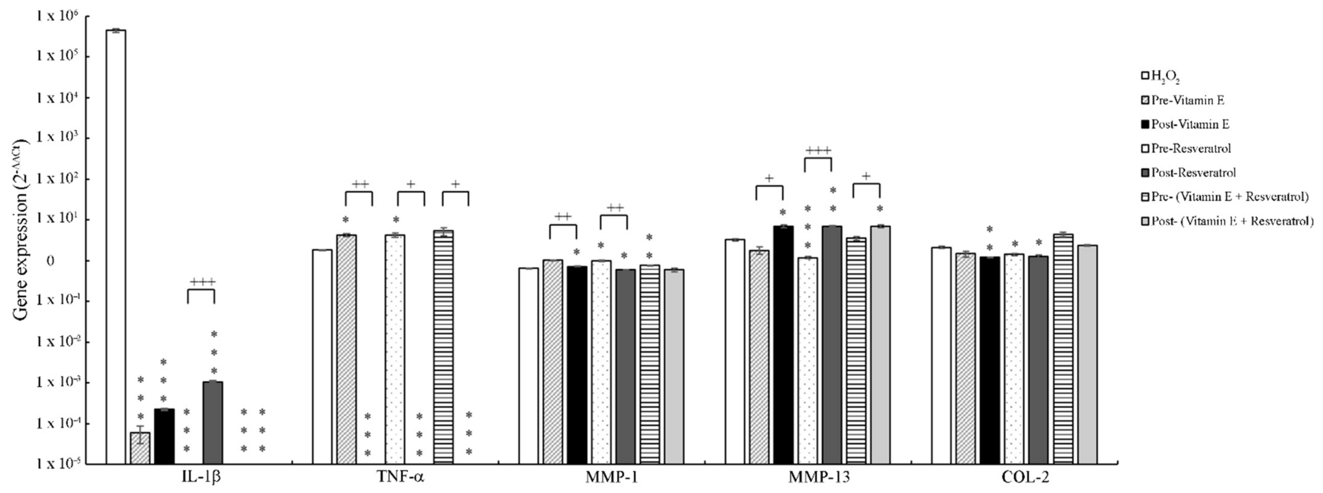
3.4. The Effect of Vitamin E and Resveratrol on Inflammatory Gene Expression

Figure 4 shows the results of gene expression. The high expression of IL-1β considerably decreased when OA-like chondrocytes were treated with vitamin E and resveratrol. The best reduction occurred when the chondrocytes were pre-treated with resveratrol, and pre-treated and post-treated with a combination of vitamin E and resveratrol. For TNF-α expression, post-treatment of vitamin E and/or resveratrol was better than pre-treatment. Post-treatment of resveratrol and the combination decreased MMP-1 expression compared with H2O2 induction, but only pre-treatment of vitamin E or resveratrol reduced the expression of MMP-13. In addition, the combination of vitamin E and resveratrol increased the expression of COL2A1 regardless of pre-treatment or post-treatment.

4. Discussion

OA patients often describe pain and stiffness in the joints and are thus recommended NSAIDs, which have some anti-inflammatory and analgesic effects [17]. However, prolonged use of NSAIDs results in adverse renal, hepatic, and cardiovascular effects as well as economic burden [18]. Alternative strategies have been proposed, including taking antioxidant supplements, which may reduce oxidative stress and consequently inflammation and pain. Vitamin E has been shown to reduce free radicals by donating its hydrogen and to modulate the activity of a transcription factor that regulates the expression of antioxidant enzymes [19,20]. In the current study, pre-treatment and post-treatment of vitamin E at 100 μM effectively reduced the level of oxygen-derived free radicals (Figure 3A). Indeed, 100 μM of vitamin E pre-treatment was shown to reverse oxidant-induced alterations in chondrocytes [21]. Our results also demonstrated that pre-treating chondrocytes with vitamin E reduces the expression of IL-1β and MMP-13. In contrast, post-treatment of vitamin E decreases IL-1β and TNF-α expression (Figure 4). However, treating chondrocytes with vitamin E before and after induction results in an increase in MMP-1 and a reduction in COL2A1. Our results suggested that although treating vitamin E in OA-like cells reduces the high expression of IL-1β and TNF-α induced by H2O2, it does not promote COL2A1 expression. Therefore, vitamin E might only play a role in reducing inflammation in OA-like chondrocytes by inhibiting the activity of IL-1β.
In addition, our results showed that resveratrol reduces the level of H2O2-induced free radicals, especially at 5 μM (Figure 2B). Resveratrol at 5 μM also influenced the expression of inflammatory genes, and pre-treatment of resveratrol had different effects than post-treatment. Pre-treatment of resveratrol resulted in a complete inhibition of IL-1β and a reduction in MMP-13, while post-treatment led to a complete inhibition of TNF-α and a decrease in IL-1β and MMP-1 (Figure 4). Resveratrol has been demonstrated to suppress IL-1β-induced proinflammatory and MMP production in cell culture models of OA [22,23,24]. Resveratrol also reduced cartilage tissue destruction in a rabbit model of OA [25]. Evidence shows that resveratrol is an activator of sirtuin 1 (SIRT 1), which plays a role in anti-inflammation along with other functions during OA progression [26]. In cell culture, resveratrol-activated SIRT 1 helps protect chondrocytes from IL-1β-mediated oxidative stress [27]. Treating human chondrocytes with resveratrol could produce an anti-inflammatory effect by affecting the TNF-α-mediated pathway [28]. Therefore, resveratrol possibly reduces H2O2-induced free radicals and the expression of IL-1β (pre-treatment) and TNF-α (post-treatment) by activating SIRT1. The actual molecular mechanism of resveratrol in our OA-like chondrocyte model will need further investigation.
Many antioxidant supplements, such as curcumin or avocado–soya extract, have been shown to be of benefit in the prevention and/or management of OA [1]. However, the effects of different combinations of antioxidant supplements on OA have rarely been investigated. We combined 100 μM vitamin E and 5 μM resveratrol to investigate their effects. Our results showed that a combination of vitamin E and resveratrol completely blocks the expression of IL-1β and increases the expression of COL2A1, demonstrating the potential for promoting collagen synthesis. Post-treatment of both antioxidant supplements inhibited TNF-α and reduced MMP-1, while pre-treatment did not (Figure 4). Our results suggested that post-treatment of OA-like chondrocytes with vitamin E and resveratrol can more effectively reduce the level of inflammation than pre-treatment as well as single treatment. However, animal verification and clinical investigation will be required to verify the clinical applications of vitamin E and resveratrol for OA treatment.

5. Conclusions

We investigated the role of vitamin E and resveratrol on the level of oxygen-derived free radicals and the expression of inflammatory genes. Both vitamin E and resveratrol reduced the level of free radicals that were induced by H2O2. Single treatment with either vitamin E or resveratrol reduced the expression of IL-1β and TNF-α. A combination of vitamin E and resveratrol not only inhibited the expression of IL-1β and TNF-α but also promoted the expression of COL2A1. The cell viability results demonstrated the biocompatibility of both antioxidant supplements, implying their potential application for reducing inflammation in OA.

Author Contributions

Conceptualization, C.-Y.S., Y.L., and H.-W.F.; methodology, C.-H.F.; validation, C.-Y.S. and C.-H.F.; formal analysis, C.-Y.S., Y.L., and C.-H.F.; investigation, C.-H.F. and H.-W.F.; data curation, C.-Y.S., Y.L., and C.-H.F.; writing—original draft preparation, C.-Y.S.; writing—review and editing, Y.L. and H.-W.F. All authors have read and agreed to the published version of the manuscript.

Funding

This research received no external funding.

Institutional Review Board Statement

Not applicable.

Informed Consent Statement

Not applicable.

Data Availability Statement

The data presented in this study are available on request from the corresponding author.

Acknowledgments

This work was supported by the Ministry of Science and Technology (MOST), Taiwan under grant number 107-2221-E-027-039-MY3, and the National Taipei University of Technology—Shenzhen University Joint Research Program (NTUT-SZU-107-05).

Conflicts of Interest

The authors declare no conflict of interest.

References

  1. Grover, A.K.; Samson, S.E. Benefits of antioxidant supplements for knee osteoarthritis: Rationale and reality. Nutr. J. 2016, 15, 1. [Google Scholar] [CrossRef] [PubMed] [Green Version]
  2. Adatia, A.; Rainsford, K.D.; Kean, W.F. Osteoarthritis of the knee and hip. Part 2: Therapy with ibuprofen and a review of clinical trials. J. Pharm. Pharmacol. 2012, 64, 626–636. [Google Scholar] [CrossRef] [PubMed]
  3. Juhl, C.; Christensen, R.; Roos, E.M.; Zhang, W.; Lund, H. Impact of exercise type and dose on pain and disability in knee osteoarthritis: A systematic review and meta-regression analysis of randomized controlled trials. Arthritis Rheumatol. 2014, 66, 622–636. [Google Scholar] [CrossRef] [PubMed]
  4. McCarberg, B.; Tenzer, P. Complexities in the pharmacologic management of osteoarthritis pain. Curr. Med. Res. Opin. 2013, 29, 539–548. [Google Scholar] [CrossRef] [PubMed]
  5. Wojdasiewicz, P.; Poniatowski, L.A.; Szukiewicz, D. The role of inflammatory and anti-inflammatory cytokines in the pathogenesis of osteoarthritis. Mediat. Inflamm. 2014, 2014, 561459. [Google Scholar] [CrossRef] [PubMed] [Green Version]
  6. Melchiorri, C.; Meliconi, R.; Frizziero, L.; Silvestri, T.; Pulsatelli, L.; Mazzetti, I.; Borzi, R.M.; Uguccioni, M.; Facchini, A. Enhanced and coordinated in vivo expression of inflammatory cytokines and nitric oxide synthase by chondrocytes from patients with osteoarthritis. Arthritis Rheum. 1998, 41, 2165–2174. [Google Scholar] [CrossRef]
  7. van de Loo, F.A.; Joosten, L.A.; van Lent, P.L.; Arntz, O.J.; van den Berg, W.B. Role of interleukin-1, tumor necrosis factor alpha, and interleukin-6 in cartilage proteoglycan metabolism and destruction. Effect of in situ blocking in murine antigen—and zymosan-induced arthritis. Arthritis Rheum. 1995, 38, 164–172. [Google Scholar]
  8. Shakibaei, M.; Schulze-Tanzil, G.; John, T.; Mobasheri, A. Curcumin protects human chondrocytes from il-l1beta-induced inhibition of collagen type ii and beta1-integrin expression and activation of caspase-3: An immunomorphological study. Ann. Anat. 2005, 187, 487–497. [Google Scholar] [CrossRef]
  9. Stove, J.; Huch, K.; Gunther, K.P.; Scharf, H.P. Interleukin-1beta induces different gene expression of stromelysin, aggrecan and tumor-necrosis-factor-stimulated gene 6 in human osteoarthritic chondrocytes in vitro. Pathobiology 2000, 68, 144–149. [Google Scholar] [CrossRef]
  10. Vincenti, M.P.; Brinckerhoff, C.E. Transcriptional regulation of collagenase (mmp-1, mmp-13) genes in arthritis: Integration of complex signaling pathways for the recruitment of gene-specific transcription factors. Arthritis Res. 2002, 4, 157–164. [Google Scholar] [CrossRef]
  11. Fridovich, I. Oxygen: How do we stand it? Med. Princ. Pract. 2013, 22, 131–137. [Google Scholar] [CrossRef] [PubMed]
  12. Walia, M.; Kwan, C.Y.; Grover, A.K. Effects of free radicals on coronary artery. Med. Princ. Pract. 2003, 12, 1–9. [Google Scholar] [CrossRef] [PubMed]
  13. Altindag, O.; Erel, O.; Aksoy, N.; Selek, S.; Celik, H.; Karaoglanoglu, M. Increased oxidative stress and its relation with collagen metabolism in knee osteoarthritis. Rheumatol. Int. 2007, 27, 339–344. [Google Scholar] [CrossRef] [PubMed]
  14. Sutipornpalangkul, W.; Morales, N.P.; Charoencholvanich, K.; Harnroongroj, T. Lipid peroxidation, glutathione, vitamin e, and antioxidant enzymes in synovial fluid from patients with osteoarthritis. Int. J. Rheum. Dis. 2009, 12, 324–328. [Google Scholar] [CrossRef] [PubMed]
  15. Chen, Y.C.; Chang, Y.W.; Tan, K.P.; Shen, Y.S.; Wang, Y.H.; Chang, C.H. Can mesenchymal stem cells and their conditioned medium assist inflammatory chondrocytes recovery? PLoS ONE 2018, 13, e0205563. [Google Scholar] [CrossRef] [PubMed]
  16. Gao, G.; Ding, H.; Zhuang, C.; Fan, W. Effects of hesperidin on H2O2-treated chondrocytes and cartilage in a rat osteoarthritis model. Med. Sci. Monit. 2018, 24, 9177–9186. [Google Scholar] [CrossRef]
  17. Hochberg, M.C. New directions in symptomatic therapy for patients with osteoarthritis and rheumatoid arthritis. Semin. Arthritis Rheum. 2002, 32, 4–14. [Google Scholar] [CrossRef]
  18. Crofford, L.J. Use of nsaids in treating patients with arthritis. Arthritis Res. Ther. 2013, 15, S2. [Google Scholar] [CrossRef] [Green Version]
  19. Dworski, R.; Han, W.; Blackwell, T.S.; Hoskins, A.; Freeman, M.L. Vitamin e prevents nrf2 suppression by allergens in asthmatic alveolar macrophages in vivo. Free Radic. Biol. Med. 2011, 51, 516–521. [Google Scholar] [CrossRef] [Green Version]
  20. Peh, H.Y.; Tan, W.S.; Liao, W.; Wong, W.S. Vitamin e therapy beyond cancer: Tocopherol versus tocotrienol. Pharmacol. Ther. 2016, 162, 152–169. [Google Scholar] [CrossRef]
  21. Bhatti, F.U.; Mehmood, A.; Wajid, N.; Rauf, M.; Khan, S.N.; Riazuddin, S. Vitamin e protects chondrocytes against hydrogen peroxide-induced oxidative stress in vitro. Inflamm. Res. 2013, 62, 781–789. [Google Scholar] [CrossRef] [PubMed]
  22. Csaki, C.; Keshishzadeh, N.; Fischer, K.; Shakibaei, M. Regulation of inflammation signalling by resveratrol in human chondrocytes in vitro. Biochem. Pharmacol. 2008, 75, 677–687. [Google Scholar] [CrossRef] [PubMed]
  23. Shakibaei, M.; Csaki, C.; Nebrich, S.; Mobasheri, A. Resveratrol suppresses interleukin-1beta-induced inflammatory signaling and apoptosis in human articular chondrocytes: Potential for use as a novel nutraceutical for the treatment of osteoarthritis. Biochem. Pharmacol. 2008, 76, 1426–1439. [Google Scholar] [CrossRef] [PubMed]
  24. Shakibaei, M.; John, T.; Seifarth, C.; Mobasheri, A. Resveratrol inhibits il-1 beta-induced stimulation of caspase-3 and cleavage of parp in human articular chondrocytes in vitro. Ann. N.Y. Acad. Sci. 2007, 1095, 554–563. [Google Scholar] [CrossRef]
  25. Elmali, N.; Esenkaya, I.; Harma, A.; Ertem, K.; Turkoz, Y.; Mizrak, B. Effect of resveratrol in experimental osteoarthritis in rabbits. Inflamm. Res. 2005, 54, 158–162. [Google Scholar] [CrossRef]
  26. Deng, Z.; Li, Y.; Liu, H.; Xiao, S.; Li, L.; Tian, J.; Cheng, C.; Zhang, G.; Zhang, F. The role of sirtuin 1 and its activator, resveratrol in osteoarthritis. Biosci. Rep. 2019, 39. [Google Scholar] [CrossRef] [Green Version]
  27. Lei, M.; Wang, J.G.; Xiao, D.M.; Fan, M.; Wang, D.P.; Xiong, J.Y.; Chen, Y.; Ding, Y.; Liu, S.L. Resveratrol inhibits interleukin 1beta-mediated inducible nitric oxide synthase expression in articular chondrocytes by activating sirt1 and thereby suppressing nuclear factor-kappab activity. Eur. J. Pharmacol. 2012, 674, 73–79. [Google Scholar] [CrossRef]
  28. Moon, M.H.; Jeong, J.K.; Lee, Y.J.; Seol, J.W.; Jackson, C.J.; Park, S.Y. Sirt1, a class iii histone deacetylase, regulates tnf-alpha-induced inflammation in human chondrocytes. Osteoarthr. Cartil. 2013, 21, 470–480. [Google Scholar] [CrossRef] [Green Version]
Figure 1. The induction of osteoarthritis by H2O2 in porcine chondrocytes. (A) The level of oxygen-derived free radicals was represented by the level of lumen density. The level of lumen density increased significantly when the chondrocytes were treated with H2O2 for 30 min and 2 h. ** p < 0.01 compared with no induction. ++ p < 0.01 compared with 30 min induction. (B) The expressions of IL-1β, TNF-α, and COL2A1 were higher when the chondrocytes were induced by H2O2 for 30 min (white bars) than for 2 h (gray bars). Gene expression with no induction is shown in black bars. * p < 0.05, ** p < 0.01, and *** p < 0.001 compared with no induction.
Applsci 11 00239 g001
Figure 2. The cell viability of vitamin E and resveratrol at various concentrations. (A) L929 cells were cultured with 5, 50, 100, or 200 μM of vitamin E for 1 (white bars), 2 (black bars), or 3 (gray bars) days. The cell viability increased when the concentration of vitamin E was higher. * p < 0.05 compared with the cell viability at 5 μM. (B) When the L929 cells were cultured with 5, 50, 100, and 200 μM of resveratrol for 1 day, the cell viability increased depending on the concentration of resveratrol.
Applsci 11 00239 g002
Figure 3. The changes in the level of lumen density when treating OA-like chondrocytes with vitamin E and resveratrol. Adding vitamin E (A) or resveratrol (B) at 5, 50, 100, and 200 μM before H2O2 induction (white bars) or after H2O2 induction significantly reduces the level of lumen density. * p < 0.05 and ** p < 0.01 compared with H2O2 induction.
Applsci 11 00239 g003
Figure 4. The inflammatory gene expression of OA-like chondrocytes when treated with vitamin E and resveratrol. The expressions of IL-1β, TNF-α, MMP-1, MMP-13, and COL2A1 when the chondrocytes were pre-treated with 100 μM of vitamin E (gray slashed bars), 5 μM of resveratrol (black bars), or their combination (dotted bars), or when the chondrocytes were post-treated with vitamin E (dark gray bars), resveratrol (horizonal striped bars), or their combination (light gray bars). * p < 0.05, ** p < 0.01, and *** p < 0.001 compared with H2O2 induction; + p < 0.05, ++ p < 0.01, and +++ p < 0.001.
Applsci 11 00239 g004
Publisher’s Note: MDPI stays neutral with regard to jurisdictional claims in published maps and institutional affiliations.

