Article

Assessment of Anthropometric and Body Composition Risk Factors in Patients with both Hypertension and Stroke in the Korean Population

National Center for Standard Reference Data, Korea Research Institute of Standards and Science, Daejeon 34113, Korea
*
Author to whom correspondence should be addressed.
Appl. Sci. 2020, 10(9), 3046; https://doi.org/10.3390/app10093046
Submission received: 2 April 2020 / Revised: 24 April 2020 / Accepted: 24 April 2020 / Published: 27 April 2020
(This article belongs to the Special Issue Data Technology Applications in Life, Diseases, and Health)

Abstract

:
The association of hypertension or stroke with anthropometric and body composition indices has been evaluated individually but not for patients with both conditions. Here, we compared these indices in patients with both hypertension and stroke and evaluated the best indicators for identifying patients with both diseases in the Korean population. Data were obtained from the Korea National Health and Nutrition Examination Survey (KNHANES) conducted from 2008 to 2011. Data analysis was carried out using a complex sampling design that considered the weighting for personal analysis to represent the whole population in Korea. Binary logistic regression was conducted for evaluating potential associations, and areas under the curve were calculated to compare the predictive power of all variables for identifying patients with hypertension or both hypertension and stroke. Among all hypertension-related factors, waist-to-height ratio (WHtR) exhibited a strong association in men (odds ratio (OR) = 1.390 [1.127–1.714]), whereas trunk-fat mass (OR = 1.613 [1.237–2.104]) and thoracic spine bone mineral density (BMD) (OR = 1.250 [1.044–1.496]) represented the best indicators in women. Comparison of anthropometric and body composition indices in patients with both diseases revealed that left arm BMD and left leg fat mass (LLF) were strongly associated in both men (OR = 0.504 [0.320–0.793]) and women (OR = 0.391 [0.208–0.734]). However, among patients with both hypertension and stroke, WHtR (OR = 1.689 [1.080–2.641]) and LLF (OR = 0.391 [0.208–0.734]) were the best risk predictors in men and women, respectively. Our findings suggested that the best indicators among patients with hypertension or both hypertension and stroke may differ according to men and women.

1. Introduction

Hypertension, a chronic disease characterized by blood pressure (BP) higher than the normal range, is a major global health concern and a risk factor for cardiovascular disease (CVD), myocardial infarction (MI), stroke, and death from its associated complications [1,2,3]. According to the World Health Organization, an estimated 1.13 billion people worldwide have hypertension, with most living in low- and middle-income countries [4]. In turn, according to the Global Burden of Disease 2016 Lifetime Risk of Stroke Collaborators, the global risk of stroke increased from 22.8% in 1990 to 24.9% in 2016 [5]. Stroke, which refers to the neurological symptoms that occur suddenly consequent to cerebral blood flow abnormalities, has thus become a major negative influence on economic and social development [6,7].
Numerous studies support the association between hypertension and vascular diseases [8,9,10,11,12,13,14,15,16,17,18]. In a US-based study, Woo et al. showed that hypertension is a highly important risk factor for stroke [8], as did Du et al. for the UK [9]. In particular, Kario et al. demonstrated that the morning systolic BP (SBP) and difference between morning and evening SBP are associated with stroke [10]. Willmot et al. showed the mean arterial BP, diastolic BP (DBP), and SBP to be associated with ischemic stroke or death [11]. Meanwhile, Qureshi et al. found that prehypertension was associated with CVD and MI but not atherothrombotic brain infarction (ABI) or stroke [12]. Furthermore, low-density lipoprotein and serum uric acid were shown to associate with hypertension by Cicero et al. [13].
The association of anthropometric and body composition indices with hypertension or stroke has also been examined [19,20,21,22,23,24,25,26,27,28]. For example, waist circumference (WC), waist-to-hip ratio (WHR), and waist-to-height ratio (WHtR) were associated with hypertension by Fuchs et al. [19], whereas associations were found with WC, WHR, and body mass index (BMI) values in a Canadian study [20]. Chandra et al. revealed that increased visceral adiposity was associated with incident hypertension [21], and abdominal fat mass was shown by Wiklund et al. to be strongly associated with hypertension in women [22]. Moreover, decreased lumbar spine bone mineral density (BMD) was associated with hypertension in a study by Tsuda et al. [23]. In turn, WC, WHR, and WHtR were shown by Winter et al. to be associated with stroke and transient ischemic attack [24]. In women, WC, WHR, and WHtR were associated with stroke but not BMI in a study by Lu et al. [25]. Toss et al. also revealed an association between WHtR and stroke, with abdominal fat mass also associated after adjustment for BMI [26]. Finally, femoral neck BMD was identified as an independent predictor of stroke [27,28]; notably, although this result retained significance after adjustment for confounding factors, the association was specific to women [28].
Nevertheless, to our knowledge no research comparing and analyzing anthropometric and body composition indices has been published regarding patients with both hypertension and stroke (hypertension + stroke). Therefore, the aim of the present study was to compare the association of these indices with hypertension and hypertension + stroke in the Korean population. Using binary logistic regression analysis by SPSS analysis software, we determined the odds ratio (OR) values and p-values for evaluating the association with hypertension and stroke among all variables. The findings of the present study will provide basic knowledge related to the associations of hypertension and stroke with anthropometric and body composition indices to facilitate the prevention and management of patients with both disorders.

2. Materials and Methods

2.1. Subjects and Definitions

The data were obtained from the Korea National Health and Nutrition Examination Survey (KNHANES IV-2, 3 and V-1, 2) from 2008 to 2011; the KNHANES is a cross-sectional survey study conducted by the Korea Centers for Disease Control and Prevention (KCDC) [29,30]. The KNHANES datasets were approved by the Korea Ministry of Health and Welfare (2008-04EXP-01-C, 2009-01CON-03-2C, 2010-02CON-21-C, 2011-02CON-06-C) and were conducted in accordance with the Declaration of Helsinki. The present study was approved by the Institutional Review Board of the Korea Research Institute of Standards and Science, including approval for the access and analysis of open source data from the KNHANES IV-2, 3 and V-1, 2, with a waiver for the documentation of informed consent (IRB No. KRISS-IRB-2019-14).
The KNHANES I–VII survey was conducted from 1998 to 2018 [31]. This is the survey study to assess and monitor health, nutritional status and health risk factors of major diseases of Koreans. These data were collected by 3 parts of survey method as health examination, health interview and nutrition survey. The body fat mass and BMD data were obtained only from 2008 to 2011. Therefore, KNHANES IV-2, 3 and V-1, 2 were selected for the association analysis with hypertension, stroke, and body composition (body fat mass and BMD). KNHANES IV-2, 3 and V-1, 2 included 37,753 subjects who participated in the health examination, blood parameters, anthropometric assessment, and body fat mass and BMD measurements. Using KNHANES data that accurately estimated the characteristics of Koreans, weighted values were used for analysis [29,30]. The weighted sample average ( Y ^ ) was calculated using the following equation to estimate the population average of specific items by reflected weights ( w i , i = 1, 2, …, n) in n subjects ( y i , i = 1, 2, …, n).
Y ^ = i = 1 n w i y i w i

2.2. Measurements

All anthropometric indices were measured using standard methods such as height, weight, WC, and BMI. Height was measured to the nearest 1 mm using a Seca 225 portable stadiometer (Seca, Hamburg, Germany). Weight was measured to the nearest 0.1 kg using an electronic scale (GL-6000-20; Caskorea, Seoul, Korea). WC was measured using a Seca 200 to the nearest 1 mm between the sternum and hips. BMI was calculated as weight/height2. The blood parameters were measured using a Hitachi Automatic Analyzer 7600 (Hitachi, Tokyo, Japan) with Pureauto SCHO-N (Sekisui, Tokyo, Japan), S GLU, and Cholestest N HDL (Sekisui, Tokyo, Japan) reagents. BMD was measured by dual energy X-ray absorptiometry (DXA) and DISCOVERY-W (Hologic, Marlborough, MA, USA). Body fat mass was measured using the same methods and equipment as for BMD measurement [29,30]. Body fat mass was measured for the head, left arm, right arm, left leg, right leg, trunk, body without the head, and total body. BMD was measured for the head, left arm, right arm, left rib, right rib, thoracic spine, lumbar spine, pelvis, left leg, right leg, total BMD without the head, and total BMD. The WHtR was calculated as WC/height as a new indicator in this study.

2.3. Statistical Analysis

Analysis was carried out using a complex sampling design. Stratified two-stage sampling was used, with weighting for personal analysis and the cluster primary sampling unit being considered. It is possible to draw biased results from the estimation and estimated variance using a simple random sampling method for the mean and prevalence rate. Therefore, complex sampling data analysis that reflects the complex sampling design factors should be performed [29,30].
Herein, statistical analysis was performed using SPSS 22 software for Windows (SPSS, Inc., Armonk, NY, USA). Binary logistic regression algorithm is used for classification or prediction model in data mining and machine learning fields, but the algorithm is mainly used to examine or analyze the association between disease and variables in medicine, epidemiology, public health studies, etc., and can provide p-values and OR values for association analysis. Therefore, the use of this algorithm is suitable for analysis of our medical data and objectives. The data preprocessing was divided into five steps. First, data integration was carried out using the measured data (fat mass and BMD) from 2008 to 2011. Second, we extracted the data according to the characteristics, such as demographics, anthropometry, blood information, and body compositions. Then, the diseases were defined by diagnosis, and data translation and cleaning was carried out. Finally, data were standardized for comparison and analysis purposes. Binary logistic regression was conducted to identify a significant association of hypertension and stroke with various measured values (anthropometry, body fat mass, BMD, etc.), and differences between normal and hypertension subjects and between normal subjects and those with hypertension + stroke. Adjusted analysis was performed using age, BMI, frequency of alcohol consumption, smoking, income, recognized stress rate, and education level. We analyzed the association between the normal and hypertension groups using binary logistic regression to obtain the p-values, OR, and confidence interval (CI). Moreover, we analyzed the association between the normal and hypertension + stroke groups using the same conditions. A t-test was conducted to evaluate the significant differences by men and women. We used the area under the receiver operating characteristic curve (AUC) to assess whether a significant improvement in the identification of hypertension and stroke was achieved based on anthropometric and body composition indices. As no method is available to calculate AUC in complex sampling analysis, we carried out the general AUC analysis based on the original data. Figure 1 shows the design of the experimental study for statistical analysis.

3. Results

In the present study, we focused on patients aged over 50 years who were recently diagnosed with hypertension and stroke by a physician, because the prevalence of hypertension and stroke increases rapidly in individuals over 50 years of age [32]. This yielded a sample size of 13,682 subjects.
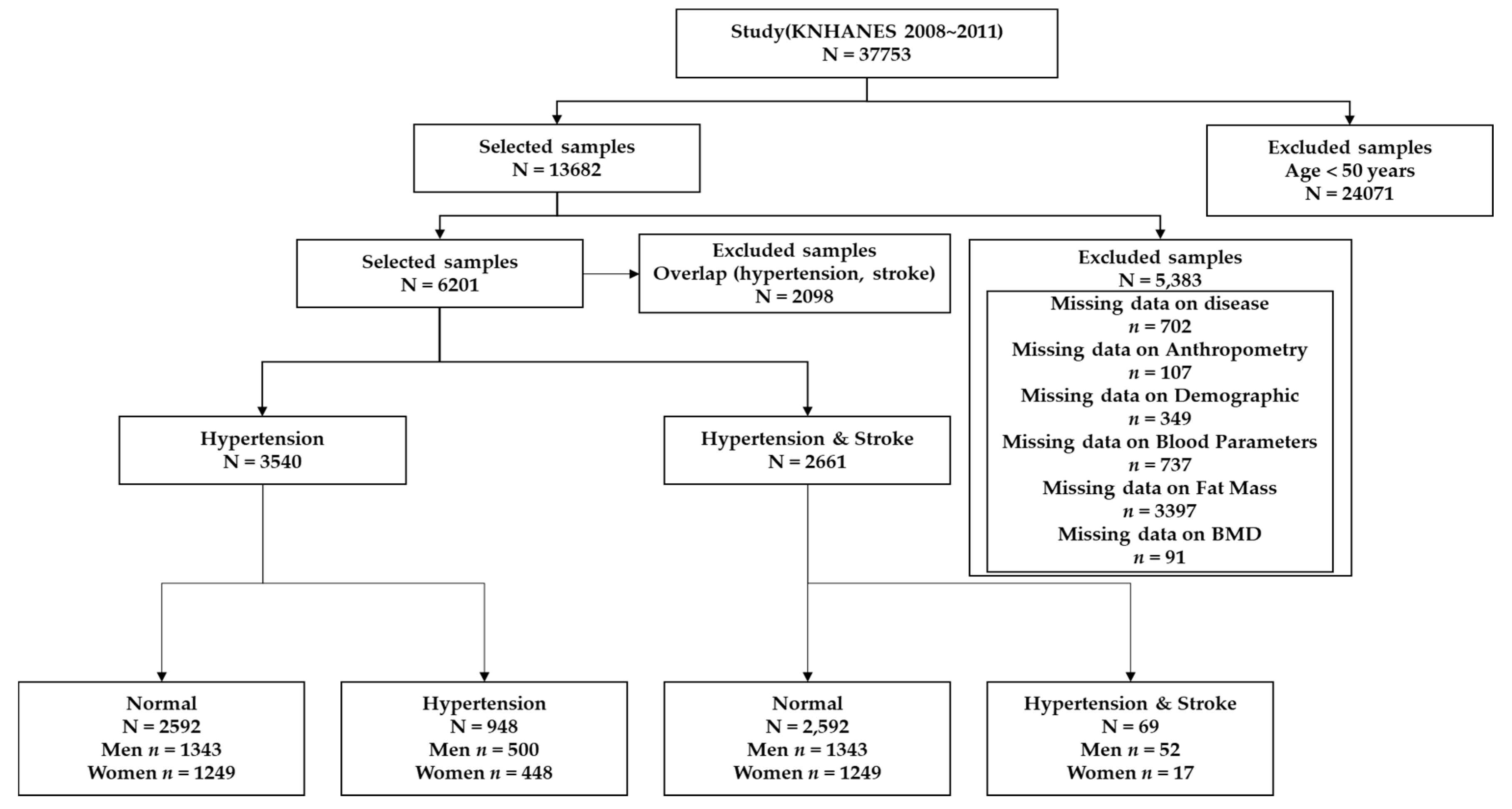
In total, missing values were excluded for disease (n = 702), anthropometry (n = 107), demographics (n = 349), blood parameters (n = 737), fat mass (n = 3397), and BMD (n = 91), resulting in the selection of a sample of 8407 subjects without overlaps (n = 2098). The final Group 1 for hypertension consisted of 2592 normal subjects (1343 men, 1249 women) and 948 with hypertension (500 men, 448 women). Group 2, for hypertension + stroke, consisted of 2592 normal subjects (1343 men, 1249 women) and 69 subjects with hypertension + stroke (52 men, 17 women). Figure 2 shows the detailed sample selection procedure of data preprocessing.
Table 1 and Table 2 show the demographic characteristics of each group and a detailed description of all experimental variables.

3.1. Associations of Hypertension with Anthropometric and Body Composition Indices

Table 3 and Table 4 represent the association of hypertension with anthropometric and body composition (body fat mass and BMD) indices in Korean men and women. Overall, 26.8% (N = 948) of 3540 participants exhibited hypertension. The prevalence of hypertension was higher in men than in women (27.1% and 26.4%, respectively).
In men, among all the variables, WHtR (p < 0.0001, OR = 1.836, 95% CI [1.627–2.071]) presented the strongest association with hypertension during crude analysis. In addition, WHtR (adjusted p = 0.0021, adjusted OR = 1.390 [1.127–1.714]) exhibited the strongest association after adjusting for age, BMI, alcohol consumption, smoking, income, recognized stress rate, and education level. Among the body fat mass variables, trunk fat mass (TF, p < 0.0001, OR = 1.626 [1.443–1.832]) was the best indicator of hypertension, followed by total body fat mass (TBF, p < 0.0001, OR = 1.561 [1.389–1.755]) in crude analysis. However, these associations disappeared after adjustment for confounders. Among the BMD variables, pelvis BMD (PlvBMD, p = 0.0079, OR = 1.183 [1.045–1.339]) showed the highest association with hypertension, followed by thoracic spine BMD (TSBMD, p = 0.0330, OR = 1.126 [1.010–1.256]) in crude analysis. These associations also disappeared following adjustment for confounders.
In women, age (p < 0.0001, OR = 1.832 [1.658–2.025]) showed the strongest association with hypertension among all variables. Moreover, TF (adjusted p = 0.0004, adjusted OR = 1.613 [1.237–2.104]) was associated in the adjusted analysis. WHtR (p < 0.0001, OR = 1.806 [1.612–2.022]) exhibited the highest association with hypertension among the anthropometric variables in crude analysis. Furthermore, WC (p < 0.0001, OR = 1.611 [1.440–1.802]) showed association with hypertension. Among these, the association with WC (adjusted p = 0.0492, adjusted OR = 1.231 [1.001–1.515]) was maintained following the adjusted analysis. Among the body fat mass variables, TF (p < 0.0001, OR = 1.499 [1.318–1.704]; adjusted p = 0.0004, adjusted OR = 1.613 [1.237–2.104]) showed the strongest association with hypertension, followed by TBFWH (p < 0.0001, OR = 1.324 [1.165–1.504]) in both crude and adjusted analyses. In the adjusted analysis, notably, right leg fat mass (RLF) and left leg fat mass (LLF) were negatively associated with hypertension (adjusted p = 0.0246, adjusted OR = 0.796 [0.652–0.971]; adjusted p = 0.0334, adjusted OR = 0.810 [0.667–0.983]). Head BMD (HDBMD, p = 0.0003, OR = 0.794 [0.703–0.898]) exhibited the highest negative association with hypertension in crude analysis. Total BMD (TBMD, p = 0.0006, OR = 0.797 [0.701–0.907]) was also associated with hypertension. In particular, TSBMD and left leg BMD (LLBMD) were associated with hypertension in the adjusted analysis (adjusted p = 0.0152, adjusted OR = 1.250 [1.044–1.496]; adjusted p = 0.0237, adjusted OR = 1.189 [1.024–1.381]).
In the overall crude analysis, WHtR presented the strongest association with hypertension in men, whereas age demonstrated the strongest association in women. WHtR was also associated in men, although TF was associated in women upon adjusted analysis. In women, TSBMD was associated with hypertension; however, this association disappeared following adjustment for confounders in men. These analysis results thus suggested a sex-based difference in hypertension risk factors.
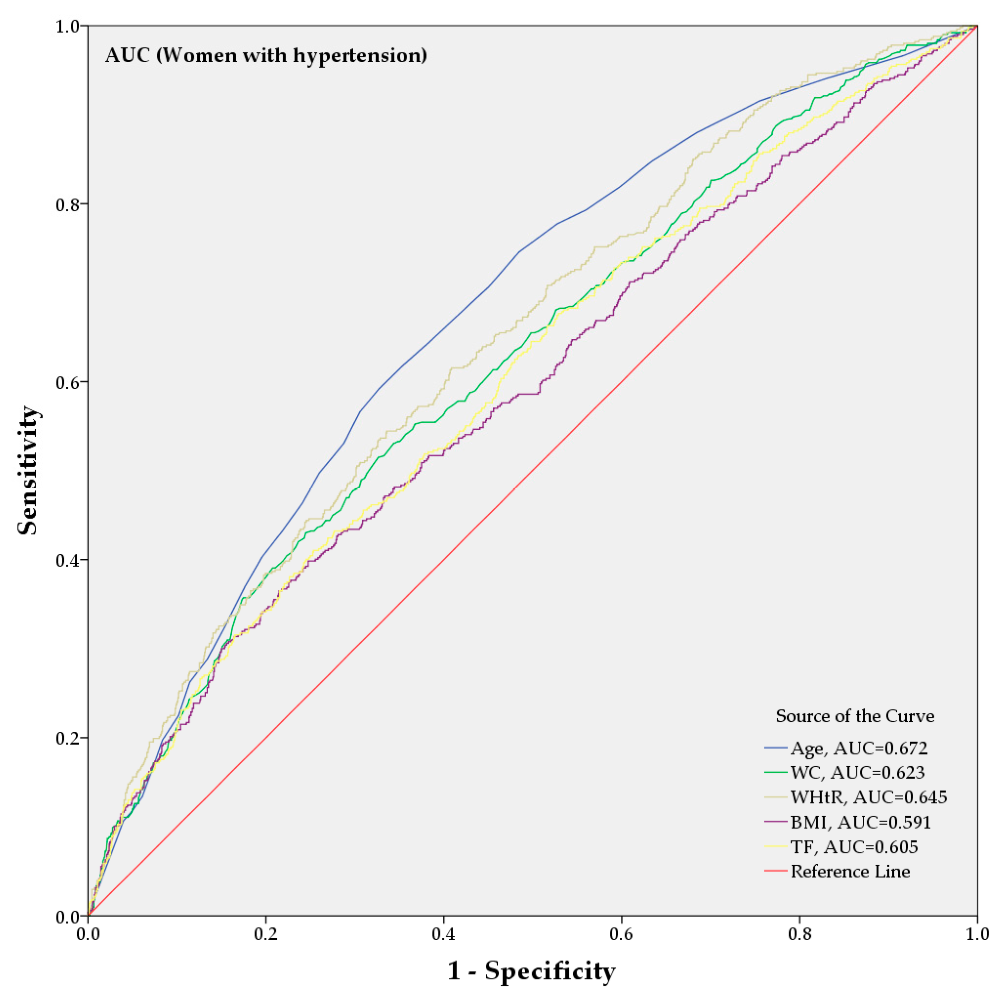
From the AUC analysis, Figure 3 and Figure 4 show the predictive power of anthropometric and body composition indicators for hypertension in Korean men and women; only the five indices with the highest AUC values are shown. Among all the variables, WHtR (AUC = 0.638 [0.610–0.666]) presented strong identification power in men, whereas age (AUC = 0.645 [0.618–0.673]) showed the highest AUC value in women. TF, TBFWH, and TBF exhibited strong identification power among the body fat mass indicators in men (AUC = 0.630 [0.602–0.657]; AUC = 0.616 [0.588–0.644]; AUC = 0.616 [0.588–0.644]) and women (AUC = 0.605 [0.576–0.634]; AUC = 0.574 [0.544–0.603]; AUC = 0.573 [0.544–0.603]). For BMD, TSBMD (AUC = 0.546 [0.517–0.575]; AUC = 0.469 [0.439–0.498]) showed the strongest identification power for men and women, respectively.

3.2. Comparison of Anthropometric and Body Composition Indices in Patients with Both Hypertension and Stroke

Table 5 and Table 6 show the association of anthropometric and body composition indices with hypertension + stroke in Korean men and women. Overall, 2.6% (N = 69) of 2661 participants had both hypertension and stroke. The prevalence of hypertension + stroke was higher in men than in women (3.7% and 1.3%, respectively).
In men, among all the variables, age (p < 0.0001, OR = 2.156 [1.644–2.828]) presented the strongest association with hypertension + stroke in crude analysis. LABMD (adjusted p = 0.0031, adjusted OR = 0.504 [0.320–0.793]) showed the strongest negative association with hypertension + stroke following confounding factor adjustment. Among the anthropometric indices, WHtR (p < 0.0001, OR = 1.797 [1.382–2.336]; adjusted p = 0.0217, adjusted OR = 1.689 [1.080–2.641]) exhibited the strongest association with hypertension + stroke in both crude and adjusted analyses; WC and BMI were also associated (p = 0.0026, OR = 1.519 [1.157–1.993]; p = 0.0336, OR = 1.329 [1.022–1.728]). Among the body fat mass variables, TF (p = 0.0265, OR = 1.506 [1.049–2.162]) showed the highest association with hypertension + stroke, followed by TBFWH (p = 0.0257, OR = 1.495 [1.050–2.128]) in crude analysis. However, these associations disappeared following adjustment for confounders. Among the BMD variables, LABMD (p < 0.0001, OR = 0.542 [0.396–0.742]; adjusted p = 0.0031, adjusted OR = 0.504 [0.320–0.793]) was the best indicator of hypertension + stroke in the crude and adjusted analysis. The right arm BMD (RABMD) and right leg BMD (RLBMD) were also shown to be associated in crude (p < 0.0001, OR = 0.531 [0.384–0.735]; p = 0.0078, OR = 0.0078 [0.570–0.862]) and adjusted analyses (adjusted p = 0.0054, adjusted OR = 0.528 [0.337–0.0827]; adjusted p = 0.0117, adjusted OR = 0.537 [0.331–0.870]).
In women, among all variables, age (p < 0.0001, OR = 2.097 [1.588–2.770]) also presented the strongest association with hypertension + stroke in crude analysis. Among the anthropometric variables, HT (p < 0.0001, OR = 0.607 [0.474–0.778]), in particular, was negatively associated with hypertension + stroke; WHtR (p = 0.0008, OR = 1.564 [1.204–2.031]) was associated with hypertension + stroke in crude analysis, although both associations disappeared following adjustment for confounders. Notably, among the body fat mass variables, LLF (adjusted p = 0.0036, adjusted OR = 0.391 [0.208–0.734]) and RLF (adjusted p = 0.0211, adjusted OR = 0.460 [0.237–0.890]) only showed association with hypertension + stroke following adjusted analysis. Among the BMD indices, PlvBMD and LLBMD exhibited negative association with hypertension + stroke in crude analysis (p = 0.0161, OR = 0.593 [0.387–0.907]; p = 0.0445, OR = 0.542 [0.298–0.985]), although these associations disappeared following adjustment for confounders.
In both men and women, age presented the strongest association with hypertension + stroke upon crude analysis. In the results of the adjusted analysis, WHtR was associated in men but not in women. The results of the body composition and anthropometric indices association analysis with hypertension + stroke also differed according to men and women.
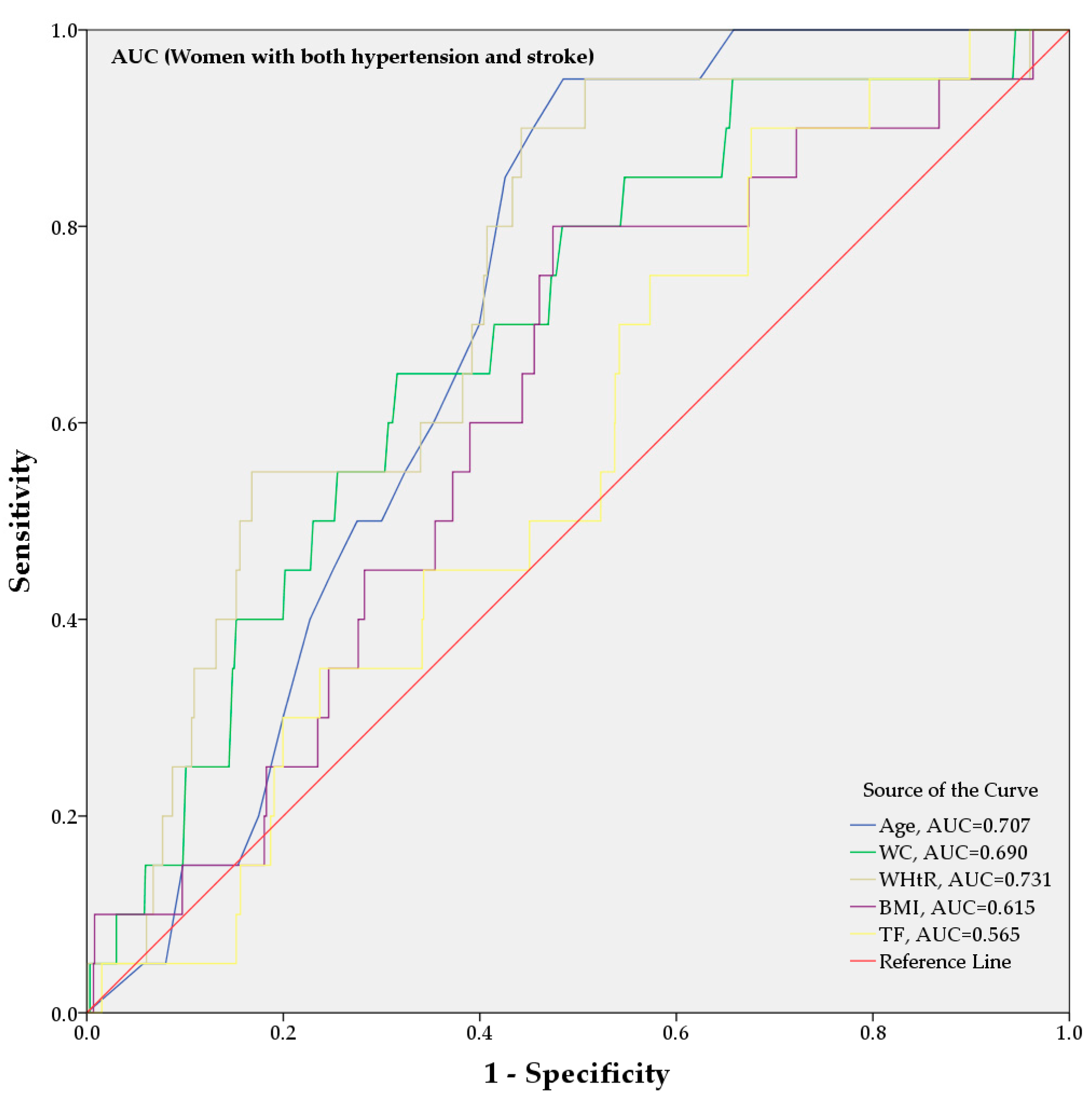
Figure 5 and Figure 6 show the hypertension + stroke identification power of anthropometric and body composition (fat mass and BMD) indicators based on the AUC analysis in Korean men and women. Only five indicators with the highest AUC values are shown. Age presented the highest AUC value among all the variables in both men and women (AUC = 0.689 [0.617–0.761]; AUC = 0.707 [0.640–0.774], respectively). WHtR demonstrated strong identification power among the anthropometric indices in both men and women (AUC = 0.593 [0.514–0.671]; AUC = 0.731 [0.632–0.829]). For body composition, TF (AUC = 0.551 [0.475–0.627]) showed an association in men, and PlvBMD (AUC = 0.375 [0.277–0.473]) exhibited strong identification power in women.

4. Discussion

In this study, anthropometric and body composition indices were examined as potential risk factors for hypertension and hypertension + stroke. In a previous study of anthropometric indices and hypertension, Fuchs et al. [19] evaluated 592 randomly selected patients and obtained the hazard ratio (HR) and p-value for evaluation. BMI (HR = 1.042, p = 0.091), WC (HR = 1.023, p = 0.028), WHtR (HR = 1.042, p = 0.013), and WHR (HR = 1.033, p = 0.006) were highlighted after adjusting for age, baseline BP, sex, and alcohol consumption, respectively. The related WC indices were associated with hypertension. Alternatively, a study based on Canadian heart health population-based cross-sectional surveys that targeted 9913 men and women aged 18–74 years, selected from among five Canada hospitals, utilized WC, WHR, and BMI for association analysis to compare various risk factors as indicators of CVD [20]. Data analysis according to sensitivity, specificity, and positive and negative predictive values using receiver operating characteristic curves identified WC as the best indicator of other cardiovascular risk factors [20]. Another study evaluated the relationship of body mass and fat distribution with hypertension through visceral and subcutaneous adipose tissue measurements obtained by magnetic resonance and proton-spectroscopic imaging, along with lower body fat ascertained by DXA [21]. Only visceral adipose tissue (relative risk (RR) = 1.22, CI [1.06–1.39]) was independently associated with hypertension; in comparison, increased visceral adiposity was robustly associated with incident hypertension but not total or subcutaneous adiposity [21]. Moreover, the association between CVD (related to hypertension) and abdominal and gynoid fat mass was evaluated in a cross-sectional study of 175 men and 417 women who were participants in a community intervention program for investigating the contribution of estimated regional fat mass [22]. In men, abdominal fat mass was the strongest predictor of most cardiovascular variables (OR [2.63–3.37]), whereas in women, gynoid fat mass showed the strongest association (OR [1.48–2.19]), suggesting that abdominal fat mass is strongly independently associated with hypertension [22]. The association of (untreated) hypertension with BMD as measured by DXA was also evaluated in 31 patients as compared with 14 control Japanese women [23]. Analysis using t-tests and regression models revealed that the lumbar spine BMD was decreased in patients with hypertension, suggesting an association of hypertension with reduced BMD [23].
Several studies on the association of stroke with risk factors have also been conducted. Evaluation of the association of obesity and abdominal fat mass with stroke using the data of patients collected in 3188 primary care offices in Germany, encompassing 6980 study participants divided into 1745 patients with stroke and 5235 control subjects, revealed WC (OR = 1.15 [1.00–1.32]), WHR (OR = 1.21 [1.05–1.38]), and WHtR (OR = 1.25 [1.09–1.44]) as associated with stroke and transient ischemic attack, albeit stronger predictors for stroke [24]. A study of body size and stroke risk using anthropometric indices incorporating 45,449 women below 60 years of age and estimated multivariate RR using Cox proportional hazards regression models identified WHR (RR = 2.4), WHtR (RR = 2.5), and WC (RR = 2.3) as associated with stroke, suggesting that in women, abdominal obesity but not BMI constituted a strong predictor of stroke [25]. Abdominal adiposity was also identified as the factor most closely related to stroke in a study of 2751 men and women aged over 40 years, selected and examined for associations of stroke with fat mass characteristics using HR and CI [26]. In that study, abdominal fat mass was the best indicator of stroke in women (HR = 1.66, CI [1.23–2.24]). Notably, after adjustment for BMI, the associations remained significant in both women (HR = 1.80, CI [1.06–3.07]) and men (HR = 1.71, CI [1.13–2.59]); however, abdominal fat was not retained as a significant factor after adjusting for diabetes, smoking, and hypertension, resulting in the identification of abdominal fat mass as the best predictor in women [26]. In a different study, BMD was associated as an independent risk factor with stroke in 4302 men and women aged 40–75 years through the analysis of HR and CI values [27]. Specifically, femoral neck BMD (HR = 1.23, CI = 1.01–1.49) was found to constitute an independent predictor of stroke following adjustment for age, sex, and BMI. Moreover, decreased BMD and osteoporosis of the femoral neck were independently associated with stroke and death [27]. Finally, evaluation of the association of stroke with low BMD in 63 patients with stroke and 188 normal subjects, including men and women aged over 60 years, identified that femoral neck BMD (p = 0.007) was 8% lower than that of female control participants. The significance of these results was maintained after adjustment for BMI, alcohol consumption, previous MI, and medication for hypertension. However, the association of stroke with BMD was not observed in men [28].
Considerable evidence supports the relationship between hypertension and stroke. Evaluation of morning hypertension and stroke in 519 elderly individuals with monitored ambulatory BP identified morning SBP (RR = 1.44) as exhibiting the strongest association with stroke; the differences between morning and evening SBP were also associated (RR = 1.24), highlighting hypertension as the strongest risk factor of stroke [10]. Nevertheless, an analysis of the relationship between prehypertension; hypertension; and CVD, including ABI, stroke, MI, and CAD found that prehypertension was associated with MI (RR = 3.5, CI [1.6–7.5]) and CAD (RR = 1.7, CI [1.2–2.4]) but not ABI (RR = 2.2, CI [0.5–9.3]), thus indicating a lack of relationship between early disease and stroke [12]. In contrast, untreated hypertension was identified as a highly prevalent and important risk factor for stroke, as shown using data collected by screening all area hospitals, emergency rooms, radiology reports, and International Classification of Diseases 9 codes in the greater Cincinnati region [8]. Specifically, untreated hypertension was a significant risk factor for hemorrhagic stroke (p < 0.0001, OR = 3.5 [2.3–5.2]) [8]. In turn, a study of the association of BP with acute stroke, in which data were analyzed as OR, weighted mean differences and CI using Cochrane Review Manager software, identified significant association of mean arterial BP (OR = 1.61 [1.12–2.31]), DBP (OR = 1.71 [1.33–2.48]), and SBP (OR = 2.69 [1.13–6.40]) with ischemic stroke or death, suggesting that decreasing BP might improve the outcome of stroke treatments [11]. Finally, evaluation of data from patients with first-ever stroke identified from a community-based stroke registry in 1994–95 in northwest England, including 267 stroke cases and 534 control subjects, showed a stroke OR of 2.6 after adjusting for hypertension along with a 6-fold increased stroke risk (OR = 6.1 [2.7–13.7]) in patients with hypertension who were smokers [9]. Furthermore, patients with a preexisting history of MI, obesity, or diabetes exhibited 3-fold increased stroke risk, with the risk steadily increased in patients with hypertension [9]. Together, these results confirm hypertension as an important risk factor for stroke.
Various studies have been conducted to predict and identify CVD, coronary heart disease, vascular disease, hypertension, and stroke using machine learning, data mining, and AI techniques [33,34,35]. Amarbayasgalan et al. [33] proposed the deep neural network for predicting risk factors of coronary heart disease and compared machine learning techniques such as naïve Bayes, k-nearest neighbor algorithm, decision tree, and support vector machine. The proposed method presented the highest AUC value among the compared techniques (AUC = 0.866 [0.852–0.880]. The risk scoring system was developed for the estimation of multivessel disease (MVD) using the univariate Cox regression approach in ST-segment elevation myocardial infarction by Ryu et al. [34]. CVD (HR = 2.19) was a significant factor by multivariate analysis for predicting 1-year mortality in MVD. Heo et al. [35] proposed a prediction model for the association of hypertension and prehypertension with risk factors using the Waikato Environment for Knowledge Analysis data mining tool and evaluated 8212 selected patients. The prediction models were developed using logistic regression, naïve Bayes, and decision tree. In prehypertension, BMI showed a high association in men (OR = 1.429 [1.304–1.462]) and women (OR = 1.428 [1.204–1.453]).
The present study yielded results similar to those of previous studies [19,20,21,22,23,24,25,26,27,28] with regard to the association of hypertension with anthropometric indices, fat mass, and BMD. Specifically, WHtR exhibited the strongest association among the anthropometric variables in both men and women. With regard to fat mass, FT exhibited a significant association in both men and women, whereas for BMD, PlvBMD and TSBMD showed a high association in men and women, respectively. Our results supported the previous findings of sex-based differences in the association of hypertension with risk factors. In addition, building upon previous findings of correlations between hypertension and stroke [8,9,10,11,12,13,14,15,16,17,18], our results of risk factor analysis in hypertension + stroke were as follows. Among all variables except age, WHtR showed the strongest association in both men and women. However, FT showed an association in men, whereas factors related to leg fat mass were indicated as significantly involved in women. For BMD, as opposed to fat mass, arm and leg-related factors were shown to be associated with hypertension + stroke in men, with PlvBMD exhibiting the strongest association in women.
Overall, the results of this study showed that body composition, fat mass, and BMD had a significant effect on hypertension and stroke. Specifically, we found that increased fat mass and decreased BMD were associated with hypertension or hypertension + stroke. Clear differences for men and women were also associated with each risk factor. To our knowledge, this is the first report of the association of anthropometric indices, fat mass, and BMD in patients with hypertension + stroke. However, this study had several limitations. First, cause and effect associations were difficult to determine because of the cross-sectional design. Second, our results were limited to Korean adults because the study was based on KNHANES data.

5. Conclusions

Hypertension and stroke constitute major global health concerns, and considerable efforts have been made toward their prevention and treatment. In this study, we examined the association of hypertension with anthropometric variables and body composition and evaluated their correlation with hypertension and hypertension + stroke. We used binary logistic regression for statistical analysis. Age, WHtR, TF, PlvBMD, and TSBMD were associated with hypertension; notably, WHtR, TF, and PlvBMD were also found to represent highly significant risk factors in patients with both hypertension and stroke. By AUC analysis, age presented strong identification power for disease among all variables. TF and PlvBMD also exhibited high identification power among body composition indices. Our findings provide fundamental clinical information necessary to support the prevention and management of hypertension and stroke and show potential for use as a large-scale screening tool.

Author Contributions

Conceptualization, S.Y.K.; formal analysis, S.Y.K., G.H.N.; investigation, S.Y.K.; validation, S.Y.K.; writing—original draft preparation, S.Y.K.; writing—review and editing, S.Y.K. All authors have read and agreed to the published version of the manuscript.

Funding

This research was supported by the National Measurement Standard Services and Technical Services for SME funded by the Korea Research Institute of Standards and Science (KRISS–2020–GP2020-0012).

Acknowledgments

We would like to thank the CDC, Division of Health and Nutrition Survey, who provided data for this study, and colleagues who provided research consultation.

Conflicts of Interest

The authors declare no conflict of interest.

References

  1. Ohishi, M.; Tatara, Y.; Ito, N.; Takeya, Y.; Onishi, M.; Maekawa, Y.; Kato, N.; Kamide, K.; Rakugi, H. The combination of chronic kidney disease and increased arterial stiffness is a predictor for stroke and cardiovascular disease in hypertensive patients. Hypertens. Res. 2011, 34, 1209–1215. [Google Scholar] [CrossRef] [Green Version]
  2. Staessen, J.A.; Wang, J.; Bianchi, G.; Birkenhäger, W.H. Essential hypertension. Lancet 2003, 361, 1629–1641. [Google Scholar] [CrossRef]
  3. Whitworth, J.A.; World Health Organization; International Society of Hypertension Writing Group. 2003 World Health Organization (WHO)/International Society of Hypertension (ISH) statement on management of hypertension. J. Hypertens. 2003, 21, 1983–1992. [Google Scholar] [CrossRef]
  4. World Health Organization. Global Action Plan for the Prevention and Control of Noncommunicable Diseases 2013–2020. World Health Organization. 2013. Available online: https://www.who.int/nmh/publications/ncd-action-plan/en/ (accessed on 24 February 2020).
  5. Virani, S.S.; Alonso, A.; Benjamin, E.J.; Bittencourt, M.S.; Callaway, C.W.; Carson, A.P.; Chamberlain, A.M.; Chang, A.R.; Cheng, S.; Delling, F.N.; et al. American Heart Association Council on Epidemiology and Prevention Statistics Committee and Stroke Statistics Subcommittee. Heart Disease and Stroke Statistics 2020 Update: A report from the American Heart Association. Circulation 2020, 141, e139–e596. [Google Scholar] [CrossRef]
  6. Hicks, K.A.; Mahaffey, K.W.; Mehran, R.; Nissen, S.E.; Wiviott, S.D.; Dunn, B.; Solomon, S.D.; Marler, J.R.; Teerlink, J.R.; Farb, A.; et al. Standardized Data Collection for Cardiovascular Trials Initiative (SCTI). 2017 Cardiovascular and stroke endpoint definitions for clinical trials. J. Am. Coll. Cardiol. 2018, 71, 1021–1034. [Google Scholar] [CrossRef] [PubMed]
  7. Mendis, S.; Davis, S.; Norrving, B. Organizational update: The World Health Organization global status report on noncommunicable diseases 2014; one more landmark step in the combat against stroke and vascular disease. Stroke 2015, 46, e121–e122. [Google Scholar] [CrossRef] [PubMed]
  8. Woo, D.; Haverbusch, M.; Sekar, P.; Kissela, B.; Khoury, J.; Schneider, A.; Kleindorfer, D.; Szaflarski, J.; Pancioli, A.; Jauch, E.; et al. Effect of untreated hypertension on hemorrhagic stroke. Stroke 2004, 35, 1703–1708. [Google Scholar] [CrossRef] [PubMed] [Green Version]
  9. Du, X.; McNamee, R.; Cruickshank, K. Stroke risk from multiple risk factors combined with hypertension: A primary care based case-control study in a defined population of Northwest England. Ann. Epidemiol. 2000, 10, 380–388. [Google Scholar] [CrossRef]
  10. Kario, K.; Ishikawa, J.; Pickering, T.G.; Hoshide, S.; Eguchi, K.; Morinari, M.; Hoshide, Y.; Kuroda, T.; Shimada, K. Morning hypertension: The strongest independent risk factor for stroke in elderly hypertensive patients. Hypertens. Res. 2006, 29, 581–587. [Google Scholar] [CrossRef] [Green Version]
  11. Willmot, M.; Leonardi-Bee, J.; Bath, P.M. High blood pressure in acute stroke and subsequent outcome. Hypertension 2004, 43, 18–24. [Google Scholar] [CrossRef]
  12. Qureshi, A.I.; Suri, M.F.K.; Kirmani, J.F.; Divani, A.A.; Mohammad, Y. Is prehypertension a risk factor for cardiovascular diseases? Stroke 2005, 36, 1859–1863. [Google Scholar] [CrossRef] [PubMed] [Green Version]
  13. Cicero, A.F.G.; Fogacci, F.; Giovannini, M.; Grandi, E.; D’Addato, S.; Borghi, C. Interaction between low-density lipoprotein-cholesterolaemia, serum uric level and incident hypertension: Data from the Brisighella Heart Study. J. Hypertens. 2019, 37, 728–731. [Google Scholar] [CrossRef] [PubMed]
  14. Verdecchia, P.; Reboldi, G.P.; Angeli, F.; Schillaci, G.; Schwartz, J.E.; Pickering, T.G.; Imai, Y.; Ohkubo, T.; Kario, K. Short- and long-term incidence of stroke in white-coat hypertension. Hypertension 2005, 45, 203–208. [Google Scholar] [CrossRef] [PubMed] [Green Version]
  15. Hu, G.; Sarti, C.; Jousilahti, P.; Peltonen, M.; Qiao, Q.; Antikainen, R.; Tuomilehto, J. The impact of history of hypertension and type 2 diabetes at baseline on the incidence of stroke and stroke mortality. Stroke 2005, 36, 2538–2543. [Google Scholar] [CrossRef] [PubMed] [Green Version]
  16. Mancia, G. Prevention and treatment of stroke in patients with hypertension. Clin. Ther. 2004, 26, 631–648. [Google Scholar] [CrossRef]
  17. Pringle, E.; Phillips, C.; Thijs, L.; Davidson, C.; Staessen, J.A.; de Leeuw, P.W.; Jaaskivi, M.; Nachev, C.; Parati, G.; O’Brien, E.T.; et al. Syst-Eur investigators. Systolic blood pressure variability as a risk factor for stroke and cardiovascular mortality in the elderly hypertensive population. J. Hypertens. 2003, 21, 2251–2257. [Google Scholar] [CrossRef]
  18. Brott, T.; Thalinger, K.; Hertzberg, V. Hypertension as a risk factor for spontaneous intracerebral hemorrhage. Stroke 1986, 17, 1078–1083. [Google Scholar] [CrossRef] [Green Version]
  19. Fuchs, F.D.; Gus, M.; Moreira, L.B.; Moraes, R.S.; Wiehe, M.; Pereira, G.M. Anthropometric indices and the incidence of hypertension: A comparative analysis. Obes. Res. 2005, 13, 1515–1517. [Google Scholar] [CrossRef] [Green Version]
  20. Dobbelsteyn, C.J.; Joffres, M.R.; MacLean, D.R.; Flowerdew, G.; The Canadian Heart Health Surveys Research Group. A comparative evaluation of waist circumference, waist-to-hip ratio and body mass index as indicators of cardiovascular risk factors. The Canadian Heart Health Surveys. Int. J. Obes. Relat. Metab. Disord. 2001, 25, 652–661. [Google Scholar] [CrossRef] [Green Version]
  21. Chandra, A.; Neeland, I.J.; Berry, J.D.; Ayers, C.R.; Rohatgi, A.; Das, S.R.; Khera, A.; McGuire, D.K.; de Lemos, J.A.; Turer, A.T. The relationship of body mass and fat distribution with incident hypertension. observations from the Dallas Heart Study. J. Am. Coll. Cardiol. 2014, 64, 997–1002. [Google Scholar] [CrossRef] [Green Version]
  22. Wiklund, P.; Toss, F.; Weinehall, L.; Hallmans, G.; Franks, P.W.; Nordstrӧm, A. Abdominal and gynoid fat mass are associated with cardiovascular risk factors in men and women. J. Clin. Endocrinol. Metab. 2008, 93, 4360–4366. [Google Scholar] [CrossRef] [PubMed] [Green Version]
  23. Tsuda, K.; Nishio, I.; Masuyama, Y. Bone mineral density in women with essential hypertension. Am. J. Hypertens. 2001, 14, 704–707. [Google Scholar] [CrossRef] [Green Version]
  24. Winter, Y.; Pieper, L.; Klotsche, J.; Riedel, O.; Wittchen, H.U. Obesity and abdominal fat markers in patients with a history of stroke and transient ischemic attacks. J. Stroke Cerebrovasc. Dis. 2016, 25, 1141–1147. [Google Scholar] [CrossRef] [PubMed]
  25. Lu, M.; Ye, W.; Adami, H.O.; Weiderpass, E. Prospective study of body size and risk for stroke amongst women below age 60. J. Int. Med. 2006, 260, 442–450. [Google Scholar] [CrossRef] [PubMed]
  26. Toss, F.; Wiklund, P.; Franks, P.W.; Eriksson, M.; Gustafson, Y.; Hallmans, G.; Nordström, P.; Nordström, A. Abdominal and gynoid adiposity and the risk of stroke. Int. J. Obes. 2011, 35, 1427–1432. [Google Scholar] [CrossRef] [PubMed] [Green Version]
  27. Nordström, A.; Eriksson, M.; Stegmayr, B.; Gustafson, Y.; Nordström, P. Low bone mineral density is an independent risk factor for stroke and death. Cerebrovasc. Dis. 2010, 29, 130–136. [Google Scholar] [CrossRef]
  28. Jørgensen, L.; Engstad, T.; Jacobsen, B.K. Bone mineral density in acute stroke patients: Low bone mineral density may predict first stroke in women. Stroke 2001, 32, 47–51. [Google Scholar] [CrossRef] [Green Version]
  29. Ministry of Health and Welfare of Korea; Korea Centers for Disease Control and Prevention. The Fourth Korea National Health and Nutrition Examination Survey Data User Guide (KNHANES IV) 2007–2009; Korea Centers for Disease Control and Prevention Press: Cheongju, Korea, 2009.
  30. Ministry of Health and Welfare of Korea; Korea Centers for Disease Control and Prevention. The Fifth Korea National Health and Nutrition Examination Survey Data User Guide (KNHANES V) 2010–2012; Korea Centers for Disease Control and Prevention Press: Cheongju, Korea, 2012.
  31. Korea Centers for Disease Control and Prevention: Korea National Health and Nutrition Examination Survey. Available online: https://knhanes.cdc.go.kr/knhanes/eng/index.do (accessed on 20 January 2020).
  32. Statistics Korea Web Sites. Available online: http://kostat.go.kr (accessed on 24 February 2020).
  33. Amarbayasgalan, T.; Park, K.H.; Lee, J.Y.; Ryu, K.H. Reconstruction error based deep neural networks for coronary heart disease risk prediction. PLoS ONE 2019, 14, e0225991. [Google Scholar] [CrossRef] [Green Version]
  34. Ryu, K.S.; Bae, J.W.; Jeong, M.H.; Cho, M.C.; Ryu, K.H.; Other Korea Acute Myocardial Infarction Registry I. Risk scoring system for prognosis estimation of multivessel disease among patients with ST-segment elevation myocardial infarction. Int. Heart J. 2019, 60, 708–714. [Google Scholar] [CrossRef] [Green Version]
  35. Heo, B.M.; Ryu, K.H. Prediction of prehypertenison and hypertension based on anthropometry, blood parameters, and spirometry. Int. J. Environ. Res. Public Health 2018, 15, 2571. [Google Scholar] [CrossRef] [Green Version]
Figure 1. Design of experimental study for statistical analysis. WHtR, waist-to-height ratio; OR, odd ratio; CI, confidence interval; AUC, area under curve.
Applsci 10 03046 g001
Figure 2. Sample selection procedure. BMD, bone mineral density.
Applsci 10 03046 g002
Figure 3. Area under the curve (AUC) analysis of hypertension based on anthropometrics and body composition in men.
Applsci 10 03046 g003
Figure 4. Area under the curve (AUC) analysis of hypertension based on anthropometrics and body composition in women.
Applsci 10 03046 g004
Figure 5. AUC analysis of both hypertension and stroke based on anthropometrics and body composition in men.
Applsci 10 03046 g005
Figure 6. AUC analysis of both hypertension and stroke based on anthropometrics and body composition in women.
Applsci 10 03046 g006
Table 1. Basic characteristics of all variables for hypertension.
VariableMenWomen
NormalHypertensionNormalHypertension
Subjects, no. (%)1343 (72.9%)500 (27.1%)1249 (73.6%)448 (26.4%)
Age, mean, (SD) 61.08 (8.43)64.72 (8.54)59.84 (8.54)65.04 (9.04)
Systolic BP (mmHg), mean, (SD) 124.90 (17.16)134.39 (16.14)122.55 (17.44)136.24 (16.39)
Diastolic BP (mmHg), mean, (SD) 80.05 (10.65)82.02 (10.68)77.03 (10.00)81.31 10.83)
Height (HT), (cm), mean, (SD) 166.84 (6.05)165.75 (5.85)153.93 (5.95)152.35 (6.10)
Weight (WT), (kg), mean, (SD) 65.09 (9.34)66.85 (9.21)55.43 (8.18)57.10 (9.00)
Waist circumference (WC), (cm), mean, (SD) 83.77 (7.93)86.70 (8.02)79.43 (8.61)83.59 (8.88)
Waist-to-height ratio (WHtR), mean, (SD) 0.502 (0.046)0.523 (0.049)0.517 (0.057)0.549 (0.059)
Body mass index (BMI) (kg/m2), mean, (SD) 23.33 (2.75)24.30 (2.84)23.36 (2.93)24.55 (3.29)
Glucose (mg/dL), mean, (SD) 97.68 (15.38)102.08 (16.62)94.83 (14.35)99.04 (15.44)
Total cholesterol (mg/dL), mean, (SD) *190.46 (35.12)185.41 (33.80)202.47 (33.82)203.99 (35.61)
High-density lipoprotein (HDL) (mg/dL), mean, (SD) 46.27 (11.63)44.35 (11.31)49.73 (11.03)47.32 (10.72)
Low-density lipoprotein (LDL) (mg/dL), mean, (SD) 113.70 (32.01)110.98 (31.16)127.70 (29.43)129.14 (29.48)
Triglycerides (mg/dL), mean, (SD) 155.98 (131.75)163.61 (123.06)121.76 (74.53)147.30 (95.62)
Body fat mass (g), mean, (SD)
Head fat mass (HDF) 976.74 (120.53)997.59 (127.47)854.72 (99.12)863.06 (100.96)
Left arm fat mass (LAF) 723.80 (249.40)794.90 (247.05)1111.48 (344.13)1198.91 (388.46)
Right arm fat mass (RAF) 734.12 (257.21)801.82 (251.37)1116.30 (352.34)1208.20 (397.23)
Trunk fat mass (TF) 7749.64 (2945.93)9012.72 (3074.65)9577.91 (3131.95)10,961.25 (3397.55)
Left leg fat mass (LLF) 1868.20 (623.90)2009.38 (618.33)2806.96 (814.50)2860.79 (862.72)
Right leg fat mass (RLF) 1910.86 (643.61)2043.92 (630.45)2875.70 (844.39)2917.87 (883.51)
Total body fat mass without
the head (TBFWH)
12,986.62 (4491.04)14,662.74 (4589.06)17,488.34 (4998.85)19,147.02 (5467.79)
Total body fat mass (TBF) 13,963.36 (4552.24)15,660.34 (4655.27)18,343.06 (5045.80)20,010.08 (5515.59)
Bone mineral density (g/cm2), mean, (SD)
Head BMD (HDBMD) 2.23 (0.38)2.20 (0.35)2.13 (0.44)2.04 (0.41)
Left arm BMD (LABMD) *0.81 (0.08)0.81 (0.08)0.64 (0.07)0.63 (0.07)
Right arm BMD (RABMD) *0.82 (0.07)0.82 (0.07)0.65 (0.07)0.64 (0.08)
Left Rib BMD (LRibBMD) *0.69 (0.09)0.70 (0.12)0.60 (0.09)0.59 (0.07)
Right Rib BMD (RRibBMD) *0.69 (0.08)0.70 (0.08)0.60 (0.08)0.59 (0.07)
Thoracic spine BMD (TSBMD) *0.92 (0.15)0.95 (0.16)0.76 (0.14)0.76 (0.20)
Lumbar spine BMD (LSBMD) *1.06 (0.24)1.09 (0.26)0.95 (0.27)0.92 (0.21)
Pelvis BMD (PlvBMD) *1.08 (0.15)1.10 (0.18)0.98 (0.16)0.97 (0.17)
Left leg BMD (LLBMD)1.23 (0.16)1.22 (0.14)1.01 (0.12)1.00 (0.18)
Right leg BMD (RLBMD) *1.22 (0.14)1.23 (0.14)1.01 (0.14)1.00 (0.16)
Total BMD without
the head (TBMDWH) *
1.00 (0.10)1.00 (0.10)0.83 (0.10)0.82 (0.10)
Total BMD (TBMD) *1.16 (0.12)1.16 (0.11)1.02 (0.13)0.99 (0.12)
* p < 0.05 and p < 0.0001 indicate significant difference between men and women using the two-sample t-test. The results are presented as the means (standard deviation [SD]) or as the number of participants N (%).
Table 2. Basic characteristics of all variables for both hypertension and stroke.
VariableMenWomen
NormalHypertension + StrokeNormalHypertension + Stroke
Subjects, no. (%)1343 (96.3%)52 (3.7%)1249 (98.7%)17 (1.3%)
Age, mean, (SD) 61.08 (8.43)68.08 (9.05)59.84 (8.54)67.00 (5.44)
Systolic BP (mmHg), mean, (SD) 124.90 (17.16)135.27 (17.87)122.55 (17.44)133.47 (13.02)
Diastolic BP (mmHg), mean, (SD)80.05 (10.65)81.60 (11.27)77.03 (10.00)80.59 (7.71)
Height (HT), (cm), mean, (SD) *166.84 (6.05)164.00 (4.87)153.93 (5.95)150.65 (5.81)
Weight (WT), (kg), mean, (SD)65.09 (9.34)63.70 (9.11)55.43 (8.18)57.10 (10.20)
Waist circumference (WC), (cm), mean, (SD) *83.77 (7.93)85.27 (8.33)79.43 (8.61)86.11 (9.61)
Waist-to-height ratio (WHtR), mean, (SD) 0.502 (0.046)0.520 (0.048)0.517 (0.057)0.572 (0.069)
BMI (kg/m2), mean, (SD) *23.33 (2.75)23.65 (2.97)23.36 (2.93)25.08 (3.66)
Glucose (mg/dL), mean, (SD)97.68 (15.38)97.94 (12.42)94.83 (14.35)96.12 (12.39)
Total cholesterol (mg/dL), mean, (SD)190.46 (35.12)181.25 (33.90)202.47 (33.82)204.29 (39.79)
High-density lipoprotein (HDL) (mg/dL), mean, (SD) *46.27 (11.63)41.61 (8.51)49.73 (11.03)51.58 (14.67)
Low-density lipoprotein (LDL) (mg/dL), mean, (SD) *111.46 (31.00)104.64 (33.78)129.95 (28.91)92.00 (43.84)
Triglycerides (mg/dL) *155.98 (131.75)136.65 (64.45)121.76 (74.53)135.00 (79.83)
Body fat mass (g), mean, (SD)
Head fat mass (HDF)976.74 (120.53)997.67 (144.56)854.72 (99.12)842.57 (112.95)
Left arm fat mass (LAF)723.80 (249.40)780.29 (283.85)1111.48 (344.13)1254.23 (510.04)
Right arm fat mass (RAF)734.12 (257.21)803.00 (318.78)1116.30 (352.34)1190.21 (534.52)
Trunk fat mass (TF) *7749.64 (2945.93)8641.31 (3435.56)9577.91 (3131.95)10,507.29 (2833.51)
Left leg fat mass (LLF)1868.20 (623.90)2001.40 (693.38)2806.96 (814.50)2620.02 (789.83)
Right leg fat mass (RLF)1910.86 (643.61)2024.60 (759.46)2875.70 (844.39)2747.47 (925.45)
Total body fat mass without the head (TBFWH) *12,986.62 (4491.04)14,250.60 (5295.67)17,488.34 (4998.85)18,319.22 (5029.55)
Total body fat mass (TBF) *13,963.36 (4552.24)15,248.26 (5352.16)18,343.06 (5045.80)19,161.78 (5111.43)
Bone mineral density (g/cm2), mean, (SD)
Head BMD (HDBMD) *2.23 (0.38)2.20 (0.36)2.13 (0.44)1.88 (0.35)
Left arm BMD (LABMD) *0.81 (0.08)0.77 (0.07)0.64 (0.07)0.61 (0.07)
Right arm BMD (RABMD) *0.82 (0.07)0.77 (0.07)0.65 (0.07)0.62 (0.06)
Left Rib BMD (LRibBMD) *0.69 (0.09)0.65 (0.08)0.60 (0.09)0.57 (0.08)
Right Rib BMD (RRibBMD)0.69 (0.08)0.66 (0.09)0.60 (0.08)0.59 (0.07)
Thoracic spine BMD (TSBMD)0.92 (0.15)0.90 (0.19)0.76 (0.14)0.69 (0.11)
Lumbar spine BMD (LSBMD)1.06 (0.24)1.04 (0.23)0.95 (0.27)1.06 (0.61)
Pelvis BMD (PlvBMD) *1.08 (0.15)1.02 (0.19)0.98 (0.16)0.91 (0.11)
Left leg BMD (LLBMD) *1.23 (0.16)1.17 (0.21)1.01 (0.12)0.94 (0.11)
Right leg BMD (RLBMD) *1.22 (0.14)1.14 (0.12)1.01 (0.14)1.02 (0.28)
Total BMD without the head (TBMDWH)1.00 (0.10)0.95 (0.11)0.83 (0.10)0.80 (0.09)
Total BMD (TBMD)1.16 (0.12)1.11 (0.12)1.02 (0.13)0.95 (0.10)
* p < 0.05 and p < 0.0001 indicate significant difference between the men and women using the two-sample t-test. The results are presented as the means (standard deviation) or as the number of participants N (%).
Table 3. Associations of hypertension with anthropometric and body composition indices in men.
VariableCrudeAdjustedAUC
p-ValueOR [95% CI]p-ValueOR [95% CI]
Age<0.00011.441 [1.301–1.597]--0.615 [0.588–0.642]
Anthropometrics
 HT0.01310.886 [0.806–0.975]0.29300.941 [0.840–1.054]0.448 [0.420–0.477]
 WT<0.00011.404 [1.267–1.556]0.22040.868 [0.691–1.089]0.559 [0.531–0.587]
 WC<0.00011.721 [1.527–1.939]0.02851.262 [1.025–1.555]0.617 [0.589–0.645]
 WHtR<0.00011.836 [1.627–2.071]0.00211.390 [1.127–1.714]0.638 [0.610–0.666]
 BMI<0.00011.652 [1.478–1.847]--0.602 [0.574–0.630]
Body fat mass
 HDF<0.00011.275 [1.136–1.430]0.09991.130 [0.977–1.306]0.546 [0.517–0.574]
 LAF<0.00011.450 [1.291–1.629]0.68161.037 [0.870–1.237]0.595 [0.567–0.623]
 RAF<0.00011.421 [1.261–1.602]0.65200.960 [0.803–1.147]0.594 [0.566–0.622]
 TF<0.00011.626 [1.443–1.832]0.07801.216 [0.978–1.512]0.630 [0.602–0.657]
 LLF<0.00011.342 [1.198–1.503]0.35350.915 [0.758–1.105]0.576 [0.548–0.604]
 RLF<0.00011.334 [1.194–1.491]0.14140.872 [0.727–1.047]0.571 [0.542–0.599]
 TBFWH<0.00011.560 [1.388–1.754]0.46531.085 [0.871–1.352]0.616 [0.588–0.644]
 TBF<0.00011.561 [1.389–1.755]0.43381.092 [0.876–1.362]0.616 [0.588–0.644]
Bone mineral density
 HDBMD0.08490.896 [0.791–1.015]0.08920.892 [0.782–1.018]0.470 [0.442–0.497]
 LABMD0.30280.941 [0.838–1.015]0.20670.910 [0.786–1.054]0.480 [0.452–0.509]
 RABMD0.66920.975 [0.865–1.097]0.35600.936 [0.814–1.077]0.486 [0.458–0.515]
 LRibBMD0.25661.064 [0.956–1.183]0.76540.981 [0.862–1.116]0.527 [0.498–0.556]
 RRibBMD0.17401.086 [0.964–1.224]0.67720.972 [0.848–1.113]0.523 [0.495–0.552]
 TSBMD0.03301.126 [1.010–1.256]0.68821.024 [0.910–1.153]0.546 [0.517–0.575]
 LSBMD0.03621.126 [1.008–1.257]0.87061.010 [0.892–1.144]0.534 [0.506–0.563]
 PlvBMD0.00791.183 [1.045–1.339]0.35261.078 [0.920–1.264]0.536 [0.507–0.565]
 LLBMD0.58950.966 [0.853–1.095]0.05150.863 [0.744–1.001]0.488 [0.459–0.517]
 RLBMD0.57891.034 [0.920–1.162]0.29690.932 [0.816–1.064]0.499 [0.470–0.528]
 TBMDWH0.63871.028 [0.915–1.155]0.24370.921 [0.801–1.058]0.505 [0.476–0.533]
 TBMD0.50880.960 [0.851–1.083]0.14290.905 [0.791–1.035]0.487 [0.459–0.516]
Adjusted for age, body mass index (BMI), alcohol consumption, smoking, income, recognized stress rate, and education level. The results are from crude and adjusted analyses using binary logistic regression. AUC, area under the curve; HT, height; WT, weight; WC, waist circumference; WHtR, waist-to-height ratio; HDF, head fat mass; LAF, left arm fat mass; RAF, right arm fat mass; TF, total fat mass, LLF, left leg fat mass; RLF, right leg fat mass; TBFWH, total body fat without the head; TBF, total body fat; HDBMD, head bone mineral density; LABMD, left arm bone mineral density; RABMD, right arm bone mineral density; LRibBMD, left rib bone mineral density; RRibBMD, right rib bone mineral density; TSBMD, thoracic spine bone mineral density; LSBMD, lumbar spine bone mineral density; PlvBMD, pelvis bone mineral density; LLBMD, left leg bone mineral density; RLBMD, right leg bone mineral density; TBMDWH, total bone mineral density without the head; TBMD, total bone mineral density.
Table 4. Associations of hypertension with anthropometric and body composition indices in women.
VariableCrudeAdjustedAUC
p-ValueOR [95% CI]p-ValueOR [95% CI]
Age<0.00011.832 [1.658–2.025]--0.672 [0.645–0.699]
Anthropometrics
 HT<0.00010.714 [0.641–0.794]0.63471.035 [0.898–1.192]0.424 [0.395–0.453]
 WT0.00341.181 [1.057–1.319]0.73771.045 [0.808–1.352]0.543 [0.513–0.573]
 WC<0.00011.611 [1.440–1.802]0.04921.231 [1.001–1.515]0.623 [0.594–0.651]
 WHtR<0.00011.806 [1.612–2.022]0.11201.200 [0.958–1.503]0.645 [0.618–0.673]
 BMI<0.00011.506 [1.344–1.686]--0.591 [0.561–0.621]
Body fat mass
 HDF0.2283 1.081 [0.952–1.228]0.99300.999 [0.854–1.169]0.514 [0.484–0.543]
 LAF0.00341.214 [1.067–1.382]0.63881.061 [0.828–1.359]0.550 [0.520–0.580]
 RAF0.00091.253 [1.097–1.432]0.35171.128 [0.875–1.454]0.551 [0.521–0.582]
 TF<0.00011.499 [1.318–1.704]0.00041.613 [1.237–2.104]0.605 [0.576–0.634]
 LLF0.8341 0.986 [0.868–1.121]0.03340.810 [0.667–0.983]0.503 [0.473–0.533]
 RLF0.7817 0.982 [0.865–1.115]0.02460.796 [0.652–0.971]0.502 [0.472–0.532]
 TBFWH<0.00011.324 [1.165–1.504]0.18451.224 [0.908–1.650]0.574 [0.544–0.603]
 TBF<0.00011.322 [1.164–1.503]0.18341.222 [0.909–1.643]0.573 [0.544–0.603]
Bone mineral density
 HDBMD0.00030.794 [0.703–0.898]0.55661.046 [0.899–1.218]0.435 [0.407–0.464]
 LABMD0.00640.818 [0.708–0.945]0.42551.076 [0.899–1.287]0.432 [0.403–0.461]
 RABMD0.01310.822 [0.704–0.959]0.11851.128 [0.970–1.311]0.427 [0.398–0.456]
 LRibBMD0.02180.855 [0.748–0.977]0.90181.010 [0.860–1.186]0.457 [0.429–0.486]
 RRibBMD0.01820.854 [0.750–0.973]0.82141.019 [0.868–1.195]0.454 [0.425–0.483]
 TSBMD0.64230.966 [0.835–1.118]0.01521.250 [1.044–1.496]0.469 [0.439–0.498]
 LSBMD0.06930.865 [0.740–1.012]0.91011.006 [0.902–1.123]0.462 [0.433–0.491]
 PlvBMD0.04800.866 [0.750–0.999]0.08251.159 [0.981–1.369]0.446 [0.417–0.476]
 LLBMD0.29980.920 [0.786–1.077]0.02371.189 [1.024–1.381]0.449 [0.419–0.478]
 RLBMD0.0573 0.848 [0.715–1.005]0.52631.049 [0.904–1.218]0.438 [0.409–0.468]
 TBMDWH0.00710.829 [0.723–0.950]0.17211.130 [0.948–1.348]0.438 [0.409–0.467]
 TBMD0.00060.797 [0.701–0.907]0.27101.096 [0.931–1.291]0.430 [0.401–0.459]
Adjusted for age, body mass index (BMI), alcohol consumption, smoking, income, recognized stress rate, and education level. The results were from crude and adjusted analysis using binary logistic regression. AUC, area under the curve; HT, height; WT, weight; WC, waist circumference; WHtR, waist-to-height ratio; HDF, head fat mass; LAF, left arm fat mass; RAF, right arm fat mass; TF, total fat mass, LLF, left leg fat mass; RLF, right leg fat mass; TBFWH, total body fat without the head; TBF, total body fat; HDBMD, head bone mineral density; LABMD, left arm bone mineral density; RABMD, right arm bone mineral density; LRibBMD, left rib bone mineral density; RRibBMD, right rib bone mineral density; TSBMD, thoracic spine bone mineral density; LSBMD, lumbar spine bone mineral density; PlvBMD, pelvis bone mineral density; LLBMD, left leg bone mineral density; RLBMD, right leg bone mineral density; TBMDWH, total bone mineral density without the head; TBMD, total bone mineral density.
Table 5. Comparison of anthropometric and body composition indices in men with both hypertension and stroke.
VariableCrudeAdjustedAUC
p-ValueOR [95% CI]p-ValueOR [95% CI]
Age<0.00012.156 [1.644–2.828]--0.689 [0.617–0.761]
Anthropometrics
 HT0.00030.666 [0.533–0.831]0.31660.859 [0.639–1.156]0.375 [0.308–0.441]
 WT0.93591.010 [0.783–1.304]0.25950.707 [0.387–1.292]0.451 [0.379–0.523]
 WC0.00261.519 [1.157–1.993]0.21111.404 [0.824–2.392]0.549 [0.470–0.627]
 WHtR<0.00011.797 [1.382–2.336]0.02171.689 [1.080–2.641]0.593 [0.514–0.671]
 BMI0.03361.329 [1.022–1.728]--0.513 [0.433–0.593]
Body fat mass
 HDF0.43691.149 [0.809–1.633]0.54651.156 [0.721–1.853]0.535 [0.455–0.615]
 LAF0.09491.319 [0.953–1.827]0.96131.012 [0.634–1.614]0.535 [0.457–0.612]
 RAF0.04711.411 [1.005–1.981]0.53631.194 [0.679–2.100]0.548 [0.469–0.628]
 TF0.02651.506 [1.049–2.162]0.15421.664 [0.825–3.354]0.551 [0.475–0.627]
 LLF0.02971.417 [1.035–1.940]0.56941.162 [0.693–1.947]0.544 [0.466–0.622]
 RLF0.04471.409 [1.008–1.969]0.63741.137 [0.667–1.937]0.527 [0.447–0.607]
 TBFWH0.02571.495 [1.050–2.128]0.25641.509 [0.741–3.075]0.547 [0.470–0.624]
 TBF0.02561.493 [1.050–2.122]0.24541.518 [0.750–3.071]0.547 [0.470–0.624]
Bone mineral density
 HDBMD0.99110.999 [0.774–1.289]0.84881.027 [0.780–1.353]0.483 [0.413–0.553]
 LABMD<0.00010.542 [0.396–0.742]0.00310.504 [0.320–0.793]0.336 [0.264–0.408]
 RABMD <0.00010.531 [0.384–0.735]0.00540.528 [0.337–0.827]0.319 [0.246–0.391]
 LRibBMD 0.05910.693 [0.474–1.014]0.06680.626 [0.379–1.033]0.373 [0.304–0.443]
 RRibBMD 0.42130.879 [0.642–1.204]0.50780.890 [0.629–1.258]0.410 [0.334–0.487]
 TSBMD0.96241.012 [0.624–1.640]0.89410.969 [0.606–1.548]0.426 [0.341–0.511]
 LSBMD0.68511.063 [0.791–1.427]0.70560.944 [0.701–1.272]0.452 [0.368–0.537]
 PlvBMD 0.49770.837 [0.499–1.403]0.59750.866 [0.507–1.479]0.389 [0.312–0.466]
 LLBMD0.57180.833 [0.442–1.571]0.62640.856 [0.456–1.605]0.343 [0.269–0.417]
 RLBMD0.00780.570 [0.377–0.862]0.01170.537 [0.331–0.870]0.318 [0.248–0.388]
 TBMDWH0.08720.692 [0.454–1.055]0.12210.689 [0.429–1.105]0.351 [0.276–0.426]
 TBMD0.20790.800 [0.564–1.133]0.30210.833 [0.589–1.179]0.389 [0.319–0.458]
Adjusted for age, body mass index (BMI), alcohol consumption, smoking, income, recognized stress rate, and education level. The results are from crude and adjusted analyses by binary logistic regression. AUC, area under the curve; HT, height; WT, weight; WC, waist circumference; WHtR, waist-to-height ratio; HDF, head fat mass; LAF, left arm fat mass; RAF, right arm fat mass; TF, total fat mass, LLF, left leg fat mass; RLF, right leg fat mass; TBFWH, total body fat without the head; TBF, total body fat; HDBMD, head bone mineral density; LABMD, left arm bone mineral density; RABMD, right arm bone mineral density; LRibBMD, left rib bone mineral density; RRibBMD, right rib bone mineral density; TSBMD, thoracic spine bone mineral density; LSBMD, lumbar spine bone mineral density; PlvBMD, pelvis bone mineral density; LLBMD, left leg bone mineral density; RLBMD, right leg bone mineral density; TBMDWH, total bone mineral density without the head; TBMD, total bone mineral density.
Table 6. Comparison of anthropometric and body composition indices in women with both hypertension and stroke.
VariableCrudeAdjustedAUC
p-ValueOR [95% CI]p-ValueOR [95% CI]
Age<0.00012.097 [1.588–2.770]--0.707 [0.640–0.774]
Anthropometrics
 HT<0.00010.607 [0.474–0.778]0.60950.922 [0.674–1.261]0.370 [0.257–0.483]
 WT0.5579 0.891 [0.604–1.313]0.80870.928 [0.506–1.701]0.521 [0.398–0.643]
 WC0.0672 1.337 [0.980–1.824]0.86471.069 [0.494–2.315]0.690 [0.583–0.798]
 WHtR0.00081.564 [1.204–2.031]0.76221.138 [0.493–2.624]0.731 [0.632–0.829]
 BMI0.2297 1.202 [0.890–1.624]--0.615 [0.503–0.728]
Body fat mass
 HDF0.7080 0.910 [0.554–1.493]0.65170.883 [0.515–1.515]0.468 [0.339–0.597]
 LAF0.3996 1.280 [0.720–2.278]0.27101.588 [0.696–3.625]0.526 [0.404–0.649]
 RAF0.6832 1.127 [0.634–2.005]0.74630.833 [0.274–2.529]0.460 [0.345–0.576]
 TF0.1588 1.270 [0.910–1.771]0.34580.627 [0.238–1.656]0.565 [0.459–0.671]
 LLF0.2851 0.785 [0.504–1.224]0.00360.391 [0.208–0.734]0.430 [0.309–0.550]
 RLF0.4357 0.825 [0.508–1.339]0.02110.460 [0.237–0.890]0.436 [0.314–0.557]
 TBFWH0.5755 1.121 [0.752–1.671]0.12210.367 [0.103–1.309]0.519 [0.409–0.629]
 TBF0.5882 1.118 [0.747–1.672]0.12190.362 [0.100–1.313]0.517 [0.407–0.628]
Bone mineral density
 HDBMD0.2313 0.689 [0.373–1.270]0.64770.809 [0.326–2.011]0.356 [0.247–0.465]
 LABMD0.2648 0.735 [0.427–1.264]0.77780.920 [0.513–1.649]0.392 [0.267–0.517]
 RABMD 0.1083 0.688 [0.436–1.086]0.68950.876 [0.458–1.678]0.395 [0.284–0.505]
 LRibBMD 0.5438 0.861 [0.532–1.396]0.95710.982 [0.498–1.936]0.426 [0.300–0.551]
 RRibBMD 0.9698 0.993 [0.676–1.458]0.34371.179 [0.838–1.660]0.472 [0.356–0.587]
 TSBMD0.0802 0.647 [0.397–1.054]0.34230.756 [0.424–1.348]0.352 [0.246–0.457]
 LSBMD0.3061 1.152 [0.878–1.510]0.11561.211 [0.954–1.539]0.430 [0.282–0.579]
 PlvBMD 0.01610.593 [0.387–0.907]0.10790.551 [0.266–1.140]0.375 [0.277–0.473]
 LLBMD0.04450.542 [0.298–0.985]0.27160.656 [0.309–1.393]0.359 [0.245–0.473]
 RLBMD0.8163 0.906 [0.391–2.095]0.57711.113 [0.764–1.620]0.388 [0.262–0.513]
 TBMDWH0.1298 0.698 [0.438–1.112]0.58300.850 [0.475–1.520]0.403 [0.285–0.521]
 TBMD0.1519 0.687 [0.411–1.149]0.59810.828 [0.411–1.670]0.374 [0.263–0.486]
Adjusted for age, body mass index (BMI), alcohol consumption, smoking, income, recognized stress rate and education level. The results are from crude and adjusted analyses by binary logistic regression. AUC, area under the curve; HT, height; WT, weight; WC, waist circumference; WHtR, waist-to-height ratio; HDF, head fat mass; LAF, left arm fat mass; RAF, right arm fat mass; TF, total fat mass, LLF, left leg fat mass; RLF, right leg fat mass; TBFWH, total body fat without the head; TBF, total body fat; HDBMD, head bone mineral density; LABMD, left arm bone mineral density; RABMD, right arm bone mineral density; LRibBMD, left rib bone mineral density; RRibBMD, right rib bone mineral density; TSBMD, thoracic spine bone mineral density; LSBMD, lumbar spine bone mineral density; PlvBMD, pelvis bone mineral density; LLBMD, left leg bone mineral density; RLBMD, right leg bone mineral density; TBMDWH, total bone mineral density without the head; TBMD, total bone mineral density.