Article

An Alternative Approach to Cataract Surgery Using BSS Temperature of 2.7 °C

1
Biomorf Department, University of Messina, 98126 Messina, Italy
2
Diapartimento di Patologia umana e dell’età evolutiva “Gaetano Barresi”, University of Messina, 98126 Messina, Italy
3
MIFT Department, University of Messina, 98124 Messina, Italy
4
IRCCS, Neurolesi Center, 98125 Messina, Italy
5
Department of Reggio Calabria, EnvironmentalProtection Agency of Calabria, Italy (ARPACal), Via Troncovito SNC, 89135 Reggio Calabria, Italy
6
Department of Veterinary Sciences, University of Messina, Polo Annunziata, 98166 Messina, Italy
*
Author to whom correspondence should be addressed.
Appl. Sci. 2020, 10(8), 2682; https://doi.org/10.3390/app10082682
Submission received: 28 January 2020 / Revised: 2 April 2020 / Accepted: 8 April 2020 / Published: 13 April 2020
(This article belongs to the Section Applied Physics General)

Abstract

:
The aim of this study is to evaluate the difference in the number of endothelial cells after cataract operations with phacoemulsification by using a balanced salt solution (BSS) at standard temperature (about 20 °C) and at 2.7 °C. Two groups, comprising 214 individuals in total, participated in this study; patients were operated on using BSS bottle at about 20 °C and 2.7 °C in the first and second groups, respectively. All operations were conducted by the same surgeon and in similar conditions. One month after the operations, endothelial cells in the two groups were checked. For patients in Group 2, an important reduction in the loss of endothelial cells was observed.

Graphical Abstract

1. Introduction

Today, cataracts are globally considered to be a significant factor in the development of blindness, and are a concern from a public health perspective [1]. Cataract surgery—despite being nowadays much more standardized and safe, and widely regarded as an ordinary practice in the ophthalmological field—still has some limitations [2]. Indeed, cataract surgery may present possible postoperative complications such as corneal decompensation [3], the possibility of a cornea with more swelling [4], anterior chamber reaction [5], cystoid macular edema [6], increased intraocular pressure, and, rarely, endophthalmitis [7]. An objective approach to preoperative evaluations is required to obtain the best results [8].
Over the years, different methods have been proposed to make cataract surgery easier and safer, but the revolutionary breakthrough in this area occurred with the development of phacoemulsification. With the use of this technique, it has been possible to achieve a good standard of low-risk efficacy compared to the techniques previously used [9]. The advent of Femto Laser-Assisted Cataract Surgery (FLACS) has indeed decreased the use of ultrasonic energy in the anterior chamber. However, this is primarily due to the laser performing nuclear fragmentation and capsulorhexis, which reduces the level of energy required for nuclear fragmentation and decreases the average loss of endothelial cells to around 20% [6]. Surgical damage to the endothelium can have severe consequences on functional outcome and on the consequent recovery of the patient [7]. For this reason, for many years, technological research has been devoted to reducing this loss, for example by using fewer ultrasounds, thanks to femtosecond lasers [10,11,12,13]. Joussen et al. [14] used a cold irrigation temperature solution during phacoemulsification. In this study, the saline bottle was cooled in a refrigerator to 8 °C and then maintained at room temperature in the operating room.
The aim of this study is to test whether lowering the temperature of the balanced salt solution (BSS) from room average temperature to 2.7 °C ± 0.1 °C (i.e., the average temperature at the tip of the phacoemulsifier handpiece), and consequently, lowering the temperature in the anterior chamber, has a protective effect on the cells of the endothelial epithelium, and whether this could prevent damage to the corneal endothelium cells during phacoemulsification. The temperature of 2.7 °C, measured at the tip of the phacoemulsifier handpiece, results from a BSS bottle kept inside a thermal “coat” of water and ice, and is a consequence of the experimental setup.
Before performing the study on patients, measurements were conducted using an artificial eye model, manufactured from lactic acid (PLA). In this manner, two objectives were reached: a) correct and reproducible temperature evaluations were achieved; b) patient safety was ensured.

2. Materials and Methods

All subjects gave their informed consent for inclusion before they participated in the study. The study was performed according to the ethical principles set in the Declaration of Helsinki [15], and the protocol was approved by the Ethics Committee of Messina, Italy (Project identification code 43/19). The study was conceived and developed in two steps: a preclinical one, simulated on experimental models, and a clinical one, conducted on 214 patients.
In the first step, tests were conducted on an artificial eye model, made of PLA using a 1:1 scale 3D printer [16] and designed for the evaluation of the temperature variation (detected by an immersion probe) near the tip of the phacoemulsifier under different conditions. A thermometer with an immersion probe (WTW pH315i), an operating range between −5.0 °C and +105.0 °C, and a resolution of 0.1 °C, was employed for in this phase [17].
Tests were carried out in the operating room (its temperature and humidity were recorded). The temperature of the BSS employed during the simulation of the intervention (for a representative time of 15 min) was monitored in two specific conditions:
  • Bottle kept in the operating room for at least three hours
  • Bottle kept in a thermal “coat” made of water and ice.
In the second step, the effects of different temperatures during cataract phacoemulsification were investigated. Two hundred and fourteen patients of both sexes, in apparent good health, were enrolled from among patients that had to undergo cataract surgery from the central vitreous cavity, aged from 55 to 75 years (mean age 69 ± 8.3 years, M:F = 130:84). Each patient was classified using an internal protocol, i.e., the degree of the cataract according to the Lens Opacities Classification System III (LOCSIII) [18]. Table 1 reports the degrees of the cataracts included in the study, for each feature proposed in [18] Patients with degree 3 or higher cataracts were included in the study.
The exclusion criteria from the study were proliferative diabetic retinopathy, previous corneal diseases or any ocular surgical procedures, glaucoma, infections, autoimmune diseases, and malignant neoplasias. All patients were pretreated three days prior to the cataract surgery with 1% sodium hypochlorite 2X/day to deteriorate periorbital skin, 0.3% ofloxacin 1 gtt (abbreviation from the Latin “guttae”) 3X/day, and bromfenac (0.9 mg/mL) 1 gtt 2X/day. Patients were treated using a stable mydriasis for successful cataract surgery and lens implantation (0.28 mg/5.4 mg) (Mydriasert®, Thèa Farma S.p.A., Milan, Italy). The 214 patients were randomly divided into two groups. Group A comprised 110 patients treated with BSS at room temperature in the operating room (i.e., about 20 °C). Group B comprised 104 patients treated with BSS at 2.7 °C.
To standardize the two groups, clinical and demographic variables were chosen. In Table 2, the demographic data and the preoperative status lens of the two groups are reported.
The use of the thermal “coat” of ice and water ensured that there was no temperature variation during the surgery.
All cataract surgeries were carried out with different intensity setups on the basis of the cataract degree by a surgeon with 10 years of experience, using a traditional OPMI Lumera 700 microscope (Carl Zeiss Vision Italia, S.p.A., Varese, Italy) and a SIGNATURE phacoemulsifier (Johnson & Johnson Vision Medical SpA, Pomezia-Roma, Italy) [19]; only the initial temperature at the handpiece was considered.
The surgical technique was performed following the routine steps: anesthesia, prepping, draping, setting up the microscope, making an incision, capsulorhexis, nuclear disassembly, cortical cleanup, implantation of the Intra Ocular Lens, and closure of the incision. During Phaco activation, the following parameters were used: flow 25 mL/min, vacuum 350 and ultrasound energy 30% (J) (the latter parameters was variable on the basis of the cataract hardness). Each surgery was conducted in a single blind manner. The patient did not know into which group she/he had been assigned. Three checks were performed: preoperatively, one week postoperatively, and thirty days postoperatively. During the preoperative inspection, the patient’s suitability for the surgical intervention and inclusion in the study after consent were assessed. The degree of cataract hardness was evaluated through a slit-lamp biomicroscopy examination [20]. Thirty days after surgery, a biomicroscopic check and an assessment of the number of endothelial cells were carried out [21].
To analyze the experimental results, a statistical analysis was performed by a medical physicist (12 years of experience; this person developed the thermal “coat”) and a mathematician (10 years of experience) using the open source R3.0 software package [22]. A 95% of confidence level was set with a 5% alpha error. Statistical significance was set at p < 0.05. In particular, the Student’s paired t-test was used to compare the pre- and post- treatment results (by intragroups analysis) for each group (A and B). The Student’s unpaired t-test was employed for intergroup analysis, specifically, to compare the pre- and post- treatment results of the two groups. Moreover, an interaction effect analysis was performed by calculating the temperature differences in endothelial cells (T0−T1) and the average amount of ultrasound used (AVG) scores, in order to determine their correlations using the Pearson correlation coefficient in each group. Subsequently, each group (A and B) was divided into four subgroups based on the different AVG ranges (0–9; 10–19; 20–29; >30). This range was set according to cataract hardness. One-way ANOVA, followed by a Tukey’s post hoc test, was applied to determine possible differences between the four subgroups. The Student’s paired and unpaired t-tests were used for each the AVG range of groups A and B for intra- and inter- group analyses, respectively. In addition, the differences between pre- and post- treatment endothelial cells for groups A and B, and for each AVG range, were analyzed using one-way ANOVA and the Student’s unpaired t-test.

3. Results and Discussion

Preclinical phase data showed that over a time of about 20 min, the temperature inside the bottle both the BSS at 18.0 ± 0.1 °C and of the BSS at 2.7 ± 0.1 °C did not change when kept at room temperature or at constant cold temperature using the thermal “coat”, respectively. Table 3 reports the temperature of the saline solution detected at the outlet of the phacoemulsifier handpiece in the absence of ultrasound (US), under conditions 1 and 2.
When the surgery was simulated using the eye model with US for about one minute (twenty seconds at the maximum power of 80 mW and forty seconds at a power of 25 mW), the saline solution temperature increased and its increments were recorded. In particular, the average temperature variation was +2.5 °C (from 19 °C to 21.5 °C) and +0.4 °C (from 7.4 °C to 7.8 °C) for conditions 1 and 2, respectively; the maximum temperature variation was +3.1°C (from 19 °C to 22.1 °C) and +1.0 °C (from 7.4 °C to 8.4 °C) for conditions 1 and 2, respectively. These results are reported in Table 4.
A preclinical study assessed the reproducibility of the cooling parameters during surgery and confirmed that the temperature variation of BSS solution is smaller when ultrasounds are used together with the cold BSS. It was determined that the temperature increases measured in conditions 1 and 2 would not harm the patients; as such the methodology was applied during “in vivo” cataract surgery.
The clinical study compared the effects on corneal endothelial cell density (ECD) with two different BSS solution temperatures, used as irrigating solutions during phacoemulsification. Patients of Group A received BSS solution at 18.0 ± 0.1 °C, whereas those of Group B received BSS solution at 2.7 ± 0.1 °C. There were no significant differences in preoperative variables between the two groups, and no surgical complications occurred in any of the patients involved in the study. With regard to ECD, the two groups did not show statistically significant differences in the number of cells per mm2 (p = 0.12) before surgery (Figure 1a), whereas the difference was evident comparing ECD data after surgery (Figure 1b). However, intragroup analyses showed a significant decrease between pre- and post- surgery in ECD, both in patients belonging to Group A (p < 0.001) and Group B (p < 0.001) (Figure 1). Intergroup analyses revealed that in Group B, ECD was significantly higher than in Group A (p < 0.001) (Figure 1). These data show that the intraoperative use of a cold eye irrigation solution reduces the loss of corneal number cells. This protection was observed among almost half of the cells.
Because in each group patients were treated with different protocols of phacoemulsification depending on the cataract degree, we further analyzed the patients of each group by evaluating the impact of phacoemulsification power on postsurgery endothelial health. A significant direct correlation between the loss of endothelial cell density from pre- to post- surgery and different amounts of phacoemulsification ultrasound energy used were observed.
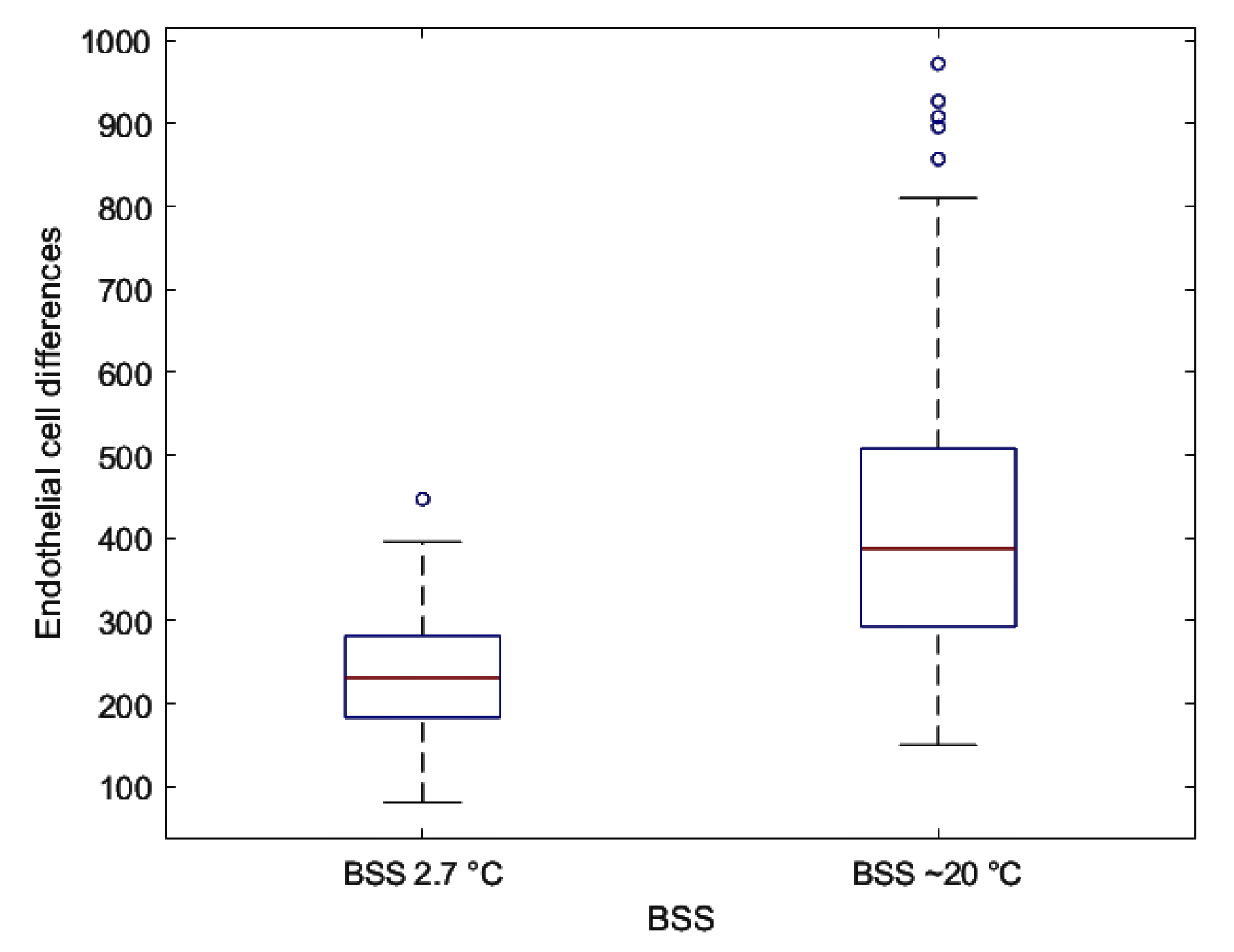
Figure 2 shows the correlation of endothelial cells and AVG, while Figure 3 shows the distribution of endothelial cell differences for BSS at 2.7 °C and at ~20 °C between pre- and post- treatment. Intragroup analyses highlighted a significant difference between pre- and post- treatment for the groups A and B (p < 0.001) (Table 5) and between the differences of the two groups (Table 4), and a significant negative correlation between endothelial cells and AVG.
In Figure 4, the percentage difference of loss cells and the corresponding error during surgery conditions 1 and 2 are depicted. This figure highlights that the use of BSS at 2.7 °C reduces the loss of endothelial cells by 50 %.
One-way ANOVA (Table 6) put in evidence the significant variation in the AVG post- treatment range in group A (p < 0.001). In particular, there was a substantial difference between the 0–9 and 10–19 AVG range (p < 0.001), the 0–9 and 20–29 AVG range (p < 0.001), and the 10–19 and 20–29 AVG range (p < 0.001). Intragroup analyses showed a highly significative difference in each AVG for both groups. An intergroup analysis highlighted a very significative difference in posttreatment in the 10–19 AVG range (p < 0.0001) and in the 20–29 AVG range (p < 0.0001) (Table 7).
One-way ANOVA also put in evidence a significant variation in AVG range in the endothelial cell differences related to the same group. Tukey’s test showed a very significant difference in endothelial cell differences between all the AVG ranges for both groups. An intergroup analysis highlighted a very significant difference in endothelial cell differences between the two groups for every AVG range (Table 8).

4. Conclusions

It is well known that the application of ultrasound during phacoemulsification can lead to endothelial cell damage in cataract surgery due to mechanical trauma from sonic waves and thermal injury [23]. In this scenario, our study demonstrated that the use of a cold irrigation solution plays a pivotal role in reducing or even preventing the damage of corneal endothelial cells during phacoemulsification.
A presurgery test conducted on an artificial eye model allowed us to set the parameters to be used during cataract surgery, to ensure that there was no potential risk for the patients.
In the preoperative period, the two groups of patients did not present statistically significant differences in numbers of endothelial cells. In the postoperative period a clear reduction, i.e., ~50 % of endothelial cells in group A with respect to those in group B (p < 0.001) was found.
We also evaluated subgroups in relation to the AVG and found that there were no significant differences in the softer cataracts, which required an ultrasound percentage of less than 10% of the maximum power. Of note is the data showing a greater reduction in subjects in group B with medium and hard cataract using ultrasounds higher than 10%, among whom we did not observe large losses of endothelial cells compared to those in group A, despite the high percentage of ultrasound used.
For these reasons, and considering the protective role of BSS at low temperature (2.7 ± 0.1 °C), we propose the use of this new technique mainly in medium and hard cataracts and in subjects at risk of corneal decompensation.
Despite these encouraging results, further studies are needed to establish the optimal BSS temperature at which the maximum anti-inflammatory effect can be achieved without causing cold damage. The reproducibility and relatively low cost of the method facilitate its widespread use.

Author Contributions

Conceptualization, A.M and G.A.; methodology, A.M., G.A., S.S.; validation, F.C., G.A.; formal analysis, L.B., S.G.; investigation, A.M., P.A., S.S.; resources, A.M., P.A., S.S.; data curation, B.T.; writing—original draft preparation, F.C., G.A.; writing—review and editing, F.C., G.A., S.G.; supervision, P.A.; All authors have read and agreed to the published version of the manuscript.

Funding

This research received no external funding.

Conflicts of Interest

The authors declare no conflict of interest.

References

  1. Thylefors, B. The World Health Organization’s programme for the prevention of blindness. Int. Ophthalmol. 1990, 14, 211–219. [Google Scholar] [CrossRef] [PubMed]
  2. Christen, W.G.; Glynn, R.J.; Ajani, U.A. Smoking cessation and risk of age-related cataract in men. JAMA 2000, 284, 713–716. [Google Scholar] [CrossRef] [PubMed] [Green Version]
  3. Doors, M.; Berendschot, T.T.J.M.; Touwslager, W.; Webers, C.A.; Nuijts, R.M.M.A. Phacopower modulation and the risk for postoperative corneal decompensation: A randomized clinical trial. JAMA Ophthalmol. 2013, 131, 1443–1450. [Google Scholar] [CrossRef] [PubMed] [Green Version]
  4. Lundberg, B.; Jonsson, M.; Behndig, A. Postoperative corneal swelling correlates strongly to corneal endothelial cell loss after phacoemulsification cataract surgery. Am. J. Ophthalmol. 2005, 139, 1035–1041. [Google Scholar] [CrossRef] [PubMed]
  5. Shah, S.M.; Spalton, D.J. Changes in anterior chamber flare and cells following cataract surgery. Br. J. Ophthalmol. 1994, 78, 91–94. [Google Scholar] [CrossRef] [PubMed] [Green Version]
  6. Gandhi, J.S. Cystoid macular edema after cataract surgery. J. Cataract Refract. Surg. 2018, 44, 1536. [Google Scholar] [CrossRef] [PubMed]
  7. Vieira, I.V.; Boianovsky, C.; Saraiva, T.J.; Godoy, R.B.; de Lake, J. Safety and efficacy of intracameral moxifloxacin injection for prophylaxis of endophthalmitis after phacoemulsification. Arq. Bras. Oftalmol. 2017, 80, 165–167. [Google Scholar] [CrossRef] [PubMed]
  8. Cho, K.; Lim, D.H.; Yang, C.M.; Chung, E.S.; Chung, T.Y. Comparison of Intraocular Lens Power Calculation Methods Following Myopic Laser Refractive Surgery: New Options Using a Rotating Scheimpflug Camera. Korean J. Ophthalmol. 2018, 32, 497–505. [Google Scholar] [CrossRef] [PubMed]
  9. Li, S.; Chen, X.; Zhao, J.; Xu, M.; Yu, Z. Isolated Capsulorhexis Flap Technique in Femtosecond Laser-Assisted Cataract Surgery to Protect the Corneal Endothelial Cells. J. Investig. Surg. 2017, 32, 35–38. [Google Scholar] [CrossRef] [PubMed]
  10. Stănilă, D.M.; Florea, A.M.; Stănilă, A.; Panga, A.A. Endothelial cells loss to the hyperopic pacients during phacoemulsification. Rom. J. Ophthalmol. 2017, 61, 256. [Google Scholar]
  11. Pajic, B.; Cvejic, Z.; Pajic-Eggspuehler, B. Cataract Surgery Performed by High Frequency LDV Z8 Femtosecond Laser: Safety, Efficacy, and Its Physical Properties. Sensors 2017, 17, 1429. [Google Scholar] [CrossRef] [PubMed] [Green Version]
  12. Agarwal, A.; Jacob, S. Current and effective advantages of femtophacoemulsification. Curr. Opin. Ophthalmol. 2017, 28, 49–57. [Google Scholar] [CrossRef] [PubMed]
  13. Kanellopoulos, A.J.; Asimellis, G. Standard manual capsulorhexis/Ultrasound phacoemulsification compared to femtosecond laser-assisted capsulorhexis and lens fragmentation in clear cornea small incision cataract surgery. Eye Vis. 2016, 3, 20. [Google Scholar] [CrossRef] [PubMed] [Green Version]
  14. Joussen, A.M.; Barth, U.; Çubuk, H.; Koch, H.R. Effect of irrigating solution and irrigation temperature on the cornea and pupil during phacoemulsification. J. Cataract Refract. Surg. 2000, 26, 392–397. [Google Scholar] [CrossRef]
  15. World Medical Association. Declaration of Helsinki: Ethical Principles for Medical Research Involving Human Subjects. JAMA 2013, 310, 2191–2194. [Google Scholar] [CrossRef] [PubMed] [Green Version]
  16. R2Evo 3DMarkerLab. Available online: http://www.3dmarketlab.it (accessed on 20 January 2020).
  17. Handheld Meter pH315i Operating Manual. Available online: http://www.globalw.com/downloads/WQ/PH3150i.pdf (accessed on 20 January 2020).
  18. Chylack, L.T.; Wolfe, J.K.; Singer, D.M.; Leske, M.C.; Bullimore, M.A.; Bailey, I.L.; Friend, J.; McCarthy, D.; Wu, S. The Lens Opacities Classification System III—Longitudinal Study of cataract study Group. Arch. Ophtalmol. 1993, 111, 831–836. [Google Scholar] [CrossRef] [PubMed]
  19. Whitestar Signature PRO—Johnson & Johnson Vision. Available online: https://www.dc-ophthalmology.com/products/193 (accessed on 20 January 2020).
  20. SL990-Series Slitlamps Operating Manual. Available online: http://www.iproweb.fr/test/0%20-%20MATOS%20GUILLAUME/CSO%20-%20SL990%20TYPE%205X%20D%20LAMPE%20A%20FENTE/SL990.pdf (accessed on 20 January 2020).
  21. Takahashi, H. Corneal Endothelium and Phacoemulsification. Cornea 2016, 35, S3–S7. [Google Scholar] [CrossRef] [PubMed]
  22. R Software Open Source. Available online: https://rextester.com/l/r_online_compiler (accessed on 8 January 2020).
  23. Walkow, T.; Anders, N.; Klebe, S. Endothelial cell loss after phacoemulsification: Relation to preoperative and intraoperative parameters. J. Cataract Refract. Surg. 2000, 26, 727–732. [Google Scholar] [CrossRef]
Figure 1. The distribution of endothelial cells with balanced salt solution (BSS) at 2.7 °C (a) and at ~20 °C (b), pre- and post- treatment.
Applsci 10 02682 g001
Figure 2. The correlation between endothelial cells (mean difference pre- vs. post- interventions) and the average amount of ultrasound used (AVG) at: (a) BSS at 2.7 °C and (b) BSS ~20 °C.
Applsci 10 02682 g002
Figure 3. The distribution of endothelial cell differences between pre- and post- treatment with BSS at 2.7 °C and at ~20 °C.
Applsci 10 02682 g003
Figure 4. Percentage differences in loss endothelial cells using BSS at 2.7 °C (Group B) and at ~20 °C (Group A), pre- and post- treatment.
Applsci 10 02682 g004
Table 1. Cataract grade of patients, according to Lens Opacities Classification System (LOCSIII) classification, included in the study. All treated cataracts were cortical/nuclear, as it is difficult to provide a clear distinction between them.
LOCS III ClassificationGrade of Opacities% of Patients
Nuclear Opalescence (NO) and Nuclear Color (NC)≥375%
Cortical Cataract (C)≥320%
Posterior Subcapsular Cataract (P)≥35%
Table 2. Table of demographic data and preoperative status of the two groups.
Group AGroup B
Number of patients110104
Males6763
Females4341
Mean age 70 ± 8.568 ± 8.2
OD5848
OS5254
Preoperative status lens
Phakic
Pseudophakic
 
88
22
 
87
17
Table 3. The temperature of the saline solution detected at the outlet of the phacoemulsifier handpiece in the absence of ultrasound (US), under conditions 1 and 2.
Surgery ConditionSolution Temperature in the Bottle (°C)Temperature Output from the Handpiece (°C)Time
118.0 ± 0.119.0 ± 0.14 min
22.7 ± 0.17.4 ± 0.14 min
Table 4. The average and the maximum temperature variation for one minute of simulated intervention under conditions 1 and 2.
Surgery ConditionAverage Temperature Variation (°C) over the TimeMaximum Temperature Variation (°C) over the TimeTime
1+2.5 (from 19 °C to 21.5 °C)+3.1 (from 19 °C to 22.1 °C)1 min
2+0.4 (from 7.4 °C to 7.8 °C)+1.0 (from 7.8 °C to 8.4 °C)1 min
Table 5. Intra- and inter- group statistical differences in endothelial cells for the groups A and B (pre- and post- treatment) and in endothelial cells differences (pre- minus post- treatment).
TreatmentEndothelial Cells at 2.7 °C
Mean ± SD
Endothelial Cells at ~20 °C
Mean ± SD
p-Value
(Student’s Unpaired t-Test)
PRE2354 ± 308.932295 ± 234.460.12
POST2113 ± 283.611877 ± 276.54<0.001 §
p-value
(Student’s paired t-test)
<0.001 §<0.001 §
Differences240.87 ± 73.09418.63 ± 185.79<0.001 §
§p < 0.001.
Table 6. One-way ANOVA results.
TreatmentAVGPatients NumberMean ± SDp
PRE 0–9452316.2 ± 255.370.7Group A (BSS ~20 °C)
10–19492275.45 ± 221.15
20–29162297.15 ± 221.47
POST 0–9452050.31 ± 240.56<0.001 §
10–19491830.79 ± 210.55
20–29161528.62 ± 132.56
PRE0–9232312.08 ± 313.020.58Group B (BSS 2.7 °C)
10–19532368.87 ± 333.57
20–29232392.35 ± 261.01
>3052215.2 ± 227.77
POST0–9232139.65 ± 290.430.13
10–19532141.3 ± 295.09
20–29232081.96 ± 242.66
>3051838.4 ± 191.84
§p < 0.001.
Table 7. Intra- and inter- group of subgroups divided by AVG range in endothelial cells for groups A (BSS ~20 °C) and B (BSS 2.7 °C) (pre- and post- treatment).
AVGTreatmentGroup A
Mean ± SD
Group B
Mean ± SD
p-Value
(Student’s Unpaired t-Test)
0–9PRE2316.2 ± 255.372312.08 ± 313.020.95
POST2050.31 ± 240.56 §2139.65 ± 290.43 §0.18
10–19PRE2275.45 ± 221.152368.87 ± 333.570.1
POST1830.79 ± 210.55 §2141.3 ± 295.09 §<0.001 §§
20–29PRE2297.15 ± 221.472392.35 ± 261.010.24
POST1528.62 ± 132.56 §2081.96 ± 242.66 §<0.001 §§
>30PRE-2215.2 ± 227.77-
POST-1838.4 ± 191.84 §-
§p < 0.05 for the Student’s paired t-test between PRE and POST. §§ p < 0.05 for the Student’s unpaired t-test between A and B.
Table 8. Intra- and inter- group statistical differences in subgroups differences divided by AVG range of endothelial cells for groups A (BSS ~20 °C) and B (BSS 2.7 °C).
AVGGroup A
Mean ± SD
Group B
Mean ± SD
p-Value
(Student’s Unpaired t-Test)
0–9268.19 ± 69.14172.44 ± 29.22<0.001 §
10–19446.17 ± 79.39227.57 ± 52.79<0.001 §
20–29768.5 ± 119.74310.39 ± 50.89<0.001 §
>30-376.8 ± 47.44-
p-value
(ANOVA)
<0.001§<0.001§
§p < 0.001.