Article

“Green” Synthesis and Antioxidant Activity of Thermally Stable Gold Nanoparticles Encapsulated in Carbon Nanosheets

1
Department of Optometry, Asia University, Taichung 41354, Taiwan
2
Genetics Center, Department of Medical Research, China Medical University Hospital, and School of Chinese Medicine, China Medical University, Taichung 40402, Taiwan
3
Department of Medical Applied Chemistry, Chung Shan Medical University, Taichung 40201, Taiwan
4
Department of Applied Materials and Optoelectronic Engineering, National Chi Nan University, Nantou 54561, Taiwan
5
Department of Medical Education, Chung Shan Medical University Hospital, Taichung 40201, Taiwan
*
Authors to whom correspondence should be addressed.
Appl. Sci. 2020, 10(7), 2272; https://doi.org/10.3390/app10072272
Submission received: 12 March 2020 / Revised: 23 March 2020 / Accepted: 24 March 2020 / Published: 26 March 2020
(This article belongs to the Special Issue Nano-Biophotonics in Sensing Using Nanostructures)

Abstract

:

Featured Application

According to our results, AuNPs with thermal stability and antioxidant property prepared through biogenic approaches have potential use in food preservation.

Abstract

We have developed a “green” method for fabricating gold nanoparticles (AuNPs) through biogenic approaches. The proposed method has the advantages of facile preparation under ecofriendly conditions. AuNPs encapsulated in carbon nanosheets, and exhibiting high thermal stability, were fabricated by autoclaving pectin-capped AuNPs, which were subsequently collected through high-speed centrifugation and redispersed in aqueous solution. The 1,1-diphenyl-2-picrylhydrazy (DPPH) radical scavenging assay indicated that our prepared AuNPs exhibited more prolonged antioxidant capacity than pristine apple extracts. Electron paramagnetic resonance (EPR) spectra showed that approximately 80% of DPPH radicals were scavenged by the pectin-capped AuNPs at a concentration of 3 mg/mL. According to our results, AuNPs prepared through biogenic approaches have potential use in the food industry.

1. Introduction

Biogenic approaches for synthesizing gold nanostructures with unique chemophysical and biological properties have received considerable attention in the last decade [1]. “Green” synthesis methods typically entail adopting environmentally friendly protocols based on natural or biological agents such as nature gums [2], fruit extracts [3,4,5,6,7,8], plant tissues (including leaf, fruit, peel, flower, and root) [9,10,11,12,13,14,15] for synthesis. For example, in one-pot single-step synthesis methods, biological extracts are mixed with an auric salt (HAuCl4) solution at either room or elevated temperature and conversion occurs within a few minutes in the absence of external additives used as reducing, stabilizing, or capping agents. Moreover, metal nanoclusters of different sizes, shapes, and morphologies can be readily prepared on a large scale by adjusting reaction conditions, such as pH and temperature [16]. However, the mechanism underlying crystal growth has yet to be adequately established because of the complex compositions of natural components. Various biological reductants present in plant extracts, such as sugars, amines, phenolic compounds, and flavonoids, can initiate the reduction of gold ions; these biochemicals also help stabilize gold colloids [17,18,19,20,21].
Among the biogenic resources, pectin is a naturally occurring gel present in fruits such as citrus peels, oranges, apples, cherries, and plums. Pectin can serve as a biological agent for preparing gold nanoparticles (AuNPs) with bioactive functions [22,23]. Pectin is mainly composed of a complex set of polysaccharides and is rich in polymers of d-galacturonic acid (GalA) with α(1−4) linkages. In general, the polygalacturonic acid chain of pectin is partly esterified with methyl groups, and free acid groups may be partly or fully neutralized with sodium, potassium, or ammonium ions. The ratio of esterified GalA groups to total GalA groups is termed as the degree of esterification (DE). Pectin can be classified into low- and high-methoxy groups according to the corresponding DE values. Pectin not only exhibits gelling, thickening, and emulsion-stabilizing properties that are commonly used in the food industry but also has potential pharmaceutical applications such as controlled drug delivery, detoxification, and antioxidation.
In this study, pectin extracted from red apples (Malus pumila) was utilized as the biogenic resource to synthesize AuNPs. The antioxidant components of apples, including polyphenols, can induce the reduction of gold ions and initiate crystal growth. Polysaccharides present in pectin can provide a gel network for stabilizing nanoparticles. Accordingly, the pectin-capped AuNPs prepared in this study exhibited remarkable thermal stability under hydrothermal carbonization and consequently formed a hybrid nanostructure of AuNPs encapsulated in carbon nanosheets. Previous studies have used complex fabrication processes, for example sintering, sputtering, multi-steps, and complex chemicals [24,25,26,27,28,29,30,31] to obtain AuNPs with high thermal stability. However, the lack of bioactive capping agents hinders the practical application of such processes in food preservation [32]. In this study, we analyzed the absorption spectra and morphology of pectin-capped AuNPs with a defined size and found that the characteristic absorption remained unaltered after the AuNPs were redispersed into the aqueous solution. The particle size of pectin-capped AuNPs changed from 10 nm to 7 nm after the AuNPs heating in an autoclave. The 1,1-diphenyl-2-picrylhydrazy (DPPH) radical scavenging assay was used to determine the antioxidant activity of AuNPs encapsulated with carbon nanosheets, indicating that our prepared AuNPs exhibited more prolonged antioxidant capacity than pristine apple extracts. The reason for using the DPPH assay is for comparison with electron paramagnetic resonance (EPR) spectra where the EPR results showed that approximately 80% of DPPH radicals were scavenged by the pectin-capped AuNPs at a concentration of 3 mg/mL. The embedded AuNPs can enhance the radical scavenging efficiency of pectin extracts, rendering organic-inorganic hybrids fabricated at high temperatures a potential dietary supplement in the food industry.

2. Materials and Methods

2.1. Materials

Red apples (Fuji 4131) were purchased in bulk from a local supermarket in Taiwan. Tetrachloroauric (III) acid trihydrate (49% Au) and sodium citrate (99%) were purchased from Acros Organics (Geel, Belgium) and Hayashi Pure Chemical Ind., Ltd (Osaka, Japan), respectively. All chemicals were used without further purification. Milli-Q water was used for all experiments.

2.2. Apple Extract Preparation

Apple extract was prepared using fresh, peeled apples. The peeled apples were chopped, and 120 g of the chopped apples blended with 100 mL of pure water. The apple juice filtered from the resulting apple slurry was centrifuged at 7000 rpm for 15 min to obtain clear apple juice at the top.

2.3. Pectin-Capped AuNP Preparation

Pectin-capped AuNPs were prepared by reducing 0.25 mM of 200 mL tetrachloroauric (III) acid trihydrate with 50 mL of apple extract heated at 100 °C, and the homogeneous mixture was stirred for 30 min. The solution was filtered using vacuum filtration after keeping the sample overnight. The resulting AuNP aqueous solution was centrifuged at 12,000 rpm for 10 min to obtain the condensed AuNPs on the bottom. The condensed AuNPs were then redispersed in 5 mL Mili-Q water using a sonicator, and finally, a freeze-drying system was used to get the powder-like AuNPs for further use.

2.4. Citrate-Capped AuNP Preparation

Citrate-capped AuNPs were prepared by reducing 0.25 mM of 200 mL tetrachloroauric (III) acid trihydrate with 50 mL of 4 mM sodium citrate heated at 100 °C, and the homogeneous mixture was stirred for 30 min.

2.5. Preparation of AuNPs Encapsulated in Carbon Nanosheets

Powder of pectin-capped AuNPs was obtained using a freeze-drying method, and was weighed for further use. AuNPs, encapsulated in carbon nanosheets, were fabricated in an autoclave at two separate temperatures of 120 and 180 °C.

2.6. Characterization

The absorption spectra of the samples were measured using a Thermo Genesys 10S UV–visible spectrometer (Thermo-Fisher Scientific, Waltham, MA, USA), operated at a resolution of 1 nm. Standard quartz cells with an optical path length of 10 mm were used for sample characterization. Scanning electron microscopy (SEM) and the corresponding elemental analysis were conducted using a JEOL JSM-7800F field-emission scanning electron microscope (JEOL, Tokyo, Japan) equipped with an X-ray energy-dispersive spectrometry (EDS) instrument. Transmission electron microscopy (TEM) was performed using a JEOL JEM-2100 transmission electron microscope (JEOL, Tokyo, Japan). High-resolution TEM (HRTEM) and the corresponding elemental analysis were performed using a Thermo Fisher Titan transmission electron microscope. The 1,1-diphenyl-2-picrylhydrazyl (DPPH) free radical scavenging assay was conducted according to procedures [33]; specifically, different concentrations (1–8 mg/mL) of AuNPs in an equal volume of 1.2 mL were added to 1 mM DPPH in a 50 μL methanol solution. After 30 min of reaction at room temperature, changes in the absorption peak of DPPH at 517 nm were measured for further calculating the clearance rate. The absorption value was incorporated into the following formula to calculate the clearance rate of DPPH free radicals: percentage of inhibition of DPPH radical scavenging activity = A o ( A f A i ) / A o , where Ao is the absorbance of pure water (blank), Af is the absorbance of a solution with DPPH, and Ai is the absorbance of the solution without DPPH. Moreover, the antioxidant properties of the prepared AuNPs were studied through EPR spectroscopy (Bruker EMSplus spectrometer, Billerica, MA, USA).

3. Results and Discussion

For comparison, AuNPs were first fabricated through a standard one-pot single-step synthesis process in the presence of sodium citrate, apple extracts (which are typically rich in natural pectin), and with the precursor, a HAuCl4 water solution. The freeze-drying method was used to make powdered-like AuNPs compared to the redispersion ability of both fabricated AuNPs. Compared with the AuNPs prepared through the standard method of using sodium citrate as the reducing agent and stabilizer, the AuNPs fabricated using our biogenic (green) approach exhibited a less resolved and red-shifted absorption band centered at 532 nm (Figure 1a; spectrum I). The spectral analysis results indicated that the AuNPs fabricated using the green method exhibited a broader particle size distribution and morphology than did those fabricated using the standard method (Figure 1b, spectrum I). Furthermore, the AuNPs were purified by collecting precipitates through high-speed centrifugation. The AuNPs were completely accumulated and collected after centrifugation. In both cases, no localized surface-plasmon resonance (LSPR) absorption bands were observed in the supernatant (Figure 1a,b, spectrum II). The AuNPs fabricated using the green method was redispersed in water at the same concentration, and two characteristic LSPR (as-prepared and redispersed) absorption bands were observed and determined to be superimposable (Figure 1a, spectra I and III). However, the sodium-citrate-stabilized AuNPs could not recover their redispersion ability (i.e., recover their LSPR absorption bands; Figure 1b, spectra I, and III); this thus indicates the instability of sodium-citrate-capped AuNPs in water solution and demonstrate that such AuNPs cannot be applied in the food industry. AuNPs are sensitive to environmental fluctuations such as changes in pH, ionic strength, and temperature. When AuNPs are capped with a thin layer of stabilizing molecules, high-speed centrifugation may lead to irreversible aggregation. This thus explains why the sodium-citrate-capped AuNPs lost their redispersion ability in the water. However, the AuNPs prepared through the green method using pectin as the promoter exhibited excellent stability; this may be because the gel-like pectin provided solid support to stabilize the AuNPs against irreversible aggregation.
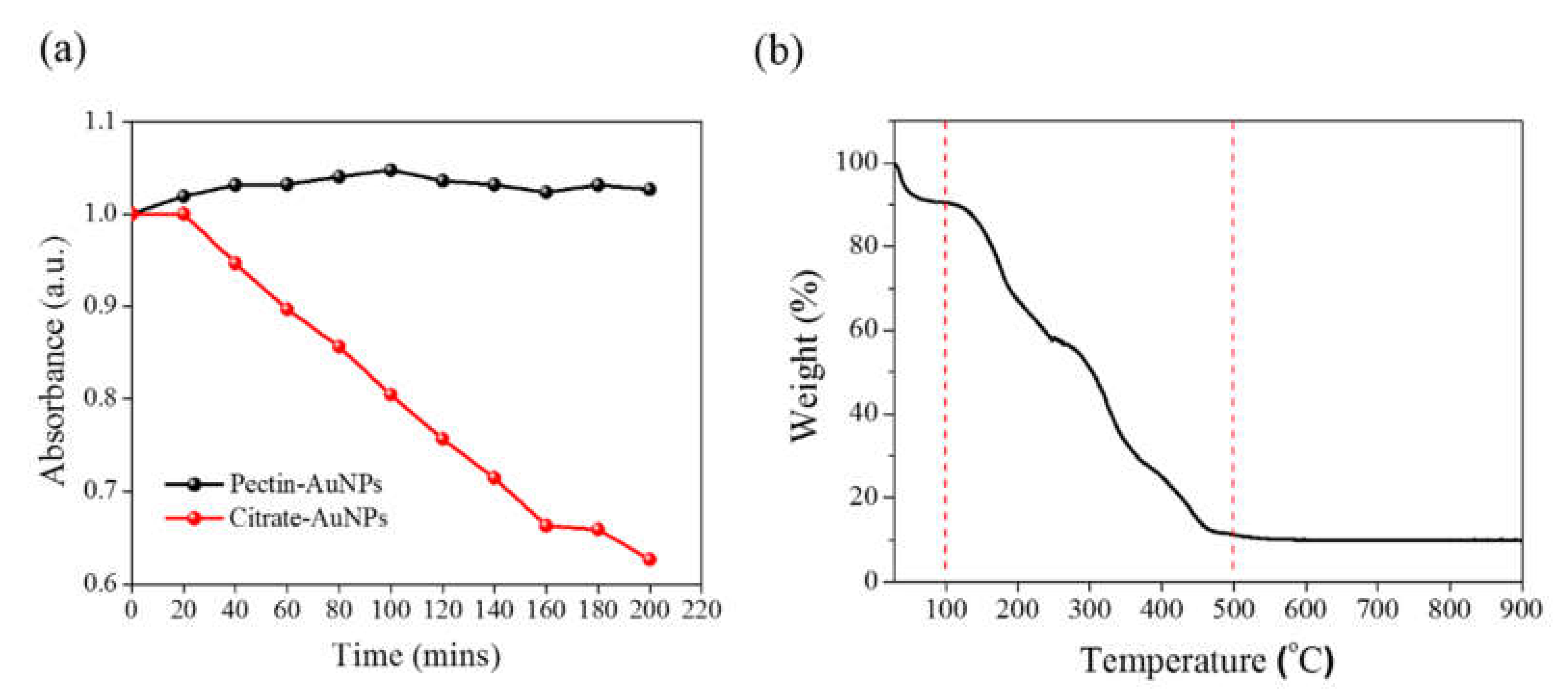
Figure 2a illustrates the variations in the absorbance of AuNPs dissolved in the water solution and heated under 80 °C for 200 min. The absorbance for the green-synthesized AuNPs remained constant after these AuNPs were heated. By contrast, the characteristic LSPR absorption peak of the sodium-citrate-capped AuNPs decreased gradually in the thermal environment. This finding suggests that the pectin-capped AuNPs were more stable than the sodium-citrate-capped AuNPs. Figure 2b presents the thermogravimetric analysis (TGA), Thermo Electron Corporation (Newington, NH, USA), results for the pectin-capped AuNPs, revealing a remarkable weight loss before 500 °C; this thus confirms that these AuNPs comprised 90% and 10% organic and inorganic contents, respectively. Accordingly, the primary constituents of the AuNPs were pectin and other organic components, and the secondary constituents were Au nanoclusters used for decoration.
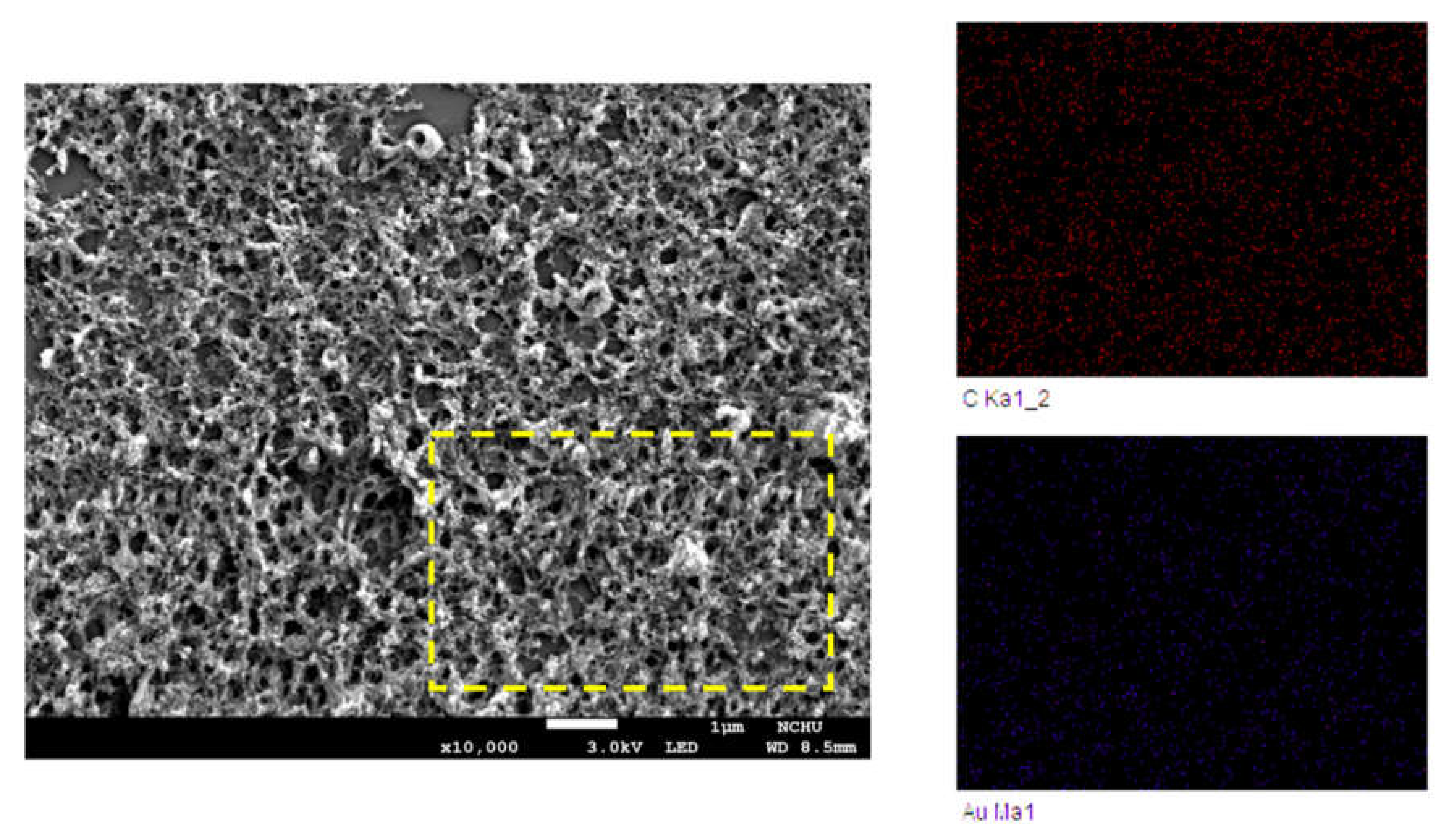
The morphology of the pectin-capped AuNPs deposited on a silicon substrate was analyzed using SEM, as shown in Figure 3, revealing a gel network engendered by the apple extract, which prevents the AuNPs further assembling into large aggregates under the thermal environment. In addition, elemental mapping revealed the distributions of both carbon and gold in a selected area of the SEM image (Figure 3). Instead of the fabrication of a thin organic layer on the nanoparticle surface, the gold nanoclusters were embedded within the apple extracts to form inorganic–organic hybrid materials.
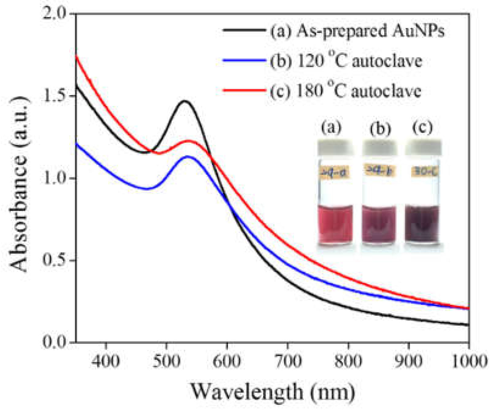
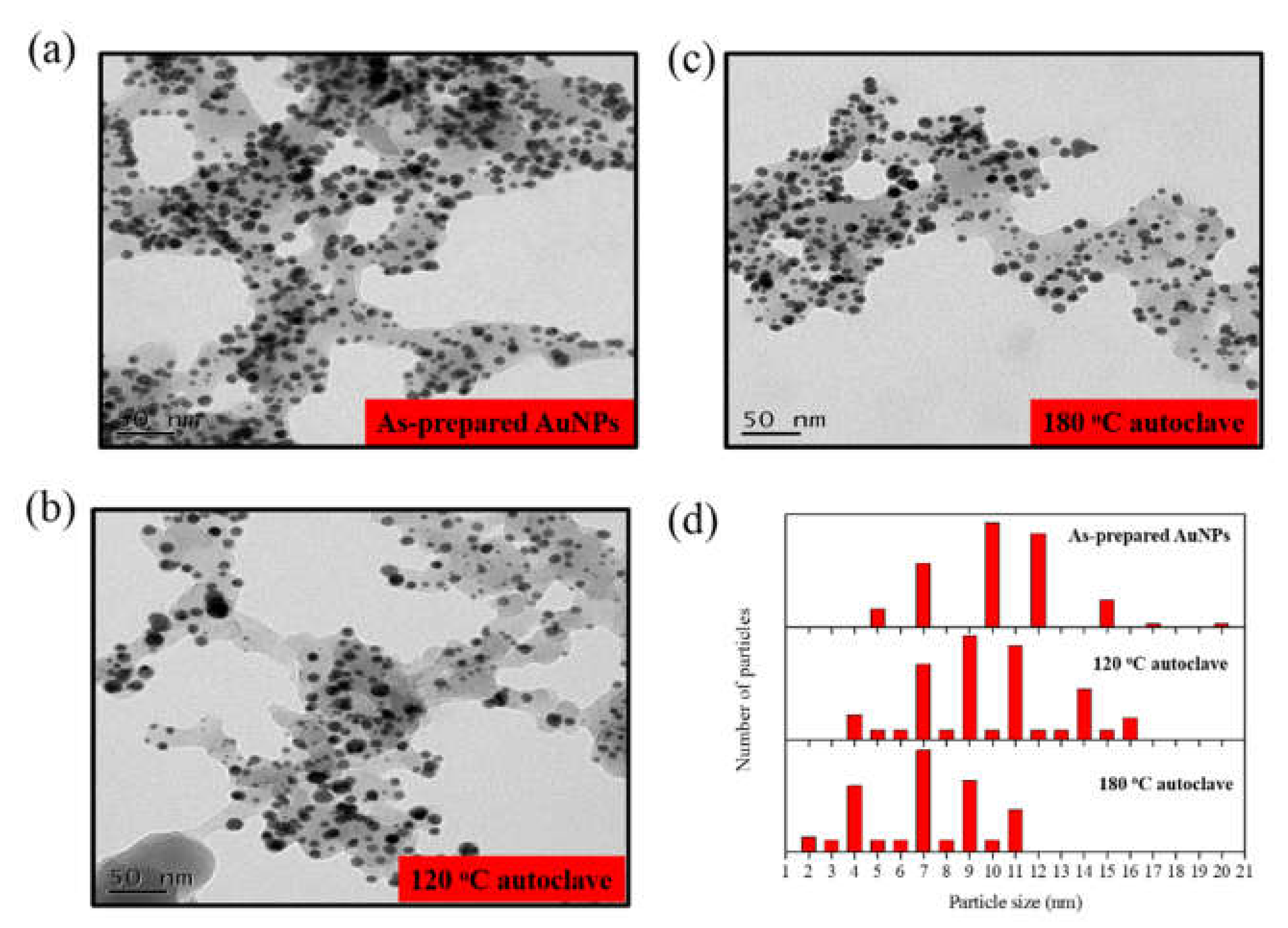
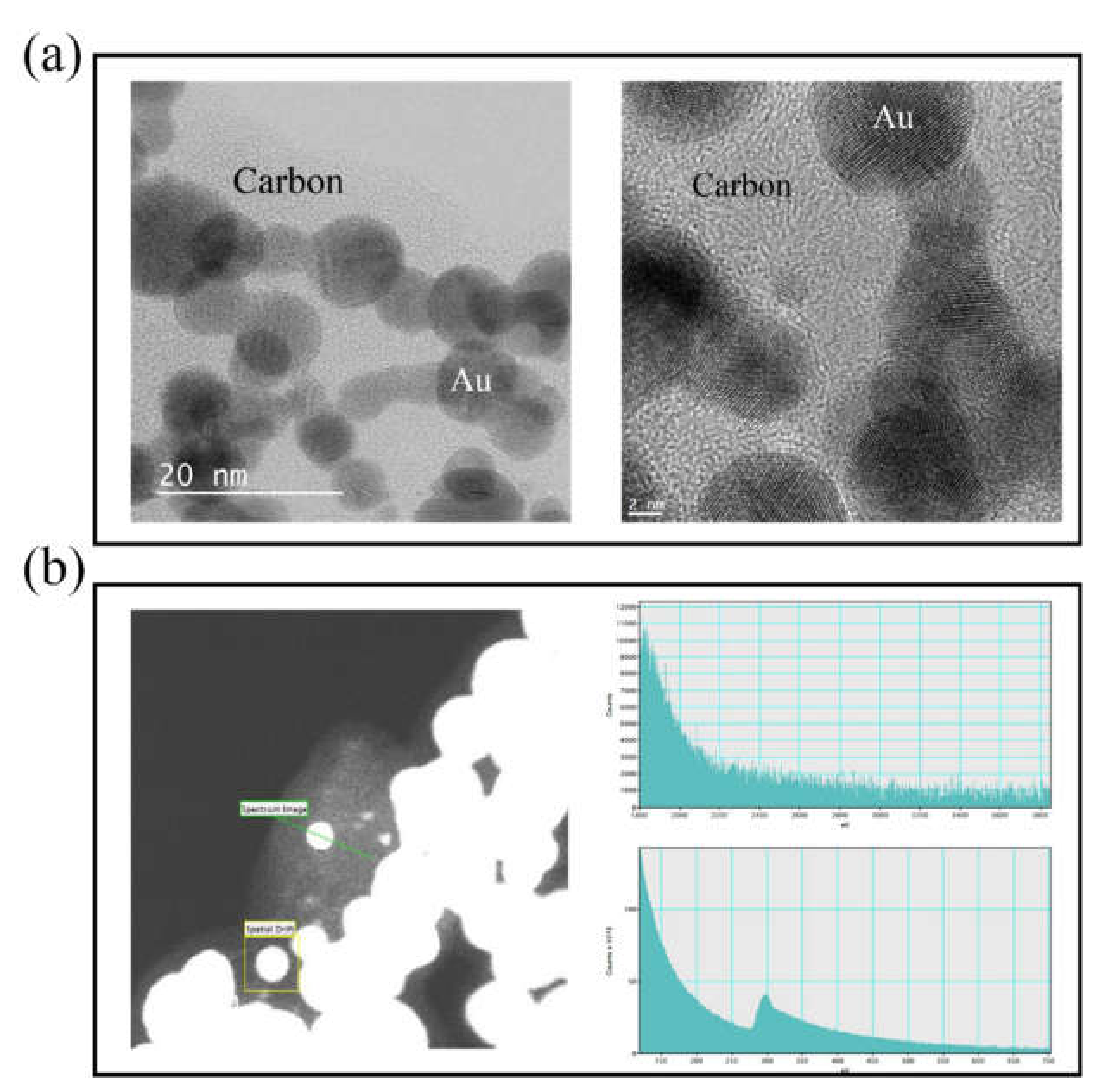
A hydrothermal process was further conducted to heat the pectin-capped AuNPs in a Teflon-lined autoclave. Carbonization under a low-temperature (120 °C) or a high-temperature (180 °C) protocol was adopted to fabricate carbon dots with partial crystallinity and unique photoluminescence. As displayed in Figure 4, the LSPR absorption bands observed for the pectin-capped AuNPs remained nearly unchanged after the hydrothermal treatment process conducted under the two protocols. Moreover, the absorption maximum was centered at approximately 530 nm, suggesting that the size and morphology of nanoclusters remained unaltered after heating in the autoclave. Figure 5a–c presents the TEM images of the AuNPs, indicating that most of the hybrid nanoclusters exhibited spherical morphologies after the hydrothermal treatment process; however, the particle size distribution of the AuNPs, calculated by manually measuring 100 AuNPs, decreased slightly at elevated temperatures (Figure 5d). Notably, all AuNPs were embedded within an irregular carbon nanosheet, confirming that the hybrid nanoclusters comprised two components: an organic component and AuNPs. The high thermal stability of the pectin-capped AuNPs may be attributable to the formation of a rigid carbon-based network against thermal-induced aggregation [27]. Figure 6a depicts high-resolution TEM images of the pectin-capped AuNPs, revealing organic (carbon) and inorganic (AuNPs) components and a clear boundary between the crystalline AuNPs and amorphous carbon. Moreover, the darker spots against the carbon background in these images (Figure 6a) represent the crystal lattice of AuNPs with a size distribution of < 20 nm. We also subjected the AuNPs to electron energy-loss spectroscopy (EELS). Figure 6b presents the EELS results, confirming the presence of carbon and gold elements. Overall, the TEM results reveal that organic extracts from red apples were wrapped around the nanoparticles rather than being deposited as a thin layer on the particle surface. We speculate that this carbon-based feature can render AuNPs extremely stable under conditions of temperature fluctuations. The fabricated hybrid materials composed of pectin extracts and metal nanoparticles have a potential for biological applications in which toxic and oxidant issues may hinder the practical use of metal nanoparticles [33].
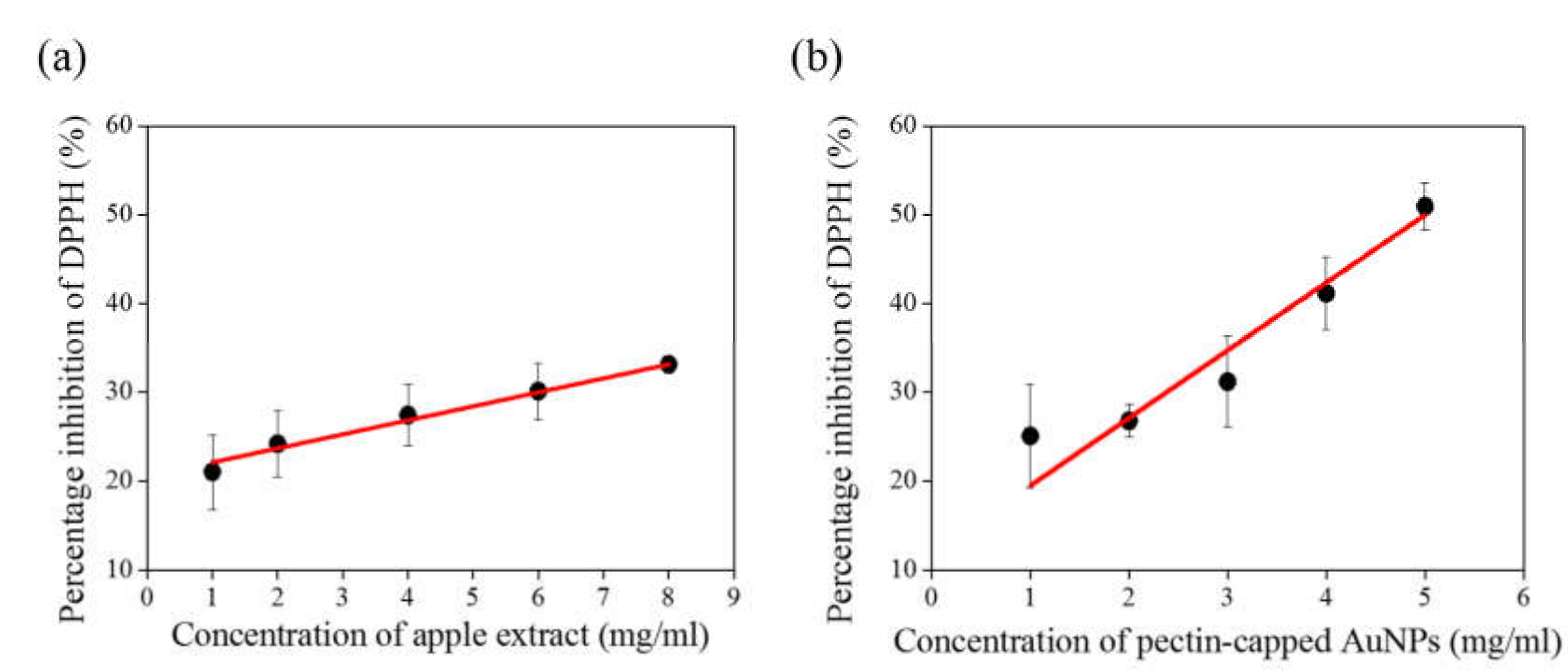
Apple extracts with antioxidant and anti-inflammatory effects may be possibly responsible for the potential cancer-preventive candidates [34]. Accordingly, we conducted the DPPH radical scavenging assay to preliminarily investigate the antioxidant capability of AuNPs encapsulated in carbon nanosheets, which were fabricated through the hydrothermal process using the pectin-capped AuNPs as precursors. DPPH is a stable nitrogen-containing free radical molecule with a dark purple appearance. DPPH can capture other free radical molecules, and their characteristic absorption peak, which can be attributed to the resonance between one nitrogen atom and three benzene rings, decreasing with the occurrence of free radical reactions. In this study, we quantified the decrease in absorbance to determine the antioxidant activity of AuNPs encapsulated in carbon nanosheets at different concentrations. Figure 7 presents a comparison of the antioxidant activity of apple extracts with that of the AuNPs/carbon nanosheets. The DPPH free radical removal rate was higher for the AuNPs.
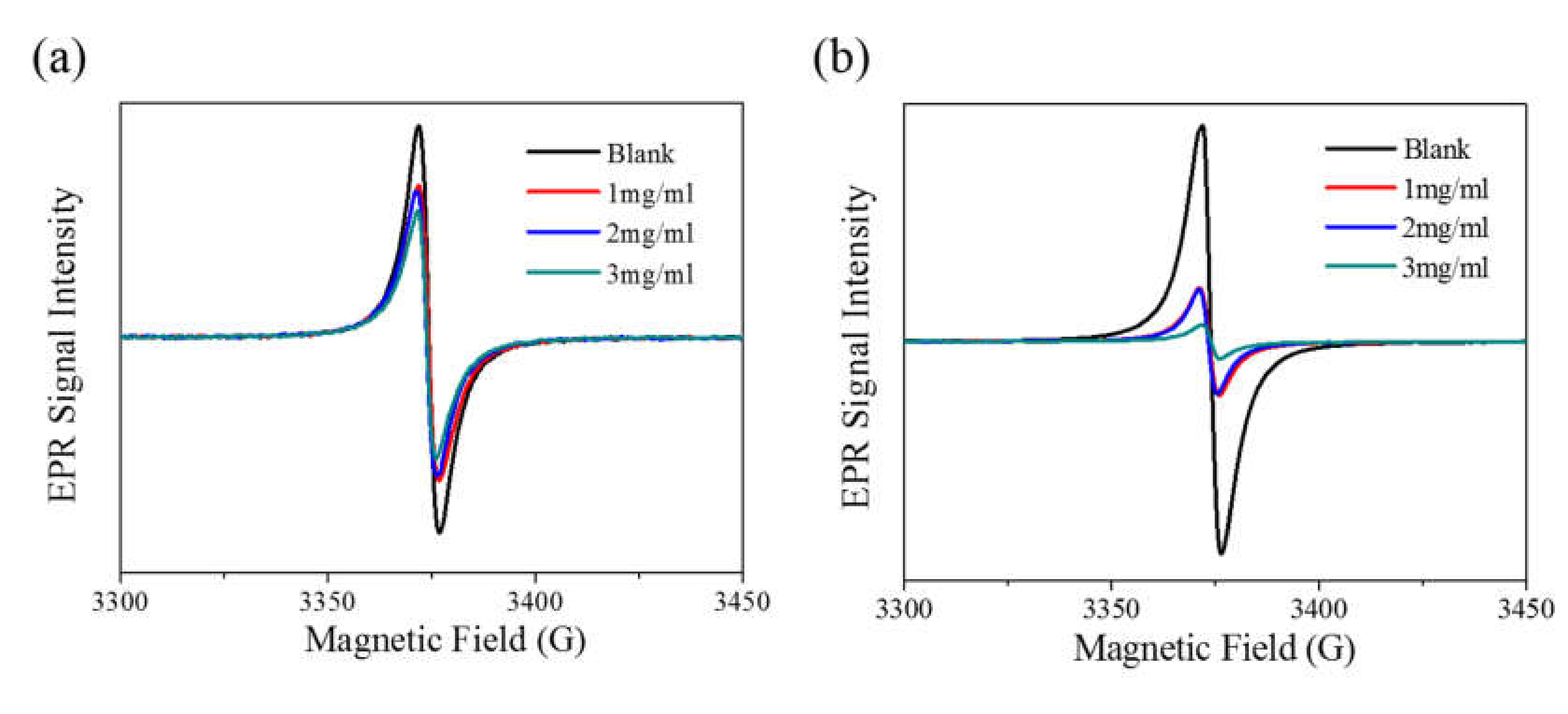
We further used an EPR spectrometer to determine the DPPH free radical removal rate. In EPR, the presence of unpaired electrons, such as free radicals, indicates the occurrence of magnetic resonance. The resonance intensity can be used to determine the concentration of DPPH free radicals. Figure 8 presents the EPR spectra for the apple extract and AuNP/carbon nanosheet solutions. Similar to the preceding comparison results, the EPR results indicated that the DPPH removal rate was higher for the AuNPs/carbon nanosheets than it was for the apple extract. Moreover, we calculated the DPPH radical removal rate by integrating the EPR spectra; the results showed a 79% removal rate for the AuNPs/carbon nanosheets and a 30% removal rate for the apple extracts at a concentration of 3 mg/mL.

4. Conclusions

In this study, we synthesized AuNPs through a green approach by using apple extract as a reducing agent. AuNPs with a particle size of approximately 12.0 ± 5.7 nm were thermally stable under an autoclaving condition of 180 °C. AuNPs encapsulated in carbon nanosheets and exhibited considerably higher thermal stability and redispersion ability than did those prepared through the traditional citrate-based method. In addition, DPPH radical scavenging assay results reveal that the pectin-capped AuNPs has higher DPPH free radical removal efficiency than did the apple extract. DPPH free radical assay and EPR spectra all indicated that the pectin-capped AuNPs exhibited satisfactory antioxidant ability. In conclusion, AuNPs with moderate to high thermal stability can be used in the food industry, like in food preservation [35] where higher temperatures are required during manufacturing.

Author Contributions

Conceptualization, V.K.S.H. and C.-C.C.; methodology and validation, J.-Y.W.; formal analysis and investigation, H.-W.L.; writing—original draft preparation, H.-W.L. and C.-C.C.; writing—review and editing, V.K.S.H.; funding acquisition, V.K.S.H. and C.-C.C. All authors have read and agreed to the published version of the manuscript.

Funding

This research was funded by Ministry of Science and Technology (MOST) of Taiwan under the project number MOST106-2113-M-040-001, MOST107-2113-M-040-003 and MOST107-2221-E-260-016-MY3.

Acknowledgments

The authors especially thanks Prof Jing-Yun Wu for the instruction and assistance on TGA measurements.

Conflicts of Interest

The authors declare no conflict of interest.

References

  1. Teimuri-Mofrad, R.; Hadi, R.; Tahmasebi, B.; Farhoudian, S.; Mehravar, M.; Nasiri, R. Green synthesis of gold nanoparticles using plant extract: Mini-review. Nanochem. Res 2017, 2, 8–19. [Google Scholar]
  2. Madhusudhan, A.; Reddy, G.B.; Krishana, I.M. Chapter 4 Green Synthesis of Gold Nanoparticles by Using Natural Gums. Nanomaterials and Plant Potential; Husen, A., Iqbal, M., Eds.; Springer: Berlin/Heidelberg, Germany, 2019; pp. 111–134. [Google Scholar]
  3. Vijayakumar, S. Eco-friendly synthesis of gold nanoparticles using fruit extracts and in vitro anticancer studies. J. Saudi Chem. Soc. 2019, 23, 753–761. [Google Scholar] [CrossRef]
  4. Devendiran, R.M.; Chinnaiyan, S.K.; Yadav, N.K.; Moorthy, G.K.; Ramanathan, G.; Singaravelu, S.; Sivagnanam, U.T.; Perumal, P.T. Green synthesis of folic acid-conjugated gold nanoparticles with pectin as reducing/stabilizing agent for cancer theranostics. RSC Adv. 2016, 6, 29757–29768. [Google Scholar] [CrossRef]
  5. Rimal Isaac, R.S.; Sakthivel, G.; Murthy, C.H. Green Synthesis of Gold and Silver Nanoparticles Using Averrhoa bilimbi Fruit Extract. J. Nanotechnol. 2013, 2013, 6. [Google Scholar] [CrossRef] [Green Version]
  6. Lee, K.X.; Shameli, K.; Miyake, M.; Kuwano, N.; Ahmad Khairudin, N.B.B.; Mohamad, S.E.B.; Yew, Y.P. Green Synthesis of Gold Nanoparticles Using Aqueous Extract of Garcinia mangostana Fruit Peels. J. Nanomater. 2016, 2016, 7. [Google Scholar] [CrossRef] [Green Version]
  7. Sujitha, M.V.; Kannan, S. Green synthesis of gold nanoparticles using Citrus fruits (Citrus limon, Citrus reticulata and Citrus sinensis) aqueous extract and its characterization. Spectrochim. Acta A 2013, 102, 15–23. [Google Scholar] [CrossRef]
  8. Sett, A.; Gadewar, M.; Sharma, P.; Deka, M.; Bora, U. Green synthesis of gold nanoparticles using aqueous extract of Dillenia indica. Adv. Nat. Sci. Nanosci. Nanotechnol. 2016, 7, 025005. [Google Scholar] [CrossRef]
  9. Ramírez Castro, J.; García Hernández, L.; Ramírez Ortega, P.A.; Arenas Islas, D. Green synthesis of gold nanoparticles (AuNPs) by Cupressus goveniana extract. ECS Transact. 2018, 84, 207–215. [Google Scholar] [CrossRef]
  10. Vijaya Kumar, P.; Mary Jelastin Kala, S.; Prakash, K.S. Green synthesis of gold nanoparticles using Croton Caudatus Geisel leaf extract and their biological studies. Mater. Lett. 2019, 236, 19–22. [Google Scholar] [CrossRef]
  11. Shabestariana, H.; Homayouni-Tabrizib, M.; Soltanic, M.; Namvard, F.; Azizif, S.; Mohamadd, R.; Shabestarianb, H. Green Synthesis of Gold Nanoparticles Using Sumac Aqueous Extract and Their Antioxidant Activity. Mater. Res. 2017, 20, 264–270. [Google Scholar] [CrossRef] [Green Version]
  12. Lal, S.; Nayak, P.L. Green synthesis of gold nanoparticles using various extract of plants and spices. IJSID 2012, 2, 325–350. [Google Scholar]
  13. Anjana, P.M.; Bindhu, M.R.; Rakhi, R.B. Green synthesized gold nanoparticle dispersed porous carbon composites for electrochemical energy storage. Mater. Sci. Energy Tech. 2019, 2, 389–395. [Google Scholar] [CrossRef]
  14. Patra, J.K.; Baek, K.-H. Novel green synthesis of gold nanoparticles using Citrullus lanatus rind and investigation of proteasome inhibitory activity, antibacterial, and antioxidant potential. Int. J. Nanomed. 2015, 10, 7253–7264. [Google Scholar]
  15. Darabdhara, G.; Das, M.R.; Singh, S.P.; Rengan, A.K.; Szunerits, S.; Boukherroub, R. Green one-pot synthesis of gold nanoparticles using Sansevieria roxburghiana leaf extract for the catalytic degradation of toxic organic pollutants. Mater. Res. Bull. 2019, 117, 18–27. [Google Scholar]
  16. Bogireddy, N.K.R.; Pal, U.; Martinez Gomezc, L.; Agarwal, V. Size controlled green synthesis of gold nanoparticles using Coffea arabica seed extract and their catalytic performance in 4-nitrophenol reduction. RSC Adv. 2018, 8, 24819. [Google Scholar] [CrossRef] [Green Version]
  17. Aljabali, A.A.A.; Akkam, Y.; Al-Zoubi, M.S.; Al-Batayneh, K.M.; Al-Trad, B.; Alrob, O.A.; Alkilany, A.M.; Benamara, M.; Evans, D.J. Synthesis of Gold Nanoparticles Using Leaf Extract of Ziziphus zizyphus and their Antimicrobial Activity. Nanomaterials 2018, 8, 174. [Google Scholar] [CrossRef] [PubMed] [Green Version]
  18. Golmoraj, V.E.; Khoshayand, M.R.; Amini, M.; Moghadamd, K.M.; Amin, G.; Shahverdi, A.R. The surface chemistry and stability of gold nanoparticles prepared using methanol extract of Eucalyptus camaldulensis. J. Exp. Nanosci. 2010, 6, 200–208. [Google Scholar] [CrossRef]
  19. Thirumalraj, B.; Rajkumar, C.; Chen, S.-M.; Palanisamy, S. One-Pot Green Synthesis of Graphene Nanosheets Encapsulated Gold Nanoparticles for Sensitive and Selective Detection of Dopamine. Sci. Rep. 2017, 7, 412213. [Google Scholar] [CrossRef] [PubMed] [Green Version]
  20. Roy, A.; Mohanta, B. Microwave-assisted green synthesis of Gold nanoparticles and its catalytic activity. Int. J. Nano Dimens. 2019, 10, 359–367. [Google Scholar]
  21. Liu, Y.; Kim, S.; Kim, Y.J.; Perumalsamy, H.; Lee, S.; Hwang, E.; Yi, T.-H. Green synthesis of gold nanoparticles using Euphrasia officinalis leaf extract to inhibit lipopolysaccharide-induced inflammation through NF-κB and JAK/STAT pathways in RAW 264.7 macrophages. Int. J. Nanomed. 2019, 14, 2945–2959. [Google Scholar] [CrossRef] [Green Version]
  22. Wang, A.; Ng, H.P.; Xu, Y.; Li, Y.; Zheng, Y.; Yu, J.; Han, F.; Peng, F.; Fu, L. Gold Nanoparticles: Synthesis, Stability Test, and Application for the Rice Growth. J. Nanomater. 2014, 2014, 6. [Google Scholar] [CrossRef]
  23. Vanitha Kumari, G.; JothiRajan, M.A.; Mathavan, T. Pectin functionalized gold nanoparticles towards singlet oxygen generation. Mater. Res. Express 2018, 5, 085027. [Google Scholar] [CrossRef]
  24. Veith, G.M.; Lupini, A.R.; Rashkeev, S.; Pennycook, S.J.; Mullins, D.R.; Schwartz, V.; Bridges, C.A.; Dudney, N.J. Thermal stability and catalytic activity of gold nanoparticles supported on silica. J. Catal. 2009, 262, 92–101. [Google Scholar] [CrossRef]
  25. Masoud, N.; Partsch, T.; de Jong, K.P.; de Jongh, P.E. Thermal stability of oxide-supported gold nanoparticles. Gold Bull. 2019, 52, 105–114. [Google Scholar] [CrossRef] [Green Version]
  26. Liu, X.; Wang, A.; Yang, X.; Zhang, T.; Mou, C.-Y.; Su, D.-S.; Li, J. Synthesis of Thermally Stable and Highly Active Bimetallic Au-Ag Nanoparticles on Inert Supports. Chem. Mater. 2009, 21, 410–418. [Google Scholar] [CrossRef]
  27. Kim, M.; Shhn, K.; Na, H.B.; Hyeon, T. Synthesis of Nanorattles Composed of Gold Nanoparticles Encapsulated in Mesoporous Carbon and Polymer Shells. Nano Lett. 2002, 2, 1383–1387. [Google Scholar] [CrossRef]
  28. Goncalves, G.; Marques, P.A.A.P.; Granadeiro, C.M.; Nogueira, H.I.S.; Singh, M.K.; Gracio, J. Urface Modification of Graphene Nanosheets with Gold Nanoparticles: The Role of Oxygen Moieties at Graphene Surface on Gold Nucleation and Growth. Chem. Mater. 2009, 21, 4796–4802. [Google Scholar] [CrossRef]
  29. Xu, D.; Lv, H.; Liu, B. Encapsulation of Metal Nanoparticle Catalysts within Mesoporous Zeolites and Their Enhanced Catalytic Performances: A Review. Front. Chem. 2018, 6, 550. [Google Scholar] [CrossRef]
  30. Xu, C.; Yang, D.; Mei, L.; Lu, B.; Chen, L.; Li, Q.; Zhu, H.; Wang, T. Encapsulating Gold Nanoparticles or Nanorods in Graphene Oxide Shells as a Novel Gene Vector. ACS Appl. Mater. Interfaces 2013, 5, 2715–2724. [Google Scholar] [CrossRef]
  31. Chen, J.; Zhang, R.; Han, L.; Tu, B.; Zhao, D. One-pot synthesis of thermally stable gold@mesoporous silica core–shell nanospheres with catalytic activity. Nano Res. 2013, 6, 871–879. [Google Scholar] [CrossRef]
  32. Kang, J.; Kim, Y.; Kim, H.; Hu, X.; Saito, N.; Choi, J.-H.; Lee, M.-H. In-situ one-step synthesis of carbon-encapsulated naked magnetic metal nanoparticles conduced with additional reductants and agents. Sci. Rep. 2016, 6, 38652. [Google Scholar] [CrossRef] [PubMed] [Green Version]
  33. Blois, M.S. Antioxidant determinations by the use of a stable free radical. Nature 1958, 181, 1199–1200. [Google Scholar] [CrossRef]
  34. Gerhauser, C. Cancer chemopreventive potential of apples, apple juice, and apple components. Planta Med. 2008, 13, 1608–1624. [Google Scholar] [CrossRef] [PubMed] [Green Version]
  35. Bajpai, V.K.; Kamle, M.; Shukla, S.; Mahato, D.K.; Chandra, P.; Hwang, S.K.; Kumar, P.; Huh, Y.S.; Han, Y.-K. Prospects of using nanotechnology for food preservation, safety, and security. J. Food Drug Anal. 2018, 26, 1201–1214. [Google Scholar] [CrossRef]
Figure 1. Absorption spectra of gold nanoparticles (AuNPs) fabricated using (a) green method and (b) standard (conventional) method with sodium citrate as the reducing agent. Spectra I, II, and III indicate the as-prepared AuNPs dissolved in a water solution, the water solution after centrifugation, and the absorption spectrum of redispersed AuNPs in the water solution, respectively. AuNPs prepared with sodium citrate exhibited no redispersion ability, whereas AuNPs prepared with the apple extract exhibited dispersion ability where spectra I and III overlap (a).
Applsci 10 02272 g001
Figure 2. (a) Time-dependent absorbance of AuNPs in the water solution under 80 °C and (b) TGA analysis of pectin-capped AuNPs.
Applsci 10 02272 g002
Figure 3. SEM image and corresponding elemental analysis, EDS, of pectin-capped AuNPs on a silicon substrate.
Applsci 10 02272 g003
Figure 4. Absorption spectra of pectin-capped AuNPs before and after the hydrothermal process at different temperatures. Inserts are images of pectin-capped AuNPs under different conditions.
Applsci 10 02272 g004
Figure 5. TEM images of (a) as-prepared AuNPs; (b) autoclaved pectin-capped AuNPs at 120 °C, and (c) autoclaved pectin-capped AuNPs at 180 °C; (d) Particle size distribution of as-prepared AuNPs, and pectin-capped AuNPs under hydrothermal process at different temperatures.
Applsci 10 02272 g005
Figure 6. (a) HRTEM images of pectin-capped AuNPs encapsulated in carbon nanosheets and (b) corresponded energy-loss spectroscopy (EELS) analysis indicating the presence of Au and carbon.
Applsci 10 02272 g006
Figure 7. Comparison of scavenging ability of (a) apple extracts and (b) AuNPs encapsulated in carbon nanosheets at different concentrations.
Applsci 10 02272 g007
Figure 8. Comparison of electron paramagnetic resonance (EPR) spectra of (a) apple extract and (b) AuNPs encapsulated in carbon nanosheets at different concentrations.
Applsci 10 02272 g008