Communication

Ion Pairs of 1-Butyl-3-Methylimidazolium Triflate Do Not Dissociate in Propan-1-ol: A Vibrational Spectroscopic Viewpoint

by
Johannes Kiefer
1,2,*,
Marcus Stuckenholz
1,
Claudia Caroline Rullich
1 and
Bernd Rathke
1
1
Technische Thermodynamik, Universität Bremen, Badgasteiner Str. 1, 28359 Bremen, Germany
2
MAPEX Center for Materials and Processes, Universität Bremen, Bibliothekstr. 1, 28359 Bremen, Germany
*
Author to whom correspondence should be addressed.
Appl. Sci. 2020, 10(5), 1620; https://doi.org/10.3390/app10051620
Submission received: 12 February 2020 / Revised: 24 February 2020 / Accepted: 25 February 2020 / Published: 29 February 2020

Abstract

:

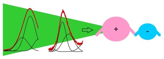
Featured Application

Understanding the structure–property relationships in mixtures of ionic liquids and molecular solvents is the key for a targeted design of optimized process fluids and electrolytes.

Abstract

Adding imidazolium ionic liquids to polar solvents such as alkyl alcohols usually results in the dissociation of ion pairs as cation–anion interactions are replaced, e.g., by ion⋯OH hydrogen bonds. In this Communication, we apply Raman scattering and infrared absorption spectroscopy to an example binary system comprising 1-butyl-3-methylimidazolium trifluoromethanesulfonate (triflate) and propan-1-ol. The spectra are analyzed using principal component analysis (PCA), excess spectroscopy, and spectral decomposition. The results provide evidence that the ion pairs of the ionic liquid do not dissociate in propan-1-ol, even at high dilution. Moreover, there are clear signs that the propan-1-ol hydrogen bonding network is disrupted in the presence of the ionic liquid as the hydroxyl groups predominantly interact with the sulfonate oxygen atoms.

Graphical Abstract

1. Introduction

Room-temperature ionic liquids (ILs) represent a very interesting class of materials because their properties can be tuned by choosing an appropriate combination of cation and anion. In this manner, tailor-made media exhibiting optimal physicochemical properties for specific applications can be synthesized, if the structure–property relationships are understood [1,2]. Adding a solvent to an IL adds another dimension to this tunability as both the chemical nature of the solvent and its concentration can be varied. However, in such a mixture, not only the interactions between the ions must be decoded, but also the impact of the solvent. The molecular interactions between conventional solvents and ILs are complicated and typically comprise a variety of mechanisms including hydrogen bonding, coulombic forces, and dispersion interactions. Their interplay determines the molecular and macroscopic behavior of the mixture.
The most common solvent considered in ILs is water. Due to the hygroscopic nature of many ILs, even careful handling under a dry atmosphere can result in contamination with water, and hence, its effects need to be studied. When water is added to, e.g., 1-alkyl-3-methylimidazolium-based ILs, the dominant interaction mechanism is the formation of hydrogen bonds (HBs). It was shown that water–ion HBs form with the anion and also with the cation, predominantly at the C(2) position, where the cation–anion interactions usually take place. As a consequence, the ion pairs dissociate and the individual ions are hydrated [3,4,5,6,7]. Mixtures with alkyl alcohols indicated the same behavior [8,9]. However, spectroscopic and computational studies aiming at insights into the molecular interactions have focused on short-chain alcohols, mainly methanol and ethanol. Hence, conclusions about the general behavior of imidazolium ILs in alcohols can hardly be drawn at this point. For completeness, the macroscopic properties and thermodynamics of alcohol–IL solutions have already been studied for longer chain lengths, and interesting correlations could be identified there [10,11,12,13]. Deducing information about the molecular scale solely from the measurement of macroscopic parameters, however, bears the danger of over-interpreting the data.
In the present Communication, we show a case in which the ion pairs of an imidazolium-based IL do not dissociate, even at high dilution by an alcohol. For this purpose, binary mixtures of 1-butyl-3-methylimidazolium trifluoromethanesulfonate (C4mimTfO) and propan-1-ol (PrOH) are studied using Raman and FTIR spectroscopy. We have recently shown that TfO ILs represent interesting model systems for spectroscopic studies as the TfO anion exhibits a single configuration, hence reducing the complexity of the spectra [14], e.g., in contrast to the common NTf2 ILs [15]. Moreover, we have chosen this combination of an IL and an alcohol as the thermodynamic behavior of their binary mixtures is well understood [11].

2. Materials and Methods

2.1. Materials

Propan-1-ol (C3H8O, CAS-No. 71-23-8, purity mass fraction w(PrOH) ≥ 0.998) was acquired from Fluka/Sigma-Aldrich (Sigma Aldrich Chemie GmbH, Munich, Germany). Prior to use, it was dried over molecular sieves (3 Å, Merck KGaA, Darmstadt, Germany) for at least 24 h and was additionally purified by vacuum distillation at p = 667 mbar ± 1 mbar and T = 358 K ± 0.5 K. Afterwards, the water content was w(H2O) < 135 ppm (mass fraction) as determined by Karl Fischer titration.
1-Butyl-3-methylimidazolium trifluoromethanesulfonate (C4mimTfO, C9H15F3N2O3S, CAS-No. 174899-66-2, purity mass fraction w(C4mimTfO) ≥ 0.99) was purchased from IoLiTec (Ionic Liquids Technologies GmbH, Heilbronn, Germany). In order to remove volatile components, reduced pressure conditions of p < 2 × 10⁻² mbar were applied for a minimum of 24 h, and a water content of w(H2O) < 50 ppm (mass fraction) was detected.
After purification, the binary mixtures (0 ≤ x(PrOH) ≤ 1 mole fraction) were prepared gravimetrically under an argon atmosphere (Ar 5.0, Air Liquide, Düsseldorf, Germany) in order to avoid contamination with water. They were filled in Duran glass tubes sealed with PTFE-protected silicon rubber seals, fixed with screw caps GL 14 (both Duran Group GmbH, Mainz, Germany) by applying a syringe pump. The mass of each component was weighed using an electronic analytical balance type BA 210 S (Sartorius, Göttingen, Germany). The mole fractions prepared were x(PrOH) = 1, 0.8984, 0.7996, 0.6992, 0.6664, 0.6024, 0.4954, 0.4029, 0.3400, 0.2948, 0.1934, 0, each with an uncertainty of σx = 0.0002.

2.2. Raman Spectroscopy

The Raman spectra were recorded in a self-built set-up with a 90° scattering geometry. The key components are a continuous-wave diode laser (Cobolt 08-NLD, 785 nm, 500 mW, Cobolt AB, Solna, Sweden), a Czerny–Turner spectrograph (Shamrock 163, 163 mm focal length, 600 grooves/mm grating, Andor Technology Ltd., Belfast, UK) and a back-illuminated charge-coupled device (CCD) camera (iDus 416, Andor Technology Ltd., Belfast, UK). The spectra were recorded with a nominal resolution of about 2 cm−1. The vertically polarized laser light was focused on the sample with an achromatic lens. The scattered Raman signal was collected in a direction perpendicular to the laser beam with an achromatic lens. In the signal collecting unit, a dielectric long-pass filter (cut-on wavelength 800 nm) and a polarizing beam splitter cube (Extinction ratio TP:TS 1000:1) were mounted. An achromatic lens focused the transmitted vertically polarized light on the slit of the spectrograph.

2.3. Infrared Spectroscopy
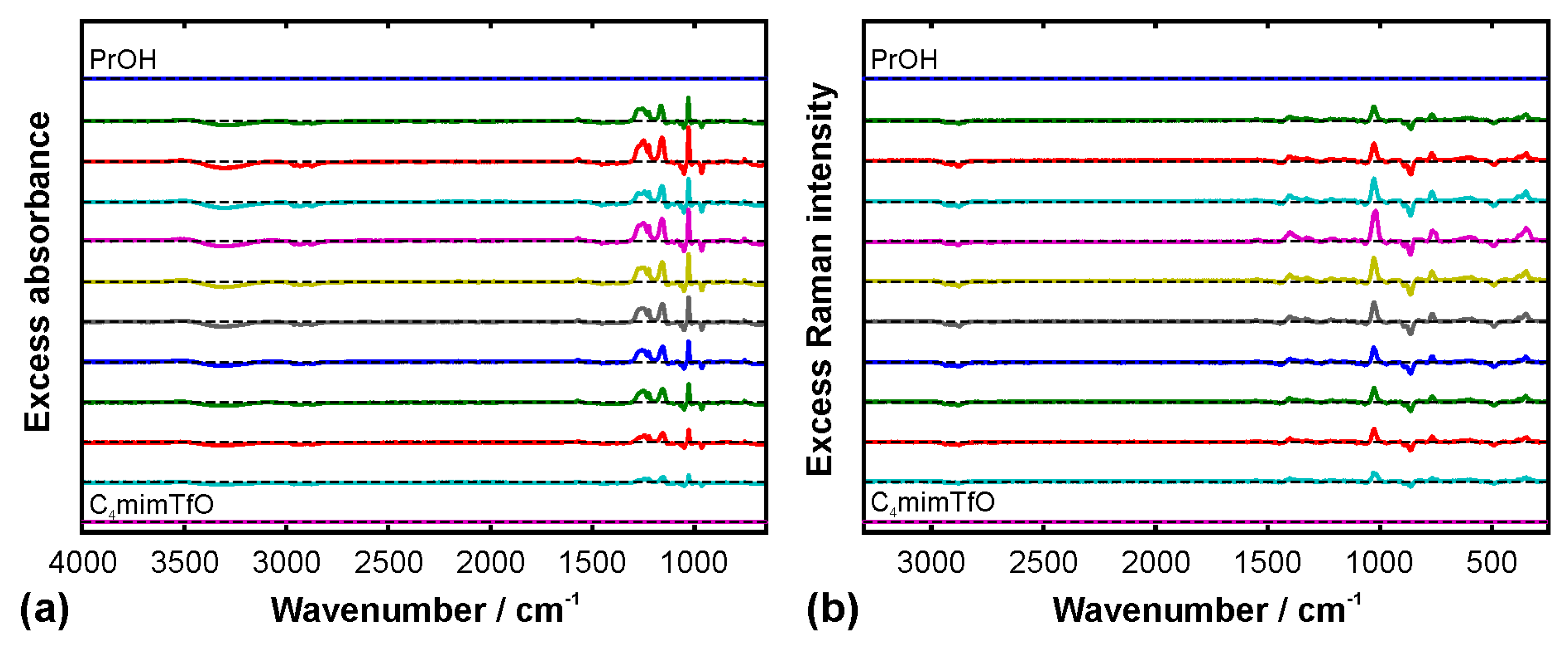
The FTIR spectra were recorded on an Agilent Cary 630 FTIR spectrometer (Agilent Technologies Inc., Santa Clara, CA, USA) equipped with a single-reflection diamond attenuated total reflection (ATR) unit at ambient temperature (T ≈ 295 K) and pressure (p ≈ 1013 mbar). The instrument exhibits a thermoelectrically cooled dTGS (deuterated triglycine sulfate) detector. The spectral range 650–4000 cm−1 was covered with a nominal resolution of 2 cm−1. For each measurement, a liquid sample was placed on the crystal and 32 scans were averaged in order to attain an appropriate signal-to-noise ratio. Contamination with water from the ambient air was minimized by operating the device in a box with a dried air atmosphere.

2.4. Excess Spectra

To calculate the excess spectrum of a mixture, the spectrum of the corresponding ideal (in a spectroscopic sense) solution is computed by adding the pure component spectra weighed by their mole fractions. This ideal spectrum is then subtracted from the experimentally measured mixture spectrum. A detailed description can be found in [16,17]. The effects of excess mixing volume were compensated using density data determined by a vibrating tube densitometer (DMA 5000, Anton Paar GmbH, Graz, Austria). The densities were converted to excess volumes and fitted by a third-order Redlich–Kister equation for further use in the calculation of the excess spectra. The resulting difference spectrum from the real and ideal spectra shows signatures that are characteristic of certain molecular interactions. For instance, peak shifts manifest as S-shaped features. Moreover, in the IR excess spectrum, net positive or net negative signatures represent changes in the IR-activity resembling changes in the dipole moment of a certain bond. On the other hand, in the Raman excess spectrum, such signatures relate to changes in the polarizability.

2.5. Decomposition of Spectra

The decomposition was performed by using a fitting algorithm implemented in Matlab R2012b. Initially, the number of Gaussian sub-peaks was varied from one to ten. The results showed that four sub-peaks are sufficient to reproduce the experimental band without systematic variations. Therefore, the detailed fitting was carried out with a fixed number of four sub-peaks. In this procedure, the peak position, the intensity, and the width were the fitting parameters.

3. Results

The Raman and FTIR spectra of the neat chemicals and their binary mixtures are displayed in Figure 1. In the first step of the evaluation of the data, the individual signatures were assigned to their corresponding modes according to the previous work of our group [14,18,19,20] and of others [21,22,23,24,25,26]. In the second step, the data were screened in order to identify interesting compositional regimes, for instance where ion pair dissociation likely takes place. For this purpose, principal component analysis (PCA) was applied to the data sets as described in reference [27]. Figure 2a shows the cumulative variance of the first five principal components (PCs) of the FTIR data. PC1-3 account for almost the full variance of the data set, and therefore the further analysis is limited to those three. Their loadings spectra are plotted in Figure 2b, revealing that they have a reasonable signal-to-noise ratio, indicating that they are physically meaningful. The loadings of the individual PCs are then plotted as a function of the mixture composition: see Panel 2c. PC1 decreases monotonously across the mole fraction range, while PC2 shows the opposite behavior. This makes sense when we look at the loadings plot in Panel 2b. PC1 predominantly contains signatures of the IL, while PC2 appears more characteristic of the alcohol spectrum. Hence, the loading-vs.-composition profiles roughly resemble the amounts of the pure components. The curves are not linear though, because the PC spectra do not exclusively represent the pure component spectra.
PC3, on the other hand, does not exhibit a monotonous behavior. It shows a clear minimum around an alcohol mole fraction of x(PrOH) = 0.66. In the previous study [27], it was shown that such extreme values are likely pointing towards stoichiometries, at which interesting phenomena can be expected. In the present case, the mole fraction of x(PrOH) = 0.66 translates to a situation in which the solution contains two alcohol molecules per ion pair. This suggests two possible, but very different, interaction scenarios: (1) The hydrogen-bonded ion pairs, which likely exist in the neat IL [19], dissociate, and the cation–anion interactions are replaced by cation–alcohol and anion–alcohol interactions. In scenario (2), the ion pair remains intact, and two alcohol molecules establish an interaction, e.g., at those sulfonate oxygen atoms that are not involved in the cation–anion hydrogen bonding. Further details cannot be deduced from this initial PCA analysis.
In order to gain further insights, the excess spectra were computed. The concept of excess spectroscopy was introduced about a decade ago [28], and was found to be very useful in the analysis of solvent mixtures, including those comprising ILs [29,30]. Figure 3a,b display the IR and Raman excess spectra, respectively. The excess spectra of the pure compounds are equal to zero by definition. In contrast, the mixture spectra show multiple signatures. Interestingly, the OH stretching region in both data sets reveals predominantly negative contributions. In other words, the dipole moment and the polarizability are reduced in the real mixture compared to the ideal counterpart. This is an indicator of changes in the hydrogen bonding interactions when the two fluids are mixed. For example, the strengthening of a hydrogen bond between two molecules usually leads to a weakening of the covalent bond. This weakening can be observed as a red-shift of the vibrational frequency of the corresponding stretching mode [31]. The excess spectra give additional information here, as they indicate how the charge distribution develops. The OH bond, therefore, appears less polarized in the mixture than in the neat alcohol. On the other hand, the excess signatures in the fingerprint region are predominantly positive. The majority of the corresponding modes originate from the triflate anion, indicating that the re-arrangement of the molecular network mainly affects the anion. This is reasonable, as the sulfonate oxygen atoms can easily establish hydrogen bonds with the hydroxyl groups of the alcohol molecules.
For a clearer picture of how the signatures vary with the alcohol content, selected representative signatures in the excess spectrum are integrated and plotted vs. the propanol mole fraction, x(PrOH). Figure 4 illustrates three example profiles: the OH stretching band from the excess IR and Raman spectra, and the SO stretching band from the excess IR spectrum. They all reveal rather similar profiles exhibiting extreme values around x(PrOH) = 0.66. The experimental data were fitted to a third-order polynomial (solid line) in which the values for x(PrOH) = 0 and x(PrOH) = 1 were set to zero. The resulting maxima and minima of these polynomials are all located around x(PrOH) = 0.66, hence pointing to the 2:1 stoichiometry again. Interestingly, this molar ratio also stood out in a previous study, in which the vapor–liquid equilibrium (VLE) of the binary mixtures was studied [11]. In that work, the VLE data were fitted using a modified Raoult’s law, and the activity coefficients were derived. The profile (see Figure 4d) also peaks around x(PrOH) = 0.66. This shows a clear relationship between the molecular and macroscopic behavior of the solution.
The above analyses of the data sets strongly indicate that the mixture stoichiometry of 2:1 PrOH:C4mimTfO plays a distinct role and is worth further investigation. In order to obtain a clearer picture of the molecular interactions in the solutions, we analyze the OH stretching band. This is due to two main reasons: first, only the alcohol exhibits an OH group and therefore the signals in the range 3200–3700 cm−1 will exclusively originate from PrOH; second, the hydroxyl group is the predominant site at which hydrogen bonding will take place.
The OH stretching band in liquid alcohols is rather broad [18]. However, the overall band can be decomposed into narrower sub-peaks that are characteristic of distinct hydrogen bonding states. The results of the spectral decomposition of five selected example mixtures are displayed in Figure 5. The red solid line represents the experimental data, the thin solid black lines are the derived sub-peaks, and the dashed line shows the sum of the sub-peaks. The two small sub-peaks at the high- and low-wavenumber ends of the OH band are of limited interest in the discussion of the molecular interactions. The high-wavenumber peak can be attributed to those OH groups that are not involved in hydrogen bonding interactions. They are commonly referred to as “free” OH groups, but it should be noted that their frequency is still affected by the dielectric surrounding medium [32]. The sub-peak at the low-wavenumber end of the OH band basically serves as a compensation for the overlap from the neighboring CH stretching band.
The two main contributions to the overall band are located at 3315 and 3431 cm−1 in the neat alcohol. The positions change only marginally in the mixtures. However, the peak intensities and the ratio between them varies substantially. The sub-peak at 3431 cm−1 can be assigned to OH groups that participate in a single hydrogen bonding interaction, while the 3315 cm−1 peak originates from OH groups involved in two hydrogen bonds. In the latter, the hydrogen acts as a HB donor while the oxygen atom acts a HB acceptor. This is very common in neat alcohols where chain- and ring-like structures can be found, forming large HB networks [18]. As a consequence, the 3315 cm−1 contribution dominates the OH band in the neat alcohol. This situation changes when the ionic liquid is added to the alcohol. At x(PrOH) = 0.8, both sub-peaks exhibit similar intensity. Further addition of IL leads to the 3431 cm−1 peak becoming the dominant contribution. The observed behavior suggests that the doubly hydrogen-bonded OH groups gradually change to groups involved in only a single HB. This observation on its own, however, could still be explained by both of the scenarios derived from the PCA and excess analyses above.
Further insights can be obtained from a closer look at the spectral profiles and the other regions of the vibrational spectrum. Analyzing the fitted sub-peaks in Figure 5 in more detail, an interesting observation of the peak assigned to singly hydrogen-bonded OH groups can be made. The full width at half maximum of this peak reduces considerably with increasing IL content, while the appearance of the other peak remains almost constant. This can be explained as follows. As aforesaid, in the neat PrOH, the majority of OH groups are involved in two hydrogen bonds. Those taking part in a single hydrogen bonding interaction can do this in two different ways: either as a HB donor, or as an acceptor. In both cases, the impact on the covalent bond will be slightly different resulting in two close but distinguishable vibrational frequencies. This results in a broadened peak. The narrowing of the peak upon the addition of C4mimTfO suggests that the mixtures predominantly contain one of the above-described configurations. In turn, this points towards the second scenario in which the ion pair remains intact and interacts with two alcohol molecules via the sulfonate oxygen of the anion. If the ion pair dissociated, the cation–anion hydrogen bonding at the C(2) position would be replaced by a cation C(2)-H⋯alcohol O-H hydrogen bond and an anion S-O⋯alcohol H-O hydrogen bond. In other words, there would be equal amounts of HB donating and accepting OH groups, which contradicts the observed peak narrowing. On the other hand, if the ion pair remains intact via a hydrogen bond at the C(2) position, the two “free” oxygen atoms of the sulfonate group can establish hydrogen bonds with a PrOH molecule each. In this case, both OH groups act as HB donors only, which is in concert with the narrowed peak.
Our hypothesis of scenario (2) is further corroborated by the CH stretching peaks. Replacing the cation–anion interactions by ion–alcohol hydrogen bonding would result in a shift of the C(2)-H stretching band as the strength of the hydrogen bond would most likely be different. The IR and Raman spectra, however, reveal that the associated band at 3114 cm−1 is independent of the mixture composition. The negligible signatures in the excess spectra at this position lend further support for this interpretation.

4. Conclusions

In conclusion, the results suggest that there are predominantly ion pairs existing in the solutions. These ion pairs establish hydrogen bonds with the alcohol molecules via the oxygen atoms of the TfO anion. This configuration was unexpected as previously studied mixtures of short-chain alkyl alcohols and imidazolium-based ionic liquids showed clear signs of ion pair dissociation. In addition, our data illustrate the strong links between the molecular scale studied by spectroscopy and the macroscopic behavior of the solution. Clear correlations between spectroscopic signatures and the thermodynamics derived from VLE data were observed. Therefore, combining vibrational spectroscopy with the analysis of the phase equilibrium is a powerful approach to unravel these structure–property relationships.
The observed behavior of stable ion pairs existing in the mixture and their solvation by alcohol molecules may be a general phenomenon in 1,3-dialkylimidazolium TfO IL–alcohol mixtures. However, further and systematic investigations are necessary to confirm this. Molecular dynamics simulations will be helpful to obtain additional insights. However, we should also note that the behavior is strongly dependent on the nature of the cation–anion combination and the solvent. For example, a previous Raman study of an imidazolium ethylsulfate IL in mixtures with methanol and ethanol [8] has shown that ion pair dissociation takes place in these solutions. Further evidence of the tendency of ion pair dissociation may also be provided by conductivity measurements in the future. For instance, Wang et al. [33] conducted such experiments in mixtures of imidazolium-based ILs with highly symmetric anions and organic solvents to derive association constants. The behavior they found, however, cannot be directly compared with our results due to the systematic difference in molecular structure. Therefore, finding universal rules and structure–property relationships for ILs and their mixtures with organic solvents remains a topic that requires further theoretical and experimental research.

Author Contributions

Conceptualization, J.K. and B.R.; methodology, J.K.; sample preparation and FTIR experiments, M.S.; Raman experiments, C.C.R.; data analysis, J.K.; data interpretation, all authors; writing—Original draft preparation, J.K.; writing—Review and editing, all authors. All authors have read and agreed to the published version of the manuscript.

Funding

Part of this work was funded by the Deutsche Forschungsgemeinschaft (DFG) through grant KI1396/4-1.

Conflicts of Interest

The authors declare no conflict of interest. The funders had no role in the design of the study, in the collection, analyses, or interpretation of data, in the writing of the manuscript, or in the decision to publish the results.

References

  1. Turner, E.A.; Pye, C.C.; Singer, R.D. Use of ab initio calculations toward the rational design of room temperature ionic liquids. J. Phys. Chem. A 2003, 107, 2277–2288. [Google Scholar] [CrossRef]
  2. Katsyuba, S.; Zvereva, E.; Vidis, A.; Dyson, P. Application of density functional theory and vibrational spectroscopy toward the rational design of ionic liquids. J. Phys. Chem. A 2007, 111, 352–370. [Google Scholar] [CrossRef]
  3. Cammarata, L.; Kazarian, S.G.; Salter, P.A.; Welton, T. Molecular states of water in room temperature ionic liquids. Phys. Chem. Chem. Phys. 2001, 3, 5192–5200. [Google Scholar] [CrossRef] [Green Version]
  4. Cha, S.; Ao, M.; Sung, W.; Moon, B.; Ahlström, B.; Johansson, P.; Ouchi, Y.; Kim, D. Structures of ionic liquid-water mixtures investigated by IR and NMR spectroscopy. Phys. Chem. Chem. Phys. 2014, 16, 9591–9601. [Google Scholar] [CrossRef]
  5. Jaeger, V.W.; Pfaendtner, J. Structure, dynamics, and activity of xylanase solvated in binary mixtures of ionic liquid and water. ACS Chem. Biol. 2013, 8, 1179–1186. [Google Scholar] [CrossRef]
  6. Köddermann, T.; Wertz, C.; Heintz, A.; Ludwig, R. The association of water in ionic liquids: A reliable measure of polarity. Angew. Chem. Int. Ed. 2006, 45, 3697–3702. [Google Scholar] [CrossRef]
  7. Zhang, Q.-G.; Wang, N.-N.; Yu, Z.W. The hydrogen bonding interactions between the ionic liquid 1-ethyl-3-methylimidazolium ethyl sulfate and water. J. Phys. Chem. B 2010, 114, 4747–4754. [Google Scholar] [CrossRef]
  8. Noack, K.; Leipertz, A.; Kiefer, J. Molecular interactions and macroscopic effects in binary mixtures of an imidazolium ionic liquid with water, methanol, and ethanol. J. Mol. Struct. 2012, 1018, 45–53. [Google Scholar] [CrossRef]
  9. Chang, H.; Jiang, J.; Liou, Y.; Hung, C.; Lai, T.; Lin, S. Effects of water and methanol on the molecular organization of 1-butyl-3-methylimidazolium tetrafluoroborate as functions of pressure and concentration. J. Chem. Phys. 2008, 129, 04452. [Google Scholar] [CrossRef]
  10. Vale, V.R.; Will, S.; Schröer, W.; Rathke, B. The general phase behavior of mixtures of 1-alkyl-3-methylimidazolium bis[(trifluoromethyl)sulfonyl]amide ionic liquids with n-alkyl alcohols. ChemPhysChem 2012, 13, 1860–1867. [Google Scholar] [CrossRef]
  11. Stuckenholz, M.; Crespo, E.A.; Vega, L.F.; Carvalho, P.J.; Coutinho, J.A.P.; Schröer, W.; Kiefer, J.; Rathke, B. Vapor Liquid Equilibria of Binary Mixtures of 1-Butyl-3-methylimidazolium Triflate (C4mimTfO) and Molecular Solvents: N-Alkyl Alcohols and Water. J. Phys. Chem. B 2018, 122, 6017–6032. [Google Scholar] [CrossRef] [PubMed]
  12. Chouireb, N.; Khan, I.; Crespo, E.A.; Oliveira, M.B.; Llovell, F.; Vega, L.F.; Tafat-Igoudjilene, O.; Kaci, A.A.; Santos, L.M.N.B.F.; Carvalho, P.J.; et al. Evaluation of the Solvent Structural Effect Upon the Vapor-Liquid Equilibrium of [C4C1im][Cl] + Alcohols. Fluid Phase Equilibria 2017, 440, 36–44. [Google Scholar] [CrossRef]
  13. Verevkin, S.P.; Safarov, J.; Bich, E.; Hassel, E.; Heintz, A. Thermodynamic Properties of Mixtures Containing Ionic Liquids Vapor Pressures and Activity Coefficients of n-Alcohols and Benzene in Binary Mixtures with 1-Methyl-3-butyl-imidazolium Bis(trifluoromethyl-sulfonyl)imide. Fluid Phase Equilibria 2005, 236, 222–228. [Google Scholar] [CrossRef]
  14. Kiefer, J.; Stuckenholz, M.; Rathke, B. Influence of the alkyl chain on the vibrational structure and interionic interactions in 1-alkyl-3-methylimidazolium trifluoromethanesulfonate ionic liquids. J. Mol. Liquids 2018, 255, 413–418. [Google Scholar] [CrossRef]
  15. Herstedt, M.; Smirnov, M.; Johansson, P.; Chami, M.; Grondin, J.; Servant, L.; Lassègues, J.C. Spectroscopic characterization of the conformational states of the bis(trifluoromethanesulfonyl)imide anion (TFSI). J. Raman Spectrosc. 2005, 36, 762–770. [Google Scholar] [CrossRef]
  16. Zhou, Y.; Gong, S.; Xu, X.; Yu, Z.; Kiefer, J.; Wang, Z. The interactions between polar solvents (methanol, acetonitrile, dimethylsulfoxide) and the ionic liquid 1-ethyl-3-methylimidazolium bis(fluorosulfonyl)imide. J. Mol. Liquids 2020, 299, 112159. [Google Scholar] [CrossRef]
  17. Zehentbauer, F.M.; Kiefer, J. Molecular solution behaviour of an intermediate biofuel feedstock: Acetone-butanol-ethanol (ABE). ChemPhysChem 2015, 16, 3846–3858. [Google Scholar] [CrossRef]
  18. Kiefer, J.; Wagenfeld, S.; Kerle, D. Chain length effects on the vibrational structure and molecular interactions in the liquid normal alkyl alcohols. Spectrochim. Acta A 2018, 189, 57–65. [Google Scholar] [CrossRef]
  19. Singh, D.K.; Rathke, B.; Kiefer, J.; Materny, A. Molecular Structure and Interactions in the Ionic Liquid 1-Ethyl-3-methylimidazolium Trifluoromethanesulfonate. J. Phys. Chem. A 2016, 120, 6274–6286. [Google Scholar] [CrossRef]
  20. Noack, K.; Schulz, P.S.; Paape, N.; Kiefer, J.; Wasserscheid, P.; Leipertz, A. The role of the C2 position in interionic interactions of imidazolium based ionic liquids: A vibrational and NMR spectroscopic study. Phys. Chem. Chem. Phys. 2010, 12, 14153–14161. [Google Scholar] [CrossRef]
  21. Michniewicz, N.; Muszyński, A.S.; Wrzeszcz, W.; Czarnecki, M.A.; Golec, B.; Hawranek, J.P.; Mielke, Z. Vibrational spectra of liquid 1-propanol. J. Mol. Struct. 2008, 887, 180–186. [Google Scholar] [CrossRef]
  22. Plyler, E.K. Infrared spectra of methanol, ethanol, and n-propanol. J. Natl. Bur. Stand. 1952, 48, 281–286. [Google Scholar] [CrossRef]
  23. Tomsic, M.; Jamnik, A.; Fritz-Popovski, G.; Glatter, O.; Vlcek, L. Structural properties of pure simple alcohols from ethanol, propanol, butanol, pentanol, to hexanol: Comparing Monte Carlo simulations with experimental SAXS data. J. Phys. Chem. B 2007, 111, 1738–1751. [Google Scholar] [CrossRef] [PubMed]
  24. Ozawa, R.; Hayashi, S.; Saha, S.; Kobayashi, A.; Hamaguchi, H. Rotational isomerism and structure of the 1-butyl-3-methylimidazolium cation in the ionic liquid state. Chem. Lett. 2003, 32, 948–949. [Google Scholar] [CrossRef]
  25. Garaga, M.N.; Nayeri, M.; Martinelli, A. Effect of the alkyl chain length in 1-alkyl-3-methylimidazolium ionic liquids on inter-molecular interactions and rotational dynamics: A combined vibrational and NMR spectroscopic study. J. Mol. Liquids 2015, 210, 169–177. [Google Scholar] [CrossRef]
  26. Holomb, R.; Martinelli, A.; Albinsson, I.; Lassègues, J.C.; Johansson, P.; Jacobsson, P. Ionic liquid structure: The conformational isomerism in 1-butyl-3-methyl-imidazolium tetrafluoroborate ([bmim][BF4]). J. Raman Spectrosc. 2008, 39, 793–805. [Google Scholar] [CrossRef]
  27. Kiefer, J.; Eisen, K. Unsupervised screening of vibrational spectra by principal component analysis for identifying molecular clusters. ChemPhysChem 2018, 19, 795–800. [Google Scholar] [CrossRef]
  28. Li, Q.; Wang, N.; Zhou, Q.; Sun, S.; Yu, Z. Excess infrared absorption spectroscopy and its applications in the studies of hydrogen bonds in alcohol-containing binary mixtures. Appl. Spectrosc. 2008, 62, 166–170. [Google Scholar] [CrossRef]
  29. Wang, N.-N.; Zhang, Q.-G.; Wu, F.-G.; Li, Q.-Z.; Yu, Z.-W. Hydrogen bonding interactions between a representative pyridinium-based ionic liquid [BuPy][BF4] and water/dimethyl sulfoxide. J. Phys. Chem. B 2010, 114, 8689–8700. [Google Scholar] [CrossRef]
  30. Kiefer, J.; Molina Martinez, M.; Noack, K. The peculiar nature of molecular interactions between an imidazolium ionic liquid and acetone. ChemPhysChem 2012, 13, 1213–1220. [Google Scholar] [CrossRef]
  31. Joseph, J.; Jemmis, E.D. Red-, blue-, or no-shift in hydrogen bonds: A unified explanation. J. Am. Chem. Soc. 2007, 129, 4620–4632. [Google Scholar] [CrossRef] [PubMed]
  32. Serra, P.B.P.; Rocha, M.A.A.; Rathke, B.; Ruzicka, K.; Fulem, M.; Kiefer, J. Infrared spectroscopy of the symmetric branched isomers of n-heptanol. J. Mol. Liquids 2017, 244, 528–532. [Google Scholar] [CrossRef]
  33. Wang, H.; Wang, J.; Zhang, S.; Pei, Y.; Zhuo, K. Ionic Association of the Ionic Liquids [C4mim][BF4], [C4mim][PF6], and [Cnmim]Br in Molecular Solvents. ChemPhysChem 2009, 10, 2516–2523. [Google Scholar] [CrossRef] [PubMed]
Figure 1. Attenuated total reflection (ATR)-FTIR spectra (a) and Raman spectra (b) of the neat substances and the binary mixtures.
Applsci 10 01620 g001
Figure 2. Principal component analysis (PCA) screening results of the FTIR data set: (a) cumulative variance of the first five principal components (PCs), (b) loadings spectra, (c) loadings as a function of composition.
Applsci 10 01620 g002
Figure 3. Excess ATR-FTIR spectra (a) and excess Raman spectra (b) of the neat substances and the binary mixtures.
Applsci 10 01620 g003
Figure 4. Panels (a–c) show the integrated excess absorbance/intensity as a function of the propanol mole fraction for selected spectral signatures. For this purpose, the band areas over characteristic spectral windows in the excess spectra were calculated. Panel (d) shows the profile of the activity coefficient derived from vapor–liquid equilibrium (VLE) data [11] for comparison.
Applsci 10 01620 g004
Figure 5. Decomposed FTIR spectra in the OH stretching region for five selected mixtures.
Applsci 10 01620 g005

