Article

Effect of Bioactivity of Surface Topography and Coating Forming by Infrared Light-Induced on Titanium for Bone Repair

1
Graduate Institute of Oral Science, Chung Shan Medical University, Taichung 40201, Taiwan
2
Department of Dentistry, Chung Shan Medical University Hospital, Taichung 40201, Taiwan
3
School of Dental Technology, College of Oral Medicine, Taipei Medical University, Taipei 11031, Taiwan
4
UniSA Clinical & Health Sciences, University of South Australia, Adelaide, SA 5001, Australia
5
Nature Dental Laboratory, School of Dental Technology, College of Oral Medicine, Taipei Medical University, Taipei 11031, Taiwan
*
Author to whom correspondence should be addressed.
These authors contributed equally to this work.
Appl. Sci. 2020, 10(22), 8158; https://doi.org/10.3390/app10228158
Submission received: 20 October 2020 / Revised: 16 November 2020 / Accepted: 17 November 2020 / Published: 18 November 2020
(This article belongs to the Special Issue Application of the Biocomposite Materials on Bone Reconstruction)

Abstract

:
Calcium ions and phosphate ions are usually present in biological organisms and human bodies. Different ratios of calcium to phosphorus result in different types of calcium-to-phosphorus crystals. Hydroxyapatite (HA) is the main component of human hard bony tissues. It has good biocompatibility and is often used in bone repair. With the addition of cobalt ions, it can act as a hypoxia-inducing factor to accelerate the regeneration of hard bony tissues. At present, the laser-assisted biomimetic (LAB) method can very quickly deposit calcium phosphate coatings, which can be used on polymer and titanium surfaces. In this study, we first used anodization treatment (with TiO2 nanotubes (TNTs)), alkali treatment (with NaOH), and acid treatment (with HCl) to form nanopore structures on titanium surfaces in the laboratory. Subsequently, LAB treatment was used to deposit calcium phosphate and cobalt-substituted hydroxyapatite onto titanium pieces with different surface treatments. The results showed that smaller holes resulted in better deposition (TNTs), and controlling the pH value in the solution changed the crystal morphology. LAB treatment imbued the titanium surface with super-hydrophilic properties and improved biocompatibility. A human osteoblast cell line (MG-63) used for the cell viability test showed that LAB treatment can improve cell growth. In particular, TNT-CoHA (cobalt-substituted hydroxyapatite) cells grew the best. Immersion in simulated bodily fluid confirmed that LAB treatment with a CoHA solution improved the hydrophilicity, biocompatibility, and bioactivity of titanium surfaces. It is hoped that this study provides useful information for surface coating of biomedical materials in the future.

Graphical Abstract

1. Introduction

Calcium phosphate (CaP) coatings are osteoconductive and are widely used in orthopedics and dental implants to increase osseointegration between the implant and hard bone [1]. In previously study, Susmita Bose et al. reported that the calcium phosphate (CaP) coating formed on the porous titanium surface can reduce the healing time by early enhance osseointegration in the body [2]. CaP can be divided into hydroxyapatite (HA), octacalcium phosphate (OCP), amorphous calcium phosphate (ACP), and dicalcium phosphate (DCPD), according to the calcium-to-phosphorus (Ca/P) ratio [3,4]. HA is the main component of human body tissues and enamel on the surface of the teeth. After HA is implanted into a hard bone defect, the surface of the material will form a chemical bond with the hard bone tissue to improve osseointegration [5]. However, the poor biodegradability of HA results in a rather slow induction of hard bone growth after implantation [6]. Therefore, the previous literature used metal ions instead of calcium ions to reduce the crystallinity and increase the degradation rate of HA. The substitution of cobalt ions not only reduces crystallinity but also acts as a hypoxia mimic, which can activate hypoxia-inducible factor (HIF)-1α in mesenchymal stem cells to help angiogenesis [7]. Previous studies showed that cobalt-substituted hydroxyapatite (CoHA) has good biological activity and can induce bone growth [8].
There are many methods for forming HA coatings such as sol–gel [9], plasma spraying [10], and laser-assisted biomimetic (LAB) [11,12]. LAB is a method of immersing a substrate in a supersaturated CaP solution and irradiating the sample with an Neodymium-dopedYttriumAluminiumGarnet laser (Nd-YAG, 200 mJ pulse−1 cm−2). The surface of the sample converts absorbed light energy into heat energy, which induces CaP to rapidly form a coating [13,14]. LAB can also be used on polymer and titanium surfaces [14,15,16]. Pecheva et al. reported the use of a high-energy laser (1 Kw mm−2) to form a CaP coating on the surface of titanium immersed in a CaP solution. The biocompatibility test with a human osteosarcoma cell line (MG-63) showed good cell adhesion [17]. Mahanti et al. found that near-infrared (NIR; λ = 1064 nm) laser pulses can replace ultraviolet (UV, λ = 355 nm) laser pulses used by LAB to avoid using too high of an energy, which can damage the surface of a sample [13].
In addition, surface treatment of titanium test pieces was proven to increase the pores on the surface of the material, thereby increasing the effect of osseointegration [11,18]. Clinical dentistry will also increase the success rate of surgery through the modification of the implant surface [19]. Surface treatment methods include plasma spray, sand-blasting, anodic treatment, and chemical etching. Among these, the modification methods of alkali treatment and acid treatment are relatively simple and can be applied to complex-shaped structures to produce many uniform nano-level holes on the surface [20,21]. Anodic oxidation treatment (with TiO2 nanotubes (TNTs)) can form an oxide film on the surface of titanium to improve biocompatibility and can attract apatite to the surface to promote the regeneration of hard bones [22].
This study investigated whether changes in the surface morphology and elemental composition of titanium after different surface treatment procedures (anodization, alkali treatment, and acid treatment) will affect LAB coatings. In addition, we used CaP and CoHA solutions with the LAB method to observe whether the structure and composition of the mineralized layer on the surface were affected, and we analyzed cell viability to evaluate the biocompatibility of test pieces. Finally, test pieces were immersed in simulated body fluid (SBF) for 7 days, and the surface morphology, crystal structure, and elemental composition were analyzed as evaluations of biological activities. This study is expected to provide useful information for developing bone repair material coatings.

2. Experimental Procedures

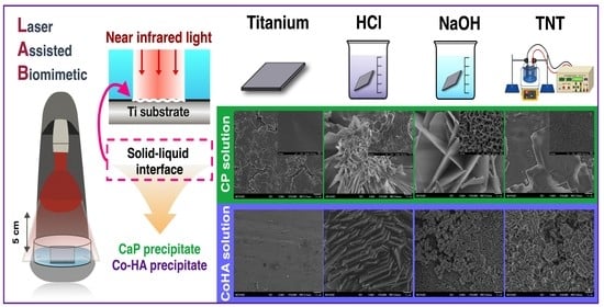
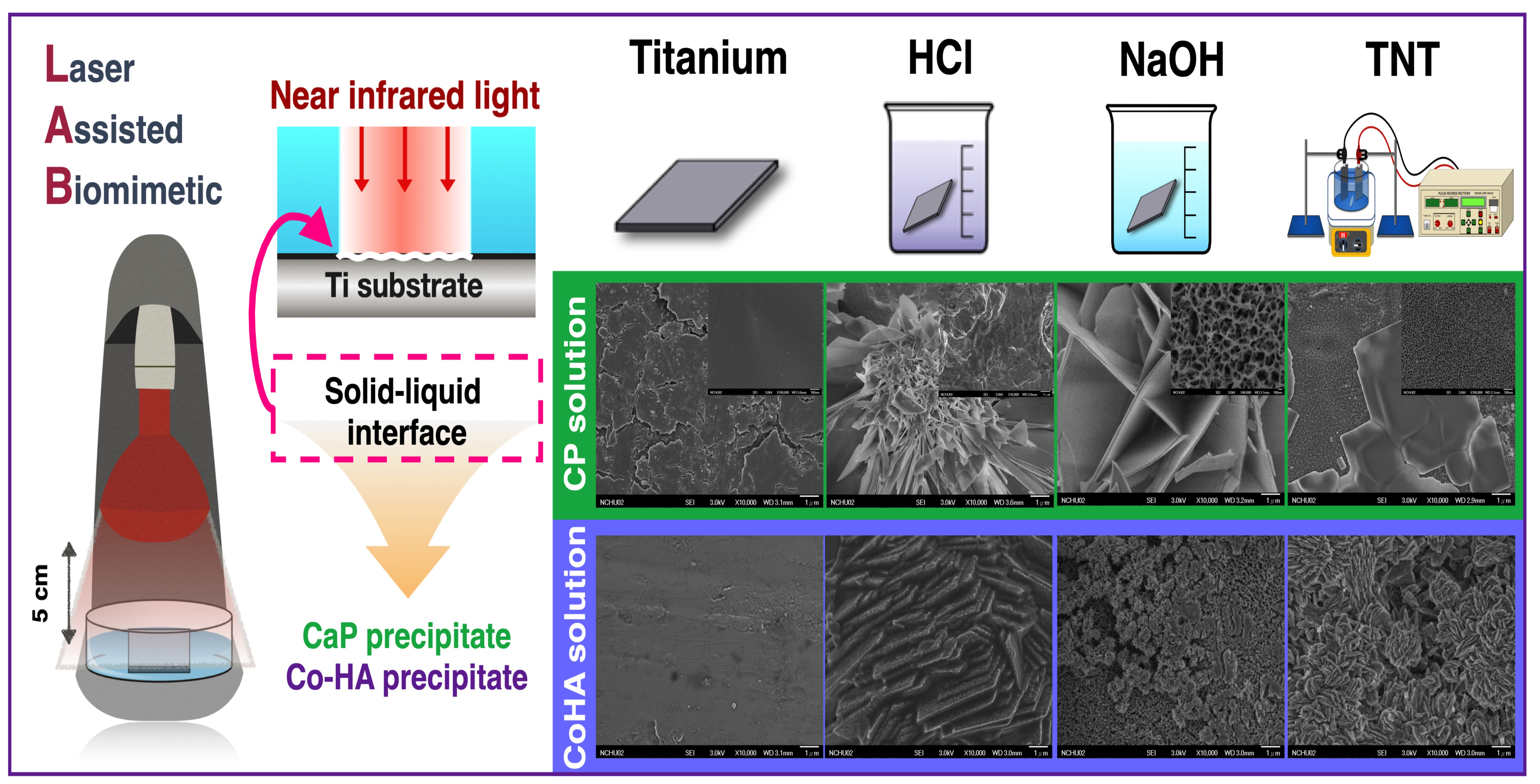
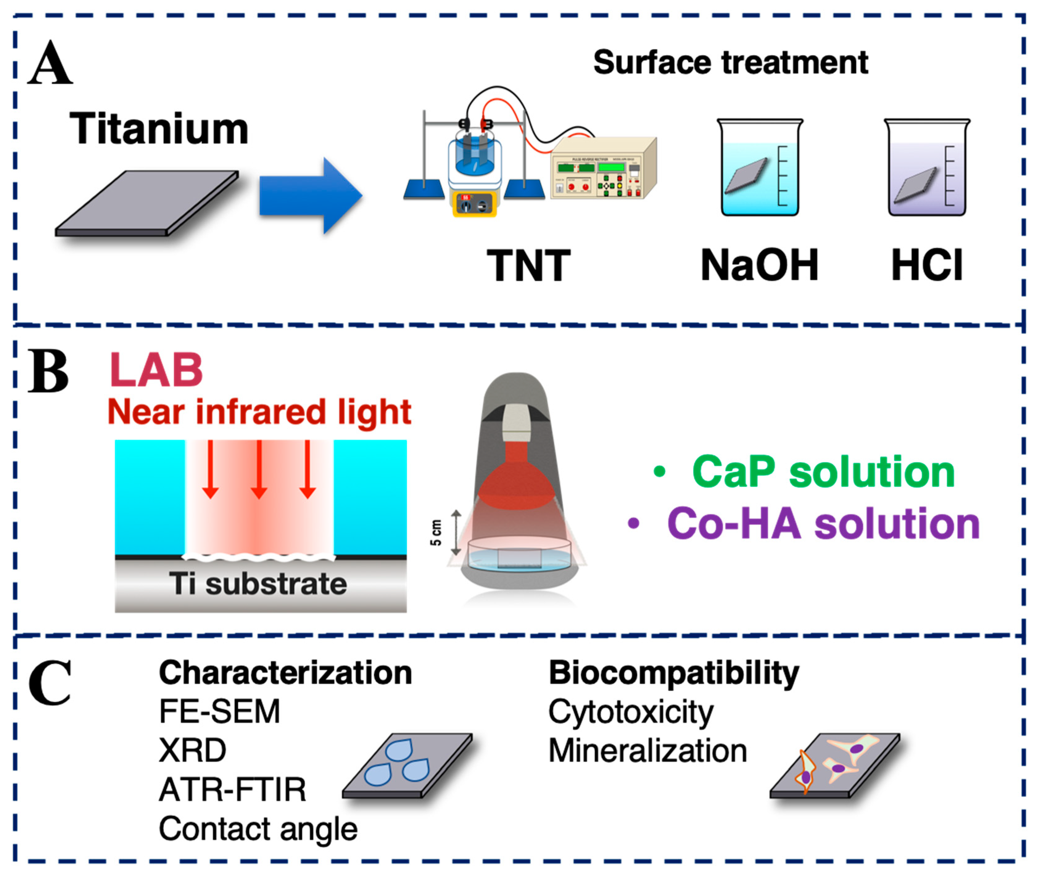
2.1. Surface Treatment of Titanium

Pure titanium pieces were cut into a fixed area (16 cm2). These pieces were washed with acetone, ethanol, and deionized water for 10 min. After that, (i) anodization, (ii) alkali treatment, and (iii) acid treatment were carried out (Figure 1A). (i) Anodic oxidation uses a pulse method for electrochemical reactions [23]. Ammonium fluoride at 2.7 g was added to 18 mL of deionized water and 900 mL of ethylene glycol. The titanium piece was used as the anode, and a stainless steel piece was used as the cathode. The electrolyte was heated to 55 °C, and a voltage of 30 V was used for 40 min (time on 150 s, time off 1 s). After the reaction, the titanium piece was immersed in ethanol and cleaned in an ultrasonic shaker for 20 min. (ii) For alkali treatment, a pure titanium sheet was immersed in 15 mL of a sodium hydroxide (NaOH) solution (5 M) and reacted in a 60 °C water bath for 24 h [20]. (iii) For acid treatment, a pure titanium sheet was immersed in 15 mL of a hydrochloric acid (HCl) solution (5 M) and reacted in a 60 °C water bath for 24 h [21]. Names of the completed samples are given in Table 1.

2.2. LAB Preparation

First, a CaP solution and a CoHA solution were prepared. The CaP solution used NaCl (142 mM), K2HPO4•3H2O (1.50 mM), HCl (40 mM), and CaCl2 (3.75 mM) added to deionized water, and the pH was adjusted to 7.4 [24]. The CoHA solution was prepared by adding calcium nitrate (42 mM) and ammonium dihydrogen phosphate (25 mM) to deionized water, adjusting the pH to 3.52, and finally adding cobalt chloride (7.9 mM) [8]. The LAB method uses NIR light as the light source. The pure titanium test pieces were placed in 70 mL of the CaP solution or CoHA solution and irradiated at 25 °C for 30 min (Figure 1B). The distance between the pure titanium sheet and the bulb was 5 cm. After irradiation, the titanium sheet was rinsed with deionized water and dried. The names of completed samples are given in Table 1.

2.3. Characterization

The surface morphology of samples was examined by field-emission scanning electron microscopy (FE-SEM) (JSM-6700F, JEOL, Tokyo, Japan). Infrared spectra of samples were recorded using a Fourier-transformed infrared (FTIR) system (FTIR-8000, Shimadzu, Tokyo, Japan) in the spectral range of 650–4000 cm−1 with a spectral resolution of 2 cm−1, using the attenuated total reflection (ATR) mode. Phase indication of samples was confirmed by X-ray diffraction (XRD) (Miniflex II, Rigaku, Tokyo, Japan), operated at 30 kV with Cu-Kα radiation within a scanning range of 10°–70° (2θ) and a scanning speed of 4° min−1.

2.4. Hydrophilicity Test

The contact angle is the angle between the liquid and the surface of a material. The contact angle is used to judge the hydrophilicity and hydrophobicity of a sample surface. A smaller contact angle indicates that the surface of a sample is hydrophilic, and a larger contact angle indicates that the surface of a sample is hydrophobic. The surface hydrophilicity of TiO2 nanotubes (TNTs) was evaluated with a contact angle meter (CA-D, Kyowa Interface Science, Tokyo, Japan). The static contact angle was determined at 25 °C and 70% relative humidity by employing drops of deionized water. The quantitative titration of the water drop used was 4 μL and the time was 1 s. After that, the image was analyzed using Drop-analysis software attached to Image J for the contact angle analysis.

2.5. Biocompatibility

We used a human osteosarcoma cell line (MG63) maintained in Dulbecco’s modified eagle medium (DMEM) supplemented with 10% fetal bovine serum (FBS) (Biological Industries, Cromwell, CT, USA) at 37 °C in a 5% CO2 incubator (310, Thermo Fisher Scientifc, Waltham, MA, USA). Samples processed by the CaP and CoHA solutions LAB were sterilized with ethanol and irradiated with UV light for 1 h. MG63 cells were seeded in 24-well plates at 5 × 10⁴ cells well−1. Finally, cells were cultured in a 37 °C, 5% CO2 cell incubator for 24 and 72 h. Biocompatibility was evaluated by a colorimetric 3-(4,5-dimethylthiazol-2-yl)-2,5-diphenyltetrazolium bromide (MTT) assay. The absorbance (O.D.) at a wavelength of 563–650 nm was read with an enzyme-linked immunosorbent assay (ELISA) reader (Sunrise, Tecan, Männedorf, Switzerland). Biocompatibility was expressed as a percentage compared to that of a control tissue culture plate (TCP).

2.6. Bioactivity

Surface-treated and LAB-treated titanium sheets were used as samples. Surface-treated titanium pieces were used as a control group. A sample was soaked in simulated body fluid (SBF) to evaluate the bioactivity of the material [25]. All samples were cut to a size of 2 × 1 cm2 and immersed in 15 mL of SBF in a water bath at 37 °C for 7 days, after which it was taken out, rinsed with deionized water, and dried. The morphology and crystalline form of apatite on the sample surface were observed by FE-SEM, ATR-FTIR, and XRD.

2.7. Statistical Analysis

All data are expressed as the mean ± standard deviation (SD) of three independent replicates. Data were analyzed with JMP 13 software (SAS Institute, Cary, NC, USA). A one-way analysis of variance (ANOVA) followed by Tukey’s honest significant difference (HSD) post-hoc test was used to determine the level of significance, and p < 0.05 was considered significant. In the figures, values with different letters significantly differ from each other (p < 0.05, mean ± SD, n = 3).

3. Results and Discussion

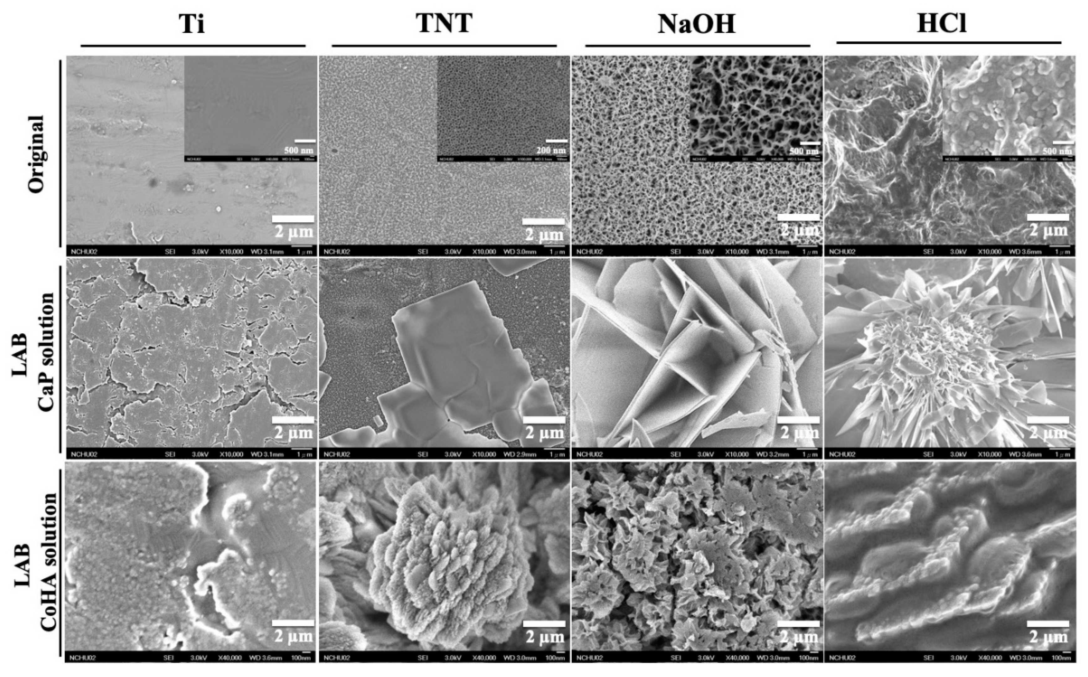
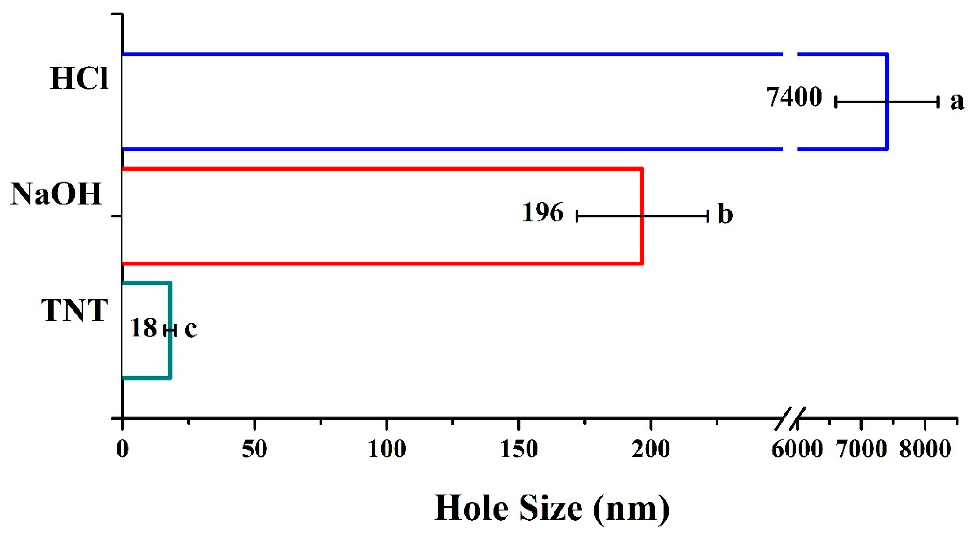
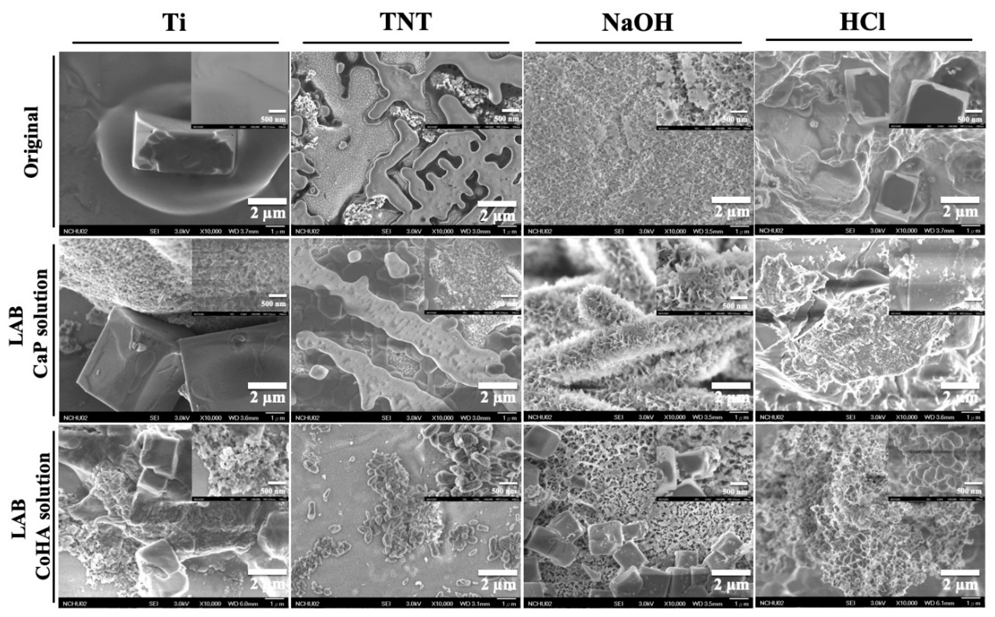
3.1. Surface Treatment of Titanium

Results of different surface treatments of samples were determined by FE-SEM (Figure 2). The untreated titanium sheet had a smooth, flat surface. After anodization (TNTs) treatment, a TNTs structure with a diameter of about 18 ± 1.1 nm appeared on the surface. The formation of the nanotube structure could be divided into the formation of titanium dioxide and the chemical attack caused by fluoride ions in the electrolyte. In past research, the diameter of TNTs could be controlled by the voltage and electrolytic solution. In this study, the conditions of the electrolysis reaction were not adjusted, so the hole sizes were not changed. After both alkali treatment and acid treatment of the titanium sheet, a uniform nanoporous network structure was observed on the surface. Calculating the large pores of the titanium sheet after different surface treatments showed that acid treatment produced the largest pore structure followed by alkali treatment and anodization (Figure 3). The results of the surface elemental analysis showed that the pure titanium sheet had only titanium and a little carbon, indicating that the sample surface was not contaminated (Table 2). A small amount of the fluorine element remained on the surface of TNTs due to erosion of fluoride ions in the electrolyte. After alkali treatment, the surface contained sodium because of the phenomenon of sodium titanate (Na2Ti5O11) being formed on the surface of titanium metal after alkali treatment [26]. Finally, only titanium was observed on the surface after acid treatment. This may have been due to corrosion of the entire surface oxide layer by hydrochloric acid [27].

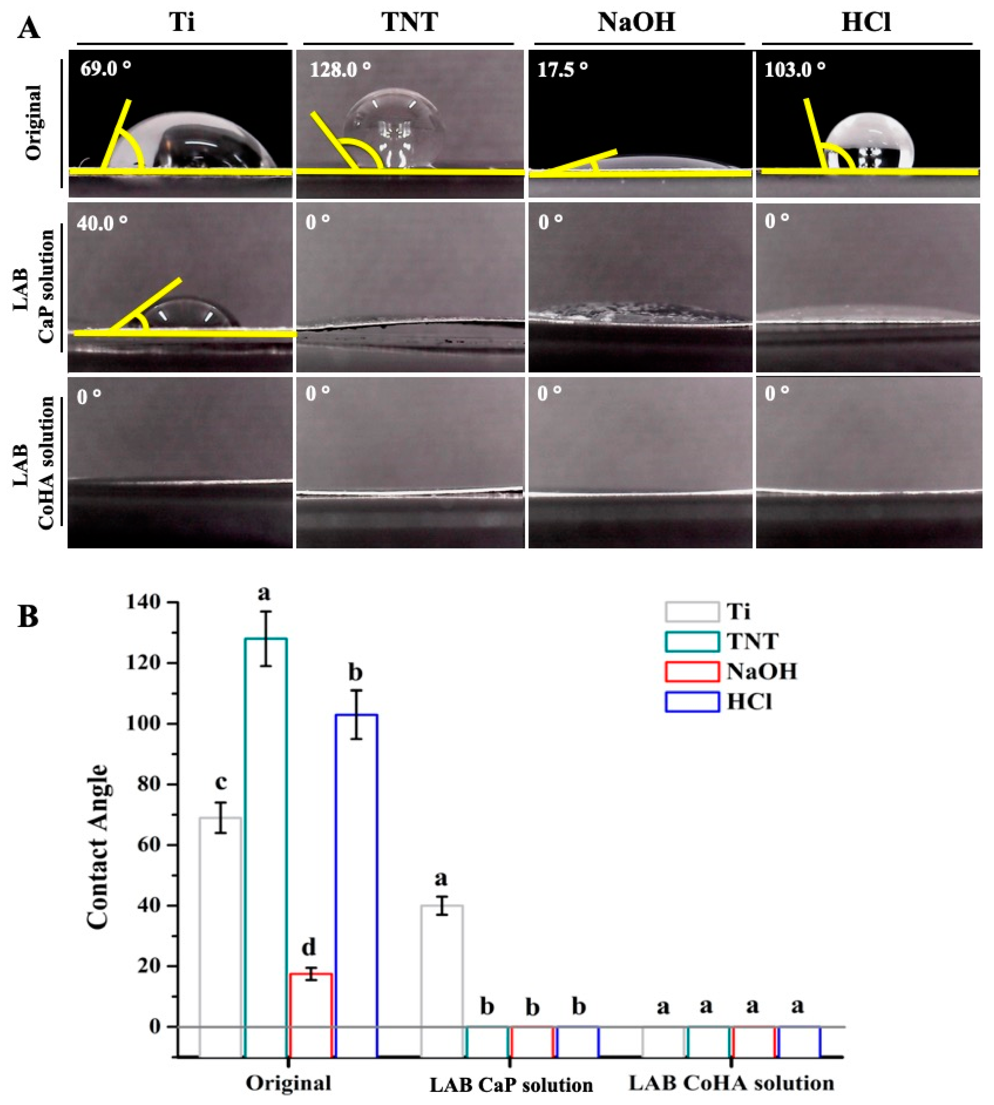
3.2. Hydrophilicity Test

Measuring the contact angle is a method of evaluating the hydrophilicity and hydrophobicity of a material surface and can be used to observe the effect of the material surface on cell adhesion. Therefore, the hydrophilicity and hydrophobicity of the material surface are two of the most important biological parameters of implants [28]. The literature points out that increasing the surface wettability of a material can improve its biological activity. In addition, a hydrophilic surface is better than a hydrophobic surface for the adhesion of bone cells in the early stage of implantation [29]. Hydrophilic and hydrophobic test results of titanium sheets after different surface treatments are shown in Figure 4. Masahashi et al. showed that TNTs produced after anodization are hydrophilic and hydrophobic. However, annealing treatment will produce highly crystalline anatase that expresses hydrophilic properties [30]. The titanium sheet had a relatively hydrophilic surface after alkali treatment (Figure 4A). These results are similar to those in the literature such as alkali treatment increasing the hydrophilicity of a metal surface [31]. Cheng et al. used NaOH to alkali-treat titanium flakes and showed that titanium flakes had better hydrophilicity as the concentration increased [20]. Results of acid treatment and anodic oxidation were similar as both were hydrophobic, which may have been caused by the same crystalline phase being formed.

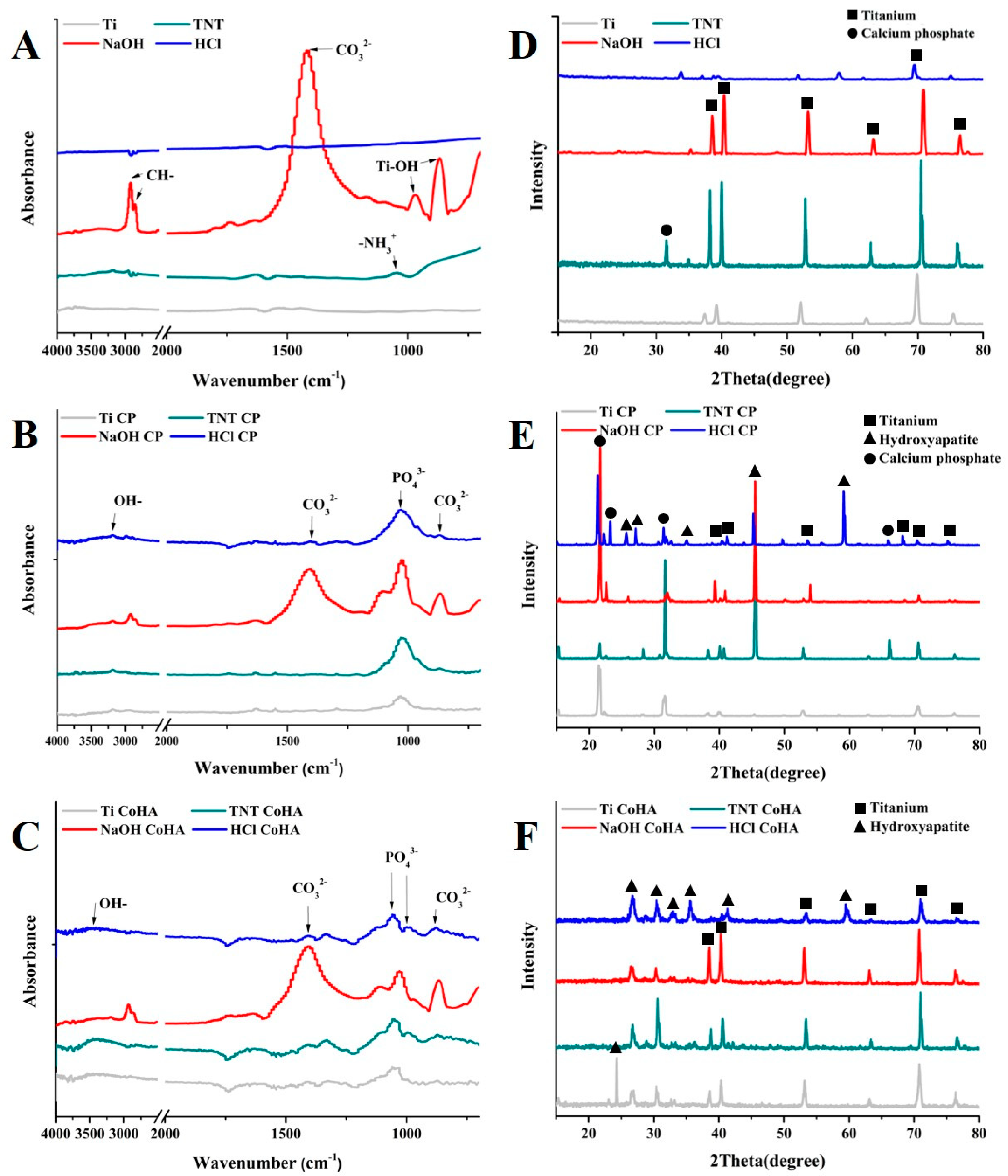
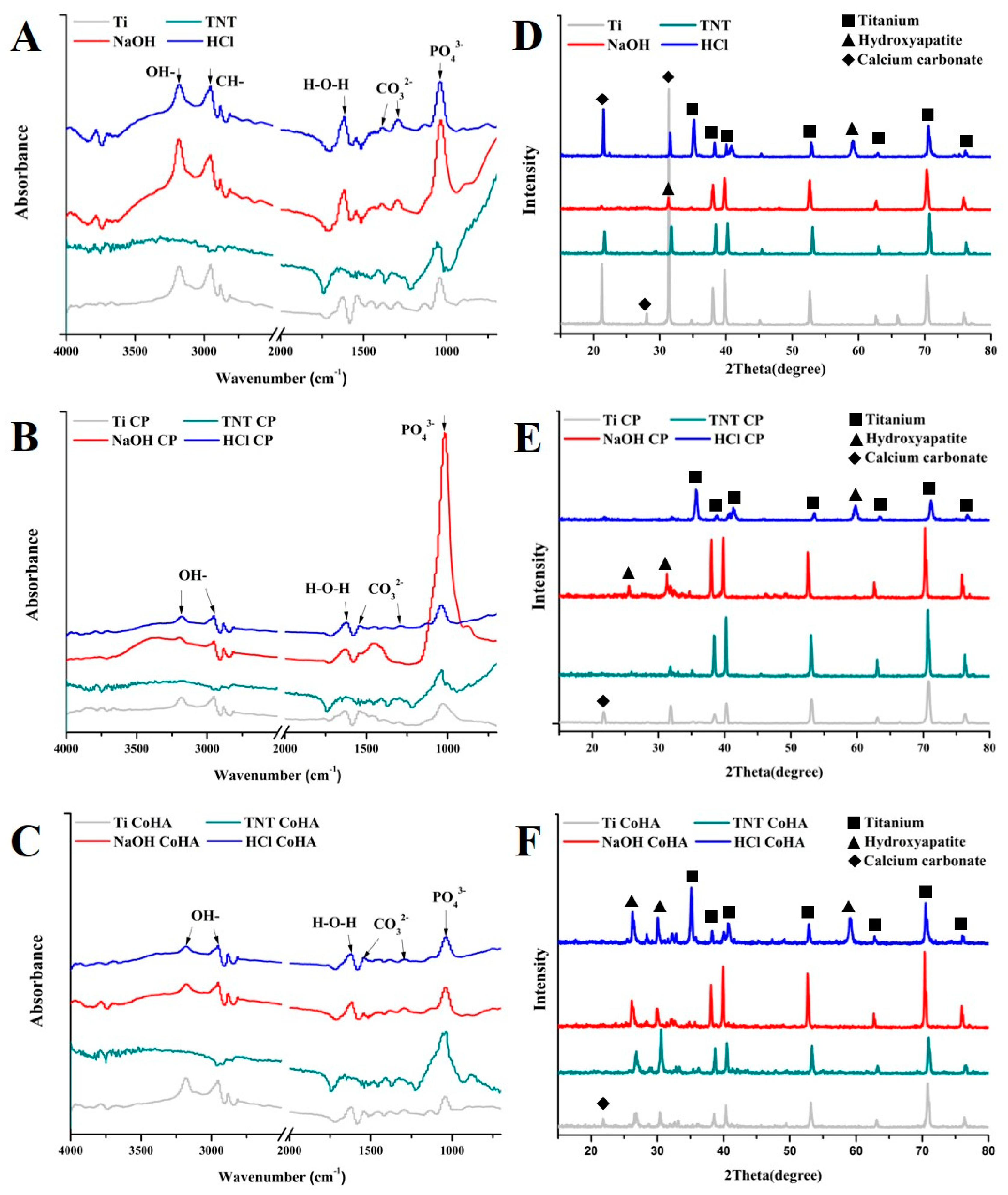
3.3. Characterization

After the CaP solution was treated with LAB, CaP crystals were found on the surface. In addition, it was found that holes of different sizes affected the crystal shape and size (Figure 2). In pure titanium and alkali-treated test pieces, flake-like CaP crystals were observed, which was similar to the results of He et al. [32]. CaP crystals treated with the same alkalinity were smaller than those of pure titanium, which may have been caused by the influence of nanopores on the surface of the test piece. CaP observed on the surface after anodization showed a plate-like crystal structure. CaP formed on the surface after acid treatment presented a flower-like crystal structure composed of needle-like crystals. The morphology of HA with different crystal structures is consistent with that reported by Sadat-Shojai et al. [33]. LAB treatment of titanium flakes soaked in a CoHA solution showed different CoHA crystals (Figure 2). No crystals were observed on the untreated titanium piece. After anodic oxidation, it was observed that CoHA in the form of plate-like aggregates was consistent with the CaP synthesized by a hydrothermal method [34]. Surfaces treated with NaOH and those with HCl respectively exhibited flaky and plate-like crystal structures.
Surface element compositions of groups of CaP solution-treated titanium sheets were analyzed by EDX. The surface was mainly composed of oxygen, calcium, and phosphorus, which indicated that HA was deposited on the surface of the titanium sheet after LAB irradiation. The test piece treated with the CoHA solution, and LAB also revealed the presence of cobalt. This proved to be cobalt-containing HA crystals. Calculating the Ca/P ratio on the surface showed that Ca/P ratios of the CP solution and the CoHA solution were both <1.67, and both were calcium-deficient HA. Previous studies indicated that calcium-deficient hydroxyapatites have higher degradability. They are easily degraded in the body, allowing absorption by surrounding tissues [35].
Functional groups on the surface of the evaluation test pieces were analyzed by ATR-FTIR. No group had characteristic peaks of HA before LAB treatment (Figure 5A). After anodization treatment, tensile vibrations of amino groups (NH3+) were detected at 1072 and 1058 cm−1 on titanium [36]. Peaks observed at 869 and 970 cm−1 after alkali treatment were Ti-OH groups. This was the result of ion exchange between sodium titanate and H3O+ produced during alkali treatment [37]. There were characteristic peaks such as P-O, C-O, and O-H functional groups on specimens treated by LAB (Figure 5B,C). Among these, several bands between 900 and 1200 cm−1 represent the stretching mode of P-O bonds. The PO43−v1 bond is at 962 cm−1, and the PO43−v3 bond is at 1038 cm−1. However, there was a bifurcation at half height of 962 cm−1. This is not a typical HA functional group. This shape indicated the presence of hydroxylapatite with lower crystallinity [38]. Obvious absorption peaks were found at 1419–1486 cm−1 to be the C-O bonding group, and the v3 bonding of CO3 is located at 1400–1580 and 870 cm−1. The presence of the two peaks indicated that the phosphate position in the HA structure was replaced by carbonate [39]. Therefore, crystals formed on the surface were carbonated HAp (CHA, Ca10(PO4)6(CO3)). This reaction was caused by the participation of carbon dioxide dissolved in the air and water. At 1595–1630 cm−1, the characteristic peak produced by the vibration of H2O molecules was the bonding functional group of symmetrical H-O-H. The asymmetrical O-H bonding functional group appeared in the range of 3800–3200 cm−1. This characteristic peak represents water molecules adsorbed onto the surface [40].
The XRD analysis of the sample before LAB treatment indicated the crystal phase structure of titanium (Figure 5D). Compared to the literature, the crystalline phase structure of the titanium plate after LAB treatment (Figure 5E,F) showed the diffraction front of HA [41]. The CaP deposited on the surface of the titanium sheet treated with the CaP solution and LAB was mainly HA. A similar HA structure was also observed on the titanium sheet after immersion in the CoHA solution and LAB. However, it may be that the Co content was too small, so the diffraction peak of cobalt oxide was not observed (Figure 5F). In addition, it was found that diffraction of the deposited HA was more pronounced in the CaP solution compared to LAB treatment of the CoHA solution. It may be that the CaP solution was a supersaturated CaP solution suitable for the hydrothermal method with a pH of 7.4, and the CoHA solution was an electrolyte with a pH of 3.52, which is an acidic solution. The principle of the LAB method used in this experiment is to use thermally induced HA deposition similar to the hydrothermal method. Therefore, the CaP solution had an obvious crystallization performance. In addition, samples were transformed into super-hydrophilic surfaces after LAB treatment (Figure 4B). This helped improve the biocompatibility of the material surface and cell adhesion.
Based on the above SEM observations, test pieces with different surface treatments were treated with the CaP solution and the CoHA solution and LAB. The results showed that in addition to pure titanium, no crystallization was observed after LAB treatment, but other groups had found the presence of CaP and CoHA crystals. The EDX analysis of the surface elemental composition had calcium and phosphorus in the CaP solution treated with LAB. In the CoHA solution treated with LAB, calcium, phosphorus, and cobalt were present, except in the untreated group. The results of ATR-FTIR confirmed that CaP and CoHA synthesized in this study were indeed composed of HA groups. XRD results showed that the surfaces of the CaP solution and CoHA solution after LAB treatment consisted of all HA structures. The titanium sheet being irradiated by LAB caused CaP and CoHA deposition on different surface-modified materials to improve the hydrophilic properties of titanium.

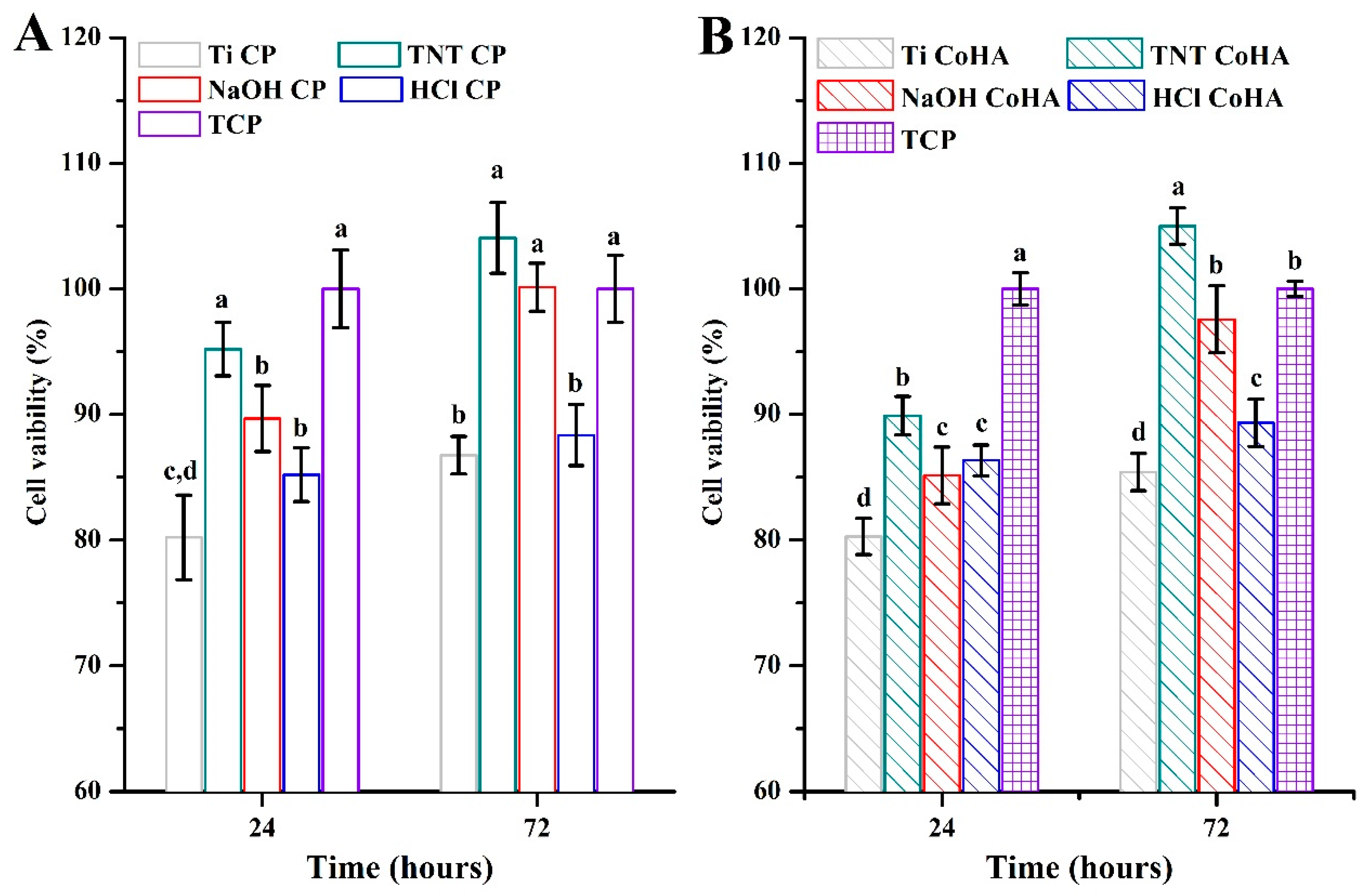
3.4. Biocompatibility

In this study, TCPs were used as the control group. The results showed that MG63 cells experienced no cytotoxicity after 24 and 72 h of culture (Figure 6). The MTT assay showed that the TNT-treated and NaOH-treated test pieces in the CaP solution group had cell survival rates significantly higher than that of pure titanium after 72 h of culture (p < 0.05). However, rates of the TNT CaP and NaOH CaP groups did not significantly differ from that of the TCPs (p < 0.05) (Figure 6A). In addition, the biocompatibility result of LAB CoHA solution is Figure 6B. Rates of the TNT CoHA, NaOH CoHA, and HCl CoHA groups were all significantly higher than that of the Ti CoHA group (p < 0.05). In particular, the cell survival rate of the TNT CoHA group after 72 h of culture was significantly higher than that of the TCPs (p < 0.05). This may have been caused by the release of cobalt ions. A previous study found that cobalt ions can mimic a hypoxic environment and activate HIF-1α to promote vascular epithelial growth factor (VEGF) production [42]. The differentiation of MG63 cells improved under the influence of cobalt ions released by TNT CoHA [43]. In addition, Ignjatovic et al. reported that synthesized CoHA was implanted in the mandible of a rat model of osteoporosis, and results showed that the mineral deposition rate of cobalt ion-filled sites was higher than that of pure HA, and cobalt promoted the rate of bone formation [44]. This is similar to results of our study, indicating that cobalt ions contributed to the growth and differentiation of MG63 cells.

3.5. Bioactivity

We uses an SBF solution in which to soak test pieces for 7 days to evaluate the bioactivity of the titanium sheet after anodization, alkali treatment, and acid treatment. The results showed that there were different mineralized crystals on the surface of the titanium sheet (Figure 7). Mineralized crystals on the surface were mainly calcium carbonate (CaCO3). Calcium carbonate has three crystal forms: rhombic calcite, needle-shaped aragonite, and spherical vaterite [45]. Diamond-shaped crystals observed in the Ti group were calcite. Mineralized crystals observed in the TNTs group were not calcium carbonate but mineralized crystals resembling mountain shapes. A mineralized layer other than calcite was observed in the alkali-treated group, and needle-shaped crystals in the mineralized layer were aragonite. In the acid treatment group, calcite deposition was observed in holes of the titanium piece. In the CaP solution group, flaky crystals that originally formed on the surface of the test piece were also transformed into needle-shaped aragonite (Figure 7). In the CoHA solution group, the calcium and phosphorus deposits deposited on the surface under the influence of cobalt ions were transformed into round and rod shapes. This was similar to a previous report on CoHA which confirmed that cobalt ions affect the morphology of surface deposits [46]. Gopi et al. reported that the circular morphology of HA can enhance cells adhesion [47]. However, it was confirmed from SEM images that irradiation via LAB induced HA deposition to increase the surface bioactivity.
The surface elemental compositions of groups of test pieces after being soaked in SBF solution for 7 days by an EDX analysis are shown in Table 3. The deposition of HA after LAB irradiation was significantly greater than that of samples without LAB treatment. The CaP solution group showed that titanium elements of TNTs, NaOH, and HCl decreased, which represented an increase in the deposition area of HA. After the CoHA solution was irradiated, cobalt was observed among the groups to show the deposition of CoHA. Comparing the Ca/P ratio on the surface of each group of test pieces showed that the Ca/P ratio increased after LAB treatment. In particular, the Ca/P ratios of the TNT CoHA and NaOH CoHA groups were both 1.67, which were similar to the Ca/P ratio of human bone (Table 3). This shows that the surface after LAB treatment was calcium-deficient HA. However, calcium-deficient HA can release ions into SBF to facilitate the deposition of HA. ATR-FTIR spectra show characteristic peaks of test pieces after soaking in the SBF solution for 7 days (Figure 8A–C). There were no significant differences in samples of each group. Functional groups such as P-O, C-O, and O-H were detected on the surface. Bifurcation (1038 cm−1) before soaking in SBF indicated that the phosphate signal was weak (Figure 5B,C). The disappearance of the branching of the PO43− group after SBF soaking indicated that the crystallinity of HA had greatly improved.
Crystal structures of different surfaces after being soaked in SBF for 7 days by LAB treatment were analyzed by XRD (Figure 5D–F). Each group of samples had a diffraction front showing HA after being soaked for 7 days. However, most of the non-irradiated and CaP solution-irradiated groups were mainly composed of calcium carbonate and titanium [48]. The sample with the CoHA solution and LAB mainly had an HA structure after being irradiated. This was similar to ATR-FTIR results. Deposition of the CoHA solution after irradiation was better dissociated in solution. Therefore, more ions were induced to bind and deposit HA. Another factor is that the ionic radius of cobalt (152 pm) is smaller than that of calcium ions (194 pm), which makes it easier for cobalt to combine with phosphorus ions [49]. This may also have resulted in samples irradiated with the CoHA solution being able to induce greater HA deposition. The effect of surface treatment on HA deposition showed that the pure titanium surface was smoother, which made it more difficult for HA to adhere. This study used various methods to make surface junctions with different pores, and results showed that smaller pores (of about 18–196 nm) had better deposition effects. In addition, the pH value of the solution and the substitution of ions also affected the type of HA.
Further, Yingchao Su et al. reported that the CaP coating on the metal implants surface is still a challenge in clinical applications, especially in controlling the coating structure and degradation rate [50]. In addition, Antonio Scarano et al. improved the integration of the implant with the surrounding bone tissue by forming a titanium dioxide layer on the surface of the titanium alloy (Ti6Al4V) [51]. Therefore, our study will evaluate the degradation and biological behavior of the coating and provide more novel clinical methods and applications.

4. Conclusions

In this study, different surface treatments were used to produce holes of different sizes in titanium sheets. We evaluated the effects of LAB treatment with different solutions on the surface structure. Results showed that all samples treated with LAB deposited CaP and CoHA. In addition, smaller holes (TNTs) on the titanium sheet exhibited better deposition, and the type of crystal could be altered by controlling the pH value of the solution. LAB treatment can imbue the surface of titanium sheets with super-hydrophilic properties and improve biocompatibility. The release of cobalt ions through surface CoHA improved the cell survival rate of TNT CoHA. Results of the final immersion in SBF confirmed that LAB treatment with a CoHA solution can improve the hydrophilicity, biocompatibility, and bioactivity of surfaces of titanium sheets. In the future, we hope to make useful contributions to surface coatings of biomedical materials.

Author Contributions

C.-M.T. and F.-Y.F. are the core first authors for contributions to the analysis and interpretation of data, drafting of the manuscript, and critical revision of the manuscript; L.W. writing—review and editing of the manuscript; W.-T.L. carried out the experiment; W.-C.L. were the corresponding authors and contributed to the design and implementation of the research, to the analysis of the results, and to the writing of the manuscript. All authors have read and agreed to the published version of the manuscript.

Funding

This research received no external funding.

Acknowledgments

This research was funded by Ministry of Taipei Medical University of Taiwan, Republic of China, via grants TMU 108-AE1-B45. The authors would like to acknowledge the office of research and development at Taipei Medical University for editing of English language and style.

Conflicts of Interest

There are no conflict to declare.

References

  1. Isa, N.N.C.; Mohd, Y.; Yury, N. Electrochemical deposition and characterization of hydroxyapatite (hap) on titanium substrate. APCBEES Procedia 2012, 3, 46–52. [Google Scholar] [CrossRef] [Green Version]
  2. Bose, S.; Banerjee, D.; Shivaram, A.; Tarafder, S.; Bandyopadhyay, A. Calcium phosphate coated 3d printed porous titanium with nanoscale surface modification for orthopedic and dental applications. Mater. Des. 2018, 151, 102–112. [Google Scholar] [CrossRef] [PubMed]
  3. Dorozhkin, S.V.; Epple, M. Biological and medical significance of calcium phosphates. Angew. Chem. 2002, 41, 3130–3146. [Google Scholar] [CrossRef]
  4. Wang, L.; Nancollas, G.H. Calcium orthophosphates: Crystallization and dissolution. Chem. Rev. 2008, 108, 4628–4669. [Google Scholar] [CrossRef] [Green Version]
  5. Deligianni, D.D.; Katsala, N.D.; Koutsoukos, P.G.; Missirlis, Y.F. Effect of surface roughness of hydroxyapatite on human bone marrow cell adhesion, proliferation, differentiation and detachment strength. Biomaterials 2001, 22, 87–96. [Google Scholar] [CrossRef]
  6. Zhou, H.; Lee, J. Nanoscale hydroxyapatite particles for bone tissue engineering. Acta Biomater. 2011, 7, 2769–2781. [Google Scholar] [CrossRef]
  7. Rath, S.; Anand, A.; Ghosh, N.; Das, L.; Kokate, S.B.; Dixit, P.; Majhi, S.; Rout, N.; Singh, S.P.; Bhattacharyya, A. Cobalt chloride-mediated protein kinase calpha (pkcα) phosphorylation induces hypoxia-inducible factor 1α (hif1α) in the nucleus of gastric cancer cell. Biochem. Biophys. Res. Commun. 2016, 471, 205–212. [Google Scholar] [CrossRef]
  8. Lin, W.-C.; Chuang, C.-C.; Wang, P.-T.; Tang, C.-M. A comparative study on the direct and pulsed current electrodeposition of cobalt-substituted hydroxyapatite for magnetic resonance imaging application. Materials 2018, 12, 116. [Google Scholar] [CrossRef] [Green Version]
  9. Niu, B.; Shi, P.; Shanshan, E.; Wei, D.; Li, Q.; Chen, Y. Preparation and characterization of ha sol–gel coating on mao coated az31 alloy. Surf. Coat. Technol. 2016, 286, 42–48. [Google Scholar] [CrossRef]
  10. De Groot, K.; Geesink, R.; Klein, C.P.A.T.; Serekian, P. Plasma sprayed coatings of hydroxylapatite. J. Biomed. Mater. Res. 1987, 21, 1375–1381. [Google Scholar] [CrossRef]
  11. Cotell, C.M. Pulsed laser deposition and processing of biocompatible hydroxylapatite thin films. Appl. Surf. Sci. 1993, 69, 140–148. [Google Scholar] [CrossRef]
  12. Torrisi, L.; Setola, R. Thermally assisted hydroxyapatite obtained by pulsed-laser deposition on titanium substrates. Thin Solid Films 1993, 227, 32–36. [Google Scholar] [CrossRef]
  13. Mahanti, M.; Nakamura, M.; Pyatenko, A.; Sakamaki, I.; Koga, K.; Oyane, A. The mechanism underlying calcium phosphate precipitation on titanium via ultraviolet, visible, and near infrared laser-assisted biomimetic process. J. Phys. D Appl. Phys. 2016, 49, 1–10. [Google Scholar] [CrossRef]
  14. Oyane, A.; Sakamaki, I.; Shimizu, Y.; Kawaguchi, K.; Koshizaki, N. Liquid-phase laser process for simple and area-specific calcium phosphate coating. J. Biomed. Mater. Res. A 2012, 100, 2573–2580. [Google Scholar] [CrossRef]
  15. Oyane, A.; Matsuoka, N.; Koga, K.; Shimizu, Y.; Nakamura, M.; Kawaguchi, K.; Koshizaki, N.; Sogo, Y.; Ito, A.; Unuma, H. Laser-assisted biomimetic process for surface functionalization of titanium metal. Colloids Interface Sci. Commun. 2015, 4, 5–9. [Google Scholar] [CrossRef] [Green Version]
  16. Oyane, A.; Sakamaki, I.; Pyatenko, A.; Nakamura, M.; Ishikawa, Y.; Shimizu, Y.; Kawaguchi, K.; Koshizaki, N. Laser-assisted calcium phosphate deposition on polymer substrates in supersaturated solutions. R. Soc. Chem. 2014, 4, 53645–53648. [Google Scholar] [CrossRef]
  17. Pecheva, E.; Petrov, T.; Lungu, C.; Montgomery, P.; Pramatarova, L. Stimulated in vitro bone-like apatite formation by a novel laser processing technique. Chem. Eng. J. 2008, 137, 144–153. [Google Scholar] [CrossRef]
  18. Ivanoff, C.-J.; Widmark, G.; Hallgren, C.; Sennerby, L.; Wennerberg, A. Histologic evaluation of the bone integration of tio2 blasted and turned titanium microimplants in humans. Clin. Oral Implants Res. 2001, 12, 128–134. [Google Scholar] [CrossRef]
  19. Afrashtehfar, K.I.; Del Fabbro, M. Clinical performance of zirconia implants: A meta-review. J. Prosthet. Dent. 2020, 123, 419–426. [Google Scholar] [CrossRef]
  20. Cheng, C.H.; HO, W.F.; Lai, C.H.; Hsu, H.C.; Wu, S.C. Apatite formation on a nano-porous titanium alloy surface treated with aqueous naoh. J. Sci. Eng. Technol. 2010, 6, 1–7. [Google Scholar]
  21. Takemoto, M.; Fujibayashi, S.; Neo, M.; Suzuki, J.; Matsushita, T.; Kokubo, T.; Nakamura, T. Osteoinductive porous titanium implants: Effect of sodium removal by dilute hcl treatment. Biomaterials 2006, 27, 2682–2691. [Google Scholar] [CrossRef] [PubMed]
  22. Oh, S.-H.; Finõnes, R.R.; Daraio, C.; Chen, L.-H.; Jin, S. Growth of nano-scale hydroxyapatite using chemically treated titanium oxide nanotubes. Biomaterials 2005, 26, 4938–4943. [Google Scholar] [CrossRef] [PubMed]
  23. Gong, J.; Lai, Y.; Lin, C. Electrochemically multi-anodized tio2 nanotube arrays for enhancing hydrogen generation by photoelectrocatalytic water splitting. Electrochim. Acta 2010, 55, 4776–4782. [Google Scholar] [CrossRef]
  24. Sasidharan, M.; Oyane, A.; Kim, H.-M.; Kokubo, T.; Ito, A. Biomimetic coating of laminin-apatite composite on titanium metal and its excellent cell-adhesive properties. Adv. Mater. 2004, 16, 1071–1074. [Google Scholar]
  25. Kokubo, T.; Ito, S.; Huang, Z.T.; Hayashi, T.; Sakka, S. Ca, p-rich layer formed on high-strenght bioactive glass-ceramic a-w. J. Biomed. Mater. Res. 1990, 24, 331–343. [Google Scholar] [CrossRef]
  26. Kim, H.M.; Miyaji, F.; Kokubo, T.; Nakamura, T. Preparation of bioactive ti and its alloys via simple chemical surface treatment. Biomed. Mater. Res. Part A 1996, 32, 409–417. [Google Scholar] [CrossRef]
  27. Liang, J.; Xu, S.; Shen, M.; Cheng, B.; Li, Y.; Liu, X.; Qin, D.; Bellare, A.; Kong, L. Osteogenic activity of titanium surfaces with hierarchical micro/nano-structures obtained by hydrofluoric acid treatment. Int. J. Nanomed. 2017, 12, 1317–1328. [Google Scholar] [CrossRef] [Green Version]
  28. Lim, Y.J.; Oshida, Y.; Andres, C.J.; Barco, M.T. Surface characterizations of variously treated titanium materials. Int. J. Oral Maxillofac. Implant. 2001, 16, 333–342. [Google Scholar]
  29. Buser, D.; Broggini, N.; Wieland, M.; Schenk, R.K.; Denzer, A.J.; Cochran, D.L.; Hoffmann, B.; Lussi, A.; Steinemann, S.G. Enhanced bone apposition to a chemically modified sla titanium surface. J. Dent. Res. 2004, 83, 529–533. [Google Scholar] [CrossRef]
  30. Masahashi, N.; Semboshi, S.; Ohtsu, N.; Oku, M. Microstructure and superhydrophilicity of anodic tio2 films on pure titanium. Thin Solid Films 2008, 516, 7488–7496. [Google Scholar] [CrossRef]
  31. Kokubo, T.; Kim, H.-M.; Kawashita, M. Novel bioactive materials with different mechanical properties. Biomaterials 2003, 24, 2161–2175. [Google Scholar] [CrossRef]
  32. He, Q.; Huang, Z. Controlled synthesis and morphological evolution of dendritic porous microspheres of calcium phosphates. J. Porous Mater. 2008, 16, 683–689. [Google Scholar] [CrossRef]
  33. Sadat-Shojai, M.; Khorasani, M.-T.; Dinpanah-Khoshdargi, E.; Jamshidi, A. Synthesis methods for nanosized hydroxyapatite with diverse structures. Acta Biomater. 2013, 9, 7591–7621. [Google Scholar] [CrossRef]
  34. Toyama, T.; Nakashima, K.; Yasue, T. Hydrothermal synthesis of β-tricalcium phosphate from amorphous calcium phosphate. J. Ceram. Soc. Jpn. 2002, 110, 716–721. [Google Scholar] [CrossRef] [Green Version]
  35. Zhang, H.; Zhang, M. Characterization and thermal behavior of calcium deficient hydroxyapatite whiskers with various ca/p ratios. Mater. Chem. Phys. 2011, 126, 642–648. [Google Scholar] [CrossRef]
  36. Lamberti, A.; Chiodoni, A.; Shahzad, N.; Bianco, S.; Quaglio, M.; Pirri, C. Ultrafast room-temperature crystallization of TiO2 nanotubes exploiting water-vapor treatment. Sci. Rep. 2015, 5, 1–6. [Google Scholar] [CrossRef] [Green Version]
  37. Chen, Y.; Zheng, X.; Ji, H.; Ding, C. Effect of ti-oh formation on bioactivity of vacuum plasma sprayed titanium coating after chemical treatment. Surf. Coat. Technol. 2007, 202, 494–498. [Google Scholar] [CrossRef]
  38. Granja, P.L.; Ribeiro, C.C.; de Jeso, B.; Baquey, C.; Barbosa, M.A. Mineralization of regenerated cellulose hydrogels. J. Mater. Sci. Mater. Med. 2001, 12, 785–791. [Google Scholar] [CrossRef]
  39. Hong, L.; Wang, Y.; Jia, S.; Huang, Y.; Gao, C.; Wan, Y. Hydroxyapatite/bacterial cellulose composites synthesized via a biomimetic route. Mater. Lett. 2006, 60, 1710–1713. [Google Scholar] [CrossRef]
  40. Koutsopoulos, S. Synthesis and characterization of hydroxyapatite crystals: A review study on the analytical methods. J. Biomed. Mater. Res. 2002, 62, 600–612. [Google Scholar] [CrossRef]
  41. Nie, L.; Suo, J.; Zou, P.; Feng, S. Preparation and properties of biphasic calcium phosphate scaffolds multiply coated with ha/plla nanocomposites for bone tissue engineering applications. J. Nanomater. 2012, 2012, 1–11. [Google Scholar] [CrossRef]
  42. Tanaka, T.; Kojima, I.; Ohse, T.; Ingelfinger, J.R.; Adler, S.; Fujita, T.; Nangaku, M. Cobalt promotes angiogenesis via hypoxia-inducible factor and protects tubulointerstitium in the remnant kidney model. Lab. Investig. 2005, 85, 1292–1307. [Google Scholar] [CrossRef] [PubMed] [Green Version]
  43. Wu, C.; Zhou, Y.; Fan, W.; Han, P.; Chang, J.; Yuen, J.; Zhang, M.; Xiao, Y. Hypoxia-mimicking mesoporous bioactive glass scaffolds with controllable cobalt ion release for bone tissue engineering. Biomaterials 2012, 33, 2076–2085. [Google Scholar] [CrossRef] [PubMed] [Green Version]
  44. Ignjatović, N.; Ajduković, Z.; Savić, V.; Najman, S.; Mihailović, D.; Vasiljević, P.; Stojanović, Z.; Uskoković, V.; Uskoković, D. Nanoparticles of cobalt-substituted hydroxyapatite in regeneration of mandibular osteoporotic bones. J. Mater. Sci. Mater. Med. 2013, 24, 343–354. [Google Scholar] [CrossRef] [Green Version]
  45. Svenskaya, Y.; Parakhonskiy, B.V.; Haase, A.; Atkin, V.; Lukyanets, E.; Gorin, D.; Antolini, R. Anticancer drug delivery system based on calcium carbonate particles loaded with a photosensitizer. Biophys. Chem. 2013, 182, 11–15. [Google Scholar] [CrossRef] [Green Version]
  46. Kramer, E.; Itzkowitz, E.; Wei, M. Synthesis and characterization of cobalt-substituted hydroxyapatite powders. Ceram. Int. 2014, 40, 13471–13480. [Google Scholar] [CrossRef]
  47. Gopi, D.; Karthika, A.; Nithiya, S.; Kavitha, L. In vitro biological performance of minerals substituted hydroxyapatite coating by pulsed electrodeposition method. Mater. Chem. Phys. 2014, 144, 75–85. [Google Scholar] [CrossRef]
  48. Rahman, M.A.; Halfar, J.; Shinjo, R. X-ray diffraction is a promising tool to characterize coral skeletons. Adv. Mater. Phys. Chem. 2013, 3, 120–125. [Google Scholar] [CrossRef] [Green Version]
  49. Lin, W.; Chuang, C.; Yao, C.; Tang, C. Effect of cobalt precursors on cobalt-hydroxyapatite used in bone regeneration and MRI. J. Dent. Res. 2020. [Google Scholar] [CrossRef]
  50. Su, Y.; Cockerill, I.; Zheng, Y.; Tang, L.; Qin, Y.-X.; Zhu, D. Biofunctionalization of metallic implants by calcium phosphate coatings. Bioact. Mater. 2019, 4, 196–206. [Google Scholar] [CrossRef]
  51. Scarano, A.; Crocetta, E.; Quaranta, A.; Lorusso, F. Influence of the thermal treatment to address a better osseointegration of ti6al4v dental implants: Histological and histomorphometrical study in a rabbit model. BioMed Res. Int. 2018, 2018, 1–7. [Google Scholar]
Figure 1. Compendium illustration showing the experimental procedures. (A) Surface treatment of titanium. (B) Preparation of laser-assisted biomimetic (LAB). (C) Characterization and biocompatibility of samples.
Applsci 10 08158 g001
Figure 2. Surface topography of the titanium sheet with different surface treatments and samples after LAB treatments (calcium-to-phosphorus (CaP) solution and cobalt-substituted hydroxyapatite (CoHA) solution) by SEM.
Applsci 10 08158 g002
Figure 3. The holes size of the titanium sheet after different surface treatments. Means with different letters were significantly different (p < 0.05, mean ± SD, n = 10).
Applsci 10 08158 g003
Figure 4. The surface wettability titanium sheet has different surface treatments. (A) Image of surface contact angle. (B) Quantitative analysis of the contact angle was performed. Means with different letters were significantly different (p < 0.05, mean ± SD, n = 3).
Applsci 10 08158 g004
Figure 5. The characteristics of the titanium sheet with different surface treatments and samples after LAB treatments. (AC) FTIR spectra obtained from original, LAB CaP solution and LAB CoHA solution. (DF) XRD patterns of the original, LAB CaP solution, and LAB CoHA solution.
Applsci 10 08158 g005
Figure 6. Proliferation determined according to 3-(4,5-dimethylthiazol-2-yl)-2,5-diphenyltetrazolium bromide (MTT) assay of MG63 cell for 24 and 72 h. (A) LAB CaP solution. (B) LAB CoHA solution. Means with different letters were significantly different (p < 0.05, mean ± SD, n = 4).
Applsci 10 08158 g006
Figure 7. The surface topography of the LAB-treated samples after soaking in simulated body fluid (SBF) for seven days by FE-SEM.
Applsci 10 08158 g007
Figure 8. The characteristics of the LAB-treated samples after soaking in SBF for seven days. (AC) FTIR spectra obtained from original, LAB CaP solution, and LAB CoHA solution. (DF) XRD patterns of the original, LAB CaP solution, and LAB CoHA solution.
Applsci 10 08158 g008
Table 1. Summary of samples for the synthesis systems.
SampleOriginalLAB
CaP SolutionCoHA Solution
Control groupTiTi CPTi CoHA
Anodic oxidationTNTTNT CPTNT CoHA
Alkali treatmentNaOHNaOH CPNaOH CoHA
Acid treatmentHClHCl CPHCl CoHA
Table 2. Surface element analysis of the titanium sheet with different surface treatments and samples after LAB treatments by EDX.
Atomic%CTiONaFCaPCa/PCoCa + Co/P
OriginalTi12.8887.12---N.D
TNT7.9229.8348.57-13.68
NaOH5.6131.4558.914.03-
HCl-100---
LAB
CaP solution
Ti11.5083.931.65--1.51.41.07N.DN.D
TNT3.0016.1037.16--2.181.811.20
NaOH7.016.6968.321.14-8.657.371.17
HCl3.6718.4263.95--7.466.501.15
LAB
CoHA solution
Ti13.2386.77--------
TNT--69.311.50-12.5215.810.790.410.82
NaOH--68.061.82-12.9715.680.830.650.87
HCl0.61-68.273.14-11.1913.170.850.660.90
Table 3. Surface element analysis of the LAB-treated samples after soaking in SBF for seven days by EDX.
Atomic%TiCaPCa/PCoCa + Co/P
OriginalTi28.710.820.771.06N.D
TNT28.020.490.411.20
NaOH14.886.886.111.13
HCl32.242.141.761.22
LAB
CaP solution
Ti23.451.171.021.15N.D
TNT1.008.676.031.44
NaOH3.489.507.801.22
HCl3.238.906.801.31
LAB
CoHA solution
Ti26.732.973.680.810.550.96
TNT15.924.223.211.311.151.67
NaOH23.032.181.751.240.781.69
HCl12.1014.2011.761.213.091.47
Publisher’s Note: MDPI stays neutral with regard to jurisdictional claims in published maps and institutional affiliations.

