Article

Photo-Biomodulation as a Prevention Modality of Oral Mucositis in Patients Undergoing Allogeneic Hematopoietic Stem Cell Transplantation

1
Department of Oral Sciences and Maxillofacial Surgery, Sapienza University of Rome, 00161 Rome, Italy
2
Department of Cellular Biotechnologies and Hematology, Policlinico Umberto I, Sapienza University of Rome, 00161 Rome, Italy
*
Author to whom correspondence should be addressed.
Appl. Sci. 2020, 10(21), 7479; https://doi.org/10.3390/app10217479
Submission received: 29 September 2020 / Revised: 21 October 2020 / Accepted: 22 October 2020 / Published: 24 October 2020
(This article belongs to the Special Issue Current Concept and Emerging Treatments in Oral Diseases)

Abstract

:
The aim of the study was to observe the effectiveness of a photo-biomodulation (PBM) protocol for the prevention of oral mucositis (OM) in patients undergoing allogeneic hematopoietic stem cell transplantation (aHSCT). A case-control study was conducted on 40 patients undergoing aHSCT. The patients were divided into two groups; the preventive group (PG) included 20 patients (7 females and 13 males) who were subjected to intra-oral PBM for five sessions a week, starting one day before the conditioning regimen and continuing until the 10th day after transplantation (D+10). In each session, ten points on the at-risk mucosal surfaces were irradiated using a double diode laser that emits two wavelengths simultaneously at 650 nm and at 904–910 nm with the following parameters at each point: energy of 4 J, and power of 88.9 mW. The control group (CG) included 20 patients (10 females and 10 males) who were not subjected to laser therapy and were selected retrospectively to compare the obtained results. For all patients, OM was assessed by the World Health Organization (WHO) grading scale. Eight patients in the PG did not experience OM during their hospitalization period (with grade 0). Severe OM was observed in 40% of the patients in the PG, while in the CG, severe OM was shown in 85% of the patients. The mean duration of OM in the PG was significantly lower than that of CG (4.7 days in the PG and 15 days in the CG) (p < 0.001). The study demonstrated that the preventive PBM protocol reduced the severity and duration of OM in patients undergoing aHSCT.

1. Introduction

Allogeneic hematopoietic stem cell transplantation (aHSCT) is the standard of care for several congenital and acquired disorders of the hematopoietic system. Patients undergoing aHSCT can develop various complications related to transplantation itself (e.g., the conditioning regimen, delayed regeneration of hematopoiesis), a hindered immune response, or treatment toxicity. The gastrointestinal (GI) tract is one of the systems most affected by HSCT complications, especially the oral cavity.
Advancements in infection control protocols, pain management modalities and reducing the intensity of conditioning regimens pre-aHSCT have minimized these complications. However, oral mucositis (OM) has remained the most persistent debilitating and challenging complication of HSCT [1,2].
OM in patients undergoing aHSCT has a high incidence and a wide range of effects in 75% to 99% of cases. This wide variation is probably due to the presence of different pre-transplant condition regimens, the presence of patients-related risk factors, the lack of standardized scoring criteria, and the presence of many complications to cancer therapy that may result in underreporting OM [3,4,5].
OM is acute inflammation and ulcerative lesions of the oral mucosa, and it is usually caused by cancer therapy [2,6]. The pathophysiology of OM is a complex biological process that can be described in five phases: the initiation, upregulation and/or activation, damage amplification phase, mucosal ulceration, and the healing phase. Proinflammatory cytokines and pathways, reactive oxygen species, and the metabolic byproducts of oral bacteria have been associated with the pathogenesis and aggravation of OM [3,4,7,8].
OM manifests as erythema and/or ulcerations conjugated with pain and difficulty eating, drinking, and talking. Severe OM may have critical clinical sequences, such as hindering normal nutrition, which leads to the need for parenteral nutrition, an increase in the incidence of secondary infections, an increase in systemic analgesic intake, interruption of cancer therapy, and prolongation of hospital stay. These consequences may have a negative impact on the patients’ quality of life and cancer prognosis [4,6,9].
Therefore, several interventions and agents have been studied and proposed for the prevention or treatment of OM [10,11,12,13,14]. Several organizations, such as the American Society of Clinical Oncology (ASCO), the Multinational Association of Supportive Care in Cancer (MASCC), and the National Comprehensive Care Network (NCCN) have evaluated these interventions by carrying out systematic reviews to prepare an evidence-based guideline [15,16,17,18].
Photo-biomodulation (PBM) is one of these evaluated interventions. PBM is the application of light at low power to utilize the effect of light energy on living cells for therapeutic purposes. The absorbed light energy triggers several pathways that eventually result in anti-inflammatory, analgesic, and biomodulatory effects [19,20,21].
Many studies have demonstrated the beneficial effect of PBM on the management of OM [22,23,24]. In 2014, the MASCC and the International Society of Oral Oncology (ISOO) published an updated clinical practice outline, recommending PBM as a method of preventing OM in patients undergoing HSCT, with or without total body irradiation (TBI), with a level of evidence II [15]. In addition, recent systematic reviews have demonstrated the effectiveness of prophylactic PBM in reducing the severity and duration of OM [25,26,27,28,29].
However, heterogeneity in the basic parameters and schedules for PBM application has been observed among previous studies [24,25,26,27,28]. It is difficult to define an optimal PBM protocol if the precise role and contribution of each parameter of PBM and the complexity of the light-tissue interactions are not fully understood [25]. It is believed that further studies that include detailed reporting of the parameters and a better understanding of the role of these variables are required to identify the optimal characteristics of PBM for the management of OM [25]. This study aimed to observe the effectiveness of a PBM protocol as a preventive modality for OM in patients undergoing a conditioning regimen for aHSCT by evaluating the impact of PBM on OM severity, duration, and pattern.

2. Materials and Methods

A case-control study was carried out in patients undergoing aHSCT in the Department of Cellular Biotechnologies and Hematology at Sapienza University of Rome with the cooperation of the Department of Oral Sciences and Maxillofacial Surgery at Sapienza University of Rome. The study is part of a project called “MoMax” (Oral Medicine and Maxillofacial Surgeons), a task force designed to provide cancer patients and high-risk patients with multidisciplinary team care, through cooperation between different health providers. All subjects gave their informed consent for inclusion in the study. The study was conducted in accordance with the Declaration of Helsinki, and the protocol was approved by the Local Ethical Committee of Sapienza University of Rome (Prot. N.264, 13 March 2017).
All patients receiving conditioning regimens for aHSCT in the age range of 6 to 80 years, were considered eligible in the study. Uncooperative patients, patients with systemic diseases that hinder wound healing (e.g., uncontrolled diabetes mellitus, etc.), patients with an oxygen mask, or orogastric catheters that might hinder the PBM application, and patients with suspicious lesions within the oral cavity were excluded from the study.
A total of 40 patients (17 females and 23 males) with a median age of 46 years, were included in the study. Twenty-three patients received HSCT from a matched unrelated donor (MUD) and 17 patients received HSCT from a matched related donor (MRD). The patient characteristics of both groups are presented in Table 1.
This case-control study analyzed two groups of patients:
The preventive group (PG): 20 patients (7 females and 13 males) were recruited prospectively and were subjected to intra-oral PBM for prevention purposes.
The retrospective control group (CG): 20 patients (10 females and 10 males) receiving aHSCT between December 2014 and October 2016, and not subjected to PBM were selected retrospectively to compare the obtained results. The aim of the study was to compare OM incidence and severity between the two groups.

2.1. Routine Oral Care

Prior to the start of the conditioning regimen, all patients received a comprehensive oral examination with panoramic radiographs. The aim of the oral examination was to optimize, treat, and eliminate all the potential sources of oral infection that might compromise the transplantation procedures. Patient education about the possible oral collateral effects due to the conditioning regimen and HSCT was provided. All patients were instructed about standardized basic oral hygiene practices during the transplantation procedures including teeth brushing 2 times daily, using soft brushes with mild toothpaste, and avoiding the use of toothpicks or dental floss. All preventive procedures were performed by the same oral surgeon at the Department of Oral Sciences and Maxillofacial Surgery at Sapienza University of Rome. A mixture of saline and sodium bicarbonate was prescribed as a mouthwash for all of the patients. In case of oral discomfort, a dentist was consulted during the hospitalization.

2.2. PBM Parameters

The patients in the PG were subjected to five sessions of intra-oral PBM per week (Monday to Friday), which started one day before the conditioning regimen, and continued until the 10th day after transplantation (D+10). The laser device that was used was a double diode laser (Lumix2®; FISIOLINE, Verduno, Cuneo, Italy), which emits two wavelengths simultaneously at 650 nm and at 904–910 nm. The technical features of this device are as follows: the visible red source (650 nm) is operated in continuous mode (CW) with an average output power (at the source) of 100 mW. The infrared GaAs source can be operated in pulsed and super-pulsed emission mode with peak power at aperture 45 W, and a pulse duration of 200 ns.
Before starting each PBM session, the device was cleaned and the hand-piece was covered with a plastic wrap (to avoid direct contact with patients). Appropriate safety glasses were used by both the operator and the patient. All the PBM sessions were performed by the same laser operator to avoid intraoperative bio-stimulation variations.
Ten points on the at-risk mucosal surfaces were irradiated in each session, including buccal mucosa, tip, ventral and marginal surface of the tongue, floor of the mouth, soft palate, and inner surfaces of the lip.
The laser device was programmed manually and the 650 nm wavelength was switched off. The only used wavelengths were the 904–910 nm and the laser light guide red 650 nm (for real visualization of the infrared beam application area). The laser was applied point by point in a defocused (non-contact) mode at a distance of ~1 cm from the mucosal tissues. The parameters that were used for each point were as follows: energy of 4 J, power of 88.9 mW, fluence of 8 J/cm2, frequency of 13 kHz, and application time of 45 s. The beam diameter was 8 mm, which is usually obtained when the handpiece lens is away from the targeted tissue of about 1 cm, and the calculated point area was ~0.5 cm2. The total energy per session was 40 J. Before the start of each session, a laser output test was performed by an optical sensor integrated into the device. The PBM parameters are reported in Table 2 as it is recommended in the literature to provide them in table [30].
In case of the development of OM, the laser sessions continued (5 sessions per week) until the complete resolution of OM, even after the 10th day after transplantation (D+10). The laser was applied (point by point) to cover all the ulcerated areas in a defocused (non-contact) mode at a distance of ~1 cm from the mucosal tissues with the following parameters per point: energy of 6 J, power of 90.9 mW, fluence of 12 J/cm2, frequency of 13 kHz, application time of 66 s, beam diameter of 8 mm, and point area of ~0.5 cm2.

2.3. Assessment of OM

For the PG, the oral examination and OM evaluation were performed for all patients before the start of each session by the same oral medicine clinical personnel. A custom-made clinical chart was used to register all patients’ findings including OM grade, pain score, and the site of the OM. The OM evaluation was performed five times a week (Monday to Friday) from one day before the conditioning regimen until the 10th day after transplantation (D+10) or continued until the complete resolution of OM.
The evaluation of the OM grade was performed using the World Health Organization (WHO) grading scale. Grade 0 was considered in case of the absence of signs and symptoms; grade I, in case of the presence of localized or diffused erythema; grade II, in case of the presence of ulcers that did not hinder eating solid food; grade III, in case of the presence of ulcers that hindered eating solid food but the patient was able to swallow liquids; and grade IV, in case where the ulcers impair eating or swallowing liquids [31]. The assessment of pain was performed using the Numeric Rating Scale (NRS).
For the retrospective CG, OM evaluation was provided as part of routine patient care at the Haematology Department, using the WHO grading scale on a daily base. The OM site and grade were collected from the Department database and registered using the same schedule as the PG (i.e., 5 days a week starting from one day before the start of the conditioning regimen until the complete OM resolution).
The total duration of OM and the duration of each grade were calculated for both groups. The maximum OM score was recorded. To evaluate the impact of PBM on the severity of OM, patients with maximum OM grade ≤I were grouped and called “non-ulcerative OM”, and patients with maximum OM grade ≥II were grouped and called “ulcerative OM”.
To evaluate the impact of PBM on the incidence of OM, patients were grouped into two groups with the first group, called “no OM”, including patients with grade 0 as a maximum score of OM and the second group, called “with OM”, including patients with ≥grade I OM.
To evaluate the impact of PBM on the OM pattern and course, OM grades were registered for all the patients at six evaluation time points (Monday to Friday). The evaluation time points (E) were fixed to better compare analyzed patients: E0 was the day of transplantation (D0); E1 was the OM value during D+1 to D+3, E2 was the OM value during D+4 to D+6, E3 was the OM value during D+7 to D+9, E4 was the OM value during D+10 to D+12, and E5 was the OM value during D+13 to D+15 [1].

2.4. Statistical Analysis

All results were databased in Excel sheet using Microsoft® Excel for Mac version 16.30. The statistical analyses were performed using SPSS software (Statistical Package for the Social Sciences) for Windows, release 20.0. The level of significance was set at p < 0.05.
The relationship between age and the set of considered variables was assessed, including the maximum score of OM, the total duration of OM, the duration of each OM grade, and the OM score at each evaluation time points; bivariate correlations were performed using the Spearman’s rank correlation. This correlation coefficient was chosen because the distribution of the various variables scores did not respect the assumptions of normality as indicated by the skewness and kurtosis, and the Shapiro test.
The Mann–Whitney test was used to compare the set of variables with the type of transplant (MUD vs. MRD) and gender (female vs. male).
Chi-square tests (with Fisher’s exact test or Montecarlo test) and student’s t-test were conducted to evaluate whether both groups (PG and retrospective CG) had the same baseline characteristics.
The Mann–Whitney test was used to compare PG and CG patients based on the same set of variables. Fisher’s exact test was used to assess and compare the incidence (no OM vs. with OM) and severity of OM (non-ulcerative OM vs. ulcerative OM) between both groups (PG vs. CG).

3. Results

There were no significant differences between the PG and CG with regard to age, gender, underlying pathology, type of aHSCT, and aHSCT conditioning regimen.
The mean duration of the PBM performed in the PG was 21 days. The average number of PBM sessions performed was 16. In the PG, the incidence of OM was 60% (12 cases) with a maximum OM grade of II. Four of them (20%) suffered from OM grade I (general erythema) with a mean duration of 2.15 days. The rest (40%) suffered from grade II (ulcerative stage) with a mean duration of 2.2 days. The mean starting time of OM was D+7 (7th day post-transplantation). The mean duration of OM was 4.7 days. No cases experienced OM grades III and/or IV. Eight cases (40%) experienced a maximum OM grade of 0. The maximum registered NRS pain value was 8 (with a mean of ~2).
The incidence of OM was 100% (20 patients) in the CG. The mean duration of OM was 15 days. The maximum OM grade was grade IV in 2 cases (10%) with a mean duration of 0.6 days. Eleven patients (55%) showed grade II as a maximum grade of OM. Only 3 patients (15%), experienced a maximum OM grade of I, with a mean duration of 6.3 days. The development of OM was observed at D+6 (6th day post-transplantation) (Figure 1 and Figure 2).
In both groups, the most frequent sites of OM were the tongue with 21 patients (52.5%) and the floor of the mouth with 10 patients (25%).
In the PG, a significant difference was observed between the type of transplantation and the total duration of OM (p = 0.036), and the duration of OM grade I (p = 0.034). Patients who received aHSCT from an MRD, showed a longer total duration of OM (MRD = 8.50 ± 4.85 days; MUD = 3.07 ± 4.32 days) and demonstrated a longer duration of OM grade I (MRD = 4.17 ± 2.93 days; MUD = 1.29 ± 2.37 days). In the CG, no significant correlation was observed between the set of variables and age, gender, and type of transplantation.
Several variables were used to compare the groups (PG vs. CG), and statistical significance was observed for the maximum grade of OM, the total duration of OM, duration of grade 0, grade I, grade II and grade III, and OM scores at evaluation time points E1, E2, E4, and E5 (p < 0.05) (Table 3) (Figure 3).
The severity of OM was observed to be significantly different between the 2 groups (PG vs. CG) (χ2 = 8424; p = 0.003): 12 patients (60%) in the PG showed a non-ulcerative or only erythema (grade ≤ 1). The incidence of OM showed a significant reduction in patients treated by PBM (χ2 = 10,000; p = 0.002): 8 patients (40%) in the PG did not show OM (grade 0) during the total duration of therapy. In contrast, all patients in the CG showed OM (grade > 0) (Table 4).

4. Discussion

Several studies have demonstrated the efficacy of PBM in decreasing the severity and duration of OM. This positive response is probably due to the action of PBM on the different phases of OM, through the decrease in the inflammatory response, the reduction of amplification, and the acceleration of the healing process through promoting growth factors release, the proliferation of fibroblasts, and deposition of collagen. The exact mechanism of PBM to explain this response is still not completely understood [32].
Several local tissue reactions have been associated with the pathogenesis of OM, including the damage caused by reactive oxygen species, inflammatory cytokines (Interleukin-6 (IL-6), tumor necrosis factor-α (TNF-α), interleukin-1 beta (IL-1β)), and submucosal connective tissue damage. Some authors have hypothesized that the reduction in the severity of OM with PBM may be due to the decrease in reactive oxygen species or the reduction of proinflammatory cytokines. This speculation was based on the premise that low-power lasers may be absorbed by specific molecules and other intracellular organelles in addition to the mitochondria [33].
Therefore, some studies have investigated the effect of PBM on the level and behavior of the inflammatory mediators in the blood and/or saliva in patients with OM. However, these studies could not find a complete link between the positive response of PBM and the modulation of the inflammatory mediators [32,34].
Silva et al. observed an elevation in the IL-6 concentration in both groups (laser and control) at the time of OM development. It was noticed that this elevation was accompanied by the increase in TNF-α and IL-1β concentration in the saliva of the laser group. The authors suggested that these findings may explain the decrease in the severity of OM, where IL-6 plays a role in the systemic response to injury by acting as a pro-inflammatory and anti-inflammatory cytokine [32]. Although too much increase in TNF-α and IL-1β may lead to mucosal damage, moderate levels are crucial for inducing the cell regeneration process [22].
Furthermore, the authors observed an increase of matrix metalloproteinase 2 (MMP-2) concentration in saliva in the laser group. They suggested that this increase may have contributed to the reduction in the severity of OM, where MMP subtypes are implicated in both pro- and anti-inflammatory pathways and play a critical role in tissue remodeling processes [32].
The mechanism of the analgesic effect of PBM has not been fully clarified yet. It is believed that the analgesic effect results from the neurophysiological effects (e.g., the release of endogenous opioids), the local circulatory, angiogenic and anti-inflammatory effects [22]. Some authors have suggested that the reduction in prostaglandin E2 (PGE-2), cyclooxygenase-2 (COX-2) expression, and plasminogen activator after PBM application might also be involved [22,35].
Recently, the Mucositis study group of MASCC/ISOO carried out a systematic review to update their evidence-based clinical practice guidelines for the use of PBM as a modality for managing OM. The authors observed an inconsistency and diversity in the laser parameters and PBM schedules, including the starting time, the total duration, and the number of sessions per week. They suggested specific settings for PBM for the prevention of OM in patients undergoing HSCT. These specific settings were based on randomized clinical trials with a high level of evidence. The authors believe that these specific PBM settings would be changed in the future based on a better understanding of the precise roles of each PBM parameter [25].
In the literature, the wavelengths used for OM ranged from 630 nm to 970 nm [27]. In our study, a double diode laser with two different wavelengths of 650 nm (red range) and 910 nm (infrared range) was employed using the laser light guide red 650 nm and the 904–910 nm. Schubert et al. carried out a study to compare two different wavelengths (650 nm and 780 nm) for the prevention of OM and pain relief. The authors found that the red range lasers (650 nm) were more effective in preventing OM lesions, while infrared lasers (780 nm) were more effective for the relief of pain [33].
The total energy deposited in the oral cavity per each PBM session should be considered as one of the most important PBM parameters for OM [22]. It was noticed that some studies with similar outcomes had the same total energy but they differed with respect to the other laser parameters [22,36]. In our study, the total irradiation energy per session was 40 J. This value increased due to the additional application of PBM on the developed OM lesions.
The timing of the start of PBM differed among previous studies. Some studies started the PBM prior to, or with the start of conditioning regimens; in others, the PBM was started on, or after the day of transplantation [1,25,27,28]. In our study, the PBM was started one day before the start of the conditioning regimen. Hodgson et al. believe that the lack of improvement in OM measurements in their study was due to the late start of PBM (the day of transplantation). They speculated that this was because the initiation phase of OM occurs with the start of the conditioning regimen; therefore, if the decrease in the severity of OM by PBM is due to a decrease in the amplification of the inflammatory process, the early start of PBM may lead to more favorable outcomes [1].
The total duration of the PBM protocol is considered to be an influencing factor on studies’ outcomes. Soto et al. compared the outcomes of their study with other studies with similar laser parameters. They found that studies with a shorter duration of the laser treatment did not show the same positive results. The mean duration of the PBM was 22 days [37]. The mean duration of PBM in our study was 21 days with an average of 16 PBM sessions.
The onset of OM has been reported to be 5–10 days from the start of the conditioning regimen with a duration of 1–2 weeks [38]. In our study, the mean duration of OM in the PG was 4.7 days, while in the CG the mean duration was 15 days.
In a study of 365 patients receiving HSCT, the incidence of severe OM was found to be 70% [39]. The incidence of severe OM has also been associated with the type of HSCT. Vera-Llonch et al. showed that of 281 patients who underwent the allogenic type of HSCT, the overall incidence of severe OM was 76%. The incidence was furthermore increased to 83.5% in patients who underwent aHSCT from an unrelated donor; moreover, a further increase was observed in patients receiving methotrexate as a graft-versus-host disease (GvHD) prophylaxis [6].
Our study has some limitations and these should be acknowledged. First, the sample size of our study was small. A larger sample size would lead to more robust conclusions. Second, due to the small sample size of the study and the heterogeneity of conditioning regimens employed, we were not able to evaluate the type of conditioning regimen as a significant variable in the development and severity of OM, as previously reported in the literature [39]. Third, the relationship between morphine dosage and OM severity was not evaluated in this study, because morphine was prescribed not only for OM, but also for mucositis occurring in anatomical sites other than mouth (pharynx, stomach, or vagina). This observation should also be considered during the scoring of OM, for example, some patients may experience an inability to drink or eat due to mucositis sites other than the oral one (e.g., gastrointestinal sites). Fourth, the use of a retrospective control group should be considered as a limitation. Both groups had the same baseline characteristics; however, a random blinded selection would be better for the control group to avoid possible selection bias. In addition, more robust results could have been obtained using a sham laser treatment control group. However, the PBM has become a standard of care for all patients in our center, which made it ethically and practically difficult to create a control group with a sham laser. Fifth, for all patients in the PG, each procedure of the study (routine oral care, OM evaluation, and PBM applications) was performed by the same operator, while this was not provided for the patients of the control group due to its retrospective nature. This issue can cause a bias and should be considered in the interpretation of the results.

5. Conclusions

A significant reduction in the severity and duration of OM was observed with the PBM protocol. PBM showed that it was a well-tolerated intervention without observing any adverse effects. The PBM protocol did not completely prevent the conditioning regimen-induced OM. Further studies with a larger number of patients are needed to evaluate the impact of PBM on OM.

Author Contributions

Conceptualization, A.M., G.T., F.R., A.D.V., R.R., W.B., C.C., A.P.I., R.P., A.P. and U.R.; methodology, A.M., G.T., F.R., A.D.V., A.P.I., R.P. and U.R.; software, A.M., G.T. and F.R.; validation, G.T., A.D.V., A.P.I., R.P. and U.R.; formal analysis, A.M., G.T., F.R., A.D.V., R.P. and U.R.; investigation, A.M., G.T., F.R., A.D.V., R.R., W.B., C.C., A.P.I., R.P., A.P. and U.R.; resources, A.M., G.T., F.R., A.D.V., R.R., W.B., C.C., A.P.I., R.P., A.P. and U.R.; data curation, A.M., G.T., F.R., A.D.V., R.R., W.B., C.C., A.P.I., R.P., A.P. and U.R.; writing—original draft preparation, A.M., G.T., F.R., A.D.V., A.P.I., R.P. and U.R.; writing—review and editing, A.M., G.T., F.R., A.D.V., R.R., W.B., C.C., A.P.I., R.P., A.P. and U.R.; visualization, A.M., G.T., F.R., A.D.V., R.R., W.B., C.C., A.P.I., R.P., A.P. and U.R.; supervision G.T., A.D.V., W.B., C.C., A.P.I., R.P., A.P. and U.R.; project administration, G.T., A.D.V., A.P.I., R.P., A.P. and U.R. All authors have read and agreed to the published version of the manuscript.

Funding

This research received no external funding.

Conflicts of Interest

The authors declare no conflict of interest.

References

  1. Hodgson, B.D.; Margolis, D.M.; Salzman, D.E.; Eastwood, D.; Tarima, S.; Williams, L.D.; Sande, J.E.; Vaughan, W.P.; Whelan, H.T. Amelioration of oral mucositis pain by NASA near-infrared light-emitting diodes in bone marrow transplant patients. Support Care Cancer 2012, 20, 1405–1415. [Google Scholar] [CrossRef] [PubMed] [Green Version]
  2. Bellm, L.A.; Epstein, J.B.; Rose-ped, A.; Martin, P.; Fuchs, H.J. Patient reports of complications of bone marrow transplantation. Support Care Cancer 2000, 8, 33–39. [Google Scholar] [CrossRef] [PubMed]
  3. Villa, A.; Sonis, S.T. Mucositis: Pathobiology and management. Curr. Opin. Oncol. 2015, 27, 159–164. [Google Scholar] [CrossRef] [PubMed]
  4. Vagliano, L.; Feraut, C.; Gobetto, G.; Trunfio, A.; Errico, A.; Campani, V.; Costazza, G.; Mega, A.; Matozzo, V.; Berni, M.; et al. Incidence and severity of oral mucositis in patients undergoing haematopoietic SCT—Results of a multicentre study. Bone Marrow Transplant. 2011, 46, 727–732. [Google Scholar] [CrossRef] [Green Version]
  5. Sonis, S.T. Pathobiology of oral mucositis: Novel insights and opportunities. J. Support Oncol. 2007, 5, 3–11. [Google Scholar]
  6. Vera-Llonch, M.; Oster, G.; Ford, C.M.; Lu, J.; Sonis, S. Oral mucositis and outcomes of allogeneic hematopoietic stem-cell transplantation in patients with hematologic malignancies. Support Care Cancer 2007, 15, 491–496. [Google Scholar] [CrossRef]
  7. Sonis, S.T. The pathobiology of mucositis. Nat. Rev. Cancer 2004, 4, 277–284. [Google Scholar] [CrossRef]
  8. Sonis, S.T. Mucositis: The impact, biology and therapeutic opportunities of oral mucositis. Oral Oncol. 2009, 45, 1015–1020. [Google Scholar] [CrossRef]
  9. De Felice, F.; de Vincentiis, M.; Valentini, V.; Musio, D.; Mezi, S.; Lo Mele, L.; Terenzi, V.; D’Aguanno, V.; Cassoni, A.; Di Brino, M.; et al. Follow-up program in head and neck cancer. Crit. Rev. Oncol. Hematol. 2017, 113, 151–155. [Google Scholar] [CrossRef]
  10. Lu, Y.; Zhu, X.; Ma, Q.; Wang, J.; Jiang, P.; Teng, S.; Zhou, L.; Wu, D.; Wang, H. Oral cryotherapy for oral mucositis management in patients receiving allogeneic hematopoietic stem cell transplantation: A prospective randomized study. Support Care Cancer 2020, 28, 1747–1754. [Google Scholar] [CrossRef]
  11. Hong, C.H.L.; Gueiros, L.A.; Fulton, J.S.; Cheng, K.K.F.; Kandwal, A.; Galiti, D.; Fall-Dickson, J.M.; Johansen, J.; Ameringer, S.; et al. Systematic review of basic oral care for the management of oral mucositis in cancer patients and clinical practice guidelines. Support Care Cancer 2019, 27, 3949–3967. [Google Scholar] [CrossRef] [PubMed] [Green Version]
  12. Logan, R.M.; Al-Azri, A.R.; Bossi, P.; Stringer, A.M.; Joy, J.K.; Soga, Y.; Ranna, V.; Vaddi, A.; Raber-Durlacher, J.E.; et al. Systematic review of growth factors and cytokines for the management of oral mucositis in cancer patients and clinical practice guidelines. Support Care Cancer 2020, 28, 2485–2498. [Google Scholar] [CrossRef] [PubMed]
  13. Kamsvåg, T.; Svanberg, A.; Legert, K.G.; Arvidson, J.; Von Essen, L.; Mellgren, K.; Toporski, J.; Winiarski, J.; Ljungman, G. Prevention of oral mucositis with cryotherapy in children undergoing hematopoietic stem cell transplantations-a feasibility study and randomized controlled trial. Support Care Cancer 2020, 28, 4869–4879. [Google Scholar] [CrossRef] [Green Version]
  14. Worthington, H.V.; Clarkson, J.E.; Bryan, G.; Furness, S.; Glenny, A.-M.; Littlewood, A.; McCabe, M.G.; Meyer, S.; Khalid, T. Interventions for preventing oral mucositis for patients with cancer receiving treatment. Cochrane Database Syst. Rev. 2011, 2011, CD000978. [Google Scholar] [CrossRef] [Green Version]
  15. Hensley, M.L.; Hagerty, K.L.; KewalRamani, T.; Green, D.M.; Meropol, N.J.; Wasserman, T.H.; Cohen, G.I.; Emami, B.; Gradishar, W.J.; Mitchell, R.B.; et al. American Society of Clinical Oncology 2008 clinical practice guideline update: Use of chemotherapy and radiation therapy protectants. J Clin. Oncol. 2009, 27, 127–145. [Google Scholar] [CrossRef]
  16. Lalla, R.V.; Bowen, J.; Barasch, A.; Elting, L.; Epstein, J.; Keefe, D.M.; McGuire, D.B.; Migliorati, C.; Nicolatou-Galitis, O.; Peterson, D.E.; et al. MASCC/ISOO clinical practice guidelines for the management of mucositis secondary to cancer therapy. Cancer 2014, 120, 1453–1461. [Google Scholar] [CrossRef] [PubMed] [Green Version]
  17. Bensinger, W.; Schubert, M.; Ang, K.-K.; Brizel, D.; Brown, E.; Eilers, J.G.; Elting, L.; Mittal, B.B.; Schattner, M.A.; Spielberger, R.; et al. NCCN Task Force Report. prevention and management of mucositis in cancer care. J. Natl. Compr. Canc. Netw. 2008, 6, S1–S21. [Google Scholar] [CrossRef]
  18. Peterson, D.E.; Bensadoun, R.J.; Roila, F. Management of oral and gastrointestinal mucositis: ESMO Clinical Practice Guidelines. Ann Oncol. 2011, 22. [Google Scholar] [CrossRef]
  19. Spanemberg, J.C.; Figueiredo, M.A.; Cherubini, K.; Salum, F.G. Low-level Laser Therapy: A Review of Its Applications in the Management of Oral Mucosal Disorders. Altern. Ther. Health Med. 2016, 22, 24–31. [Google Scholar]
  20. Romeo, U.; Palaia, G.; Tenore, G.; Del vecchio, A.; Nammour, S. Excision of oral mucocele by different wavelength lasers. Indian J Dent Res. 2013, 24, 211–215. [Google Scholar] [CrossRef]
  21. Romeo, U.; Palaia, G.; Nardo, A.; Tenore, G.; Telesca, V.; Kornblit, R.; Del Vecchio, A.; Frioni, A.; Valenti, P.; Berlutti, F. Effectiveness of KTP laser versus 980 nm diode laser to kill Enterococcus faecalis in biofilms developed in experimentally infected root canals. Aust. Endod. J. 2015, 41, 17–23. [Google Scholar] [CrossRef] [PubMed]
  22. Ferreira, B.; da Motta Silveira, F.M.; de Orange, F.A. Low-level laser therapy prevents severe oral mucositis in patients submitted to hematopoietic stem cell transplantation: A randomized clinical trial. Support Care Cancer 2016, 24, 1035–1042. [Google Scholar] [CrossRef] [PubMed]
  23. Silva, L.C.; Sacono, N.T.; Freire Mdo, C.; Costa, L.R.; Batista, A.C.; Silva, G.B. The Impact of Low-Level Laser Therapy on Oral Mucositis and Quality of Life in Patients Undergoing Hematopoietic Stem Cell Transplantation Using the Oral Health Impact Profile and the Functional Assessment of Cancer Therapy-Bone Marrow Transplantation Questionnaires. Photomed. Laser Surg. 2015, 33, 357–363. [Google Scholar] [PubMed]
  24. Silva, G.B.; Mendonça, E.F.; Bariani, C.; Antunes, H.S.; Silva, M.A. The prevention of induced oral mucositis with low-level laser therapy in bone marrow transplantation patients: A randomized clinical trial. Photomed. Laser Surg. 2011, 29, 27–31. [Google Scholar] [CrossRef]
  25. Zadik, Y.; Arany, P.R.; Fregnani, E.R.; Bossi, P.; Antunes, H.S.; Bensadoun, R.-J.; Gueiros, L.A.; Majorana, A.; Nair, R.G.; Ranna, V.; et al. Systematic review of photobiomodulation for the management of oral mucositis in cancer patients and clinical practice guidelines. Support Care Cancer 2019, 27, 3969–3983. [Google Scholar] [CrossRef] [Green Version]
  26. Anschau, F.; Webster, J.; Capra, M.E.Z.; de Azeredo da Silva, A.L.F.; Stein, A.T. Efficacy of low-level laser for treatment of cancer oral mucositis: A systematic review and meta-analysis. Lasers Med. Sci. 2019, 34, 1053–1062. [Google Scholar] [CrossRef]
  27. Peng, J.; Shi, Y.; Wang, J.; Wang, F.; Dan, H.; Xu, H.; Zeng, X. Low-level laser therapy in the prevention and treatment of oral mucositis: A systematic review and meta-analysis. Oral Surg. Oral Med. Oral Pathol. Oral Radiol. 2020, 130, 387–397.e9. [Google Scholar] [CrossRef]
  28. De Lima, V.H.S.; de Oliveira-Neto, O.B.; da Hora Sales, P.H.; da Silva Torres, T.; de Lima, F.J.C. Effectiveness of low-level laser therapy for oral mucositis prevention in patients undergoing chemoradiotherapy for the treatment of head and neck cancer: A systematic review and meta-analysis. Oral Oncol. 2020, 102, 104524. [Google Scholar] [CrossRef]
  29. Oberoi, S.; Zamperlini-Netto, G.; Beyene, J.; Treister, N.S.; Sung, L. Effect of prophylactic low level laser therapy on oral mucositis: A systematic review and meta-analysis. PLoS ONE 2014, 9, e107418. [Google Scholar] [CrossRef]
  30. Hamblin, M.R. How to Write a Good Photobiomodulation Article. Photobiomodul. Photomed. Laser Surg. 2019, 37, 325–326. [Google Scholar] [CrossRef] [Green Version]
  31. Sonis, S.T.; Elting, L.S.; Keefe, D.; Peterson, D.E.; Schubert, M.; Hauer-Jensen, M.; Bekele, B.N.; Raber-Durlacher, J.; Donnelly, J.P.; Rubenstein, E.B.; et al. Perspectives on cancer therapy-induced mucosal injury: Pathogenesis, measurement, epidemiology, and consequences for patients. Cancer 2004, 100, 1995–2025. [Google Scholar] [CrossRef]
  32. Silva, G.B.L.; Sacono, N.T.; Othon-Leite, A.F.; Mendonça, E.F.; Arantes, A.M.; Bariani, C.; Duarte, L.G.L.; Abreu, M.H.N.; Queiroz-Júnior, C.M.; Silva, T.A.; et al. Effect of low-level laser therapy on inflammatory mediator release during chemotherapy-induced oral mucositis: A randomized preliminary study. Lasers Med. Sci. 2015, 30, 117–126. [Google Scholar] [CrossRef] [PubMed]
  33. Schubert, M.M.; Eduardo, F.P.; Guthrie, K.A.; Franquin, J.-C.; Bensadoun, R.-J.J.; Migliorati, C.A.; Lloid, C.M.E.; Eduardo, C.D.P.; Walter, N.-F.; Marques, M.M.; et al. A phase III randomized double-blind placebo-controlled clinical trial to determine the efficacy of low level laser therapy for the prevention of oral mucositis in patients undergoing hematopoietic cell transplantation. Support Care Cancer 2007, 15, 1145–1154. [Google Scholar] [CrossRef] [PubMed]
  34. Rezk-Allah, S.S.; Abd Elshaf, H.M.; Farid, R.J.; Hassan, M.A.E.; Alsirafy, S.A. Effect of Low-Level Laser Therapy in Treatment of Chemotherapy Induced Oral Mucositis. J. Lasers Med. Sci. 2019, 10, 125–130. [Google Scholar] [CrossRef] [PubMed] [Green Version]
  35. Bjordal, J.M.; Johnson, M.I.; Iversen, V.; Aimbire, F.; Lopes-Martins, R.A. Low-level laser therapy in acute pain: A systematic review of possible mechanisms of action and clinical effects in randomized placebo-controlled trials. Photomed. Laser Surg. 2006, 24, 158–168. [Google Scholar] [CrossRef] [Green Version]
  36. Cowen, D.; Tardieu, C.; Schubert, M.; Peterson, D.; Resbeut, M.; Faucher, C.; Franquin, J.-C. Low energy Helium-Neon laser in the prevention of oral mucositis in patients undergoing bone marrow transplant: Results of a double blind randomized trial. Int. J. Radiat. Oncol. Biol. Phys. 1997, 38, 697–703. [Google Scholar] [CrossRef]
  37. Soto, M.; Lalla, R.V.; Gouveia, R.V.; Zecchin, V.G.; Seber, A.; Lopes, N.N. Pilot study on the efficacy of combined intraoral and extraoral low-level laser therapy for prevention of oral mucositis in pediatric patients undergoing hematopoietic stem cell transplantation. Photomed. Laser Surg. 2015, 33, 540–546. [Google Scholar] [CrossRef]
  38. Cidon, E.U. Chemotherapy induced oral mucositis: Prevention is possible. Chin. Clin. Oncol. 2018, 7, 6. [Google Scholar] [CrossRef]
  39. Wardley, A.M.; Jayson, G.C.; Swindell, R.; Morgenstern, G.R.; Chang, J.; Bloor, R.; Fraser, C.J.; Scarffe, J.H. Prospective evaluation of oral mucositis in patients receiving myeloablative conditioning regimens and haemopoietic progenitor rescue. Br. J. Haematol. 2000, 110, 292–299. [Google Scholar] [CrossRef]
Figure 1. Distribution of cases according to the maximum oral mucositis grade.
Applsci 10 07479 g001
Figure 2. Mean duration of each oral mucositis grade in both the preventive group and the control group.
Applsci 10 07479 g002
Figure 3. Mean oral mucositis score at the evaluation time points (E).
Applsci 10 07479 g003
Table 1. Clinical characteristics and patients preview.
CharacteristicsPreventive Group (PG)Control Group (CG)
Mean age (range)46.5 (7–66)45.6 (19–65)
Gendern (%)n (%)
  Male13 (65)10 (50)
  Female7 (35)10 (50)
Type of aHSCT
  Matched Unrelated Donor (MUD)14 (70)9 (45)
  Matched Related Donor (MRD)6 (30)11 (55)
Underlying Pathology
  Acute Lymphocytic Leukemia3 (15)4 (20)
  Acute Myelogenous Leukemia (AML)3 (15)7 (35)
  Myelofibrosis3 (15)3 (15)
  Myelodisplastic Syndrome3 (15)1 (5)
  Chronic Myelogenous Leukemia2 (10)-
  Non-Hodgkin’s Lymphoma-2 (10)
  Multiple Myeloma1 (5)-
  Myeloid Sarcoma (MS)1 (5)-
  Mixed Phenotype Acute Leukemia1 (5)-
  Chronic Lymphocytic Leukemia-1 (5)
  T-cell prolymphocytic leukemia-1 (5)
  Aplastic Anemia-1 (5)
  MS and AML1 (5)-
  Others2 (10)-
aHSCT Conditioning Regimen
  Thio, BUS, FLU, Meth14 (70)11 (55)
  Thio, BUS, FLU, Cyclo4 (20)5 (25)
  TBI, Thio, FLU, Meth1 (5)-
  Thio, FLU, and Meth1 (5)-
  Treo, FLU-1 (5)
  Rituximab, Cyclo, Thio, Cyclosporine-1 (5)
  Cyclo, Meth-1 (5)
  Cyclosporine, Thio, FLU, Cyclo-1 (5)
Thiotepa (Thio); Busulphan (BUS); Fludarabine (FLU); Methotrexate (Meth); Total Body Irradiation (TBI); Treosulfan (Treo); Cyclophosphamide (Cyclo).
Table 2. Irradiation and photo-biomodulation parameters.
ManufacturerFISIOLINE
Model identifierLumix2® (Double diode laser)
Number and type of emittersTwo wavelengths, visible GaAs and infrared GaAs
Wavelength and bandwidth650 nm, and 904–910 nm
Pulse modeFor visible 650 nm: continuous mode and for 904–910 nm: 13 kHz
Beam spot size at target~0.5 cm2
Exposure duration45 s per point
Number of points irradiated10 points intraorally including buccal mucosa, tip, ventral and marginal surface of the tongue, floor of the mouth, soft palate, and inner surfaces of the lip.
Area irradiated~5 cm2
Application techniquePoint by point in a defocused mode
Total irradiation energy per session40 J
Number and frequency of treatment sessions5 sessions per week (Monday to Friday) starting one day before the conditioning regimen and continued till the 10th day after transplantation (D+10)
Table 3. Comparison between groups (preventive group vs. control group).
VariablesPreventive Group (PG)Median ± SDControl Group (CG)Median ± SDp-Value
Age46.5 ± 14.4345.55 ± 16.010.914
Max grade of oral mucositis1.00 ± 0.922.25 ± 0.85<0.001 *
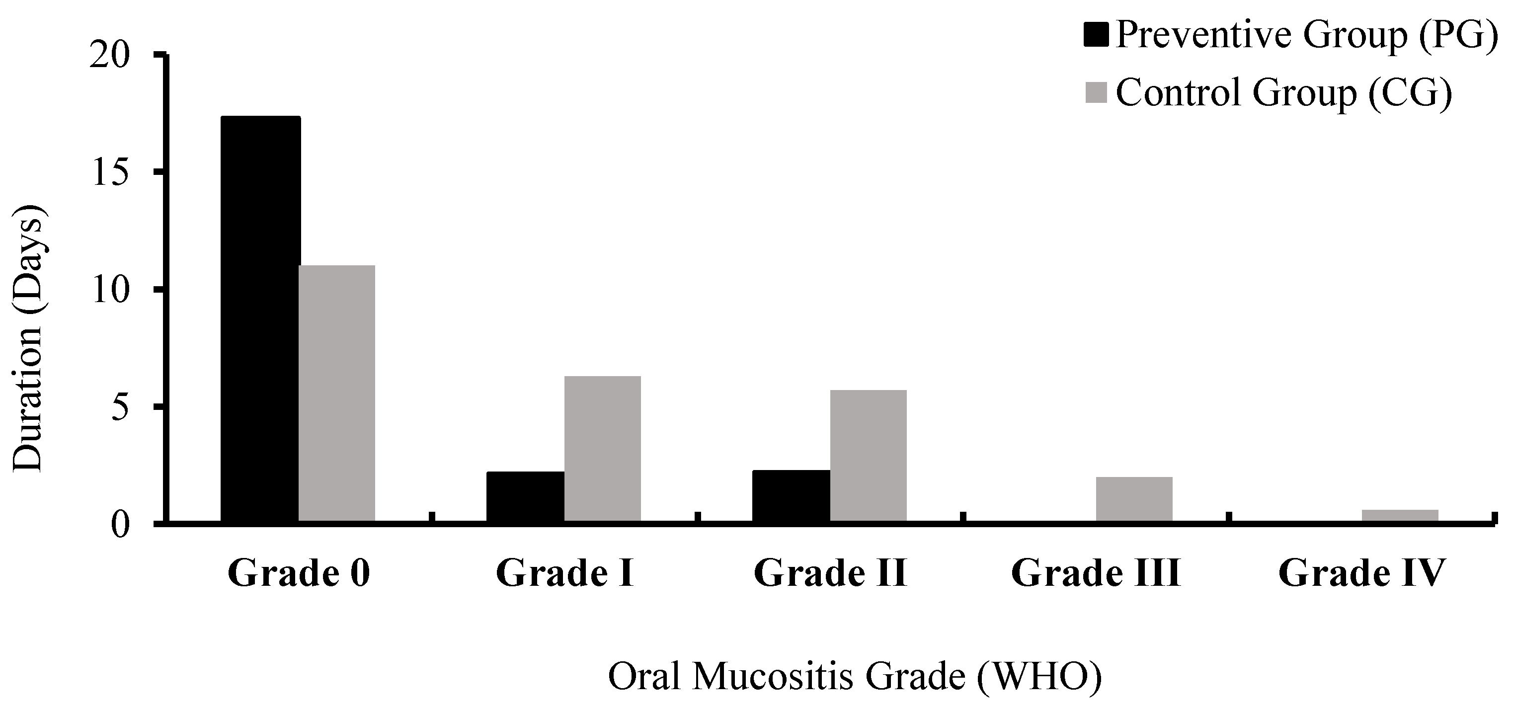
Duration of oral mucositis of:
  Grade 017.30 ± 3.0311.00 ± 5.20<0.001 *
  Grade I2.15 ± 2.816.30 ± 3.42<0.001 *
  Grade II2.20 ± 2.975.70 ± 6.160.025 *
  Grade III0.00 ± 0.002.0 ± 4.360.009 *
  Grade IV0.00 ± 0.000.55 ± 1.760.152
Total duration of oral mucositis4.70 ± 5.0515.00 ± 8.83<0.001 *
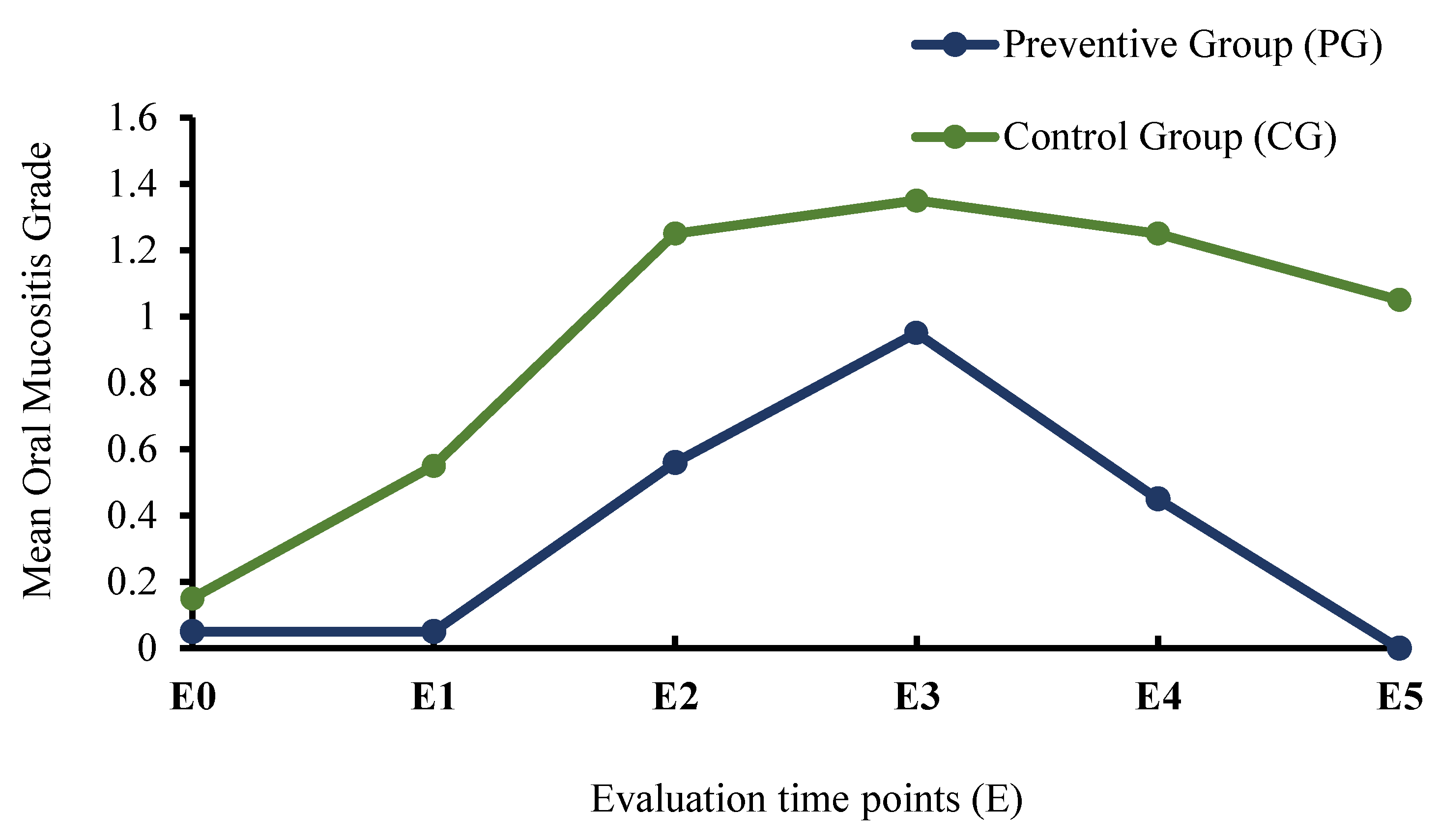
Mean oral mucositis score at Evaluation time points (E):
  E00.05 ± 0.220.15 ± 0.370.298
  E10.05 ± 0.220.55 ± 0.830.008 *
  E20.56 ± 0.681.25 ± 1.120.005 *
  E30.95 ± 0.941.35 ± 1.140.307
  E40.45 ± 0.691.25 ± 1.210.022 *
  E50.00 ± 0.001.05 ± 0.94<0.001 *
* p < 0.05.
Table 4. Comparison between groups for the incidence and severity of oral mucositis.
VariablesPreventive Group (PG)Control Group (CG)χ2p-Value
Severity of Oral Mucositis (OM)
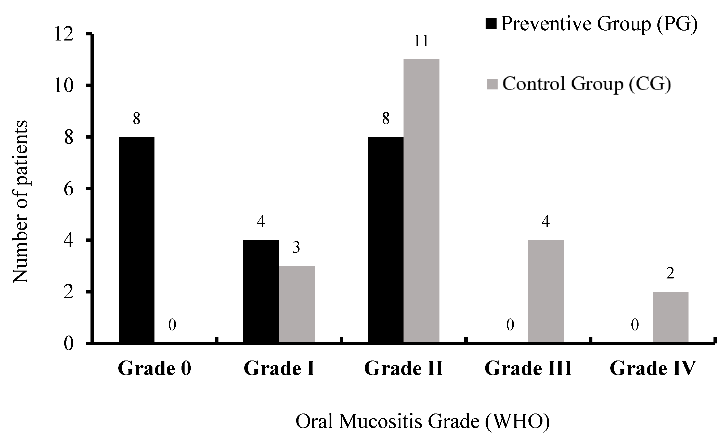
  Non-ulcerative OM (≤ Grade I)12384240.003 *
  Ulcerative OM (≥ Grade II)817
Incidence of OM
  No OM (Grade 0)8010,0000.002 *
  With OM (≥ Grade I)1220
* p < 0.05.
Publisher’s Note: MDPI stays neutral with regard to jurisdictional claims in published maps and institutional affiliations.

