Potential Use of Environmental Biological Samples for Retrospective Electron Paramagnetic Resonance Dosimetry of Radiation Accidents

 ,
, 
Abstract
1. Introduction
2. Materials and Methods
2.1. Preparation of Biological Sample
2.2. EPR Measurement
2.3. Experimental Design and Sample Irradiation
3. Results
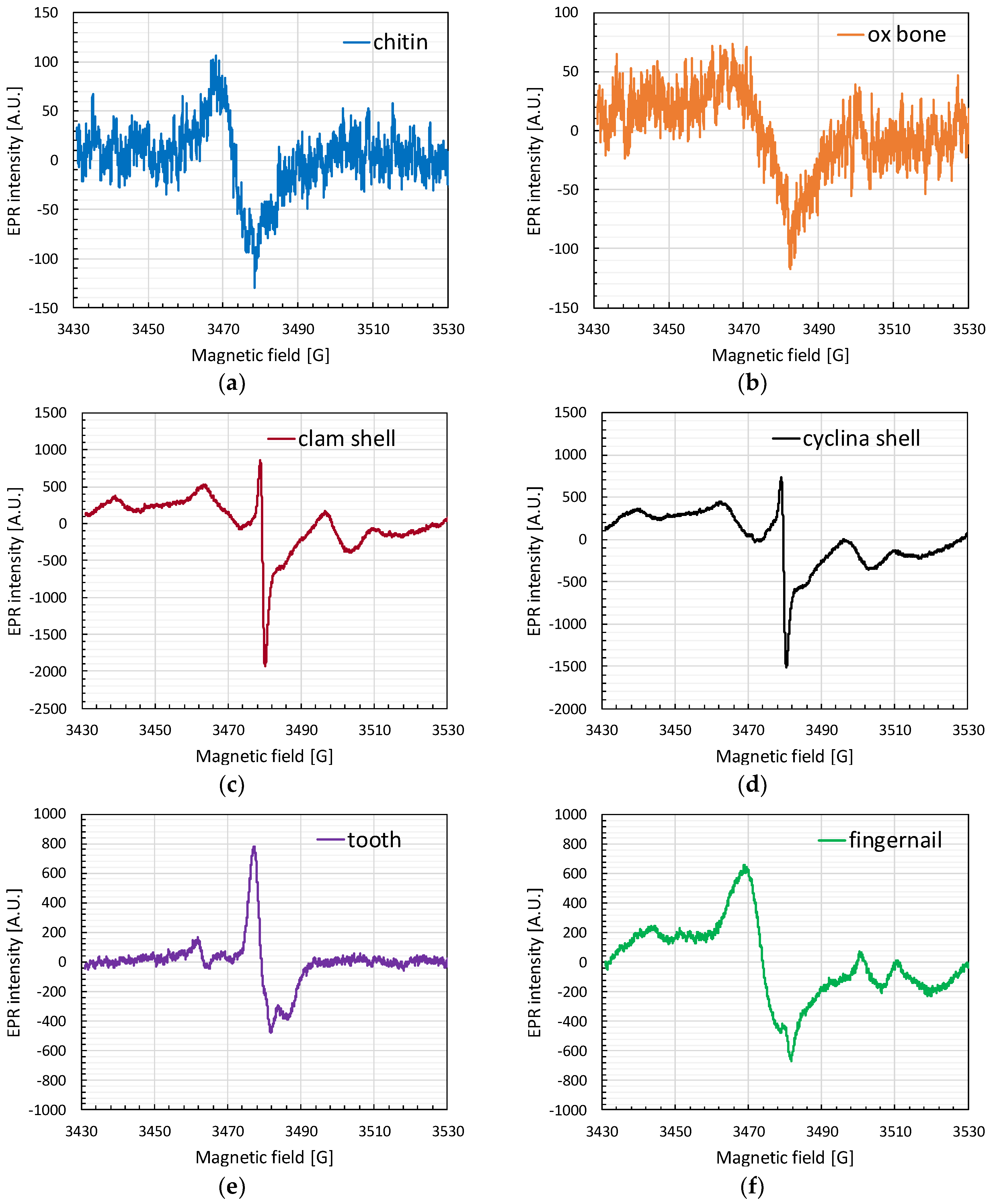
3.1. EPR Spectra
3.2. Dose-Response Curve
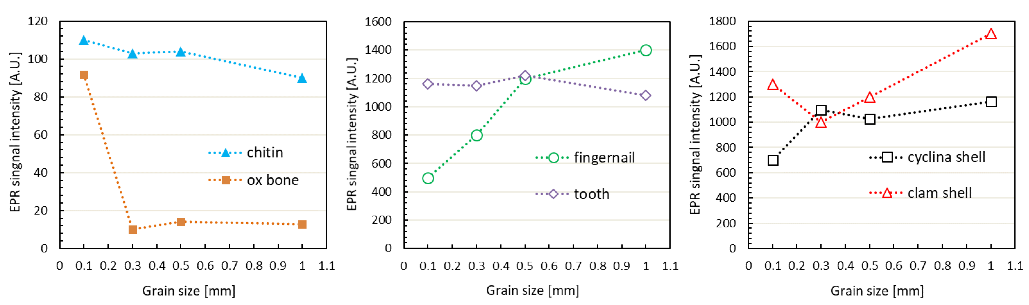
3.3. Grain Size
3.4. Storage Temperature
4. Discussion and Conclusions
Author Contributions
Funding
Conflicts of Interest
References
- Liu, C.Y.; Lai, L.H.; Wang, C.F.; Chuang, K.S.; Lu, C.C.; Lin, J.P.; Lin, H.H. Establishment of conversion coefficient of whole body effective dose by human tissue of electron paramagnetic resonance (EPR). Radiat. Phys. Chem. 2019, 155, 82–88. [Google Scholar] [CrossRef]
- Desrosiers, M.F.; Puhl, J.M. Absorbed-dose/dose-rate dependence studies for the alanine-EPR dosimetry system. Radiat. Phys. Chem. 2009, 78, 461–463. [Google Scholar] [CrossRef]
- Ikeya, M.; Miyajima, J.; Okajima, S. ESR dosimetry for atomic bomb survivors using shell buttons and tooth enamel. Jpn. J. Appl. Phys. 1984, 23, L697. [Google Scholar] [CrossRef]
- Sudprasert, W.; Monthonwattana, S.; Vitittheeranon, A. Identification of irradiated rice noodles by electron spin resonance spectroscopy. Radiat. Meas. 2012, 47, 640–643. [Google Scholar] [CrossRef]
- Trompier, F.; Kornak, L.; Calas, C.; Romanyukha, A.; LeBlanc, B.; Mitchell, C.A.; Swartz, H.M.; Clairand, I. Protocol for emergency EPR dosimetry in fingernails. Radiat. Meas. 2007, 42, 1085–1088. [Google Scholar] [CrossRef][Green Version]
- Yordanov, N.D.; Aleksieva, K. Preparation and applicability of fresh fruit samples for the identification of radiation treatment by EPR. Radiat. Phys. Chem. 2009, 78, 213–216. [Google Scholar] [CrossRef]
- Yordanov, N.D.; Mladenova, B. EPR studies on gamma-irradiated snails hard tissues. Radiat. Phys. Chem. 2001, 60, 191–193. [Google Scholar] [CrossRef]
- Soliman, Y.; Ali, L.I.; Moustafa, H.; Tadros, S.M. EPR dosimetric properties of 2-methylalanine pellet for radiation processing application. Radiat. Phys. Chem. 2014, 102, 11–15. [Google Scholar] [CrossRef]
- Desrosiers, M.F. gamma.Irradiated seafoods: Identification and dosimetry by electron paramagnetic resonance spectroscopy. J. Agric. Food Chem. 1989, 37, 96–100. [Google Scholar] [CrossRef]
- Stachowicz, W.; Burlinska, G.; Michalik, J. EPR detection of foods preserved with ionizing radiation. Radiat. Phys. Chem. 1998, 52, 157–160. [Google Scholar] [CrossRef]
- Anton, M. Uncertainties in alanine/ESR dosimetry at the Physikalisch-Technische Bundesanstalt. Phys. Med. Biol. 2006, 51, 5419–5440. [Google Scholar] [CrossRef] [PubMed]
- Jacob, P.; Bailiff, I.K.; Balonov, M.A.; Bauchinger, M.; Bouville, A.; Haskell, E.; Nakamura, N.; Romanyukha, A. Retrospective assessment of exposures to ionising radiation: Abstract. ICRU 2002, 2, 9. [Google Scholar] [CrossRef]
- Cano, N.F.; Munita, C.S.; Watanabe, S.; Barbosa, R.F.; Chubaci, J.F.; Tatumi, S.H.; Neves, E.G. OSL and EPR dating of pottery from the archaeological sites in Amazon Valley, Brazil. Quat. Int. 2014, 352, 176–180. [Google Scholar] [CrossRef]
- Egersdörfer, S.; Wieser, A.; Müller, A. Tooth enamel as a detector material for retrospective EPR dosimetry. Appl. Radiat. Isot. 1996, 47, 1299–1303. [Google Scholar] [CrossRef]
- Fattibene, P.; Callens, F. EPR dosimetry with tooth enamel: A review. Appl. Radiat. Isot. 2010, 68, 2033–2116. [Google Scholar] [CrossRef]
- Ivannikov, A.I.; Sanin, D.; Nalapko, M.; Skvortsov, V.F.; Stepanenko, V.F.; Tsyb, A.F.; Trompier, F.; Zhumadilov, K.; Hoshi, M. Dental enamel EPR dosimetry: Comparative testing of the spectra processing methods for determination of radiation-induced signal amplitude. Health Phys. 2010, 98, 345–351. [Google Scholar] [CrossRef]
- Chandra, H.; Symons, M.C. Sulphur radicals formed by cutting α-keratin. Nature 1987, 328, 833–834. [Google Scholar] [CrossRef]
- Sholom, S.; McKeever, S. Stability of X-band EPR signals from fingernails under vacuum storage. Radiat. Phys. Chem. 2017, 141, 78–87. [Google Scholar] [CrossRef]
- Chumak, V.; Bailiff, I.; Baran, N.; Bugai, A.; Dubovsky, S.; Fedosov, I.; Finin, V.; Haskell, E.; Hayes, R.; Ivannikov, A.; et al. The first international intercomparison of EPR-dosimetry with teeth: First results. Appl. Radiat. Isot. 1996, 47, 1281–1286. [Google Scholar] [CrossRef]
- Haskell, E.H.; Hayes, R.B.; Kenner, G.H.; Wieser, A.; Aragno, D.; Fattibene, P.; Onori, S. Achievable precision and accuracy in EPR dosimetry of tooth enamel. Radiat. Prot. Dosim. 1999, 84, 527–535. [Google Scholar] [CrossRef]
- Swartz, H.M.; Flood, A.B.; Williams, B.B.; Dong, R.; Swarts, S.G.; He, X.; Grinberg, O.; Sidabras, J.; Demidenko, E.; Gui, J.; et al. Electron paramagnetic resonance dosimetry for a large-scale radiation incident. Health Phys. 2012, 103, 255–267. [Google Scholar] [CrossRef] [PubMed]
- Symons, M.C.R.; Chandra, H.; Wyatt, J.L. Electron paramagnetic resonance spectra of irradiated finger-nails: A possible measure of accidental exposure. Radiat. Prot. Dosim. 1995, 58, 11–15. [Google Scholar]
- Sadło, J.; Michalik, J.; Stachowicz, W.; Strzelczak, G.; Dziedzic-Gocławska, A.; Ostrowski, K. EPR study on biominerals as materials for retrospective dosimetry. Nukleonika 2006, 51, 95–100. [Google Scholar]
- Trompier, F.; Romanyukha, A.; Reyes, R.; Vezin, H.; Queinnec, F.; Gourier, D. State of the art in nail dosimetry: Free radicals identification and reaction mechanisms. Biophysik 2014, 53, 291–303. [Google Scholar] [CrossRef]
- Hong, D.; Lee, K. Grain size effect of tooth enamel to electron paramagnetic resonance spectrum. Nucl. Sci. Technol. 2004, 41, 200–202. [Google Scholar]
- Liu, C.R.; Yin, G.M.; Han, F. Effects of grain size on quartz ESR dating of Ti–Li center in fluvial and lacustrine sediments. Quat. Geochronol. 2015, 30, 513–518. [Google Scholar] [CrossRef]
- Romanyukha, A.; Trompier, F.; LeBlanc, B.; Calas, C.; Clairand, I.; Mitchell, C.A.; Smirniotopoulos, J.G.; Swartz, H.M. EPR dosimetry in chemically treated fingernails. Radiat. Meas. 2007, 42, 1110–1113. [Google Scholar] [CrossRef] [PubMed][Green Version]
- Blanchard, S.C.; Chasteen, N.D. Electron paramagnetic resonance spectrum of a sea shell. Mytilus edulis. J. Phys. Chem. 1976, 80, 1362–1367. [Google Scholar] [CrossRef]
- Viscomi, D.; Fattibene, P. Radiation-induced signals analysed by EPR spectrometry applied to fortuitous dosimetry. Annali dell’Istituto Superiore di Sanità 2009, 45, 287–296. [Google Scholar]
- Bougai, A.; Brik, A.; Chumak, V.; Desrosiers, M.; Dubovski, S.; Fattibene, P.; Romanyukha, A. Use of Electron Paramagnetic Resonance Dosimetry with Tooth Enamel for Retrospective Dose Assessment; TECDOC-1331; International Atomic Energy Agency: Vienna, Austria, 2002. [Google Scholar]


© 2020 by the authors. Licensee MDPI, Basel, Switzerland. This article is an open access article distributed under the terms and conditions of the Creative Commons Attribution (CC BY) license (http://creativecommons.org/licenses/by/4.0/).
Share and Cite
Lu, C.-C.; Lin, H.-H.; Hsu, C.-H.; Wang, F.-N.; Lin, J.-P.; Lai, L.-H. Potential Use of Environmental Biological Samples for Retrospective Electron Paramagnetic Resonance Dosimetry of Radiation Accidents. Appl. Sci. 2020, 10, 6867. https://doi.org/10.3390/app10196867
Lu C-C, Lin H-H, Hsu C-H, Wang F-N, Lin J-P, Lai L-H. Potential Use of Environmental Biological Samples for Retrospective Electron Paramagnetic Resonance Dosimetry of Radiation Accidents. Applied Sciences. 2020; 10(19):6867. https://doi.org/10.3390/app10196867
Chicago/Turabian StyleLu, Chia-Chun, Hsin-Hon Lin, Ching-Han Hsu, Fu-Nien Wang, Jao-Perng Lin, and Lu-Han Lai. 2020. "Potential Use of Environmental Biological Samples for Retrospective Electron Paramagnetic Resonance Dosimetry of Radiation Accidents" Applied Sciences 10, no. 19: 6867. https://doi.org/10.3390/app10196867
APA StyleLu, C.-C., Lin, H.-H., Hsu, C.-H., Wang, F.-N., Lin, J.-P., & Lai, L.-H. (2020). Potential Use of Environmental Biological Samples for Retrospective Electron Paramagnetic Resonance Dosimetry of Radiation Accidents. Applied Sciences, 10(19), 6867. https://doi.org/10.3390/app10196867