Article

Fermented Rhus Verniciflua Stokes Extract Alleviates Nonalcoholic Fatty Liver through the AMPK/SREBP1/PCSK9 Pathway in HFD-Induced Nonalcoholic Fatty Liver Animal Model

1
Department of Science in Korean medicine, College of Korean Medicine, Graduate School, Kyung Hee University, Hoegi-dong, Dongdaemun-gu, Seoul 02435, Korea
2
Department of Cancer Preventive Material Development, College of Korean Medicine, Graduate School, Kyung Hee University, Hoegi-dong, Dongdaemun-gu, Seoul 02435, Korea
3
Fermented Food Science Division, National Institute of Agricultural Sciences, 166, Nongsaengmyeongro, Iseo-myeon, WanjuGun, Jeollabuk-do 55365, Korea
4
Department of Food and nutrition, Eulji University, Seongnam, Kyungido 13135, Korea
*
Author to whom correspondence should be addressed.
Appl. Sci. 2020, 10(19), 6833; https://doi.org/10.3390/app10196833
Submission received: 7 September 2020 / Revised: 25 September 2020 / Accepted: 27 September 2020 / Published: 29 September 2020
(This article belongs to the Special Issue Functional Foods and Human Health)

Abstract

:
Background: We have previously reported the anti-hepatic lipogenic effect of fermented Rhus verniciflua stokes extract (FRVE) in an oleic-acid-treated HepG2 cell model. Methods: Herein, we advanced our understanding and evaluated the impact of FRVE in HFD-fed C57BL/6 mice using an animal model of nonalcoholic fatty liver disease (NAFLD). Milk thistle extract was used as a positive control to compare the effects of FRVE. Results: FRVE decreased body weight, intra-abdominal fat weight, and liver weight. Furthermore, FRVE decreased HFD-induced elevated serum levels of ALT, AST, TC, and TG, and downregulated the increase in hepatic lipid accumulation and TG levels. FRVE reduced hepatic SREBP-1, PCSK-9, SREBP-2, and ApoB mRNA levels. IHC data showed that FRVE reduced the levels of nucleic SREBP-1, increased the levels of LDLR, and upregulated the expression of p-AMPK. Conclusion: Overall, these results demonstrate the anti-hepatic lipidemic effect of FRVE in an animal model. These findings are consistent with our previous study and strongly suggest that FRVE exerts anti-hepatic lipogenic effects by activating AMPK.

Graphical Abstract

1. Introduction

An increase in the prevalence of obesity and nonalcoholic fatty liver disease (NAFLD) is a leading cause of chronic liver disease worldwide [1,2]. NAFLD has a clinicopathological spectrum of liver diseases ranging from steatosis, nonalcoholic steatohepatitis (NASH), and cirrhosis through fibrosis. Diabetes, obesity, hyperlipidemia, and excess soft drink consumption are the high-risk factors for NAFLD [3]. Patients with NAFLD are at a higher risk of developing cardiovascular diseases, which are the leading cause of death worldwide [4].
We have studied natural products that exhibit an anti-hepatic lipidemic effect. Fermented Rhus verniciflua stokes extract (FRVE) exerts an anti-hepatic lipidemic effect by upregulating AMP-activated protein kinase (AMPK) in oleic acid-induced HepG2 cells in an in vitro nonalcoholic fatty liver model [5].
AMPK activation is reported to reduce NAFLD, suppress de novo lipogenesis (DNL), and increase fatty acid oxidation, and mitochondrial function and integrity in adipose tissue [6]. AMPK regulates lipid metabolism-related transcription factors like sterol regulatory element-binding protein-1 (SREBP-1), which modulates the expression of genes involved in cholesterol synthesis and lipid accumulation. AMPK inhibits degradation of low-density lipoprotein receptor (LDLR) by reducing the expression of proprotein convertase subtilisin/kexin type 9 (PCSK9) [7]. PCSK9 is a target gene of SREBPs and the subtilisin-related serin endoprotease protein K subfamily. PCSK9 regulates co-endocytosis and lysosomal degradation of hepatic LDLR by combining with LDLR on the hepatocyte surface [8]. This molecular crosstalk among AMPK, SREBPs, PCSK9, and LDLR could be a novel pharmacologic target for the treatment of obesity and NAFLD.
Rhus verniciflua stokes (RV) is reported to have anticancer [9,10,11,12,13], anti-adipogenic [14,15], antioxidant [16], and anti-inflammatory effects [17,18,19,20,21]. Despite these beneficial effects, RV utilization is limited due to the presence of allergen urushiol in it. Hence, various methods for removing urushiol have been described [22,23,24]. However, several detoxification methods to remove urushiol lead to the loss of bioactive compounds in RV [25]. FRVE is a microbe-detoxified RV, whose anti-hepatic lipidemic effects and bioactive compounds were previously reported by us [5]. This article presents the outcomes of an advanced preclinical study using an in vivo model for testing FRVE. The objective of this study was to evaluate the anti-hepatic lipidemic effect of FRVE in a high-fat diet (HFD)-induced fatty liver mouse model.

2. Materials and Methods

2.1. Preparation of FRVE

Lyophilized fermented Rhus verniciflua stokes extract (FRVE) powder purchased from Okkane (Seoul, Korea) was stored in a moisture-proof container and refrigerated until use. FRVE was obtained by freeze-drying the extract obtained from water extraction at 130 °C for 8 h, followed by filtration and fermentation (at 37 °C for 72 h) with Saccharomyces carlsbergensis.

2.2. High-Performance Liquid Chromatography

Urushiol and two main components (fustin and fisetin) in the FRVE were analyzed by high-performance liquid chromatography (HPLC, Agilent Technologies, Santa Clara, CA, USA) using Hichrome HPLC columns (5 μm, 250 mm × 4.6 mm, Hichrome, Ltd., Theale, UK). The mobile phases were composed of 0.1% formic acid in water (solvent A) and 100% methanol (solvent B), delivered at a flow rate of 0.7 mL/min. The gradient conditions used were 0–17 min, 100% B; 17–20 min, 100% B; 20–23 min, 0% B; and 23–30 min, 0% B. The detection wavelength was set to 254 nm, and the injection volume was 10 μL.

2.3. Ethics Statement

The IACUC of the University of Kyung Hee approved the protocol for animal use (KHUASP[SE]-17-150-1, 12/Feburary/2018) for the present study. Mice handling was performed following the IACUC recommendations and protocol.

2.4. Animal Experiment

Twenty-four-week-old C57BL/6J (DBL, Chungcheong bukdo, Korea) male mice (six mice per group), the diet formulation (HFD TD.06414, Table 1), and normal control die formulation (NCD) were purchased from DBL (Chungcheongbukdo, Korea). The mice were maintained at the Kyung Hee University animal facility in HEPA-filter top covered cages and were quarantined for one week in a limited-access specific pathogen-free room. Two weeks after the HFD, the mice were administered with milk thistle extract (MLB Biotrade, Kowanowo, Poland) (30 mg/kg), FRVE (800 mg/kg), or vehicle by oral gavage once a day for 12 weeks, and their body weights were determined once per week.

2.5. Biochemical Analyses

Heparin-containing blood was centrifuged at 2500× g for 10 min to obtain plasma. Liver tissues (50 mg) were homogenized in normal saline and the supernatant was used for biochemical analyses. Isolated plasma and supernatant from liver tissue were used to measure the levels of triglycerides (TG), total cholesterol (TC), alanine aminotransferase (ALT), and aspartate aminotransferase (AST) using a biochemical analyzer (Dri-chem NX500i, Sunkyung Medical, Seoul, Korea).

2.6. Histological Examination

For histological examination, sections of 10% formalin-fixed paraffin embedded liver tissues were stained with hematoxylin and eosin (H&E). The histological features of liver were examined using a camera (Koptic, Gyeonggi-do, Korea) connected to the light microscopy (Olympus, Tokyo, Japan).

2.7. Oil Red O Staining

Oil red O staining was performed as described previously [5]. Briefly, the frozen liver tissue specimens were fixed with 4% formalin and stained with 0.6% Oil Red O stain for 1 h. The sections were imaged using a camera (Nikon, Tokyo, Japan) connected to the microscope (Nikon, Tokyo, Japan).

2.8. RNA Isolation and Real-Time RT-PCR

Total RNA was isolated from tissues using TRIzol (Invitrogen, Carlsbad, CA, USA). cDNA was synthesized from the purified total RNA using a High-Capacity cDNA Reverse Transcription Kit (4368814, Promega, Madison, WI, USA). Real-Time reverse transcription-polymerase chain reaction (RT-PCR) was performed using a SYBR green RT-PCR kit (K-6254, Bioneer, Daejeon, Korea) and custom-designed primers (Table 2) for qPCR with sequence detection system (Takara, Shiga, Japan).

2.9. Immunohistochemistry

Paraffin sections (4 μm) prepared from liver tissues were stained with H&E. Immunohistochemical staining using anti-p-AMPK (Cell Signaling Technology, MA, USA), anti-SREBP-1 (Santa Cruz, Dallas, TX, USA), and anti-LDLR antibodies (Abcam, Cambridge, UK) was performed using the indirect avidin/biotin-enhanced horseradish peroxidase method. Antigen retrieval was carried out after dewaxing and dehydrating the tissue sections followed by heating in microwave for 15 min in 10 mM citrate buffer. The sections were quenched with 0.3% hydrogen peroxide in methanol for 10 min and blocked with 6% horse serum for 30 min in a humidity chamber. The sections were incubated with primary antibodies (anti-p-AMPK, anti-LDLR, and anti-SREBP-1) at 4 °C overnight in a humidity chamber. The antibodies were detected with the Vector ABC complex/horseradish peroxidase (HRP) kit (PK-6100, Vector Laboratories, Burlingame, CA, USA), and the color was developed with 3,3-diaminobenzidine tetrahydrochloride (DAB) (Sigma, St. Louis, MO, USA). For semi quantitative analysis, ten photomicrographs (200×) were taken with a CCD camera to avoid gross necrotic areas.

2.10. Statistical Analysis

Mean and standard deviations of the analyzed samples were determined, and the groups were compared using one-way ANOVA, followed by a t-test using Prism Pad 8. Results with p < 0.05 were considered statistically significant.

3. Results

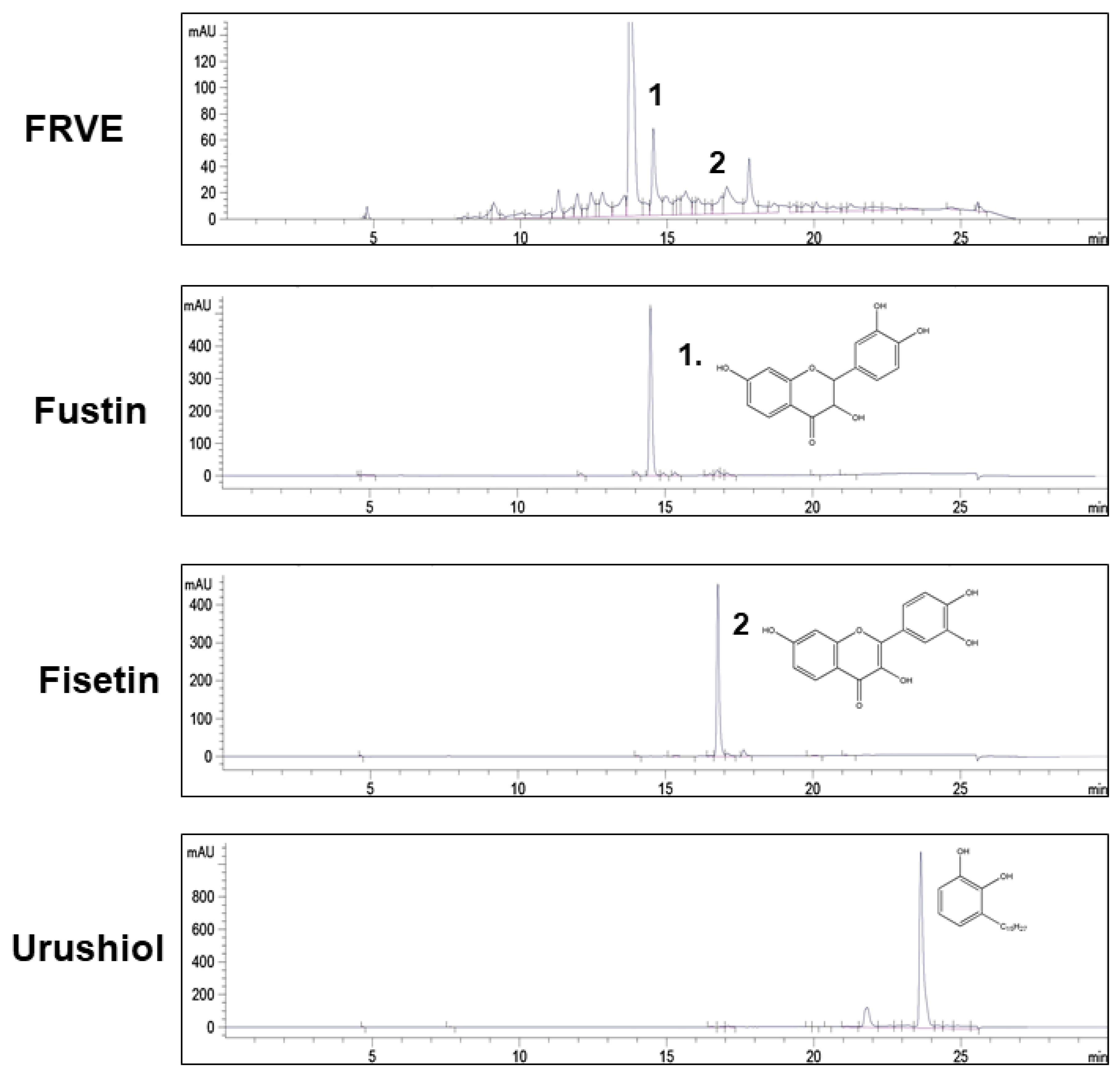
3.1. Confirmation of FRVE Detoxification and Main Components (Fustin and Fisetin) of FRVE

To confirm FRVE detoxification, the presence of the allergen urushiol was determined by high-performance liquid chromatography (HPLC). Urushiol was not detected, thereby confirming the successful detoxification of FRVE (Figure 1), whereas the urushiol peak appeared in the standard at 21.818 min.
Fustin (PubChem CID 5317435) and fisetin (PubChem CID 5281614) were the main active components present in FRVE (Figure 1). The peaks showed retention times of fustin and fisetin as 14.501 min and 16.778 min, respectively.

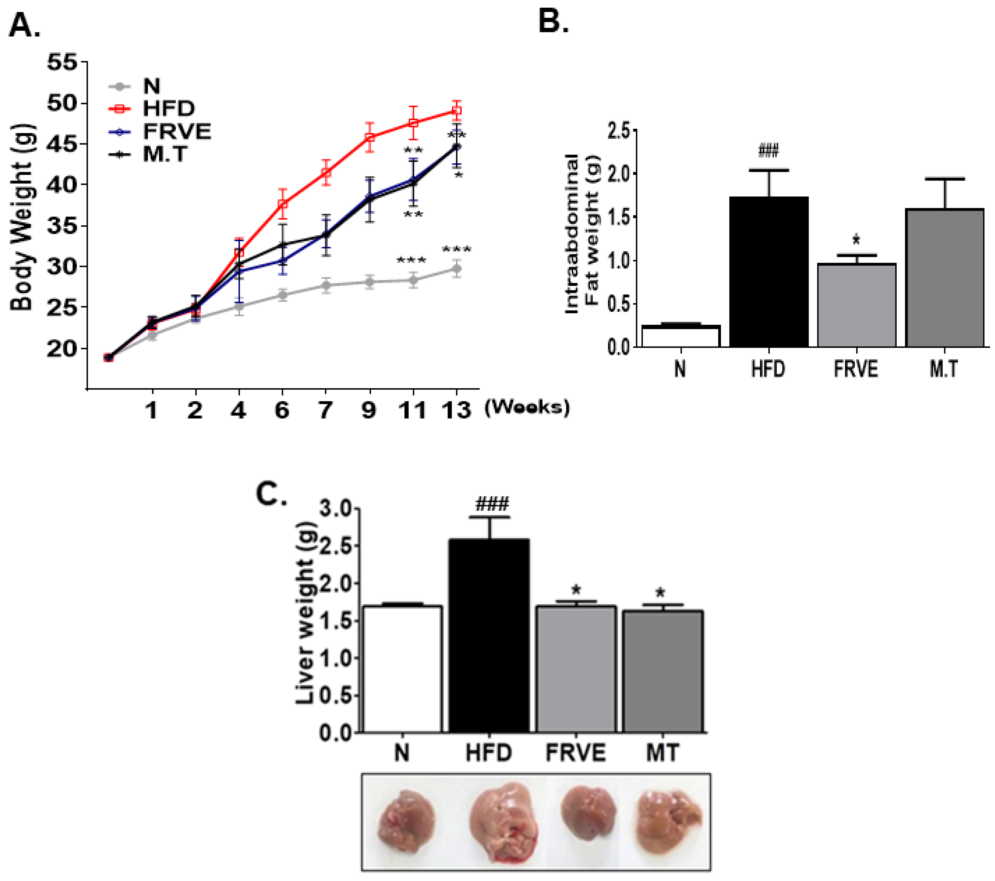
3.2. FRVE Decreased Body Weight, Intra-Abdominal Fat Weight, and Liver Weight in the HFD-Induced NAFLD Model

The bodyweights of the NCD, HFD-fed, FRVE, and MT (milk thistle extract)-treated groups were measured once a week for 13 weeks. As shown in Figure 2A, the bodyweight in the HFD-fed control group increased significantly after two weeks of feeding when compared to the bodyweight of the NCD group. In contrast, the FRVE and MT treated groups abrogated the bodyweight gain from the third week of FRVE and MT treatment. Eleven weeks after treatment, the mean bodyweights of HFD-fed control group (49.1 ± 1.18 g) were notably higher than that of the NCD group (29.75 ± 1.03 g). However, treatment with FRVE and MT suppressed the bodyweight to 44.6 ± 2.07 g and 44.76 ± 2.65 g, respectively (Figure 2A). The mean intra-abdominal fat pad weights were significantly high in HFD-fed mice (1.8 ± 0.31 g) as compared to the NCD group mice (0.25 ± 0.02 g). In contrast, FRVE and MT-treated mice exhibited decreased intra-abdominal fat pad weights of 0.97 ± 0.09 g and 1.15 ± 0.34 g, respectively (Figure 2B). Furthermore, HFD-fed mice presented an increased liver size and solid pink-colored livers. The liver weights of HFD-fed mice (2.58 ± 0.3 g) were 1.53 times higher than the NCD group. In contrast, despite being on HFD, the FRVE (1.69 ± 0.07 g) and MT-treated (1.63 ± 0.03 g) groups showed decreased liver weights and size of bright pink color, similar to the NCD group (Figure 2C) (Supplementary Figure S1).

3.3. FRVE Downregulated Serum Liver Function and Lipid Markers in an HFD-Induced Nonalcoholic Fatty Liver Model

As shown in Figure 3A,B, activities of the hepatotoxicity markers alanine aminotransferase (ALT) and aspartate aminotransferase (AST) were significantly increased in the HFD-fed mice group. FRVE treatment downregulated the enhanced ALT and AST activities induced by HFD. The levels of ALT and AST were lower in the FRVE treated group than in the positive control group. In addition, FRVE treatment inhibited the HFD-induced elevation in serum total cholesterol (TC) and triglycerides (TG) levels (Figure 3C,D).

3.4. FRVE Inhibited Hepatic Steatosis and Lipid Accumulation in an HFD-Induced Nonalcoholic Fatty Liver Model

We confirmed HFD-induced hepatic steatosis in liver tissue sections by H&E staining. Histological analysis revealed a significant increase in the typical histological features of a nonalcoholic fatty liver, such as macro- or micro-vesicular fatty changes, and large swollen hepatocytes with ballooning degeneration in HFD-fed mice (Figure 4A). Notably, the FRVE and MT-treated groups showed significantly reduced histological features of nonalcoholic fatty liver compared to HFD-fed mice (Figure 4A). Furthermore, Oil-red O staining showed that hepatic lipid accumulation significantly increased in HFD-fed mice than in the NCD group. In contrast, FRVE and MT treatment markedly ameliorated hepatic intracellular lipid accumulation in HFD-fed mice (Figure 4B). Consistently, we observed elevated hepatic TG levels in HFD-fed mice and reduced TG levels in FRVE and MT-treated groups (Figure 4C).

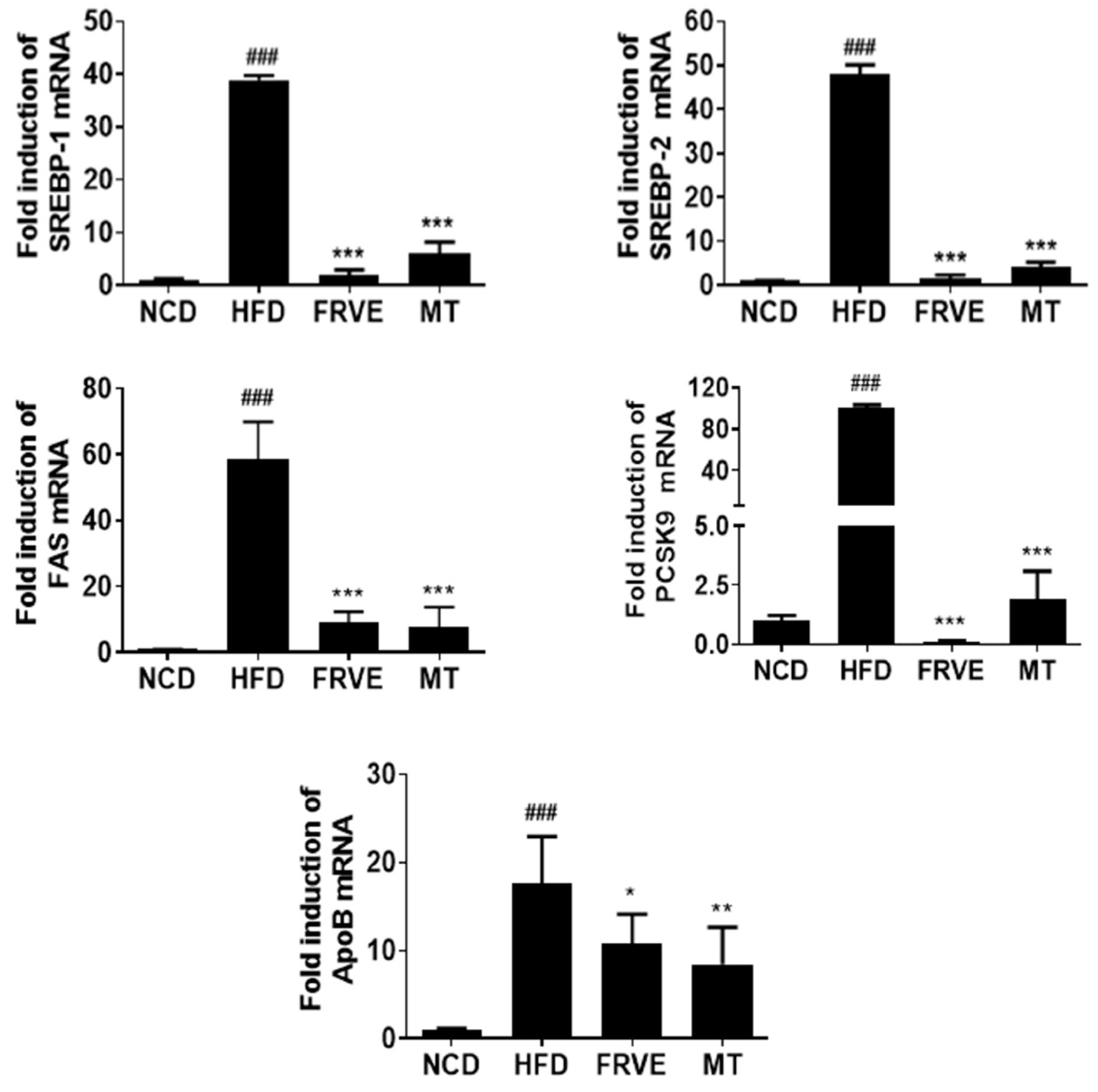
3.5. FRVE Downregulated the mRNA Levels of Lipogenesis-Related Genes in HFD-Induced Nonalcoholic Fatty Liver Model

RT-qPCR was used to determine the expression levels of proprotein convertase subtilisin/kexin type 9 (PCSK-9), apolipoprotein B (APOB), sterol regulatory element-binding protein 1 (SREBP-1), fatty acid synthase (FAS), and sterol element-binding protein 2 (SREBP-2) in the liver tissue to assess the regulation of lipogenesis-related key factors by FRVE. Our results showed that the mRNA expression levels of PCSK-9, APOB, SREBP-1, FAS, and SREBP-2 were higher in the HFD group than in the NCD group, whereas the mRNA levels were markedly downregulated in FRVE and MT treated groups (Figure 5).

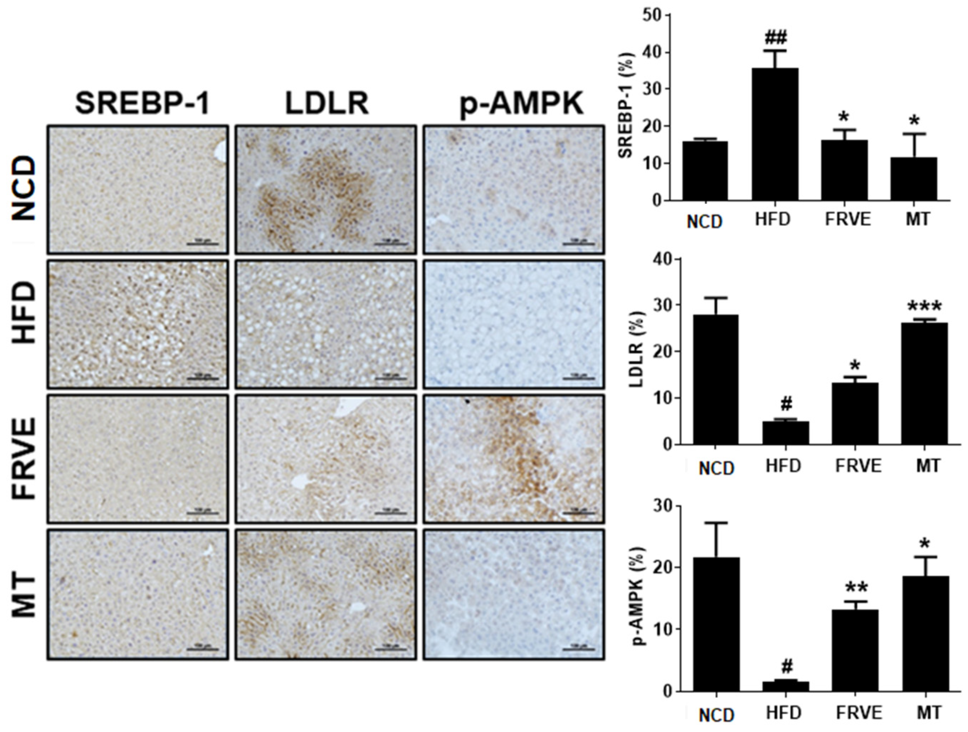
3.6. FRVE Inhibited HFD-Induced Hepatic Steatosis via AMPK Activation
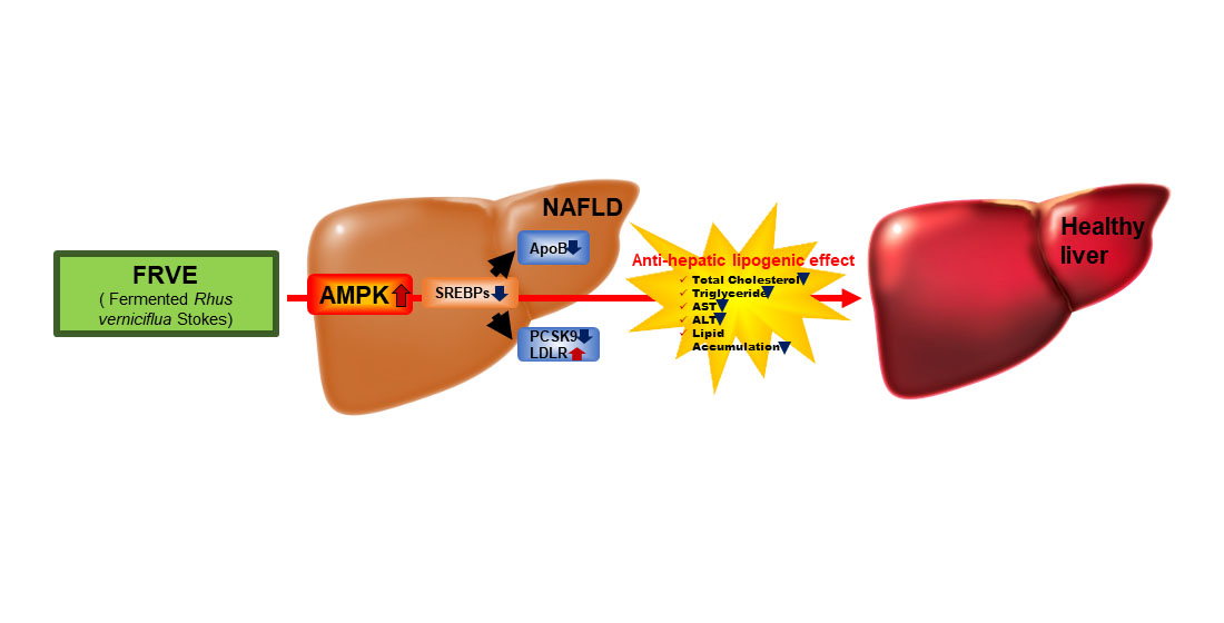
We performed immunohistochemistry to confirm the expression of AMP-activated protein kinase (AMPK) and lipid metabolic proteins regulated by AMPK in the liver tissue. SREBP-1 showed nuclear expression in hepatocytes, with an increased expression in the HFD-fed group (Figure 6). In contrast, SREBP-1 showed weak cytoplasmic expression in the other groups. Presence of low-density lipoprotein receptor (LDLR) expression was observed at the cell membrane of the hepatocytes, with elevated expression in the NCD group as compared to the other groups. The FRVE and MT-treated groups showed increased LDLR expression compared to the HFD-fed group. Further, phosphorylated AMPK was expressed in the cytoplasm of hepatocytes; with a significantly increased p-AMPK expression in the FRVE treated group (Figure 6).

4. Discussion

In 2012, the Korea Ministry of Food and Drug Safety approved the use of Rhus verniciflua stokes (RV) extracts devoid of urushiol, which causes allergic contact dermatitis known as urushiol-induced contact dermatitis. RV extracts are commercially available in various forms as food and medicine in Korea. Although RV contains urushiol, it is useful as food and medicine due to its wide range of biological activities. We have previously reported the presence of many bioactive compounds, including gallic acid, fustin, fisetin, 1-hexacosanol, and quercetin in fermented Rhus verniciflua stokes (FRVE). These compounds act on nonalcoholic fatty liver disease (NAFLD) by upregulating AMP-activated protein kinase (AMPK) [5]. Here, we performed a preclinical study to further confirm the anti-hepatic lipogenic effect of FRVE and observed results consistent with our previous in vitro study. Outcomes of our present study showed anti-nonalcoholic fatty liver effect and safety of FRVE usage in an HFD-induced NAFLD animal model.
As demonstrated in Figure 1, urushiol was not detected in the FRVE used in the animal study by HPLC. We confirmed the safety of FRVE and the presence of the main active ingredients (fisetin and fustin).
Obesity is associated with the development of NASH, NAFLD, liver fibrosis, cirrhosis, and hepatocellular carcinoma [26,27]. In a HFD, 30–75% of the total calories are derived from saturated fatty acids, and HFD mouse model is a useful system to study metabolic alterations and NAFLD. HFD-fed rodents develop metabolic diseases such as obesity, dyslipidemia, and diabetes and exhibit changes in lipogenic regulators such as sterol regulatory element-binding protein 1 (SREBP-1), liver X receptor, and inflammatory cytokines, depending on the duration of the HFD intake; these pathologies closely resembles the pathological (liver inflammation and fibrosis) and molecular alterations observed in human NAFLD [28,29].
Interestingly, FRVE decreased the body weight, intra-abdominal fat weight, and liver weight in the HFD-induced NAFLD model, without inducing toxicity; status of liver damage was confirmed by testing serum alanine aminotransferase (ALT) and aspartate aminotransferase (AST) levels (Figure 2). Serum AST and ALT levels are widely used as serum biomarkers of liver injury like NASH. Increased ALT and AST are hallmark features of NAFLD, and their levels also increase in obesity in humans [30] as well as rodents [31]. Our results showed that FRVE protected against liver injury by decreasing the levels of ALT and AST, which were induced by HFD (Figure 3A,B).
The reduction of body weight by FRVE treatment in HFD-fed mice is associated with the inhibition of lipid synthesis. FRVE decreased the levels of TG and TC in both the serum and liver, and decreased hepatic lipid accumulation as observed by oil-red O staining (Figure 3C,D and Figure 4). These results are in concordance with a previous study, such as a hyperlipidemia study of western HFD for two weeks with the use of allergen-free RVS obtained by boiling, demonstrated a decrease in hepatic lipid accumulation, body weight, and total cholesterol in the RVS-treated group [32].
As shown in Figure 5 and Figure 6, the downregulation of lipogenesis-related factors such as proprotein convertase subtilisin/kexin type 9 (PCSK-9), apolipoprotein B (APOB), sterol regulatory element-binding protein 1 (SREBP-1), fatty acid synthase (FAS), sterol regulatory element-binding protein 2 (SREBP-2), and low-density lipoprotein receptor (LDLR) by FRVE inhibited lipid accumulation and synthesis in liver tissue. In contrast to the FRVE-treated group, the HFD-fed control group overexpressed all these lipogenic factors. SREBPs are transcription factors that bind to the sterol regulatory element DNA sequence, and SREBP-1 and -2 regulate the lipogenesis targeting genes [33,34,35]. SREBPs upregulate the expression of HMG-CoA reductase (HMGR) required for cholesterol synthesis and fatty acid synthase (FAS) required for fatty acid synthesis [36]. Furthermore, SREBP protects apolipoprotein B from proteasomal degradation, thereby increasing hepatic apoB lipoprotein [37].
ApoB is expressed in the liver and is associated with cardiovascular disease, hyperlipidemia, and NAFLD [38]. ApoB is the structural protein of very-low-density lipoprotein cholesterol (VLDL), intermediate-density lipoprotein (IDL), and low-density lipoprotein (LDL), which are incorporated into chylomicrons [39].
PCSK9, one of the factors regulated by SREBP, is secreted by hepatocytes and inhibits the uptake of LDL by targeting LDLR degradation, and possibly lipogenesis [40]. Two clinical studies have demonstrated that patients with NAFLD show increased levels of PCSK9 [40,41]. In this regard, we evaluated the expression of LDLR and SREBP-1 along with PCSK9 in livers of HFD-fed mice by IHC. As shown in Figure 6, the HFD-fed control group showed high expression of PCSK9 mRNA and low levels of LDLR in the liver tissue. In contrast, the FRVE-treated group showed increased protein levels of LDLR and decreased protein levels of SREBP-1 in the liver tissue (Figure 6). Moreover, we observed an increase in the phosphorylation of AMPK in the FRVE-treated group but not in the HFD-fed control group, which was consistent with our previous data, where FRVE increased the levels of phosphorylated AMPK in OA-induced HepG2 cells [5].
Phosphorylated AMPK plays an essential role in regulating lipid metabolism. Activated AMPK leads to the suppression of lipogenesis-enhancing factors like SREBP-1 [42,43,44]. In our previous study, we demonstrated attenuation of FRVE-induced AMPK activation and recovery of FRVE-induced decrease in SREBP-1 levels by the AMPK antagonist compound C in OA-induced HepG2 cells [5].

5. Conclusions

The results in the current study are consistent with our previous in vitro data performed in an OA-induced HepG2 cell model. The outcomes of the present study show that FRVE alleviates NAFLD in HFD-fed mice. FRVE activates AMPK, which in turn regulates SREBP-1, PCSK9, ApoB, and LDLR expression, leading to inhibition of hepatic lipogenesis in HFD-fed C57BL/6J mice. These results confirm the outcomes of our previous in vitro study and extend our understanding of anti-nonalcoholic fatty liver effect of FRVE.

Supplementary Materials

The following are available online at https://www.mdpi.com/2076-3417/10/19/6833/s1, Figure S1: Food intake.

Author Contributions

S.-O.L., Y.X., H.H., Y.-K.L., J.-S.C., and S.-T.J. performed experiments. J.K.P. analyzed the data. H.-J.L. supervised the project. All authors have read and agreed to the published version of the manuscript.

Funding

This work was supported by a grant from the Cooperative Research Program for Agriculture Science and Technology Development (Project No. PJ01313501 and Project No. PJ01313503) Rural Development Administration, Republic of Korea.

Conflicts of Interest

The authors declare no conflict of interest.

References

  1. Younossi, Z.M.; Stepanova, M.; Younossi, Y.; Golabi, P.; Mishra, A.; Rafiq, N.; Henry, L. Epidemiology of chronic liver diseases in the USA in the past three decades. Gut 2020, 69, 564–568. [Google Scholar] [CrossRef] [PubMed]
  2. Collaborators, G.B.D.O.; Afshin, A.; Forouzanfar, M.H.; Reitsma, M.B.; Sur, P.; Estep, K.; Lee, A.; Marczak, L.; Mokdad, A.H.; Moradi-Lakeh, M.; et al. Health Effects of Overweight and Obesity in 195 Countries over 25 Years. N. Engl. J. Med. 2017, 377, 13–27. [Google Scholar] [CrossRef] [PubMed]
  3. Assy, N.; Kaita, K.; Mymin, D.; Levy, C.; Rosser, B.; Minuk, G. Fatty infiltration of liver in hyperlipidemic patients. Dig. Dis. Sci. 2000, 45, 1929–1934. [Google Scholar] [CrossRef]
  4. Patil, R.; Sood, G.K. Non-alcoholic fatty liver disease and cardiovascular risk. World J. Gastrointest. Pathophysiol. 2017, 8, 51–58. [Google Scholar] [CrossRef]
  5. Lee, M.-S.; Kim, J.-S.; Cho, S.-M.; Lee, S.O.; Kim, S.-H.; Lee, H.-J. Fermented Rhus verniciflua Stokes extract exerts an Antihepatic Lipogenic effect in oleic-acid-induced HepG2 cells via Upregulation of AMP-activated protein kinase. J. Agric. Food Chem. 2015, 63, 7270–7276. [Google Scholar] [CrossRef] [PubMed]
  6. Smith, B.K.; Marcinko, K.; Desjardins, E.M.; Lally, J.S.; Ford, R.J.; Steinberg, G.R. Treatment of nonalcoholic fatty liver disease: Role of AMPK. Am. J. Physiol. Endocrinol. Metab. 2016, 311, E730–E740. [Google Scholar] [CrossRef] [PubMed] [Green Version]
  7. Li, Y.; Xu, S.; Mihaylova, M.M.; Zheng, B.; Hou, X.; Jiang, B.; Park, O.; Luo, Z.; Lefai, E.; Shyy, J.Y.-J. AMPK phosphorylates and inhibits SREBP activity to attenuate hepatic steatosis and atherosclerosis in diet-induced insulin-resistant mice. Cell Metab. 2011, 13, 376–388. [Google Scholar] [CrossRef] [Green Version]
  8. Duff, C.J.; Hooper, N.M. PCSK9: An emerging target for treatment of hypercholesterolemia. Expert Opin. Ther. Targets 2011, 15, 157–168. [Google Scholar] [CrossRef]
  9. Kang, Y.; Yoon, S.W.; Park, B. Allergen-removed Rhus verniciflua Stokes suppresses invasion and migration of pancreatic cancer cells through downregulation of the JAK/STAT and Src/FAK signaling pathways. Oncol. Rep. 2018, 40, 3060–3068. [Google Scholar] [CrossRef] [Green Version]
  10. Lee, K.-W.; Um, E.-S.; Jung, B.-B.; Choi, E.-S.; Kim, E.-Y.; Lee, S.; Jang, E.; Lee, J.-H.; Kim, Y. Rhus verniciflua Stokes extract induces inhibition of cell growth and apoptosis in human chronic myelogenous leukemia K562 cells. Oncol. Rep. 2018, 39, 1141–1147. [Google Scholar] [CrossRef]
  11. Choi, H.S.; Kim, M.K.; Choi, Y.K.; Shin, Y.C.; Cho, S.-G.; Ko, S.-G. Rhus verniciflua Stokes (RVS) and butein induce apoptosis of paclitaxel-resistant SKOV-3/PAX ovarian cancer cells through inhibition of AKT phosphorylation. BMC Complement. Altern. Med. 2016, 16, 122. [Google Scholar] [CrossRef] [PubMed] [Green Version]
  12. Chae, J.; Lee, S.; Lee, S. Potential efficacy of allergen removed Rhus verniciflua Stokes extract to maintain progression-free survival of patients with advanced hepatobiliary cancer. Explore 2018, 14, 300–304. [Google Scholar] [CrossRef] [PubMed]
  13. Kim, M.S.; Lee, C.W.; Kim, J.-H.; Lee, J.-C.; An, W.G. Extract of Rhus verniciflua Stokes Induces p53-Mediated Apoptosis in MCF-7 Breast Cancer Cells. Evid.-Based Complement. Altern. Med. 2019, 2019, 9407340. [Google Scholar] [CrossRef] [PubMed] [Green Version]
  14. Lamichhane, R.; Kim, S.-G.; Kang, S.; Lee, K.-H.; Pandeya, P.R.; Jung, H.-J. Exploration of underlying mechanism of anti-adipogenic activity of sulfuretin. Biol. Pharm. Bull. 2017, 40, 1366–1373. [Google Scholar] [CrossRef] [Green Version]
  15. Song, N.-J.; Yoon, H.-J.; Kim, K.H.; Jung, S.-R.; Jang, W.-S.; Seo, C.-R.; Lee, Y.M.; Kweon, D.-H.; Hong, J.-W.; Lee, J.-S.; et al. Butein is a novel anti-adipogenic compound. J. Lipid Res. 2013, 54, 1385–1396. [Google Scholar] [CrossRef] [Green Version]
  16. Choi, D.R.; Jeong, J.H.; Yu, K.S.; Lee, N.S.; Jeong, Y.G.; Kim, D.K.; Na, C.S.; Na, D.S.; Hwang, W.M.; Han, S.; et al. Extract of Rhus verniciflua stokes protects against renal ischemia-reperfusion injury by enhancing Nrf2-mediated induction of antioxidant enzymes. Exp. Ther. Med. 2018, 15, 3827–3835. [Google Scholar]
  17. Kim, B.-G.; Song, Y.; Lee, M.-G.; Ku, J.-M.; Jin, S.-J.; Hong, J.-W.; Lee, S.; Kang, H. Macrophages from mice administered rhus verniciflua stokes extract show selective anti-inflammatory activity. Nutrients 2018, 10, 1926. [Google Scholar] [CrossRef] [Green Version]
  18. Kim, J.H.; Shin, Y.C.; Ko, S.-G. Integrating traditional medicine into modern inflammatory diseases care: Multitargeting by Rhus verniciflua Stokes. Mediat. Inflamm. 2014, 2014, 154561. [Google Scholar] [CrossRef] [Green Version]
  19. Moon, J.E.; Shin, J.-H.; Kwon, O.; Kim, J.Y. A standardized extract of rhus verniciflua stokes protects wistar rats against lipopolysaccharide-induced acute inflammation. J. Med. Food 2015, 18, 1223–1230. [Google Scholar] [CrossRef]
  20. Oh, P.-S.; Lee, S.-J.; Lim, K.-T. Glycoprotein Isolated from Rhus verniciflua S TOKES Inhibits Inflammation-Related Protein and Nitric Oxide Production in LPS-Stimulated RAW 264.7 Cells. Biol. Pharm. Bull. 2007, 30, 111–116. [Google Scholar] [CrossRef] [Green Version]
  21. Jung, C.H.; Kim, J.H.; Kim, J.H.; Chung, J.H.; Choi, H.S.; Seo, J.B.; Shin, Y.C.; Kim, S.H.; Ko, S.G. Anti-inflammatory effect of Rhus verniviflua Stokes by suppression of iNOS-mediated Akt and ERK pathways: In-vitro and in-vivo studies. J. Pharm. Pharmacol. 2011, 63, 679–687. [Google Scholar] [CrossRef] [PubMed]
  22. Choi, H.-S.; Yeo, S.-H.; Jeong, S.-T.; Choi, J.-H.; Park, H.-S.; Kim, M.-K. Preparation and characterization of urushiol free fermented Rhus verniciflua stem bark (FRVSB) extracts. Korean J. Food Sci. Technol. 2012, 44, 173–178. [Google Scholar] [CrossRef]
  23. Choi, H.-S.; Kim, M.-K.; Park, H.-S.; Yun, S.-E.; Mun, S.-P.; Kim, J.-S.; Sapkota, K.; Kim, S.; Kim, T.-Y.; Kim, S.-J.; et al. Biological detoxification of lacquer tree (Rhus verniciflua Stokes) stem bark by mushroom species. Food Sci. Biotechnol. 2007, 16, 935–942. [Google Scholar]
  24. Jeong, H.Y.; Lee, T.H.; Lee, H.J.; Cho, J.-Y.; Moon, J.-H. Ionization Neutralizes the Allergy-Inducing Property of 3-Pentadecylcatechol: A Urushiol Derivative. J. Med. Food 2020. [Google Scholar] [CrossRef] [PubMed]
  25. Lee, S.-O.; Kim, S.-J.; Kim, J.-S.; Ji, H.; Lee, E.-O.; Lee, H.-J. Comparison of the main components and bioactivity of Rhus verniciflua Stokes extracts by different detoxification processing methods. BMC Complement. Altern. Med. 2018, 18, 242. [Google Scholar] [CrossRef]
  26. Czaja, A.J. Hepatic inflammation and progressive liver fibrosis in chronic liver disease. World J. Gastroenterol. 2014, 20, 2515. [Google Scholar] [CrossRef]
  27. Sun, B.; Karin, M. Obesity, inflammation, and liver cancer. J. Hepatol. 2012, 56, 704–713. [Google Scholar] [CrossRef] [Green Version]
  28. Takahashi, Y.; Soejima, Y.; Fukusato, T. Animal models of nonalcoholic fatty liver disease/nonalcoholic steatohepatitis. World J. Gastroenterol. 2012, 18, 2300. [Google Scholar] [CrossRef]
  29. Buettner, R.; Schölmerich, J.; Bollheimer, L.C. High-fat diets: Modeling the metabolic disorders of human obesity in rodents. Obesity 2007, 15, 798–808. [Google Scholar] [CrossRef]
  30. Stranges, S.; Dorn, J.M.; Muti, P.; Freudenheim, J.L.; Farinaro, E.; Russell, M.; Nochajski, T.H.; Trevisan, M. Body fat distribution, relative weight, and liver enzyme levels: A population-based study. Hepatology 2004, 39, 754–763. [Google Scholar] [CrossRef]
  31. Fraulob, J.C.; Ogg-Diamantino, R.; Fernandes-Santos, C.; Aguila, M.B.; Mandarim-de-Lacerda, C.A. A mouse model of metabolic syndrome: Insulin resistance, fatty liver and non-alcoholic fatty pancreas disease (NAFPD) in C57BL/6 mice fed a high fat diet. J. Clin. Biochem. Nutr. 2010, 46, 212–223. [Google Scholar] [CrossRef] [PubMed] [Green Version]
  32. Jeong, S.-J.; Park, J.-G.; Kim, S.; Kweon, H.Y.; Seo, S.; Na, D.-S.; Lee, D.; Hong, C.Y.; Na, C.-S.; Dong, M.-S. Extract of Rhus verniciflua stokes protects the diet-induced hyperlipidemia in mice. Arch. Pharmacal Res. 2015, 38, 2049–2058. [Google Scholar] [CrossRef] [PubMed]
  33. Edwards, P.A.; Tabor, D.; Kast, H.R.; Venkateswaran, A. Regulation of gene expression by SREBP and SCAP. Biochim. Biophys. Acta 2000, 1529, 103–113. [Google Scholar] [CrossRef]
  34. Sakakura, Y.; Shimano, H.; Sone, H.; Takahashi, A.; Inoue, K.; Toyoshima, H.; Suzuki, S.; Yamada, N. Sterol regulatory element-binding proteins induce an entire pathway of cholesterol synthesis. Biochem. Biophys. Res. Commun. 2001, 286, 176–183. [Google Scholar] [CrossRef]
  35. Guo, D.; Bell, E.H.; Mischel, P.; Chakravarti, A. Targeting SREBP-1-driven lipid metabolism to treat cancer. Curr. Pharm. Des. 2014, 20, 2619–2626. [Google Scholar] [CrossRef] [Green Version]
  36. Madison, B.B. Srebp2: A master regulator of sterol and fatty acid synthesis. J. Lipid Res. 2016, 57, 333–335. [Google Scholar] [CrossRef] [Green Version]
  37. Wang, S.-L.; Du, E.Z.; Martin, T.D.; Davis, R.A. Coordinate regulation of lipogenesis, the assembly and secretion of apolipoprotein B-containing lipoproteins by sterol response element binding protein 1. J. Biol. Chem. 1997, 272, 19351–19358. [Google Scholar] [CrossRef] [Green Version]
  38. Lin, M.-S.; Guo, S.-E.; Lin, H.-S.; Hsu, J.-T.; Lin, Y.-S.; Lin, T.-H.; Huang, T.-J.; Chen, M.-Y.; Chung, C.-M. Impact of apolipoprotein B on hepatosteatosis in a population infected with Hepatitis C virus: A cross-sectional observational study. Obes. Facts 2016, 9, 101–111. [Google Scholar] [CrossRef]
  39. Bayly, G.R. Lipids and disorders of lipoprotein metabolism. In Clinical Biochemistry: Metabolic and Clinical Aspects; Elsevier: Amsterdam, The Netherlands, 2014; pp. 702–736. [Google Scholar]
  40. Ruscica, M.; Ferri, N.; Macchi, C.; Meroni, M.; Lanti, C.; Ricci, C.; Maggioni, M.; Fracanzani, A.L.; Badiali, S.; Fargion, S. Liver fat accumulation is associated with circulating PCSK9. Ann. Med. 2016, 48, 384–391. [Google Scholar] [CrossRef]
  41. Lakoski, S.G.; Lagace, T.A.; Cohen, J.C.; Horton, J.D.; Hobbs, H.H. Metabolism. Genetic and metabolic determinants of plasma PCSK9 levels. J. Clin. Endocrinol. Metab. 2009, 94, 2537–2543. [Google Scholar] [CrossRef]
  42. Zhao, Y.; Peng, L.; Yang, L.-C.; Xu, X.-D.; Li, W.-J.; Luo, X.-M.; Jin, X. Wedelolactone regulates lipid metabolism and improves hepatic steatosis partly by AMPK activation and up-regulation of expression of PPARα/LPL and LDLR. PLoS ONE 2015, 10, e0132720. [Google Scholar] [CrossRef] [PubMed] [Green Version]
  43. Kim, D.Y.; Yuan, H.D.; Chung, I.K.; Chung, S.H. Compound K, intestinal metabolite of ginsenoside, attenuates hepatic lipid accumulation via AMPK activation in human hepatoma cells. J. Agric. Food Chem. 2009, 57, 1532–1537. [Google Scholar] [CrossRef] [PubMed]
  44. Kahn, B.B.; Alquier, T.; Carling, D.; Hardie, D.G. AMP-activated protein kinase: Ancient energy gauge provides clues to modern understanding of metabolism. Cell Metab. 2005, 1, 15–25. [Google Scholar] [CrossRef] [PubMed] [Green Version]
Figure 1. HPLC chromatograms of FRVE. Peaks of two main components (fustin (1) and fisetin (2)) and allergen urushiol. Mobile phase: 0.1% (v/v) Formic acid in water (solvent A) and 100% (v/v) Methanol (solvent B) at a flow rate of 0.7mL/min, with gradient as follows: 0–17 min, 100% B; 17–20 min, 100% B; 20–23 min, 0% B; 23–30 min, 0% B. and detected at 254 nm.
Applsci 10 06833 g001
Figure 2. Effect of FRVE on the body, intraabdominal fat, and liver weights in HFD-induced NAFLD model. (A) Bodyweight. * p < 0.05, ** p < 0.01 and *** p < 0.001 (compared to HFD control group). (B) intraabdominal fat. (C) liver weight. Data are expressed as mean ± SD. * p < 0.05 (compared to HFD control group). ### p < 0.001 (compared to NCD control group).
Applsci 10 06833 g002
Figure 3. Effect of FRVE on serum liver function and lipid markers in HFD-induced nonalcoholic fatty liver model. Plasma (A) ALT, (B) AST, (C) TC, and (D) TG levels were analyzed by biochemistry analyzer. Values were expressed as mean ± SD in each group (n = 6). * p < 0.05 and *** p < 0.001 (compared to HFD control group). # p < 0.05 and ### p < 0.001 (compared to NCD control group).
Applsci 10 06833 g003
Figure 4. Effect of FRVE on hepatic steatosis and lipid accumulation in HFD-induced nonalcoholic fatty liver model. (A) The difference between each group by the morphology of the liver. Hepatic lipid droplet accumulation was assessed via H&E and Oil Red-O (ORO) staining. (B) Bar graph showed a percentage of hepatic lipid accumulation by Oil Red-O staining. * p < 0.05 (compared to HFD control group). ### p < 0.001 (compared to NCD control group). (C) Hepatic TG levels were analyzed by the biochemistry analyzer. Values were expressed as mean ± SD in each group (n = 6). * p < 0.05 and (compared to HFD control group). ### p < 0.001 (compared to NCD control group).
Applsci 10 06833 g004
Figure 5. Effect of FRVE on the mRNA levels of lipogenesis-related genes in HFD-induced nonalcoholic fatty liver model. Analysis of mRNA transcript abundance of established lipid modulators SREBP1 and 2, FAS, PCSK9, and ApoB in liver tissue. Values were expressed as mean ± SD in each group (n = 6). * p < 0.05, ** p < 0.01, and *** p < 0.001 (compared to HFD control group). ### p < 0.001 (compared to NCD control group).
Applsci 10 06833 g005
Figure 6. Effect of FRVE on SPREBP-1, LDLR, and AMPK protein expression in HFD-induced nonalcoholic fatty liver model. Representative pictures of SREBP-1, LDLR, and AMPK expression in the liver tissue section via immunohistochemical staining. Staining intensity was quantified using Image pro plus program. Values were expressed as mean ± SD in each group (n = 6). * p < 0.05, ** p < 0.01, and *** p < 0.001 (compared to HFD control group). # p < 0.05, ## p < 0.01 (compared to NCD control group).
Applsci 10 06833 g006
Table 1. Adjusted calories Diet (60/Fat).
Ingredient(g/kg)
Casein265.0
l-Cystine4.0
Maltodextrin160.0
Sucrose90.0
Lard310.0
Soybean Oil30.0
Cellulose65.5
Mineral mix, AIN-93-VX (94047)48.0
Calcium Phosphate, dibasic3.4
Vitamin Mix, AIN-93-VX (94047)21.0
Choline Bitartrate3.0
Blue Food Color0.1
Table 2. Primers.
Table 2. Primers.
PrimerForward Primer (5′-3′)Reverse Primer
SREBP-1GGAGCCATGGATTGCACATTGGCCCGGGAAGTCACTGT
SREBP-2GCGTTCTGGAGACCATGGAACAAAGTTGCTCTGAAAACAAATCA
FASGCGATGAAGAGCATGGTTTAGGGCTCAAGGGTTCCATGTT
PCSK9TTGCAGCAGCTGGGAACTTCCGACTGTGATGACCTCTGGA
ApoBAAGCACCTCCGAAAGTACGTGCTCCAGCTCTACCTTACAGTTGA

