Article

Adaptability Evaluation of Metal-Ceramic Crowns Obtained by Additive and Subtractive Technologies

by
Cristina Elena Savencu
1,*,
Costela Șerban
2 and
Liliana Porojan
1
1
Department of Dental Prostheses Technology, Faculty of Dentistry, Victor Babeş University of Medicine and Pharmacy, 9 Revolutiei 1989 Blv., 300041 Timişoara, Romania
2
Department 3 Functional Sciences, Victor Babes University of Medicine and Pharmacy Timişoara, 14 Spl. Tudor Vladimirescu, 300172 Timişoara, Romania
*
Author to whom correspondence should be addressed.
Appl. Sci. 2020, 10(16), 5563; https://doi.org/10.3390/app10165563
Submission received: 2 July 2020 / Revised: 6 August 2020 / Accepted: 7 August 2020 / Published: 11 August 2020
(This article belongs to the Special Issue Smart Additive Manufacturing, Design and Evaluation)

Abstract

:
(1) Background: Traditional metal-ceramic restorations are considered as a standard in the evaluation of new technologies. A critical factor in their longevity is represented by their adaptability; The purpose of this study was to evaluate the marginal and internal gap of ceramic-fused to metal crowns with frameworks obtained by additive manufacturing (AM) technologies and the influence of veneering process on their fit; (2) Methods: Metal-ceramic crowns have been produced by conventional lost-wax technology (T), digital milling (F), selective laser sintering (SLS) and selective laser melting (SLM). The adaptability was assessed using silicone replicas before and after ceramic veneering; (3) Results: The best values were obtained for the milled group followed closely by SLM and SLS, and a significantly higher gap for casted copings. The veneering process did not significantly influence the adaptability of the crowns, regardless of the manufacturing process used for frameworks. The present study promotes additive technologies (AT) as a fast, efficient, and cost-effective alternative to traditional technology. There are fewer steps in which errors can occur when digital technologies are used and the risk of distortion is diminished. (4) Conclusions: CAD/CAM technologies, both additive and subtractive, represent an excellent option to produce time-effective, precise metal-ceramic crowns with excellent adaptation.

1. Introduction

This research theme was chosen due to the dynamic evolution of additive manufacturing technologies and also to the exponential development of new materials that can be processed through these technologies in comparison with subtractive techniques. The intense growth of rapid prototyping technologies in industry and their entrance in the dental field led to the necessity of new studies to investigate their behavior, proper applications, and further necessary improvements.
Metal-ceramic restorations have long been considered the gold standard for dental restorations, and they still represent the landmark for integral ceramic crowns evaluation [1,2,3]. They are preferred, especially for posterior teeth, because of their higher loading forces and the possibility to produce precise structures [3,4]. The longevity of dental restorations is dependent on numerous factors, one of the most important being their adaptability [5]. Many studies investigate the results for traditional technologies, but fewer investigated both additive and subtractive techniques, compared with traditional ones [6,7].
A key factor for long-term survival of fixed dental prostheses is represented by marginal gap, which is responsible for 10% of failures. A significant marginal discrepancy exposes the luting agent to saliva, promoting its dissolution in time. This permits bacterial infiltration to occur, leading to dental decay with pulp complications. The space created facilitates food retention causing periodontal sufferance, with inflammation, and gum bleeding. Cement dissolution can cause loss of retention with loosening of the restoration, ceramic chipping, and marginal discolorations. All these elements are translated into functional or aesthetic failure of dental crowns [8,9,10,11,12,13,14]. Previous studies propose various limits as acceptable for marginal discrepancies, Mclean recommended 120 μm while von Fraunhoder recommended 100 μm. Other authors suggested a range from 10 μm to 160 μm for marginal gap and from 81 μm to 136 μm for internal gap [3]. While big gaps compromise the resistance and retention of crowns, internal gap is still mandatory to provide space for the luting agent. A perfect fit will impede the proper sit of the crown due to hydrostatic pressure induced in the cement. This space is measured by tracing a perpendicular line between the inner aspect of the coping and the prepared surface of the tooth on the axial walls or incisal/occlusal surface [3]. Recently introduced technologies such as digitized technologies gained popularity due to the continuous advancements in metallurgy [15]. CAD/CAM marginal adaptability is reported between 10 and 50 μm, in the range of clinical acceptance. These restorations are produced following industrial standards, so they are not subjected to variations of traditional slip-casting manufactured restorations [16].
There are many methods to investigate restoration adaptability. One of the most used is a silicone replica technique. This is a popular method because it is non-destructive, which can be easily applied in clinical use. It consists of placing light-body silicon between coping and abutment, which is afterward removed and embedded in a heavy-body material. This complex is sectioned and analyzed. Besides the fact that the crown is intact after this procedure, it allows marginal and internal discrepancies measurements in multiple points [17].
The purpose of this research was to assess the adaptability of ceramic-fused to metal crowns with frameworks fabricated by modern additive technologies compared to subtractive and traditional technologies. Adaptability was measured before and after ceramic veneering, to evaluate how porcelain firing is influencing ceramic fit.

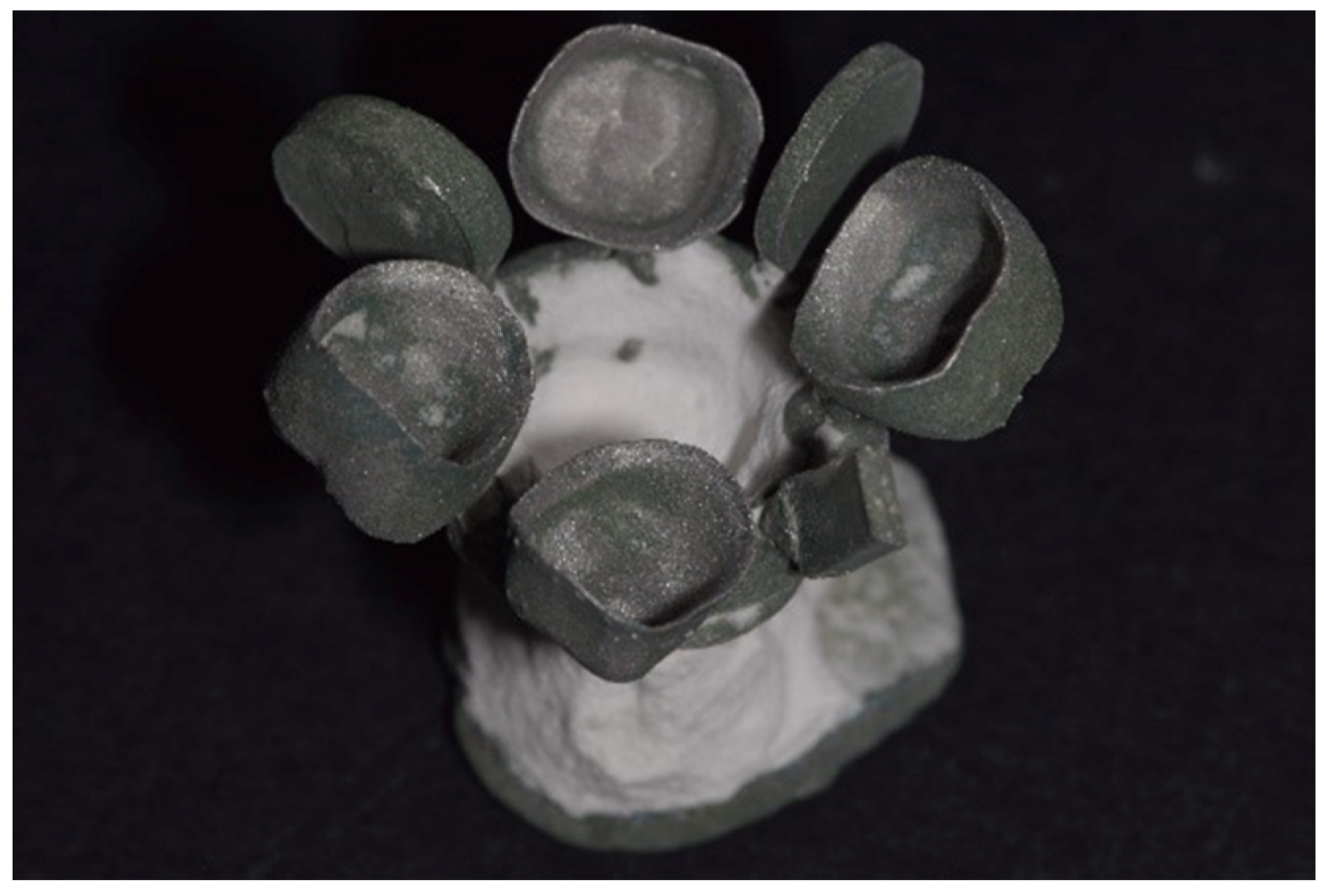
2. Materials and Methods

The study evaluated marginal and internal discrepancies of metal-ceramic crowns with cobalt-chromium frameworks obtained by the conventional casting of printed resin patterns, SLS, SLM, and CAD/CAM milling. Framework design was a “cut-back,” and it was the same for all samples. All restorations were made for a resin molar. The marginal and internal fit evaluation was evaluated using the non-destructive method, silicon replica technique, before and after ceramic over pressing. Silicon replicas were sectioned and photographed under the microscope with 20× magnification, and images were analyzed using image processing software (Figure 1). The measurements were performed in nine points divided as follows: marginal gap (MG), cervical gap (CG), axial wall at internal gap (AG), axio-occlusal angle (AOG), and occlusal wall at internal gap (OG) (Figure 2). Silicon replica technique was used to measure the space between the tooth and the framework. The resulting data were statistically analyzed.
A resin first upper molar was reduced for a full-coverage ceramic fused to metal crown with a 1 mm chamfer shoulder, tapered of axial walls of 6° and a 1.5 mm occlusal reduction, with beveled support cusps. The prepared tooth was replicated after silicon impression (Fegurasil AD Special, Feguramed, Germany), and resin models have been obtained using a provisional restorations resin (Structur 3, Voco, Cuxhaven, Germany). The resin was chosen to simulate the tooth as it has similar mechanical properties to natural dentine tissue. These abutments have been divided into four groups. Two groups, forty-eight prepared teeth, were scanned with D700 3D Scanner (3Shape, Copenhagen, Denmark) to produce laser-sintered copings (SLS) (n = 24) and cast copings (T) (n = 24). 3Shape software was used to create the cut-down design, to obtain a uniform porcelain veneering thickness (Figure 3). Twenty-four frameworks were fabricated by selective laser sintering (SLS) (PXS Dental, Phenix Systems, Riom, France) using cobalt-chromium dental alloy powder (Starbond CoS Powder 16, S&S Sheftner GmbH, Mainz, Germany). The powder layer was of 30 µm, with a compaction rate of 30%, in the controlled atmosphere of N2 with a gas flow of 5 L/min, using a Class 1 laser with a wavelength of 1070 nm and 50 W power.
Twenty-four patterns were printed from castable-resin to fabricate the casted group (E-Partial Press-E-Cast, EnvisionTEC, Dearborn, Michigan, MI, USA) using Perfactory 4 Digital Printer P4DDP (EnvisionTEC, Dearborn, Michigan, MI, USA) (Figure 4) to eliminate operator-induced flows. After printing, patterns were removed from the printer, cleaned by immersing in pure Isopropyl alcohol with 99%, dried and processed by further photopolymerization in the Otoflash unit (EnvisionTEC, Dearborn, Michigan, MI, USA) according to manufacturer instructions.
Patterns were prepared for casting, invested (Bellavest SH, BEGO, Bremen, Germany), and cast with a cobalt-chromium alloy, Wirobond 280 (BEGO, Bremen, Germany), by negative-pressure casting (Nautilus, Bego, Bremen, Germany). Frameworks were divested (Figure 5), cleaned with airborne particles (200 μm, and 75 μm), and prepared for veneering.
An oxide firing was performed. The other group of forty-eight abutments was scanned with Dental Wings 7Series Scanner (Dental Wings INC., Montreal, QC, Canada) for milled (F) (n = 24) and selective laser melted (SLM) (n = 24) copings. An anatomic-reduced framework design has been made using DWOS software (Dental Wings INC., Montreal, QC, Canada) using the same parameters used for the other two groups. Using this design, twenty-four anatomic copings were fabricated using cobalt-chromium bonding alloy for the manufacturing of removable and fixed restorations by selective laser melting (SLM) using the same powder used for SLS copings (Starbond CoS Powder 30, S&S Sheftner GmbH, Mainz, Germany) free of beryllium and nickel. Laser power was 80 W, powder layer 20 μm, under-protected N2 atmosphere <0.3 L/min, using Mysint 100 (Sisma, Piovene Rocchette, Italy). The other set of twenty-four copings were milled from a cobalt-chromium metal disc (White Peaks Dental Systems GmbH & Co. KG, Essen, Germany) using a 5-axis milling machine (Datron D5, Datron AG, Mühltal-Traisa, Germany) and the same design.
Relief-firing was performed for SLS and SLM copings (Figure 6): They were heated to 450° within 60 min, held at this temperature for 45 min, heated to 800 °C within 45 min, kept for 60 min, then slowly cooled, under nitrogen atmosphere.
SLS and SLM frameworks were placed on the building platform with the occlusal face parallel with and towards the plate, and the post-processing step was performed with the copings still attached to the plate.
For porcelain veneering, the same design was made for all ninety-six copings, which were scanned with a D700 3D scanner (3Shape, Copenhagen, Denmark). All copings were prepared for veneering by sandblasting (75 μm alumina particles), degreased using steam, and dried. Opaque firings were conducted after two applications of Opaquer paste IPS InLine System (Ivoclar Vivadent, Schaan, Principality of Liechtenstein), following manufacturers indications. Ceramic veneering was made by over-pressing, to eliminate the subjectivity and variations generated by conventional technology. For that, veneering copings were obtained by milling of Harvest ZCAD Wax Press (Harvest Dental Laboratory Products) using Zenotec (Wieland Dental + Technik GmbH & Co. KG, Pforzheim, Germany) unit. These patterns were attached to previously prepared frameworks, sprues were attached, and the weighted ensemble was invested in specific material PressVEST Speed (Ivoclar Vivadent, Schaan, Principality of Liechtenstein).
After preheating the molds at 850 °C for 75 min, the ceramic (IPS InLine PoM, Ivoclar Vivadent, Schaan, Principality of Liechtenstein) was over-pressed using the specific program of the furnace Programat EP 3010 (Ivoclar Vivadent, Schaan, Principality of Liechtenstein). The crowns were divested, processed, and glazed according to the material’s recommendations.
The initial and final gap was evaluated to establish if and how veneering firings affect internal and marginal adaptation. Measurements were made using silicon replicas for each framework before ceramic opaquer appliance and after glazing for each crown. Low viscosity silicone used for this purpose Black Fit Checker (GC Corporation, Tokyo, Japan) was inserted inside frameworks and crowns to mimic the cement, under constant pressure, until set. This silicone layer was placed between a layer of light-consistency silicone Oranwash L (Zhermack¸ Badia Polesine, Italy) and a putty Zetalabor (Zhermack, Badia Polesine, Italy). Silicone blocks obtained have been sectioned and photographed under an optical microscope (Leica DM500, Leica, Wetzlar, Germany) with a hundred micron scale for image calibration at 20× magnification. ImageJ software was used for measurements after calibration using the scale (Figure 2).
For the gap measurements, nine different points were analyzed for each specimen, for the following sites: marginal gap (MG), cervical gap (CG), axial internal gap in the middle of axial wall (AG), and occlusal internal gap wall on cusps and in the central fossa (OG).

3. Results

Mean gap values were calculated for each point, for each group, before and after ceramic firing.
The mean values for discrepancies for both internal and marginal levels are listed in Table 1. The analysis of variance with Games–Howell post-hoc adjustment showed that the cast T group had significantly higher values, as compared to other procedures, in all point measurements, p > 0.001. The significance of differences between methods is shown in Figure 7.
In Table 2 are included the mean differences between initial and final measurement, for all procedures and all points. Paired differences using t-test showed statistical significance only in the SLM group for the axial wall gap, axio-occlusal angle, occlusal gap and for T group in axial wall gap. All other comparisons were not statistically significant (p > 0.05). Differences between initial and final measurements per each point between types of procedures were analyzed using analysis of variance with Games–Howell post hoc. Significant differences were found only in the occlusal gap between SLM and F procedures, p = 0.001.
The results of this research are presented in Figure 7.
All parts were produced according to computer-aided design, and patterns for casting were printed to eliminate human errors, using a standardized, uniform thickness design. The present study concludes and sustains that modern additive technologies are faster, more reliable and cheaper as compared to the traditional fabrication of wax-patterns. The process has fewer errors, and the deformation risk is low [1,2].

4. Discussion

There is no established standard method for the examination of marginal and internal fit of dental crowns. The most frequently used is the silicon replica, as it is quick, useful, non-destructive, accessible, and fit for all types of restorations. Newly digitized versions of this technique were developed, implying the scanning of the abutment and the silicon replica on the abutment. These images are superimposed and analyzed with specific software. This is a good alternative for traditional silicon replica but is more expensive because it requires an intra-oral scanner for clinical use [18,19]. The best evaluating method for crown adaptation is the invasive method, well known but with some critical issues which limit its applicability. This method implies a section in the already cemented crown embedded in resin, allowing the assessment of both internal and marginal fit, but it is suitable only for research. It was replaced by the silicon replica technique [9,20], a non-invasive and non-destructive technique, ideal for clinical applicability. It allows the clinician to perform several evaluations and measurements during the manufacturing process [1,21,22]. Recent studies investigated the differences between investigation methods. They found similar results for the cross-sectional method and silicon replica technique, with different values when compared with the triple scan method, micro-computer tomography and optical coherence tomography [23]. The researchers concluded that when evaluating the accuracy, the evaluation method must be taken into consideration for a better interpretation of the results.
The prime objective of any prosthodontic treatment is to provide the patient with restorations or prostheses as precisely fitting as possible. In the case of fixed prosthodontics, the internal and marginal gaps are the essential factors to determine the long-term success of the restoration. This is why the prostheses adaptation is a subject that is still of interest for the prosthodontist and researchers [19,24,25].
A marginal opening of 120 μm or less has been considered clinically acceptable, while most of the clinicians would prefer a vertical cervical gap of 60 μm or less. Ideally, cemented crown margins join prepared tooth limits in perfect non-detectable junctions. Clinical perfection is not only a goal difficult to achieve, but also difficult to verify. Hence, a minimal marginal gap is nominally approximately acceptable [26]. In this study, all fabrication methods produced a clinically acceptable marginal gap with almost every value under 100 μm. At the same time, SLM and milled samples reached the goal of under 60 μm marginal gap, as shown in Table 1. Older studies showed higher differences between digital and conventional technologies, while recent ones suggest similar results to ours [27].
The terms marginal gap and internal gap do not have a single definition. Holmes et al., who established several gap definitions according to contour difference between the crown and tooth margin, states that “the perpendicular measurement from the inner surface of casting to the axial wall of preparation is called internal gap, and the same measurement at the margin is called marginal gap” [28]. The measurements used in this study were made according to this definition.
Ultimately, the marginal and internal fit of a dental restoration provides the overarching factors for its success and longevity [5]. For instance, a superior marginal fit markedly reduces the recurrence of dental caries and the development of periodontal diseases and extends the survival of the restoration. Furthermore, an excellent internal fit is necessary to maintain and support restorations [29,30].
Marginal fit and internal fit depends on both clinical, and laboratory procedures and numerous parameters, but this in vitro study investigated only the laboratory procedures, represented by additive manufacturing (AM) technologies, lately entered in the dental field, compared to traditional casting and CAD/CAM milling [16,31].
The 3D printed medical models represent a very controversial factor because several authors considered that errors might occur in the creation process, segmentation, processing, and also during the printing process [32]. Although it is a thorough process, and it needs a lot of attention for every step, including the used software, which may have some inaccuracies, the 3D process is one of the most widespread techniques. The results allowed us to obtain precise patterns for traditional casting [33,34,35,36].
The prepared teeth may have complex shapes with grooves, boxes, and sharp edges, which are difficult to mill. On top of that, the milling process is relatively complicated, costly, and time-consuming. Additionally, grinding a small structure from a large block of material creates a substantial amount of waste material [37]. The smaller diameter milling tool is the one that dictates the accuracy of the final product, so a larger burr than the details that must be reproduced can lead to a loose restoration [38]. The discrepancy between bur and details is the reason why the AM technologies are gaining popularity among practitioners, being able to produce complex structures with internal morphology, and allowing unused material to be processed again. Complex 3-dimensional [3D] shapes can be formed quickly and precisely by the additive process, without unnecessary waste. Sometimes standard technologies cannot solve the patient problem, and digital workflow is the only suitable for the clinical case [39].
CAD/CAM technologies can be considered a good alternative for the conventional wax-up technique to produce patterns for traditional alloys casting and pressing of ceramic ingots. The previous study assessed traditional casting technology, and measurements were made for internal and marginal fit using silicon replica for patterns obtained by 3D printing, cast frameworks, and final ceramic fused to metal crowns. The best adaptation was found in the resin-pattern group, with small increases of gap after casting and after ceramic pressing [34]. This suggests that further studies are necessary to develop a better workflow to increase the accuracy after investing and casting the resin patterns. The problems that can occur during AM fabrication consist of shrinkage that occurs during the manufacturing or in post-processing stages. This is why the specimens fabricated for this study had so many support structures spread across all occlusal surfaces [38].
Frameworks fabricated with CAD/CAM systems demonstrate better fit and passivity than those made by conventional castings. Laboratory variables involved in traditional casting generated by the inconsistency of volumetric and linear expansion of the materials used, including impression material, gypsum products, waxes, investments, and metal casting, are eliminated with CAD/CAM technologies [40,41].
The present study used a 50 μm space for cement in the preparation process of all structures. CAD/CAM systems offer the possibility to use a constant, established in advance, die spacer [42].
Differences in the accuracy of fit between CAD/CAM and cast frameworks have been observed in studies with implant-supported prostheses [43], showing the superiority of CAD/CAM results. However, these studies compared different types of restorations obtained by CAD/CAM, over-casting, traditional casting without a consistency regarding the fabrication process, or research methodology [44,45,46].
Furthermore, although some scientific evidence has shown that layering procedures can distort frameworks [36,47,48], in this study, the same veneering material was applied to all structures. The same method was used to investigate if and how ceramic veneering affects the adaptability of crowns with different copings. Even though some statistically significant differences were found in the SLM group for the axial wall gap, axio-occlusal angle, occlusal gap, and in T group in the axial wall gap, these differences have no clinical significance.
It is clear that CAD/CAM processes still need improvements and additional research because optimal and accurate results are dependent on specialist knowledge [49]. All technological steps of this research, evaluation and interpretation were performed by the same operator to avoid biases as much as possible.
The gap between the copings and abutment teeth in this study generally tended to increase from the marginal towards the internal surface, with the gap being the widest at internal gap. Although this finding is comparable to previous studies, the discrepancies observed in this instance were relatively narrower, thus indicating a better fit across all groups. Wide gaps at internal points may cause copings to fracture when completed restorations are bonded in the mouth without the proper intervention of cement. Factors in automated fabrication that can influence the fit include the input of information and the accuracy of its processing. Errors arising from such processes are likely to cause an increase in the internal gap. Clinician interventions during the clinical adaptation process can also influence internal and marginal adaptation [50].
The accuracy of restorations fabricated with CAD/CAM technology may not be as consistent as in previous dental manufacturing processes (e.g., casting). However, limited information is available regarding the marginal discrepancy of the alloys fabricated by such new manufacturing techniques.

5. Conclusions

The association of silicon replica technique with an image analysis software is a useful investigation method for clinical use and not only for research purposes. Because this is a conservative method it allows the clinician to perform the adaptability assessment at every step of restoration and also before the final fixation of the crown.
The best marginal and internal fit was observed at restorations with milled Cobalt-Chromium frameworks, closely followed by the SLM group.
All restorations shown gap values smaller than the clinically accepted range (120 µm), cast copings being placed at the superior limit of the interval.
3D printing of resin patterns represents a viable alternative for traditional manufacturing of wax patterns, being cost-effective, less time-consuming, and precise, having the potential to replace in a few years the conventional technique completely.
New additive manufacturing technologies can provide pieces with excellent adaptation.
Ceramic over pressing did not influence significantly marginal and internal gaps of restorations, regardless of the manufacturing method used to produce a framework.

Author Contributions

Conceptualization, C.E.S. and L.P.; methodology, L.P.; software, C.Ș.; validation, L.P., C.E.S. and C.Ș.; formal analysis, C.Ș.; investigation, C.E.S.; resources, L.P. and C.E.S.; data curation, C.E.S.; writing—original draft preparation, C.E.S.; writing—review and editing, C.E.S.; visualization, L.P.; supervision, L.P.; project administration, C.E.S.; funding acquisition, L.P. All authors have read and agreed to the published version of the manuscript.

Funding

This research was funded by the Romanian National Authority for Scientific Research and Innovation CNCS-UEFISCDI, grant number PN-II-RU-TE-2014-4-0476.

Conflicts of Interest

The authors declare no conflict of interest.

References

  1. Kim, M.-J.; Choi, Y.-J.; Kim, S.-K.; Heo, S.-J.; Koak, J.-Y. Marginal Accuracy and Internal Fit of 3D Printing Laser-Sintered Co-Cr Alloy Copings. Materials 2017, 10, 93. Available online: https://www.ncbi.nlm.nih.gov/pmc/articles/PMC5344574/ (accessed on 6 May 2019). [CrossRef] [PubMed] [Green Version]
  2. Ishida, Y.; Miyasaka, T. Dimensional accuracy of dental casting patterns created by 3D printers. Dent. Mater. J. 2016, 35, 250–256. [Google Scholar] [CrossRef] [PubMed] [Green Version]
  3. Vojdani, M.; Torabi, K.; Farjood, E.; Khaledi, A. Comparison the Marginal and Internal Fit of Metal Copings Cast from Wax Patterns Fabricated by CAD/CAM and Conventional Wax up Techniques. J. Dent. 2013, 14, 118–129. [Google Scholar]
  4. Porcelain-Fused-to-Metal Crowns Versus All-Ceramic Crowns: A Review of the Clinical and Cost-Effectiveness; CADTH Rapid Response Reports; Canadian Agency for Drugs and Technologies in Health: Ottawa, ON, Canada, 2015. Available online: http://www.ncbi.nlm.nih.gov/books/NBK304693/ (accessed on 1 July 2020).
  5. Kim, K.-B.; Kim, J.-H.; Kim, W.-C.; Kim, J.-H. Three-dimensional evaluation of gaps associated with fixed dental prostheses fabricated with new technologies. J. Prosthet. Dent. 2014, 112, 1432–1436. [Google Scholar] [CrossRef] [PubMed]
  6. Lo Russo, L.; Caradonna, G.; Biancardino, M.; De Lillo, A.; Troiano, G.; Guida, L. Digital versus conventional workflow for the fabrication of multiunit fixed prostheses: A systematic review and meta-analysis of vertical marginal fit in controlled in vitro studies. J. Prosthet. Dent. 2019, 122, 435–440. [Google Scholar] [CrossRef] [PubMed]
  7. Benic, G.I.; Sailer, I.; Zeltner, M.; Gütermann, J.N.; Özcan, M.; Mühlemann, S. Randomized controlled clinical trial of digital and conventional workflows for the fabrication of zirconia-ceramic fixed partial dentures. Part III: Marginal and internal fit. J. Prosthet. Dent. 2019, 121, 426–431. [Google Scholar] [CrossRef] [Green Version]
  8. Bhaskaran, E.; Azhagarasan, N.S.; Miglani, S.; Ilango, T.; Krishna, G.P.; Gajapathi, B. Comparative Evaluation of Marginal and Internal Gap of Co-Cr Copings Fabricated from Conventional Wax Pattern, 3D Printed Resin Pattern and DMLS Tech: An In Vitro Study. J. Indian Prosthodont. Soc. 2013, 13, 189–195. [Google Scholar] [CrossRef]
  9. In Vitro Assessment of the Marginal and Internal Fits of Interim Implant Restorations Fabricated with Different Methods. J. Prosthet. Dent. Available online: https://www.thejpd.org/article/S0022-391330009-9/fulltext (accessed on 1 July 2020).
  10. Pompa, G.; Di Carlo, S.; De Angelis, F.; Cristalli, M.P.; Annibali, S. Comparison of Conventional Methods and Laser-Assisted Rapid Prototyping for Manufacturing Fixed Dental Prostheses: An In Vitro Study. BioMed. Res. Int. 2015, 2015, 318097. [Google Scholar] [CrossRef] [Green Version]
  11. Nesse, H.; Ulstein, D.M.Å.; Vaage, M.M.; Øilo, M. Internal and marginal fit of cobalt-chromium fixed dental prostheses fabricated with 3 different techniques. J. Prosthet. Dent. 2015, 114, 686–692. [Google Scholar] [CrossRef]
  12. Ng, J.; Ruse, D.; Wyatt, C. A comparison of the marginal fit of crowns fabricated with digital and conventional methods. J. Prosthet. Dent. 2014, 112, 555–560. [Google Scholar] [CrossRef]
  13. Costea, C.M.; Badea, M.E.; Vasilache, S.; Mesaroş, M. Effects of CO-CR discrepancy in daily orthodontic treatment planning. Clujul Med. 2016, 89, 279–286. [Google Scholar] [CrossRef] [PubMed] [Green Version]
  14. Douglas, R.D.; Hopp, C.D.; Augustin, M.A. Dental students’ preferences and performance in crown design: Conventional wax-added versus CAD. J. Dent. Educ. 2014, 78, 1663–1672. [Google Scholar] [CrossRef] [PubMed]
  15. Li, K.C.; Prior, D.J.; Waddell, J.N.; Swain, M.V. Comparison of the microstructure and phase stability of as-cast, CAD/CAM and powder metallurgy manufactured Co-Cr dental alloys. Dent. Mater. 2015, 31, 306–315. [Google Scholar] [CrossRef] [PubMed]
  16. Porojan, L.; Topală, F.; Porojan, S.; Savencu, C. Effect of frame design and veneering material on biomechanical behavior of zirconia dental crowns veneered with overpressing ceramics. Dent. Mater. J. 2017, 36, 275–281. [Google Scholar] [CrossRef] [PubMed] [Green Version]
  17. Xu, D.; Xiang, N.; Wei, B. The marginal fit of selective laser melting-fabricated metal crowns: An in vitro study. J. Prosthet. Dent. 2014, 112, 1437–1440. [Google Scholar] [CrossRef] [PubMed]
  18. Koç, E.; Öngül, D.; Şermet, B. A comparative study of marginal fit of copings prepared with various techniques on different implant abutments. Dent. Mater. J. 2016, 35, 447–453. [Google Scholar] [CrossRef] [Green Version]
  19. Digital Evaluation of Marginal and Internal Fit of Single-Crown Fixed Dental Prostheses. Eur. J. Oral Sci. 2018, 126, 517. [CrossRef]
  20. Güngör, M.B.; Doğan, A.; Bal, B.T.; Nemli, S.K. Evaluation of marginal and internal adaptations of posterior all-ceramic crowns fabricated with chair-side CAD/CAM system: An in vitro study. Acta Odontol. Turc. 2017, 35, 1–8. [Google Scholar]
  21. Nawafleh, N.A.; Mack, F.; Evans, J.; Mackay, J.; Hatamleh, M.M. Accuracy and Reliability of Methods to Measure Marginal Adaptation of Crowns and FDPs: A Literature Review: Methods to Measure Marginal Adaptation of Crowns and FDPs. J. Prosthodont. 2013, 22, 419–428. [Google Scholar] [CrossRef] [Green Version]
  22. Tamac, E.; Toksavul, S.; Toman, M. Clinical marginal and internal adaptation of CAD/CAM milling, laser sintering, and cast metal ceramic crowns. J. Prosthet. Dent. 2014, 112, 909–913. [Google Scholar] [CrossRef]
  23. Son, K.; Lee, S.; Kang, S.H.; Park, J.; Lee, K.-B.; Jeon, M.; Yun, B.J. A Comparison Study of Marginal and Internal Fit Assessment Methods for Fixed Dental Prostheses. J. Clin. Med. 2019, 8, 785. [Google Scholar] [CrossRef] [PubMed] [Green Version]
  24. Halawani, S.; Al-Harbi, S. Marginal adaptation of fixed prosthodontics. Int. J. Med. Dev. Ctries. 2017, 1, 78–84. [Google Scholar] [CrossRef]
  25. Riccitiello, F.; Amato, M.; Leone, R.; Spagnuolo, G.; Sorrentino, R. In vitro Evaluation of the Marginal Fit and Internal Adaptation of Zirconia and Lithium Disilicate Single Crowns: Micro-CT Comparison between Different Manufacturing Procedures. Open Dent. J. 2018, 12, 160–172. [Google Scholar] [CrossRef] [PubMed]
  26. Evaluation of Marginal and Internal Gaps of Ni-Cr and Co-Cr Alloy Copings Manufactured by Microstereolithography. Abstract Europe PMC. Available online: https://europepmc.org/article/med/28680548 (accessed on 1 July 2020).
  27. Dauti, R.; Cvikl, B.; Lilaj, B.; Heimel, P.; Moritz, A.; Schedle, A. Micro-CT evaluation of marginal and internal fit of cemented polymer infiltrated ceramic network material crowns manufactured after conventional and digital impressions. J. Prosthodont. Res. 2019, 63, 40–46. [Google Scholar] [CrossRef]
  28. Holmes, J.R.; Bayne, S.C.; Holland, G.A.; Sulik, W.D. Considerations in measurement of marginal fit. J. Prosthet. Dent. 1989, 62, 405–408. [Google Scholar] [CrossRef]
  29. Harish, V.; Mohamed Ali, S.A.; Jagadesan, N.; Mohamed Ifthikar, S.S.; Debasish Basak, F.H. Evaluation of Internal and Marginal Fit of Two Metal Ceramic System—In Vitro Study. J. Clin. Diagn. Res. 2014, 8, ZC53. Available online: http://jcdr.net/article_fulltext.asp?issn=0973-709x&year=2014&volume=8&issue=12&page=ZC53&issn=0973-709x&id=5300 (accessed on 1 July 2020).
  30. Shokry, T.E.; Attia, M.; Mosleh, I.; Elhosary, M.; Hamza, T.; Shen, C. Effect of metal selection and porcelain firing on the marginal accuracy of titanium-based metal ceramic restorations. J. Prosthet. Dent. 2010, 103, 45–52. [Google Scholar] [CrossRef]
  31. Yajvinder, G.V.; Agrawal, A.; Singh, B. Evaluation of Influence of Finish line Design on Marginal Discrepancy of All-ceramics Lithium disilicate Crown restorations using μ-CT. IOP Conf. Ser. Mater. Sci. Eng. 2020, 802, 012003. [Google Scholar] [CrossRef]
  32. George, E.; Liacouras, P.; Rybicki, F.J.; Mitsouras, D. Measuring and Establishing the Accuracy and Reproducibility of 3D Printed Medical Models. Radiographics 2017, 37, 1424–1450. [Google Scholar] [CrossRef]
  33. Huang, Z.; Zhang, L.; Zhu, J.; Zhang, X. Clinical marginal and internal fit of metal ceramic crowns fabricated with a selective laser melting technology. J. Prosthet. Dent. 2015, 113, 623–627. [Google Scholar] [CrossRef]
  34. Savencu, C.E.; Porojan, S.; Porojan, L. Analysis of Internal and Marginal fit of Metal-ceramic Crowns During Processing, Using Conventional and Digitized Technologies. Rev. Chim. 2018, 69, 1699–1701. [Google Scholar] [CrossRef]
  35. US4863538A-Method and Apparatus for Producing Parts by Selective Sintering-Google Patents. Available online: https://patents.google.com/patent/US4863538A/en (accessed on 2 July 2020).
  36. Akova, T.; Ucar, Y.; Tukay, A.; Balkaya, M.C.; Brantley, W.A. Comparison of the bond strength of laser-sintered and cast base metal dental alloys to porcelain. Dent. Mater. 2008, 24, 1400–1404. [Google Scholar] [CrossRef] [PubMed]
  37. Park, J.-K.; Lee, W.-S.; Kim, H.-Y.; Kim, W.-C.; Kim, J.-H. Accuracy evaluation of metal copings fabricated by computer-aided milling and direct metal laser sintering systems. J. Adv. Prosthodont. 2015, 7, 122. [Google Scholar] [CrossRef] [PubMed] [Green Version]
  38. Alshalan, A.; Almutair, A.; Awad, D.; Alshmlani, M.; Abbas, S.B.; Asif, Z. Marginal and Internal Fit of CAD CAM System. Int. J. Dent. Oral Health 2019, 5, 9. [Google Scholar]
  39. Gouveia, D.N.M.; Razzoog, M.E.; Alfaro, M.F. A fully digital approach to fabricating a CAD-CAM ceramic crown to fit an existing removable partial denture. J. Prosthet. Dent. 2019, 121, 571–575. [Google Scholar] [CrossRef]
  40. Haider, Y.; Dimashkieh, M.; Rayyan, M. Survey of Dental Materials Used by Dentists for Indirect Restorations in Saudi Arabia. Int. J. Prosthodont. 2017, 30, 83–85. [Google Scholar] [CrossRef]
  41. Persson, A.S.K.; Andersson, M.; Odén, A.; Sandborgh-Englund, G. Computer aided analysis of digitized dental stone replicas by dental CAD/CAM technology. Dent. Mater. 2008, 24, 1123–1130. [Google Scholar] [CrossRef]
  42. Lopez-Suarez, C.; Gonzalo, E.; Pelaez, J.; Serrano, B.; Suarez, M. Marginal Vertical Discrepancies of Monolithic and Veneered Zirconia and Metal-Ceramic Three-Unit Posterior Fixed Dental Prostheses. Int. J. Prosthodont. 2016, 29, 256–258. [Google Scholar] [CrossRef]
  43. De França, D.G.B.; Morais, M.H.S.T.; das Neves, F.D.; Barbosa, G.A.S. Influence of CAD/CAM on the fit accuracy of implant-supported zirconia and cobalt-chromium fixed dental prostheses. J. Prosthet. Dent. 2015, 113, 22–28. [Google Scholar] [CrossRef]
  44. Moris, I.C.M.; Monteiro, S.B.; Martins, R.; Ribeiro, R.F.; Gomes, E.A. Influence of Manufacturing Methods of Implant-Supported Crowns on External and Internal Marginal Fit: A Micro-CT Analysis. BioMed. Res. Int. 2018, 2018, 1–6. [Google Scholar] [CrossRef] [Green Version]
  45. Mostafa, N.Z.; Ruse, N.D.; Ford, N.L.; Carvalho, R.M.; Wyatt, C.C.L. Marginal Fit of Lithium Disilicate Crowns Fabricated Using Conventional and Digital Methodology: A Three-Dimensional Analysis: Conventionally, Digitally Fabricated LD Crown Marginal Fit. J. Prosthodont. 2018, 27, 145–152. [Google Scholar] [CrossRef] [PubMed]
  46. Bhering, C.L.B.; Marques, I.d.S.V.; Takahashi, J.M.F.K.; Barão, V.A.R.; Consani, R.L.X.; Mesquita, M.F. Fit and Stability of Screw-Retained Implant-Supported Frameworks Under Masticatory Simulation: Influence of Cylinder Type: Stability of Cast Multiunit Implant-Supported Frameworks. J. Prosthodont. 2016, 25, 459–465. [Google Scholar] [CrossRef] [PubMed]
  47. Fonseca, J.C.; Henriques, G.E.P.; Sobrinho, L.C.; de Góes, M.F. Stress-relieving and porcelain firing cycle influence on marginal fit of commercially pure titanium and titanium–aluminum–vanadium copings. Dent. Mater. 2003, 19, 686–691. [Google Scholar] [CrossRef]
  48. Hong, M.-H.; Min, B.K.; Lee, D.-H.; Kwon, T.-Y. Marginal fit of metal-ceramic crowns fabricated by using a casting and two selective laser melting processes before and after ceramic firing. J. Prosthet. Dent. 2019, 122, 475–481. [Google Scholar] [CrossRef]
  49. Moldovan, O.; Luthardt, R.G.; Corcodel, N.; Rudolph, H. Three-dimensional fit of CAD/CAM-made zirconia copings. Dent. Mater. 2011, 27, 1273–1278. [Google Scholar] [CrossRef]
  50. Tabata, L.F.; de Lima Silva, T.A.; de Paula Silveira, A.C.; Ribeiro, A.P.D. Marginal and internal fit of CAD-CAM composite resin and ceramic crowns before and after internal adjustment. J. Prosthet. Dent. 2020, 123, 500–505. [Google Scholar] [CrossRef]
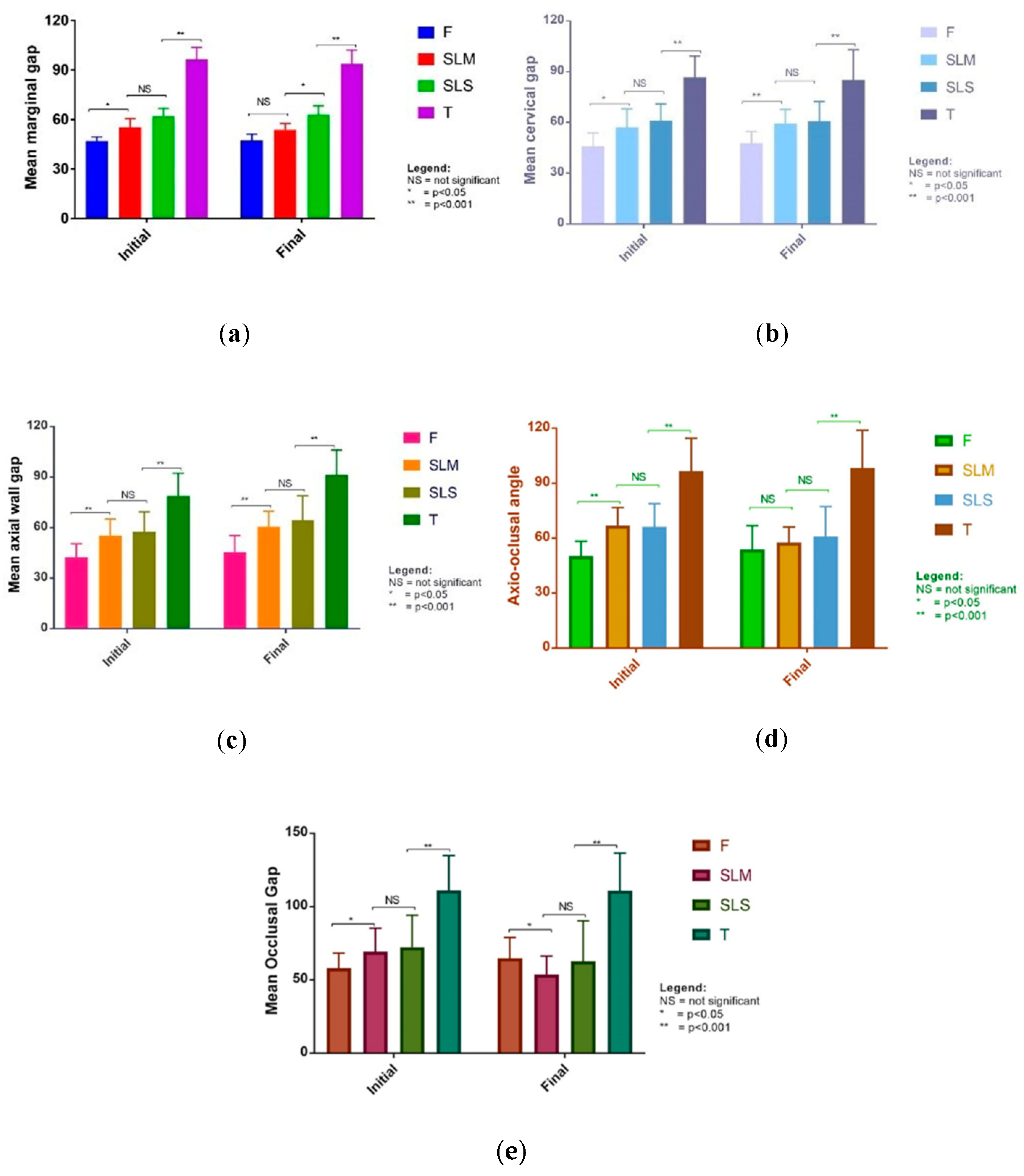
Figure 1. Measurements of silicon replica with ImageJ software (NIH, Bethesda, MA, USA).
Applsci 10 05563 g001
Figure 2. Measurement points.
Applsci 10 05563 g002
Figure 3. Framework design.
Applsci 10 05563 g003
Figure 4. 3D resin patterns for casted frameworks.
Applsci 10 05563 g004
Figure 5. Divested cast samples.
Applsci 10 05563 g005
Figure 6. SLM frameworks after relief firing.
Applsci 10 05563 g006
Figure 7. Obtained results: initial and final measurements (a) mean marginal gap; (b) mean cervical gap; (c) mean axial wall gap; (d) mean axio-occlusal angle; (e) mean occlusal gap.
Applsci 10 05563 g007
Table 1. Mean values for measured points for each procedure.
Measurement PointTime PointProcedurep-Value **
SLS *SLM *F *T *
Marginal gapinitial62.0 ± 11.2755.3 ± 12.7546.9 ± 6.2096.4 ± 17.34<0.001
final63.0 ± 12.6453.8 ± 8.9147.2 ± 9.3693.8 ± 19.71<0.001
Cervical gapinitial61.3 ± 9.6457.0 ± 11.1245.8 ± 7.9786.6 ± 12.67<0.001
final60.9 ± 11.4459.3 ± 8.4047.6 ± 7.1385.2 ± 17.76<0.001
Axial wall gapinitial57.5 ± 11.9055.3 ± 9.8342.5 ± 7.9279.0 ± 13.36<0.001
final64.4 ± 14.5860.7 ± 9.1145.5 ± 9.8491.5 ± 14.64<0.001
Axio-occlusal angleinitial66.2 ± 12.6467.0 ± 9.8350.2 ± 8.1096.6 ± 17.98<0.001
final61.0 ± 16.1557.6 ± 8.5353.9 ± 13.0198.4 ± 20.53<0.001
Occlusal gapinitial72.4 ± 21.7569.3 ± 16.0158.0 ± 10.24111.1 ± 23.69<0.001
final62.7 ± 27.5353.8 ± 12.5064.8 ± 14.09110.9 ± 25.62<0.001
* Data represent mean ± standard deviation. ** Analysis of variance, with post hoc tests using Games–Howell.
Table 2. Mean difference between initial and final measurement.
Difference between Initial and Final MeasurementSLSSLMFTBetween Groups p-Value ***
Mean Difference ± SD *p Value **Mean Difference ± SD *p Value **Mean Difference ± SD *p Value **Mean Difference ± SD *p-Value **
Marginal gap −1.05 ± 19.60.7951.53 ± 16.20.327−0.21 ± 9.90.9202.52 ± 24.70.6220.905
Cervical gap−4.22 ± 11.70.091−1.85 ± 1.60.443−0.61 ± 15.20.697−6.68 ± 17.00.0670.810
Axial wall gap−6.98 ± 18.00.069−5.38 ± 2.60.048−2.96 ± 8.50.101−12.49 ± 17.60.0020.149
Axio-oclusal angle5.21 ± 20.20.2189.45 ± 11.80.001−3.76 ± 14.10.203−1.79 ± 24.80.7260.052
Occlusal gap9.75 ± 34.70.18215.55 ± 20.80.001−6.73 ± 17.80.0770.19 ± 36.80.9810.042
* Data represent mean ± standard deviation. ** paired samples t-test results in bold are statistically significant. *** Analysis of variance with Games–Howell post hoc.

