Review

Selenium Deficiency—From Soil to Thyroid Cancer

by
Latifa Sarra Kazi Tani
1,2,*,
Nouria Dennouni-Medjati
2,
Benoit Toubhans
1 and
Laurent Charlet
1,*
1
ISTerre, University of Grenoble-Alpes and CNRS, BP 53, 38041 Grenoble CEDEX 9, France
2
Analytical Chemistry and Electrochemistry Laboratory, Abou Bekr BELKAID University of Tlemcen, Tlemcen 13000, Algeria
*
Authors to whom correspondence should be addressed.
Appl. Sci. 2020, 10(15), 5368; https://doi.org/10.3390/app10155368
Submission received: 15 June 2020 / Revised: 10 July 2020 / Accepted: 12 July 2020 / Published: 4 August 2020
(This article belongs to the Special Issue Selenium: Properties and Sources in the Food Industry)

Abstract

:

Featured Application

The paper underlines the link between soil, food and human health, with a particular emphasis on thyroid cancer in case of Se deficiency, in order to provide a scientific basis to Public Health Recommendations.

Abstract

Selenium (Se) is an essential micronutrient present in human diet, entering in the composition of selenoproteins as selenocysteine (Se-Cys) amino acid. At the thyroid level, these proteins play an important role as antioxidant and in hormone metabolism. Selenoproteins are essential for the balance of redox homeostasis and antioxidant defense of mammalian organisms, while the corresponding imbalance is now recognized as the cause of many diseases including cancer. The food chain is the main source of Se in human body. Dietary intake is strongly correlated with Se content in soil and varies according to several factors such as geology and atmospheric input. Both Se deficiency and toxicity have been associated with adverse health effects. This review synthesizes recent data on the transfer of Se from soil to humans, Se U-shaped deficiency and toxicity uptake effects and particularly the impact of Se deficiency on thyroid cancer.

1. Introduction

Selenium is one of the most intriguing trace elements of the periodic table. After its discovery in 1818 by the Swedish Berzelius in a sulfuric acid plant, it was used for its electric and photoelectric properties [1]. Its first medical application dates back to 1911, when an unidentified Se compound had caused necrosis and the disappearance of Ehrlich’s carcinoma and sarcoma in mice [1]. A year later, a publication by Delbet in 1912 reported the death of patients who received high doses of sodium selenite [2]. However, all therapeutic applications of Se ceased when, in 1943, Nelson et al. had declared the element itself carcinogenic [3]. It was not until 1957 that scientists collected evidence for the other face of the moon (Selen in Greek), that is, for the essentiality of the trace element Se for humans and animals [1,4].
Despite the importance of selenium in human health, the major impact of supply, speciation and restricted uptake of this micronutrient, is currently not well understood [5]. Much research is still needed to improve our understanding of optimized requirements, taking into account the very narrow range between the beneficial and toxic effects of the mineral [5].
Selenium is essential for many cellular functions including redox homeostasis and antioxidant defense [6]. In the body, Se is ingested from food, absorbed in the gastrointestinal tract (GIT), transported to the liver and kidney for metabolism and distribution to the body tissues [7].
In humans, Se is integrated into 25 selenoproteins in the form of the amino acid Se-Cys. Some of them such as glutathione peroxidases (GPx), iodothyronine deiodinases (DIO) and thioredoxine reductases (TRx) play an important role in the metabolism of the thyroid gland [8]. Se is present at higher concentrations in the thyroid than in other organs and is an essential element for the biosynthesis of thyroid hormones [9]. Another important role of selenoproteins in the thyroid is the detoxification by GPx of hydrogen peroxide (H2O2) massively synthetized in order to oxidize iodide (I) and introduce it into thyroglobulin, a precursor of L-3,5,3′,5′-tetraiodothyronine (thyroxine; T4) and L-3,5,3′-triiodothyronine (T3) [10]. Therefore, Se is considered to be the second most important element in thyroid metabolism after iodine [11].
The results of initial epidemiological studies have suggested a significant association between low Se status and cancer, suggesting that Se may have anticarcinogenic effect [12]. Surprisingly, recent studies have shown an increased risk of some cancers such as prostate cancer with high Se levels [13] and the chemotherapeutic effect of Se have not been proven at safe and tolerated doses (<90 µg/day) [14].
However, in the case of thyroid cancer, serum Se deficiency may be considered a risk factor but the results are inconclusive [15,16].
This review synthesizes the speciation-controlled transport of Se from soil to humans via food and its absorption in human gastrointestinal tract. We then focus on the use of Se by thyroid gland and on the relationship between Se and thyroid cancer, according to the latest knowledge produced on this pathology.

2. Selenium from Soil to Food Bowl

2.1. Selenium in Soil and Surface Waters

Selenium is a common element found in nature, in the Earth’s atmosphere, lithosphere, biosphere and hydrosphere [17]. Se is present in the soil in inorganic forms, such as selenide (Se2−), elemental selenium (Se(0)), selenate (SeO42−) and selenite (SeO32−) and in organic forms (selenocysteine “Se-Cys” and selenomethionine “Se-Met”) [18]. The availability of selenium for plant absorption is highly dependent on the chemical and physical conditions of the soil. The concentration of Se in the soil is very different from one country to another and even from one region to another within the same country [19]. Together with atmospheric input, bedrocks are the main source of Se in soil, with varying levels depending on their nature, sedimentary rocks (e.g., shale, limestone and sandstone) being the richest in Se [20]. Thus, the concentration of Se in soils is strongly dependent on the concentrations of the bedrock [21].
In water, Se is present in trace amounts, mainly in the form of selenate and selenite, where Se is present in +VI and +IV oxidation state, respectively [22]. Se is more abundant in groundwater than in seawater, due to the elution of Se from source rocks and over-fertilization of soils with mixtures rich in Se compounds [21,23].
Another source of Se in the soil is due to natural and anthropogenic input from the atmosphere [24]. The combustion of coal, crude oil and the use of agro-technical processes mainly in fertilization contribute enormously to the content of Se in the soil [25]. It has been reported that Se levels are important in people living near processing plants, for example, sulfuric acid production or coking [26,27]. Volcanic gases are also a major source of Se to the atmosphere, together with Se methylation and volatilization by ocean algal bloom [24]. Biomethylation of Se by microorganisms and the decomposition of Se-rich organic matter are additional factors that contribute to the enrichment of the atmosphere with Se [28]. In these cases, volatile selenium compounds such as dimethylselenium (DMeSe), hydrogen selenide (H2Se) and selenium oxide (SeO2) are produced [29].

2.2. Transfer and Absorption of Se from Soil to Plant

Each plant variety has the ability to accumulate Se at the root and shoot level, depending on the concentration of Se in the soil and the ability of the plant species to absorb, accumulate and metabolize Se [30]. There are several types of plants, so-called hyper-accumulators that grow only in soils very rich in Se “seleniferous” and accumulate between 1000 and 15,000 mg kg−1, including species of the genera Astragalus, Stanleya, Morinda, Neptunia, Oonopsis and Xylorhiza. Secondary accumulators such as cereals, some plants of the cruciferous family (rape, broccoli) and Allium species such as garlic and onion grow in soils with variable Se content and can accumulate 30 to 1000 mg kg−1. Non-accumulating plants generally grown on “selenium-poor” soils and cannot accumulate high concentrations of Se in their tissues and contain less than 30 mg kg−1. This is the case for most plants used for food and feed (forage, vegetables, fruits, etc.) [30].
Absorption of organic selenium by plants is more efficient than of inorganic forms such as Se (IV) or Se (VI) [31]. Less than 5% of the Se present in the soil is used by plants [32]. Other forms of Se such as selenide and elemental Se are not absorbed by plants since they are insoluble in water [31] (Figure 1).
The accumulation of Se from soil to plants requires several processes and depends on the speciation of Se in the soil. Sulfate (S) and phosphate (P), present in the soil could affect the absorption of Se by plants. As selenate is structurally and chemically similar to sulfate, both ions follow the same metabolic pathways during the translocation process in plants [34]. The high-affinity H+/sulfate symporters, homologous to sulfate transporter, AtSULTR1;1 and AtSULTR1;2, catalyze the influx of selenate into root cells from the rhizosphere [35,36]. The preference for selenate uptake over sulfate varies from plant to plant and is influenced by various factors for example, soil salinity and pH [37], the two parameters affecting Se(IV) and Se(VI) adsorption on soil particles [31]. In other non-accumulating plants, other transporters behave similarly. In hyperaccumulative plants, the genes of AtSULTR1;1 and AtSULTR1;2 have a high expression [36].
Selenite is absorbed by roots as HSeO3 by members of the phosphate transporter family [38] or as H2SeO3 by aquaporins [39]. After being absorbed, selenate moves from the root symplast to the stele and is then transferred to the shoot, while selenite is converted into organoselenated compounds [34,39,40]. In the xylem, selenate is the dominant form of Se, however minor amounts of Se-Met and selenomethionine Se-oxide (SeOMet) have been reported. After being delivered to the shoot by the xylem, selenate enters leaf cells by SULTR transporters [35,40,41].

2.3. Metabolism and Se Speciation in Plants

Selenate reduction to selenite then to selenide is coupled to an oxidation of glutathione [35,37]. The Selenide is converted to Se-Cys in a manner analogous to sulfur metabolism. Se-Cys is converted by reverse trans-sulfuration to Se-Met and the two amino acids can be incorporated nonspecifically into proteins in the place of methionine and cysteine [33].
Selenomethionine can be further metabolized to Adenosyl-Se-Met (SeAM), DMeSe and then converted to MeSeCys and γ–glutamine–Se–MethylselenoCysteine (γ-glu-SeMeSeCys) [34,42]. At high levels of Se, MeSeCys becomes the main Se compound, although other compounds are present but at low content [43].
Figure 1 also shows the process of biogenic volatilization of Se from soil and plants, which is in fact a detoxification process [33]. It is used to decontaminate soils rich in Se, such as oil refinery effluent fields [33]. Since some plants can absorb large amounts of Se from the soil, it is therefore very important that plants exhale various volatile Se compounds. The main product of plant volatilization is DMeSe [33]. Heat-treated food products can lose up to 10% of total Se due to the formation of volatile Se compounds (DMeSe) [23].
Different factors can influence the ability of plants to volatilize Se. First, the concentration of Se in the roots, the Se species as well as the concentration of sulfate compared to selenate (they can compete for particular enzymes for the process of volatilization) [33].

2.4. Selenium in Food and Dietary Intake

Food is the principal source of Se. In many populations, plant foods are the main dietary source of Se (mostly as Se-Met), followed by meat and seafood [44,45]. The concentration of Se in food depends on the type of soil, the geographical area and the capacity of plants to accumulate it [23]. In addition, other factors can influence the Se content in the diet, such as consumption of local food or imported food products [19] for example, USA Se-rich cereals [23,46].
The bioavailability of Se in the diet depends on factors such as proteins, fats and heavy metals. Thus, food rich in protein and in the presence of vitamins A, C, E contain higher levels of Se but heavy metals and sulfur reduce Se [47,48].
Cereals provide 50% of daily Se intake. While meat, poultry and fish group provide about 35%. Fresh water and beverages contribute to the daily intake with only 5–25% [23]. Fresh and non-heat-treated fruits and vegetables contain small amounts of Se about 11% in a balanced diet; this is due to their low protein content and high water content [23].
Another selenium-rich source is found in sea salt, offal, yeast (selenium-containing yeast), garlic, asparagus, kohlrabi (enriched with Se) [6,47,49]. High concentrations of Se have, exceptionally, also been found in Brassica genus (broccoli, cabbage, cauliflower) and onions [23]. Brazil nuts and mushrooms have extremely high concentrations of this element [6,50].
The reference values for dietary Se intake were estimated on the basis of the intake necessary for saturation of the selenoproteins; GPx in plasma or erythrocyte or selenoprotein P (SePP) in plasma [51,52]. The Recommended Dietary Allowances (RDA) of Se in the United States, based on the activity maximization of GPx are around 55 µg/day in adults [53]. Based on the dietary Se intake required for saturation of plasma SePP, the nutrition societies of Germany, Austria and Switzerland (D-A-CH) have recommended a Se intake reference value of 70 µg/day and 60 µg/day for men and women, respectively, for normal body weight, at a rate of approximately 1 µg of Se per kg of body weight [54]. The World Health Organization recommends that the intake of Se should not exceed 70 μg/day [55]. A dietary intake above 700 µg/day can be toxic to adults [6]. Se intake in the European population varies from 30 μg/day and 50 μg/day [55].
Furthermore, the biological activities of the several selenium species have not been fully characterized in order to assess their specific toxicity in nutrition, hence defining dietary windows for each of them [14,56,57,58].

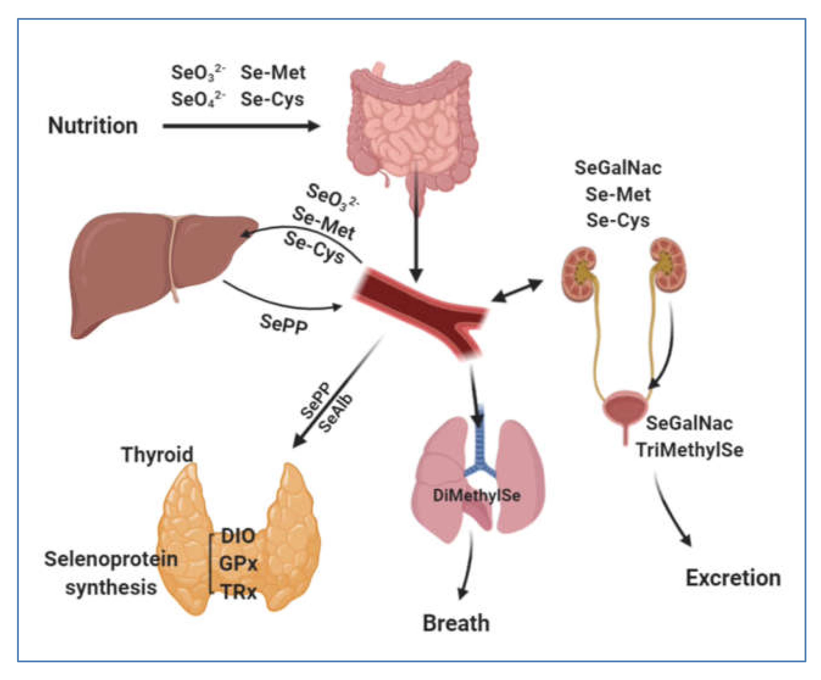
3. Selenium Human Uptake and Distribution

3.1. Gastrointestinal Uptake

Selenium is present in diet in the forms of selenomethionine, selenocysteine, selenite and selenate (Figure 2).
Selenomethionine represents the main chemical form of Se in the human diet. Once ingested, Se-Met is absorbed in the same sodium transport system as methionine [60]. It is transported to liver where it is metabolized following the same pathway as methionine and can be transformed into Se-Cys following the trans-sulfuration pathway [60]. The concentration of Se-Cys is lower in plant proteins than Se-Met, while its abundant in diets containing products of animal origin [59]. Se-Cys can also be absorbed in the same way and in completion with its analogue cysteine [61].
The human genome codes for 25 selenoproteins in which the stop codon UGA specifically insert the selenocysteine. This amino acid is not charged as such on its dedicated tRNA but is instead synthesized onto a specific Se-Cys-tRNA[Ser]Se-Cys from the serine [62].
Selenocysteine is catalyzed by the enzyme Se-Cys β-lyase to elemental Se [63]. The latter is further reduced to H2Se. The addition of phosphate to Selenide by the selenophosphate 2 synthetase produce a selenophosphate (H2SePO3) [64], which can be used as a precursor for selenocysteine synthesis [62].
In the case of excess Se, H2Se is methylated to DMeSe and trimethylselenonium (TriMethylSe) or converted to selenosugar (SeGalNac) and exhaled or excreted [59,60].
Inorganic selenium such as Selenate passes through the membrane at the edge of the intestinal brush, where absorption is via the sodium-facilitated and energy-dependent system utilized by sulfate, due to their structural analogies and reduced to selenite by the sulfate reduction pathway [33]. Selenite is absorbed by non-mediated passive diffusion. Absorption of selenite ranges from 50 to 90%, being affected by dietary constituents on which it is strongly adsorbed, while selenate absorption is almost complete [65,66].
Despite the use of inorganic forms in Se supplementation their absorption is not as high as that of Se-Met [66]. A supplementation of Se-Met at 37 μg/day increases the activity of GPx in human plasma up to its saturation level of 150 U/L, whereas for sodium selenite, 66 μg/day are necessary to reach the same level [67,68].

3.2. Blood Transfer and Distribution of Se to Tissues via Selenoproteins P

The supply of Se to tissues depends on the plasma Se transporter, SePP or serum albumin (SeAlb) [59] (Figure 2). SePP is synthesized mainly in the liver, which is the key organ for the homeostasis of Se in the body. SePP is a secreted glycoprotein with a C-terminal domain composed of nine Se-Cys residues in mice and humans. It also has another Se-Cys residue in the N-terminal region in a redox active motif [69,70]. In rats, studies have identified four types of SePP isoforms. However, in humans plasma, only two isoforms were identified; a complete 60 kDa form and a smaller 50 kDa form [71]. The 60 kDa isoform serves as a transporter of Se to body tissues and the 50 kDa isoform is involved in redox and signaling reactions [72]. The SePP knockout mice show a loss of motor coordination and reduced viability. Their survival time is about 15 days after weaning [73].
In the body, there is a hierarchy of Se distribution in different tissues or organs for example, Se level is highly conserved in the brain, thyroid and testes. This hierarchy give priority to the synthesis of the most important selenoproteins for the organ [7,74,75].
In humans, a recent study has shown that SePP gene variations affect levels of selenium biomarkers after intake of food with a high content of selenium. Indeed, in middle-aged Danes, CC homozygotes of the SePP/rs3877899 polymorphism have higher levels of selenoprotein P and whole blood Se compared to T-allele, after consumption of selenium-rich foods [76]. These results emphasize the importance of a more personalized approach to Se requirements [76].

4. Selenium in Thyroid Gland

Selenium is required at all stages of embryological development of the thyroid gland [77,78]. Deficiency of Se can aggravate the abnormalities induced by iodine deficiency [79]. Hence, the influence of Se on thyroid function is closely related to the state of iodine.
During biosynthesis of thyroid hormones, high concentrations of H2O2 are generated in order to oxidize iodide (I). The iodination of tyrosyl residues on thyroglobulin give rise to the iodine-containing thyroid hormones; T4 and T3 [10]. The DIO selenoprotein family plays an important role in activation or inactivation of thyroid hormones [80]. Excess hydrogen peroxide could be a major source of free radicals and reactive oxygen species (ROS). These molecules can significantly damage the cell and DNA. Indeed, a greater amount of DNA modified by oxidation was observed in follicular cells of the thyroid compared to the spleen, lung and liver [81]. Thus, Se via selenoproteins plays an important role in the detoxification of H2O2 as well as ROS and provides antioxidant protection to thyroid gland, against oxidative stress [81].

4.1. Role of Selenoproteins in Thyroid Gland

The DIO family consists of 3 types of DIO (DIO1, DIO2 and DIO3), that are integral membrane dimeric selenoproteins, each having different catalytic properties in specific tissue and developmental expressions [80] (Table 1). DIO convert pro-hormone T4 to active T3 and reverse T3 (rT3) as well as to diiodothyronine (T2) [80]. They contain a thyroredoxin fold that catalyzes the stereospecific and sequential elimination of iodine atoms from tyrosine residues. Their reactions are complementary and essential to the metabolism and activities of Thyroid hormones [82]. Table 1 summarizes the main functions of selenoproteins in thyroid.
Both DIO1 and DIO2 are hosted in the thyrocytes and provide T3 obtained by deiodination of T4. T3 is the main mediator of the effects of thyroid hormones. Extra-thyroid DIO1 is involved in the conversion in the liver of T4 to T3. DIO2 controls the intracellular activation of T4 into T3 in target tissues and into endocrine cells [86]. Sensitive cells are protected from biologically active T3 thyrotoxic concentrations, so DIOs control the regulation of the thyroid axis [87].
The thyroid physiological role of DIO1 and DIO2 expression is not fully understood. The hierarchy that controls the expression of selenoproteins gives priority to the distribution of Se to essential selenoenzymes and to the most physiologically important tissues preserving their metabolism under Se deficiency conditions [80]. In cultured cells and rodent tissues, the activities of DIO are protected against Se deficiency at the expense of other selenoproteins. Similar compensation pathways also function in humans, as there is no report of an association between limited Se intake and general defects in the central nervous system and in vision or hearing development under otherwise normal conditions [80].
Glutathione peroxidase is the best characterized selenoprotein family in human. Among the selenoproteins, the GPx family constitutes the main components of human antioxidant defense with five isozymes (GPx1–GPx4, GPx6) [83]. The role of the GPx is to reduce H2O2 and organic hydroperoxides to protect cells from the effects of reactive oxygen species (ROS) [82].
This group of selenoproteins is largely involved in thyroid gland function [87]. GPx3 is the most actively expressed isoform in thyroid gland and kidneys [87]. Probably its role in thyroid colloid is to degrade the large amount of H2O2 produced during the iodination of thyroid hormones [88]. GPx4 isoform is present in mitochondria of thyrocyte, its role is the protection of membrane lipids by reducing phospholipid hydroperoxides while, GPx1 is a cytosolic enzyme [87]. Among the thyroid selenoproteins, GPx1 and GPx3 are the most sensitive to the Se level [89].
The family of TRx play an important role in thyroid metabolism, mainly its members; TRx1 and TRx2 [84]. TRx1 is an intracellular enzyme while TRx2 is mitochondrial ensuring the reduction of oxidative stress in mitochondria [82].
Others selenoproteins are involved in the function of thyroid gland including SePP, Selenoprotein S and K provide quality control in endoplasmic reticulum [82].
The relationship between Se and thyroid diseases is complex. The hierarchy in the distribution of Se protects the thyroid from small fluctuations in Se intake [90]. Two isoforms of Se-Cys-tRNA[Ser]Se-Cys have been described. One of them is involved in the biosynthesis of the most important selenoproteins (TRx1, TRx2 and GPx4). The second ensures the synthesis of less essential proteins (GPx1, GPx3, selenoprotein S) [91]. Several studies have shown that Se supplementation has given positive results in certain autoimmune thyroid diseases such as Hashimoto’s thyroiditis and Graves’ disease [90].

4.2. Selenium and Thyroid Cancer

Thyroid cancer is the most common endocrine tumor, responsible for more than half a million new cases per year, ranking 9th place in cancers prevalence worldwide [92,93]. It remains rare in children and adolescent, the median age of diagnostic is 45 to 50 years old [94], it is diagnosed three times more often in women than in men [95]. Thyroid cancer includes three main types of tumors: medullary thyroid carcinoma, anaplastic thyroid carcinoma and differentiated thyroid carcinoma [96]. Differentiated carcinoma alone accounts for about 90% of thyroid cancers. It is derived from the follicular cells of the thyroid, which are responsible for the production of thyroid hormones [95].
The relationship between Se status and cancer has been debated for a long time. Observational studies and randomized controlled trials have shown conflicting results. In a meta-analysis and meta-regression conducted by Cai et al. [97], the results were in favor of a significant association between Se and cancer. High Se exposure may reduce risk of cancer, especially those of: lung, breast, esophagus, stomach and prostate [97]. On the other hand, Vinceti et al. [57] and Jablonska and Vinceti [98] published a review that reports the results of various trials suggesting dramatic effects of Se on cancer development.
The Selenium and Vitamin E Cancer Prevention Trial (SELECT) is one of the largest intervention studies, launched in 2001 and involving more than 32,000 American males. The trial investigated the effect of vitamin E and/or L-selenomethionine supplementations, primarily against the development of prostate cancer and other type of cancers, that is, colorectal cancer, lung cancer and bladder cancer [14]. The results of SELECT showed an increase in prostate cancer risk for patients supplemented with the highest Se levels [13]. However, the results of this study should be interpreted taking into account certain limitations. The possibility that the supplements given to men exceeded the adequate doses to prevent prostate cancer and on the other hand, men selected were characterized by a relatively high baseline selenium status, which suggests that selenium only reduces the risk of prostate cancer in selenium-deficient men and not in the general population [14].This may explain why certain clinical trials have not shown any side effects in cancer patients after intravenous administration of sodium selenite [99].
In a review of Murdolo et al., different information was collected to explain the divergence concerning the role of Se in the pathophysiology of cancer [100]. First: the effects of Se may be more effective against the progression of cancer, at advanced stages of the disease rather than at early stages. Its role could therefore be more important in preventing cancer progression than in its development. Second: the effects of Se are observed at concentrations lower or higher than the required concentrations to optimize selenoprotein activities especially GPx and SePP. The effects of selenium status on cancer show a U-shape curve. Third: genetic variability could also be of importance. Several single nucleotide polymorphisms (SNP) of certain selenoprotein genes have been linked to different types of cancer.
A change in the status of Se has been demonstrated in the pathogenesis of thyroid cancer [101,102]. Significantly lower levels of Se in thyroid cancer patients were found compared to the control group in several studies [10,101,102,103,104].
In contrast, other studies do not provide evidence of a significant association between Se deficiency and thyroid cancer [105,106]. Table 2 presents some studies and results conducted on Se and/or selenoproteins in serum, tissue, fingernail and urine of thyroid cancer subjects.
In Table 2, we highlight that most studies indicate low Se levels in the patients with thyroid cancer. Se deficiency in various diseases, including cancer, could be related to a high level of free radicals caused by oxidative stress [95]. Significant increase in production of ROS is observed in thyroid tumor tissue samples vs. healthy tissue [81,103]. Se is present in high concentrations in the thyroid and plays an important role in the elimination of ROS. Therefore, a fluctuation in its level could affect the expression of antioxidant selenoproteins, sensitive to the intake of Se in the thyroid (GPx1 and GPx3). In the review by Olivera et al., studies corroborate the reduction in the activity of selenoproteins in thyroid cancer, in case of Se deficiency [95].
In primary papillary thyroid carcinoma (PTC) samples, reduced or even absent expression of GPx3 has been found in patients and it has been correlated with lymph node metastasis and increased tumor size [112]. In thyroid cancer cell lines TPC-1 and FTC133, GPx3 could inhibit Wnt/β-catenin signaling and thereby suppress metastasis of thyroid cancer [112]. The anticarcinogenic effect of Se during the initiation phase of tumor development is the increased expression of antioxidant selenoproteins. Numerous cohort studies have shown that individuals with plasma selenium levels below 100–120 μg/L might benefit by increasing their selenium intake. This concentration is the amount needed to reach a plateau in SePP level and beyond, an increase of selenium concentrations no longer provides protective effects on the development of cancer [92].
In cancer cells, abnormal redox regulation is observed at different stage of tumor progression [99]. Tumor cells require antioxidant molecules such as selenoproteins to maintain the redox balance [92]. The expression of antioxidant proteins increases in many types of cancer and decreases in others [99]. Indeed, tumor cells present major differences in their selenoprotein expression pattern such as the GPx gene [92]. In colorectal cancer, 15 selenoprotein genes were analyzed in two cohorts. Both selenoproteins TRx3 and GPx2 were upregulated in adenoma and carcinoma, while SePP and selenoprotein S were down regulated [113]. The increased gene expression of GPx2 and TRx3 can be explained by the fact that both are target genes for Wnt signaling. This signaling pathway is activated in most colorectal cancer tissues [114]. However, there are not many studies regarding the overall changes in selenoprotein genes in thyroid cancer. Selenoproteins GPx1 and TRx1 in thyroid cancer tissue are lower in patients versus controls, while DIO3 mRNA levels and activity were increased in PTC [111,115]. This increase in DIO3 mRNA levels was correlated with distant metastasis or lymph nodes. Thus, it appears that some selenoproteins fight the growth of tumor cells, while others support it, which underlines the fact that the carcinogenesis mechanisms linked to the Se status are far from being elucidated [115].
Experiments have suggested that selenoproteins can act to modulate the susceptibility of the malignancy by acting on tumor suppressor gene pathways. It has been observed in breast and prostate cancer that selenoproteins carry on, for example, control in the checkpoint kinase-2 (CHEK2) gene, a suppressor tumor gene which is involved in the signal transduction in cellular response to DNA damage who is associated with thyroid malignancy [116]. Different mutations in the tumor suppressor gene CHEK2, such as 1100delC, IVS2 1 1G > A, del5395 and I157T, are associated with multi-organ cancers including breast and papillary thyroid cancer [117]. In addition, Se could act as an anti-mutagenic agent with toxicity against cancer cells [118]. Se acts by inducing cell death by production of superoxide radicals thus triggering the mitochondrial pathway of apoptosis, while sparing healthy cells [118]. It is clear that Se has anticarcinogenic properties, linked to its valence states. Selenite has the capacity to intervene in redox reactions, while selenate is completely devoid of this ability [119]. It is reported in a review by Kieliszek et al. that only selenite ions react with the –SH groups of proteins and prevent the formation of protein polymers rich in disulfides [119]. Indeed, a barrier made up of blood proteins with fibrin properties, protects the membranes of cancer cells from recognition by the immune system. Sodium selenite inhibits the protein disulfide exchange on the surface of cancer cell membranes and thus makes the tumor sensitive to the destructive activity of phagocytic cells [119].
Selenium supplementation has positive results in autoimmune thyroid disease and may improve thyroid cancer outcome, however the results are not conclusive in the majority of cases [95]. The question that remains is whether a deficiency in this micronutrient is a consequence of thyroid cancer or a risk factor. The example of hypoxia, which can influence selenoproteins biosynthesis, the expression of SePP is reduced noting a decrease in the distribution of Se by hepatocytes causing a general decline in selenoproteins expression [120,121,122].
To conclude, several studies have highlighted a Se deficiency in thyroid cancer patients. However, in the lack of evidence to this relationship, many studies are needed to confirm and to explain this hypothesis.

5. Conclusions

More than two centuries after the discovery of the trace element selenium, the roles of this mineral in human health and disease have not been fully elucidated. The relationship between Se status and cancer has been debated for a long time and the results of epidemiological studies are contradictory. In thyroid cancer, most studies indicate a significant association between Se deficiency and the risk of this cancer. However, recent studies have shown that certain selenoproteins fight the growth of tumor cells, while others support it, which underlines the fact that the carcinogenesis mechanisms linked to Se status are not completely understood in thyroid cancer. Different approaches are still needed to clarify the link between Se status and thyroid cancer; genetic association studies, large-scale population-based studies and other omics-based analysis.

6. Future Perspectives

At present, the mechanisms that influence the development of thyroid cancer and the anticancer effects of Se have not been fully elucidated. Therefore, analytical and experimental studies on the role of selenoproteins must be conducted using thyroid cancer stem cells. It is also important to have a better knowledge of selenoproteins polymorphisms and their regulation involved in thyroid cancer.
Further research should focus on studies using data on dietary behavior as well as epidemiological data to better understand the effects of selenium deficiency on thyroid cancer.

Author Contributions

L.S.K.T. and N.D.-M. have collected the data and written the review, B.T. draws the figures. B.T. and L.C. have contributed to the review and corrected the English. All authors have read and agreed to the published version of the manuscript.

Funding

This research was partially funded by a Profas B+ scholarship granted by the France Embassy in Alger to L.S.K.T.

Acknowledgments

We acknowledge the above grant which allowed an active collaboration between the French University Grenoble Alpes and the Algerian University Abou Bekr Belkaid, Tlemcen, Algeria.

Conflicts of Interest

The authors declare no conflict of interest.

References

  1. Schrauzer, G.N.; Surai, P.F. Selenium in human and animal nutrition: Resolved and unresolved issues. A partly historical treatise in commemoration of the fiftieth anniversary of the discovery of the biological essentiality of selenium, dedicated to the memory of Klaus Schwarz (1914–1978) on the occasion of the thirtieth anniversary of his death. Crit. Rev. Biotechnol. 2009, 29, 2–9. [Google Scholar]
  2. Delbet, P. Tentatives de traitement de cancer par selenium. Bull. De l’Assoc. Francaise pour l’Etude de Cancer 1912, 5, 121–125. [Google Scholar]
  3. Nelson, A.A. Liver Tumors Following Cirrhosis Caused by Selenium in Rats. Cancer Res. 1943, 3, 230–236. [Google Scholar]
  4. Rayman, M.P. The importance of selenium to human health. Lancet 2000, 356, 233–241. [Google Scholar] [CrossRef] [Green Version]
  5. Rayman, M.P. Selenium and human health. Lancet 2012, 379, 1256–1268. [Google Scholar] [CrossRef]
  6. Kieliszek, M.; Błażejak, S. Current Knowledge on the Importance of Selenium in Food for Living Organisms: A Review. Molecules 2016, 21, 609. [Google Scholar] [CrossRef] [Green Version]
  7. Kato, T.; Read, R.; Rozga, J.; Burk, R.F. Evidence for intestinal release of absorbed selenium in a form with high hepatic extraction. Am. J. Physiol. Gastrointest. Liver Physiol. 1992, 262, G854–G858. [Google Scholar] [CrossRef]
  8. Duntas, L.H.; Benvenga, S. Selenium: An element for life. Endocrine 2015, 48, 756–775. [Google Scholar] [CrossRef] [PubMed]
  9. Köhrle, J. The trace element selenium and the thyroid gland. Biochimie 1999, 81, 527–533. [Google Scholar] [CrossRef]
  10. Mehl, S.; Sun, Q.; Görlich, C.L.; Hackler, J.; Kopp, J.F.; Renko, K.; Mittag, J.; Schwerdtle, T.; Schomburg, L. Cross-sectional analysis of trace element status in thyroid disease. J. Trace Elem. Med. Biol. 2020, 58, 126430. [Google Scholar] [CrossRef]
  11. Sakız, D.; Kaya, A.; Kulaksizoglu, M. Serum Selenium Levels in Euthyroid Nodular Thyroid Diseases. Biol. Trace Elem. Res. 2016, 174, 21–26. [Google Scholar] [CrossRef] [PubMed]
  12. Novotny, L.; Rauko, P.; Kombian, S.; Edafiogho, I. Selenium as a chemoprotective anti-cancer agent: Reality or wishful thinking? Neoplasma 2010, 57, 383–391. [Google Scholar] [CrossRef]
  13. Kristal, A.R.; Darke, A.K.; Morris, J.S.; Tangen, C.M.; Goodman, P.J.; Thompson, I.M.; Meyskens, F.L.; Goodman, G.E.; Minasian, L.M.; Parnes, H.L.; et al. Baseline Selenium Status and Effects of Selenium and Vitamin E Supplementation on Prostate Cancer Risk. JNCI J. Natl. Cancer Inst. 2014, 106. [Google Scholar] [CrossRef] [PubMed] [Green Version]
  14. Vinceti, M.; Filippini, T.; Del Giovane, C.; Dennert, G.; Zwahlen, M.; Brinkman, M.; Zeegers, M.P.; Horneber, M.; D’Amico, R.; Crespi, C.M. Selenium for preventing cancer. Cochrane Database Syst. Rev. 2018. [Google Scholar] [CrossRef]
  15. Rasmussen, L.B.; Schomburg, L.; Köhrle, J.; Pedersen, I.B.; Hollenbach, B.; Hög, A.; Ovesen, L.; Perrild, H.; Laurberg, P. Selenium status, thyroid volume, and multiple nodule formation in an area with mild iodine deficiency. Eur. J. Endocrinol. 2011, 164, 585–590. [Google Scholar] [CrossRef] [Green Version]
  16. Giray, B.; Arnaud, J.; Sayek, İ.; Favier, A.; Hıncal, F. Trace elements status in multinodular goiter. J. Trace Elem. Med. Biol. 2010, 24, 106–110. [Google Scholar] [CrossRef]
  17. Reich, H.J.; Hondal, R.J. Why Nature Chose Selenium. ACS Chem. Biol. 2016, 11, 821–841. [Google Scholar] [CrossRef]
  18. Hu, T.; Liang, Y.; Zhao, G.; Wu, W.; Li, H.; Guo, Y. Selenium Biofortification and Antioxidant Activity in Cordyceps militaris Supplied with Selenate, Selenite, or Selenomethionine. Biol. Trace Elem. Res. 2019, 187, 553–561. [Google Scholar] [CrossRef] [PubMed]
  19. Dumont, E.; Vanhaecke, F.; Cornelis, R. Selenium speciation from food source to metabolites: A critical review. Anal. Bioanal. Chem. 2006, 385, 1304–1323. [Google Scholar] [CrossRef] [PubMed]
  20. Fernández-Martínez, A.; Charlet, L. Selenium environmental cycling and bioavailability: A structural chemist point of view. Rev. Env. Sci. Biotechnol. 2009, 8, 81–110. [Google Scholar] [CrossRef]
  21. Shahid, M.; Niazi, N.K.; Khalid, S.; Murtaza, B.; Bibi, I.; Rashid, M.I. A critical review of selenium biogeochemical behavior in soil-plant system with an inference to human health. Environ. Pollut. 2018, 234, 915–934. [Google Scholar]
  22. He, Y.; Xiang, Y.; Zhou, Y.; Yang, Y.; Zhang, J.; Huang, H.; Shang, C.; Luo, L.; Gao, J.; Tang, L. Selenium contamination, consequences and remediation techniques in water and soils: A review. Environ. Res. 2018, 164, 288–301. [Google Scholar] [CrossRef]
  23. Kieliszek, M. Selenium–Fascinating Microelement, Properties and Sources in Food. Molecules 2019, 24, 1298. [Google Scholar] [CrossRef] [PubMed] [Green Version]
  24. Wen, H.; Carignan, J. Reviews on atmospheric selenium: Emissions, speciation and fate. Atmos. Environ. 2007, 41, 7151–7165. [Google Scholar] [CrossRef]
  25. Söderlund, M.; Virkanen, J.; Holgersson, S.; Lehto, J. Sorption and speciation of selenium in boreal forest soil. J. Environ. Radioactiv. 2016, 164, 220–231. [Google Scholar] [CrossRef] [PubMed]
  26. Cao, S.; Duan, X.; Zhao, X.; Ma, J.; Dong, T.; Huang, N.; Sun, C.; He, B.; Wei, F. Health risks from the exposure of children to As, Se, Pb and other heavy metals near the largest coking plant in China. Sci. Total Environ. 2014, 472, 1001–1009. [Google Scholar] [CrossRef] [PubMed]
  27. Dennouni-Medjati, N.; Dali-Sahi, M.; Harek, H.; Harek, Y. Statut en sélénium de la population d’une région côtière de l’Ouest algérien à forte activité anthropique. Environ. Risque Sante 2019, 18, 8. [Google Scholar]
  28. Vriens, B.; Lenz, M.; Charlet, L.; Berg, M.; Winkel, L.H.E. Natural wetland emissions of methylated trace elements. Nat. Commun. 2014, 5, 3035. [Google Scholar] [CrossRef] [Green Version]
  29. Winkel, L.; Vriens, B.; Jones, G.; Schneider, L.; Pilon-Smits, E.; Bañuelos, G. Selenium Cycling Across Soil-Plant-Atmosphere Interfaces: A Critical Review. Nutrients 2015, 7, 4199–4239. [Google Scholar] [CrossRef] [Green Version]
  30. Zhu, Y.-G.; Pilon-Smits, E.A.H.; Zhao, F.-J.; Williams, P.N.; Meharg, A.A. Selenium in higher plants: Understanding mechanisms for biofortification and phytoremediation. Trends Plant Sci. 2009, 14, 436–442. [Google Scholar] [CrossRef]
  31. Manceau, A.; Charlet, L. The Mechanism of Selenate Adsorption on Goethite and Hydrous Ferric Oxide. J. Colloid Interface Sci. 1994, 168, 87–93. [Google Scholar] [CrossRef]
  32. Haug, A.; Graham, R.D.; Christophersen, O.A.; Lyons, G.H. How to use the world’s scarce selenium resources efficiently to increase the selenium concentration in food. Microb. Ecol. Health Dis. 2007, 19, 209–228. [Google Scholar] [PubMed] [Green Version]
  33. Lazard, M.; Dauplais, M.; Blanquet, S.; Plateau, P. Recent advances in the mechanism of selenoamino acids toxicity in eukaryotic cells. Biomol. Concepts 2017, 8, 93–104. [Google Scholar] [CrossRef] [PubMed]
  34. Sors, T.G.; Ellis, D.R.; Salt, D.E. Selenium uptake, translocation, assimilation and metabolic fate in plants. Photosynth. Res. 2005, 86, 373–389. [Google Scholar] [CrossRef] [PubMed]
  35. White, P.J. Selenium metabolism in plants. Biochim. Et Biophys. Acta (BBA) Gen. Subj. 2018, 1862, 2333–2342. [Google Scholar] [CrossRef]
  36. White, P.J. Selenium accumulation by plants. Ann. Bot. 2015, mcv180. [Google Scholar] [CrossRef] [Green Version]
  37. González-Morales, S.; Pérez-Labrada, F.; García-Enciso, E.; Leija-Martínez, P.; Medrano-Macías, J.; Dávila-Rangel, I.; Juárez-Maldonado, A.; Rivas-Martínez, E.; Benavides-Mendoza, A. Selenium and Sulfur to Produce Allium Functional Crops. Molecules 2017, 22, 558. [Google Scholar] [CrossRef] [Green Version]
  38. Zhang, L.; Hu, B.; Li, W.; Che, R.; Deng, K.; Li, H.; Yu, F.; Ling, H.; Li, Y.; Chu, C. OsPT2, a phosphate transporter, is involved in the active uptake of selenite in rice. New Phytol. 2014, 201, 1183–1191. [Google Scholar] [CrossRef] [Green Version]
  39. Zhao, X.Q.; Mitani, N.; Yamaji, N.; Shen, R.F.; Ma, J.F. Involvement of Silicon Influx Transporter OsNIP2;1 in Selenite Uptake in Rice. Plant Physiol. 2010, 153, 1871–1877. [Google Scholar] [CrossRef] [Green Version]
  40. Li, H.-F.; McGrath, S.P.; Zhao, F.-J. Selenium uptake, translocation and speciation in wheat supplied with selenate or selenite. New Phytol. 2008, 178, 92–102. [Google Scholar] [CrossRef]
  41. Wang, P.; Menzies, N.W.; Lombi, E.; McKenna, B.A.; James, S.; Tang, C.; Kopittke, P.M. Synchrotron-based X-ray absorption near-edge spectroscopy imaging for laterally resolved speciation of selenium in fresh roots and leaves of wheat and rice. EXBOTJ 2015, 66, 4795–4806. [Google Scholar] [CrossRef] [PubMed] [Green Version]
  42. Van Hoewyk, D. A tale of two toxicities: Malformed selenoproteins and oxidative stress both contribute to selenium stress in plants. Ann. Bot. 2013, 112, 965–972. [Google Scholar] [CrossRef] [PubMed] [Green Version]
  43. Grant, T.D.; Montes-Bayón, M.; LeDuc, D.; Fricke, M.W.; Terry, N.; Caruso, J.A. Identification and characterization of Se-methyl selenomethionine in Brassica juncea roots. J. Chromatogr. A 2004, 1026, 159–166. [Google Scholar] [CrossRef] [PubMed]
  44. Office of Dietary Supplements—Selenium. Available online: https://ods.od.nih.gov/factsheets/Selenium-HealthProfessional/ (accessed on 30 May 2020).
  45. Bañuelos, G.S.; Freeman, J.; Arroyo, I. Accumulation and speciation of selenium in biofortified vegetables grown under high boron and saline field conditions. Food Chem. X 2020, 5, 100073. [Google Scholar] [CrossRef] [PubMed]
  46. Fraczek, A. Selenium in medicine and treatment. J. Elementol. 2013, 18, 145–163. [Google Scholar] [CrossRef]
  47. Kieliszek, M.; Błażejak, S. Selenium: Significance, and outlook for supplementation. Nutrition 2013, 29, 713–718. [Google Scholar] [CrossRef] [PubMed]
  48. Fairweather-Tait, S.J.; Collings, R.; Hurst, R. Selenium bioavailability: Current knowledge and future research requirements. Am. J. Clin. Nutr. 2010, 91, 1484S–1491S. [Google Scholar] [CrossRef] [Green Version]
  49. Smrkolj, P.; Pograjc, L.; Hlastanribi, C.; Stibilj, V. Selenium content in selected Slovenian foodstuffs and estimated daily intakes of selenium. Food Chem. 2005, 90, 691–697. [Google Scholar] [CrossRef]
  50. Ariane, M.K.; Maristela, M.; Silmara, M.M.; Renata, H.S.; Karine, S.N.; Helyde, A.M.; Augusto, K.J. Properties of Brazil nuts: A review. Afr. J. Biotechnol. 2015, 14, 642–648. [Google Scholar] [CrossRef] [Green Version]
  51. Hurst, R.; Collings, R.; Harvey, L.J.; King, M.; Hooper, L.; Bouwman, J.; Gurinovic, M.; Fairweather-Tait, S.J. EURRECA—Estimating Selenium Requirements for Deriving Dietary Reference Values. Crit. Rev. Food Sci. Nutr. 2013, 53, 1077–1096. [Google Scholar] [CrossRef]
  52. Kipp, A.P.; Strohm, D.; Brigelius-Flohé, R.; Schomburg, L.; Bechthold, A.; Leschik-Bonnet, E.; Heseker, H. Revised reference values for selenium intake. J. Trace Elem. Med. Biol. 2015, 32, 195–199. [Google Scholar] [CrossRef] [PubMed] [Green Version]
  53. Dietary Reference Intakes for Vitamin C, Vitamin E, Selenium, and Carotenoids; Institute of Medicine (U.S.); Panel on Dietary Antioxidants and Related Compounds (Eds.) National Academy Press: Washington, DC, USA, 2000; ISBN 978-0-309-06949-6. [Google Scholar]
  54. Referenzwerte für die Nährstoffzufuhr. Available online: https://www.dge.de/wissenschaft/referenzwerte/ (accessed on 13 June 2020).
  55. World Health Organization; Food and Agricultural Organisation. Vitamin and Mineral Requirements in Human Nutrition; World Health Organization: Geneva, Switzerland, 2004; ISBN 978-92-4-154612-6. [Google Scholar]
  56. Hazane-Puch, F.; Arnaud, J.; Trocmé, C.; Faure, P.; Laporte, F.; Champelovier, P. Sodium Selenite Decreased HDAC Activity, Cell Proliferation and Induced Apoptosis in Three Human Glioblastoma Cells. ACAMC 2016, 16, 490–500. [Google Scholar] [CrossRef] [PubMed]
  57. Vinceti, M.; Filippini, T.; Cilloni, S.; Bargellini, A.; Vergoni, A.V.; Tsatsakis, A.; Ferrante, M. Health risk assessment of environmental selenium: Emerging evidence and challenges. Mol. Med. Rep. 2017, 15, 3323–3335. [Google Scholar] [CrossRef] [PubMed] [Green Version]
  58. Mandrioli, J.; Michalke, B.; Solovyev, N.; Grill, P.; Violi, F.; Lunetta, C.; Conte, A.; Sansone, V.A.; Sabatelli, M.; Vinceti, M. Elevated Levels of Selenium Species in Cerebrospinal Fluid of Amyotrophic Lateral Sclerosis Patients with Disease-Associated Gene Mutations. Neurodegener. Dis. 2017, 17, 171–180. [Google Scholar] [CrossRef]
  59. Gammelgaard, B.; Jackson, M.I.; Gabel-Jensen, C. Surveying selenium speciation from soil to cell—Forms and transformations. Anal. Bioanal. Chem. 2011, 399, 1743–1763. [Google Scholar] [CrossRef] [PubMed]
  60. Schrauzer, G.N. Selenomethionine: A Review of Its Nutritional Significance, Metabolism and Toxicity. J. Nutr. 2000, 130, 1653–1656. [Google Scholar] [CrossRef] [Green Version]
  61. Nickel, A.; Kottra, G.; Schmidt, G.; Danier, J.; Hofmann, T.; Daniel, H. Characteristics of transport of selenoamino acids by epithelial amino acid transporters. Chem. Biol. Interact. 2009, 177, 234–241. [Google Scholar] [CrossRef]
  62. Vindry, C.; Ohlmann, T.; Chavatte, L. Translation regulation of mammalian selenoproteins. Biochim. Et Biophys. Acta (BBA) Gen. Subj. 2018, 1862, 2480–2492. [Google Scholar] [CrossRef]
  63. Esaki, N.; Karai, N.; Nakamura, T.; Tanaka, H.; Soda, K. Mechanism of reactions catalyzed by selenocysteine β-Lyase. Arch. Biochem. Biophys. 1985, 238, 418–423. [Google Scholar] [CrossRef]
  64. Cupp-Sutton, K.; Ashby, M. Biological Chemistry of Hydrogen Selenide. Antioxidants 2016, 5, 42. [Google Scholar] [CrossRef] [Green Version]
  65. Mangels, A.R.; Moser-Veillon, P.B.; Patterson, K.Y.; Veillon, C. Selenium utilization during human lactation by use of stable-isotope tracers. Am. J. Clin. Nutr. 1990, 52, 621–627. [Google Scholar] [CrossRef]
  66. Ha, H.Y.; Alfulaij, N.; Berry, M.J.; Seale, L.A. From Selenium Absorption to Selenoprotein Degradation. Biol. Trace Elem. Res. 2019, 192, 26–37. [Google Scholar] [CrossRef]
  67. Burk, R.F. Effects of Chemical Form of Selenium on Plasma Biomarkers in a High-Dose Human Supplementation Trial. Cancer Epidemiol. Biomark. Prev. 2006, 15, 804–810. [Google Scholar] [CrossRef] [Green Version]
  68. Xia, Y.; Hill, K.E.; Byrne, D.W.; Xu, J.; Burk, R.F. Effectiveness of selenium supplements in a low-selenium area of China. Am. J. Clin. Nutr. 2005, 81, 829–834. [Google Scholar] [CrossRef] [Green Version]
  69. Burk, R.F.; Hill, K.E. Selenoprotein P—Expression, functions, and roles in mammals. Biochim. Et Biophys. Acta (BBA) Gen. Subj. 2009, 1790, 1441–1447. [Google Scholar] [CrossRef] [Green Version]
  70. Burk, R.F.; Hill, K.E. SELENOPROTEIN P: An Extracellular Protein with Unique Physical Characteristics and a Role in Selenium Homeostasis. Annu. Rev. Nutr. 2005, 25, 215–235. [Google Scholar] [CrossRef]
  71. Méplan, C.; Nicol, F.; Burtle, B.T.; Crosley, L.K.; Arthur, J.R.; Mathers, J.C.; Hesketh, J.E. Relative Abundance of Selenoprotein P Isoforms in Human Plasma Depends on Genotype, Se Intake, and Cancer Status. Antioxid. Redox Signal. 2009, 11, 2631–2640. [Google Scholar] [CrossRef] [PubMed]
  72. Baclaocos, J.; Santesmasses, D.; Mariotti, M.; Bierła, K.; Vetick, M.B.; Lynch, S.; McAllen, R.; Mackrill, J.J.; Loughran, G.; Guigó, R.; et al. Processive Recoding and Metazoan Evolution of Selenoprotein P: Up to 132 UGAs in Molluscs. J. Mol. Biol. 2019, 431, 4381–4407. [Google Scholar] [CrossRef] [PubMed]
  73. Hill, K.E.; Zhou, J.; McMahan, W.J.; Motley, A.K.; Atkins, J.F.; Gesteland, R.F.; Burk, R.F. Deletion of Selenoprotein P Alters Distribution of Selenium in the Mouse. J. Biol. Chem. 2003, 278, 13640–13646. [Google Scholar] [CrossRef] [PubMed] [Green Version]
  74. Burk, R.F.; Hill, K.E. Regulation of Selenium Metabolism and Transport. Annu. Rev. Nutr. 2015, 35, 109–134. [Google Scholar] [CrossRef] [PubMed]
  75. Berry, M.J. Insights into the hierarchy of selenium incorporation. Nat. Genet. 2005, 37, 1162–1163. [Google Scholar] [CrossRef]
  76. Méplan, C.; Crosley, L.K.; Nicol, F.; Beckett, G.J.; Howie, A.F.; Hill, K.E.; Horgan, G.; Mathers, J.C.; Arthur, J.R.; Hesketh, J.E. Genetic polymorphisms in the human selenoprotein P gene determine the response of selenoprotein markers to selenium supplementation in a gender-specific manner (the SELGEN study). FASEB J. 2007, 21, 3063–3074. [Google Scholar] [CrossRef] [Green Version]
  77. Valea, A.; Georgescu, C.E. Selenoproteins in human body: Focus on thyroid pathophysiology. Hormones 2018, 17, 183–196. [Google Scholar] [CrossRef] [PubMed]
  78. Lacka, K.; Szeliga, A. Significance of selenium in thyroid physiology and pathology. Pol. Merkur. Lek. 2015, 38, 348–353. [Google Scholar]
  79. Köhrle, J. Selenium and the thyroid. Curr. Opin. Endocrinol. Diabetes Obes. 2015, 22, 392–401. [Google Scholar] [CrossRef] [PubMed]
  80. Orozco, A.; Valverde-R, C.; Olvera, A.; García-G, C. Iodothyronine deiodinases: A functional and evolutionary perspective. J. Endocrinol. 2012, 215, 207–219. [Google Scholar] [CrossRef] [PubMed] [Green Version]
  81. Krohn, K.; Maier, J.; Paschke, R. Mechanisms of Disease: Hydrogen peroxide, DNA damage and mutagenesis in the development of thyroid tumors. Nat. Rev. Endocrinol. 2007, 3, 713–720. [Google Scholar] [CrossRef] [PubMed]
  82. Schomburg, L. Selenium, selenoproteins and the thyroid gland: Interactions in health and disease. Nat. Rev. Endocrinol. 2012, 8, 160–171. [Google Scholar] [CrossRef]
  83. Guillin, O.M.; Vindry, C.; Ohlmann, T.; Chavatte, L. Selenium, Selenoproteins and Viral Infection. Nutrients 2019, 11, 2101. [Google Scholar] [CrossRef] [Green Version]
  84. Hu, S.; Rayman, M.P. Multiple Nutritional Factors and the Risk of Hashimoto’s Thyroiditis. Thyroid 2017, 27, 597–610. [Google Scholar] [CrossRef] [Green Version]
  85. Kurokawa, S.; Berry, M.J. Selenium. Role of the Essential Metalloid in Health. Met. Ions Life Sci. 2013, 13, 499–534. [Google Scholar] [PubMed] [Green Version]
  86. Dayan, C.M.; Panicker, V. Novel insights into thyroid hormones from the study of common genetic variation. Nat. Rev. Endocrinol. 2009, 5, 211–218. [Google Scholar] [CrossRef]
  87. Köhrle, J.; Gärtner, R. Selenium and thyroid. Best Pract. Res. Clin. Endocrinol. Metab. 2009, 23, 815–827. [Google Scholar] [CrossRef]
  88. Schmutzler, C.; Mentrup, B.; Schomburg, L.; Hoang-Vu, C.; Herzog, V.; Köhrle, J. Selenoproteins of the thyroid gland: Expression, localization and possible function of glutathione peroxidase 3. Biol. Chem. 2007, 388. [Google Scholar] [CrossRef]
  89. Brigelius-Flohé, R. Glutathione peroxidases and redox-regulated transcription factors. Biol. Chem. 2006, 387. [Google Scholar] [CrossRef]
  90. Schomburg, L. The other view: The trace element selenium as a micronutrient in thyroid disease, diabetes, and beyond. Hormones 2020, 19, 15–24. [Google Scholar] [CrossRef]
  91. Schomburg, L.; Schweizer, U. Hierarchical regulation of selenoprotein expression and sex-specific effects of selenium. Biochim. Et Biophys. Acta (BBA) Gen. Subj. 2009, 1790, 1453–1462. [Google Scholar] [CrossRef]
  92. Kipp, A.P. Selenium in colorectal and differentiated thyroid cancer. Hormones 2020, 19, 41–46. [Google Scholar] [CrossRef]
  93. Ferlay, J.; Soerjomataram, I.; Dikshit, R.; Eser, S.; Mathers, C.; Rebelo, M.; Parkin, D.M.; Forman, D.; Bray, F. Cancer incidence and mortality worldwide: Sources, methods and major patterns in GLOBOCAN 2012: Globocan 2012. Int. J. Cancer 2015, 136, E359–E386. [Google Scholar] [CrossRef] [PubMed]
  94. Schlumberger, M. Cancer papillaire et folliculaire de la thyroïde. Ann. D’endocrinologie 2007, 68, 120–128. [Google Scholar] [CrossRef] [PubMed]
  95. De Oliveira Maia, M.; Batista, B.A.M.; Sousa, M.P.; de Souza, L.M.; Maia, C.S.C. Selenium and thyroid cancer: A systematic review. Nutr. Cancer 2019, 1–9. [Google Scholar] [CrossRef] [PubMed]
  96. Cabanillas, M.E.; McFadden, D.G.; Durante, C. Thyroid cancer. Lancet 2016, 388, 2783–2795. [Google Scholar] [CrossRef]
  97. Cai, X.; Wang, C.; Yu, W.; Fan, W.; Wang, S.; Shen, N.; Wu, P.; Li, X.; Wang, F. Selenium Exposure and Cancer Risk: An Updated Meta-analysis and Meta-regression. Sci. Rep. 2016, 6, 19213. [Google Scholar] [CrossRef] [PubMed] [Green Version]
  98. Jablonska, E.; Vinceti, M. Selenium and Human Health: Witnessing a Copernican Revolution? J. Environ. Sci. Healthpart C 2015, 33, 328–368. [Google Scholar] [CrossRef]
  99. Wallenberg, M.; Misra, S.; Björnstedt, M. Selenium Cytotoxicity in Cancer. Basic Clin. Pharm. Toxicol. 2014, 114, 377–386. [Google Scholar] [CrossRef] [Green Version]
  100. Murdolo, G.; Bartolini, D.; Tortoioli, C.; Piroddi, M.; Torquato, P.; Galli, F. Selenium and Cancer Stem Cells. In Advances in Cancer Research; Elsevier: Amsterdam, The Netherlands, 2017; Volume 136, pp. 235–257. ISBN 978-0-12-812016-3. [Google Scholar]
  101. Kucharzewski, M.; Braziewicz, J.; Majewska, U.; Gózdz, S. Concentration of Selenium in the Whole Blood and the Thyroid Tissue of Patients with Various Thyroid Diseases. BTER 2002, 88, 25–30. [Google Scholar] [CrossRef]
  102. Moncayo, R.; Kroiss, A.; Oberwinkler, M.; Karakolcu, F.; Starzinger, M.; Kapelari, K.; Talasz, H.; Moncayo, H. The role of selenium, vitamin C, and zinc in benign thyroid diseases and of selenium in malignant thyroid diseases: Low selenium levels are found in subacute and silent thyroiditis and in papillary and follicular carcinoma. BMC Endocr. Disord. 2008, 8, 2. [Google Scholar] [CrossRef] [Green Version]
  103. Baltaci, A.K.; Dundar, T.K.; Aksoy, F.; Mogulkoc, R. Changes in the Serum Levels of Trace Elements Before and After the Operation in Thyroid Cancer Patients. Biol. Trace Elem. Res. 2017, 175, 57–64. [Google Scholar] [CrossRef]
  104. Kucharzewski, M.; Braziewicz, J.; Majewska, U.; Góźdź, S. Copper, Zinc, and Selenium in Whole Blood and Thyroid Tissue of People with Various Thyroid Diseases. BTER 2003, 93, 9–18. [Google Scholar] [CrossRef]
  105. Jonklaas, J.; Danielsen, M.; Wang, H. A Pilot Study of Serum Selenium, Vitamin D, and Thyrotropin Concentrations in Patients with Thyroid Cancer. Thyroid 2013, 23, 1079–1086. [Google Scholar] [CrossRef] [Green Version]
  106. Ren, Y.; Kitahara, C.M.; de Gonzalez, A.B.; Clero, E.; Brindel, P.; Maillard, S.; Cote, S.; Dewailly, E.; Rachedi, F.; Boissin, J.-L.; et al. Lack of Association between Fingernail Selenium and Thyroid Cancer Risk: A Case-Control Study in French Polynesia. Asian Pac. J. Cancer Prev. 2014, 15, 5187–5194. [Google Scholar] [CrossRef] [PubMed] [Green Version]
  107. Przybylik-Mazurek, E.; Zagrodzki, P.; Kuźniarz-Rymarz, S.; Hubalewska-Dydejczyk, A. Thyroid Disorders—Assessments of Trace Elements, Clinical, and Laboratory Parameters. Biol. Trace Elem. Res. 2011, 141, 65–75. [Google Scholar] [CrossRef] [PubMed]
  108. Shen, F.; Cai, W.S.; Li, J.L.; Feng, Z.; Cao, J.; Xu, B. The Association Between Serum Levels of Selenium, Copper, and Magnesium with Thyroid Cancer: A Meta-analysis. Biol. Trace Elem. Res. 2015, 167, 225–235. [Google Scholar] [CrossRef] [PubMed]
  109. Chung, H.-K.; Nam, J.S.; Ahn, C.W.; Lee, Y.S.; Kim, K.R. Some Elements in Thyroid Tissue are Associated with More Advanced Stage of Thyroid Cancer in Korean Women. Biol. Trace Elem. Res. 2016, 171, 54–62. [Google Scholar] [CrossRef]
  110. Zhang, C.; Wu, H.B.; Cheng, M.X.; Wang, L.; Gao, C.B.; Huang, F. Association of exposure to multiple metals with papillary thyroid cancer risk in China. Environ. Sci. Pollut. Res. 2019, 26, 20560–20572. [Google Scholar] [CrossRef]
  111. Metere, A.; Frezzotti, F.; Graves, C.E.; Vergine, M.; De Luca, A.; Pietraforte, D.; Giacomelli, L. A possible role for selenoprotein glutathione peroxidase (GPx1) and thioredoxin reductases (TrxR1) in thyroid cancer: Our experience in thyroid surgery. Cancer Cell Int. 2018, 18, 7. [Google Scholar] [CrossRef]
  112. Zhao, H.; Li, J.; Li, X.; Han, C.; Zhang, Y.; Zheng, L.; Guo, M. Silencing GPX3 Expression Promotes Tumor Metastasis in Human Thyroid Cancer. CPPS 2015, 16, 316–321. [Google Scholar] [CrossRef]
  113. Hughes, D.; Kunická, T.; Schomburg, L.; Liška, V.; Swan, N.; Souček, P. Expression of Selenoprotein Genes and Association with Selenium Status in Colorectal Adenoma and Colorectal Cancer. Nutrients 2018, 10, 1812. [Google Scholar] [CrossRef] [Green Version]
  114. Kipp, A.P.; Müller, M.F.; Göken, E.M.; Deubel, S.; Brigelius-Flohé, R. The selenoproteins GPx2, TrxR2 and TrxR3 are regulated by Wnt signalling in the intestinal epithelium. Biochim. Et Biophys. Acta (BBA) Gen. Subj. 2012, 1820, 1588–1596. [Google Scholar] [CrossRef]
  115. Romitti, M.; Wajner, S.M.; Zennig, N.; Goemann, I.M.; Bueno, A.L.; Meyer, E.L.S.; Maia, A.L. Increased Type 3 Deiodinase Expression in Papillary Thyroid Carcinoma. Thyroid 2012, 22, 897–904. [Google Scholar] [CrossRef]
  116. Gupta, S.; Jaworska-Bieniek, K.; Lubinski, J.; Jakubowska, A. Can selenium be a modifier of cancer risk in CHEK2 mutation carriers? Mutagenesis 2013, 28, 625–629. [Google Scholar] [CrossRef] [Green Version]
  117. Siołek, M.; Cybulski, C.; Gąsior-Perczak, D.; Kowalik, A.; Kozak-Klonowska, B.; Kowalska, A.; Chłopek, M.; Kluźniak, W.; Wokołorczyk, D.; Pałyga, I.; et al. CHEK 2 mutations and the risk of papillary thyroid cancer: Genetic association between thyroid and breast cancer. Int. J. Cancer 2015, 137, 548–552. [Google Scholar] [CrossRef] [PubMed]
  118. Weekley, C.M.; Jeong, G.; Tierney, M.E.; Hossain, F.; Maw, A.M.; Shanu, A.; Harris, H.H.; Witting, P.K. Selenite-mediated production of superoxide radical anions in A549 cancer cells is accompanied by a selective increase in SOD1 concentration, enhanced apoptosis and Se–Cu bonding. J. Biol. Inorg. Chem. 2014, 19, 813–828. [Google Scholar] [CrossRef] [PubMed]
  119. Kieliszek, M.; Lipinski, B.; Błażejak, S. Application of Sodium Selenite in the Prevention and Treatment of Cancers. Cells 2017, 6, 39. [Google Scholar] [CrossRef] [PubMed] [Green Version]
  120. Becker, N.-P.; Martitz, J.; Renko, K.; Stoedter, M.; Hybsier, S.; Cramer, T.; Schomburg, L. Hypoxia reduces and redirects selenoprotein biosynthesis. Metallomics 2014, 6, 1079–1086. [Google Scholar] [CrossRef] [Green Version]
  121. Dreher, I.; Jakobs, T.C.; Köhrle, J. Cloning and Characterization of the Human Selenoprotein P Promoter: Response of selenoprotein p expression to cytokines in liver cells. J. Biol. Chem. 1997, 272, 29364–29371. [Google Scholar] [CrossRef] [Green Version]
  122. Renko, K.; Hofmann, P.J.; Stoedter, M.; Hollenbach, B.; Behrends, T.; Köhrle, J.; Schweizer, U.; Schomburg, L. Down-regulation of the hepatic selenoprotein biosynthesis machinery impairs selenium metabolism during the acute phase response in mice. FASEB J. 2009, 23, 1758–1765. [Google Scholar] [CrossRef]
Figure 1. Selenium speciation from soil to plant metabolism (Adapted from Winkel et al. [29] and Lazar et al. [33]). Se(0) = elemental selenium, Se2− = selenide, SeO42− = selenate, SeO32− = selenite, H2Se = hydrogen selenide, Se-Cys = selenocysteine, Se-Met = selenomethionine.
Applsci 10 05368 g001
Figure 2. Main selenium species in various human organs and in blood (adapted from Gammelgaard et al. [59]). SeO32− = selenite, SeO42− = selenate, Se-Met = selenomethionine, Se-Cys = selenocysteine, SePP = selenoprotein P, GPx = glutathione peroxydase, DIO = iodothyronine deiodinase, TRx = thioredoxine reductase, SeAlb = serum albumin, TriMethylSe = trimethylselenonium, SeGalNac = selenosugar.
Applsci 10 05368 g002
Table 1. Main selenoproteins in thyroid and their functions.
SelenoproteinesLocalizationFunctionsReferences
Deiodinases (DIO)[80]
DIO1Liver, kidney, thyroid gland, lung, eyes, pituitary, CNSConversion of T4 into T3and rT3 and T3 into rT3 or T2
DIO2Thyroid gland, pituitary gland, skeletal, heart muscles, brain, fat tissue, spinal cord, placentaConversion of T4 into T3 and of rT3 into T2
DIO3Gravid uterus, placenta, fetus liver, fetal and neonatal brain, skinConversion of T4 into T3 and of rT3 into T2
Gluthatione peroxidases (GPx)[82,83]
GPx1Cytoplasm, ubiquitousCytosol Antioxidant
GPx3Plasma and thyroid folliclePlasma and extracellular antioxidant
GPx4Mitochondrial membraneMembrane antioxidant
Thioredoxin reductase (TRx)[82,84]
TRx1Principally cytosolic, ubiquitousInhibition of apoptosis, redox state of transcription factors
TRx2Mitochondrial, ubiquitousReduce basal oxidative stress,
TRx3Principally mitochondrial, ubiquitousRegulation of apoptosis and signaling pathway
Selenoprotein P (SePP)Blood and thyroidTransportation of selenium and storage, endothelial antioxidant[85]
DIO = iodot hyronine deiodinase, T2 = diiodothyronine, T3 = triiodothyronine, rT3 = reverse T3, T4 = thyroxine, GPX = glutathione peroxydase, TRx = Thioredoxin reductase, SePP = selenoprotein P.
Table 2. Recent clinical studies conducted on the link between Se and thyroid cancer.
AnalysisSampleOutcomeReferences
Serum Se and GPx3 concentration.25 patients with PTC
13 patients with FTC
20 Control
No significant differences in Se and GPx3 concentrations among groups.[107]
Pre-surgery evaluation of serum Se and vit D3 at different stages of disease.35 patients with PTC
12 patients with FTC
17 patients with goiter
No significant differences among groups.[105]
Fingernail Se level215 patients diagnosed with various thyroid cancer
331 controls
No significant association between fingernail Se levels in patients vs controls, at different thyroid cancer stage[106]
Serum concentrations of Se, Cu, MnMeta-analysis on 1291 subjects performed in Norwegian, Austrian and Polish populationsSignificantly lower levels of Se and Mn and higher level of Cu, in patients with thyroid carcinoma[108]
Serum Zn and Se concentration before and after surgery or two weeks later, as well as in thyroid tissues.50 women and men with thyroid cancerLower serum Se and Zn concentrations before and after surgery but higher concentrations in thyroid tissue in the 2 groups[103]
Pre-surgery Se, Cd, Zn serum concentration and correlation to cancer stage92 Korean women, of PTCSe, Cd, Zn concentrations were significantly higher in cancer stages III and IV[109]
Serum and urine concentration in eleven metals and in Se262 patients with PTC
262 controls
Se concentrations significantly lower in PTC.
Urinary Se concentration negatively associated to PTC risk
[110]
GPx1 and TRx1 expression and analysis of free radicals tumor vs. healthy tissues20 samples of thyroid tumor
20 samples of healthy thyroid tissue
GPx1 and TRx1 in thyroid cancer tissue are lower in patients vs. controls
Significant increase in production of free radicals in all thyroid tumor tissue samples vs. healthy tissue
[111]
Se, GPx3, SePP, Cu, Zn serum concentrationPatients with various thyroid pathologies including thyroid cancer (n = 323)
200 Controls
Significantly lower serum Se and Zn levels in patients vs. controls (particular the patients with thyroid malignancy).[10]
PTC: papillary thyroid carcinoma; FTC follicular thyroid carcinoma; GPx: glutathione peroxydase; Vit = vitamin, TRx = thioredoxine reductase; SePP = selenoprotein P.

