Article

Evaluation of the Effect of Freezing and Gamma Irradiation on Different Types of Tendon Allografts by DIC Assisted Tensile Testing

1
Department of Mechatronics, Optics and Mechanical Engineering Informatics, Faculty of Mechanical Engineering, Budapest University of Technology and Economics, 1111 Budapest, Hungary
2
Biomechanical Research Centre, Faculty of Mechanical Engineering, Budapest University of Technology and Economics, 1111 Budapest, Hungary
3
Department of Polymer Engineering, Faculty of Mechanical Engineering, Budapest University of Technology and Economics, 1111 Budapest, Hungary
4
Orthopaedic and Trauma Department, Uzsoki Hospital, 1145 Budapest, Hungary
5
Department of Traumatology, Semmeweis University, 1145 Budapest, Hungary
*
Author to whom correspondence should be addressed.
Appl. Sci. 2020, 10(15), 5369; https://doi.org/10.3390/app10155369
Submission received: 8 July 2020 / Revised: 28 July 2020 / Accepted: 31 July 2020 / Published: 4 August 2020
(This article belongs to the Section Applied Biosciences and Bioengineering)

Abstract

:

Featured Application

The replacement of autografts with allografts from organ banks in ligament reconstruction is a hot topic in medicine. The demand for ACL allografts has increased in the last decade, and postoperative results are promising. This is visible in the increase in allograft use from 2% (between 1986 and 1996) to 14% (between 1996 and 2001), and it further increased around 2007 as its number reached 20–30% in the US. Orthopedic surgeons do not have to struggle with the problems of the donor site, size and the number of the grafts. In the case of multiple ligament reconstruction or ACL revision operations, the surgeons cannot remove enough tendons from the patients to replace the injured ligaments. Our work can help operating surgeons in the selection of proper allografts based on precise mechanical properties provided by our statistically evaluated measurements.

Abstract

This study aimed to evaluate the changes in the endurance properties of four types of tendons caused by freezing and gamma irradiation. Four types of grafts were harvested: quadriceps, semitendinosus + gracilis, tibialis anterior, and peroneus longus. These were put into three groups: Group A was the control group, tested without freezing or irradiation. Grafts in Group B were frozen and irradiated (target dose: 21 kGy), while grafts in Group C were again frozen and irradiated (target dose: 42 kGy). Maximum load, tensile modulus, tensile strength, strain at maximum force and fracture strain were calculated from the force-elongation graphs of cyclic load tests. The higher gamma irradiation dose (Group C) significantly decreased the strains at tensile strength of the quadriceps tendons (p = 0.0004–0.0237), compared to the other two groups. In the case of the quadricep tendons (p = 0.0151), there is a significant decrease in Young’s modulus after gamma irradiation with the dose of 42 kGy. According to the results of the study, the tibialis anterior and the peroneus longus are recommended in ACL reconstruction when gamma irradiation is required, while quadricep tendons, which are usually used for an autograph, are not suitable for allograft reconstruction after gamma irradiation from the viewpoint of mechanical properties.

1. Introduction

The demand for anterior cruciate ligament (ACL) allografts (knee ligament replacements sourced from different patients) has changed in the last few years [1], and the postoperative results are promising. Its increased availability made allograft tissue an appealing alternative to autograft (where the ligament to replace the damaged ligament is sourced from the same patient’s body) for primary ACL reconstruction, especially for ACL revision [2]. The use of musculoskeletal graft allografts has grown continuously in recent years. The American Academy of Orthopaedic Surgeons estimated that nowadays, approximately 60,000 allografts are used in knee reconstruction procedures [3,4]. However, 27% of primary ACL reconstruction operations and 57% of ACL revisions used allografts [5].
The tendons are usually stored as deep-frozen (−80 °C). In practice, there is no significant change in the mechanical properties of the tendons, but this has not been systematically investigated in the literature. Freezing as a method of sterilization is not tested due to storage temperature. Allografts should be sterilized before implantation as they may carry bacterial or viral diseases. [1,6,7]. In most cases, gamma radiation is used for sterilization. A radio-protectant solution is used to avoid side effects and protect tissues. A lower dose of gamma radiation (10–15 kGy) is bactericidal, whereas higher doses (30–50 kGy) are antiviral [7,8,9,10,11].
In our preliminary study [12], we showed that the effect of radiation on the mechanical properties of tendons depends on the type of tendon. The aim of this study was biomechanical evaluation of the changes caused by freezing and gamma irradiation in a higher sample size in the mechanical properties in four different types of tendon allografts used in knee ligamentous reconstruction In this study, doubled semitendinosus and gracilis (STG), doubled tibialis anterior (TA), doubled peroneus longus (PL), and quadricep tendons were prepared and tested. The mechanical properties of the tendons, such as tensile modulus, maximum load, strain at maximum force and fracture strain were determined. We hypothesized that the type of tendon influences the effect of freezing and gamma irradiation on mechanical properties. The results of the present research confirm the results of our preliminary study [12], and can help to select the most appropriate tendons for ACL reconstruction.

2. Materials and Methods
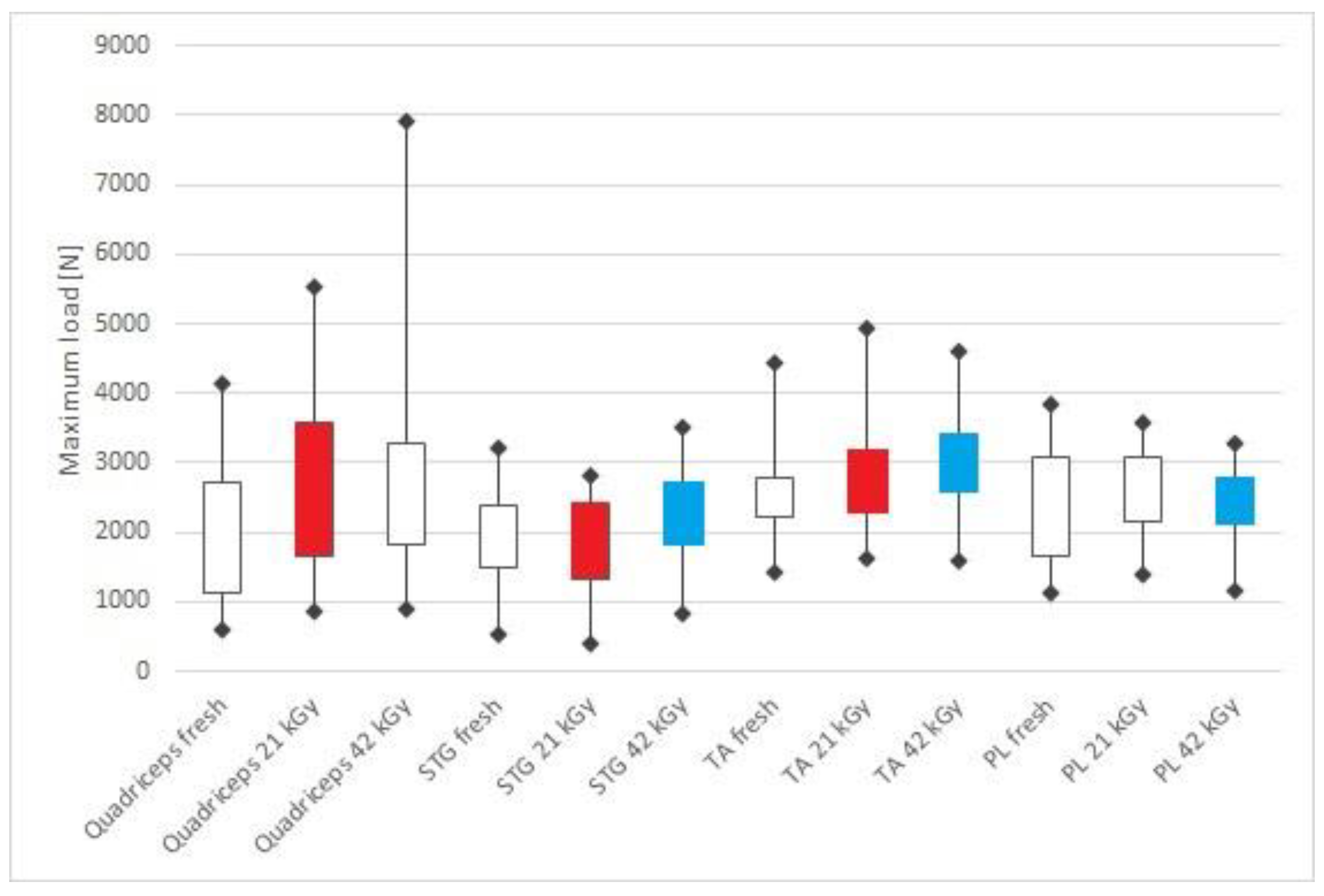
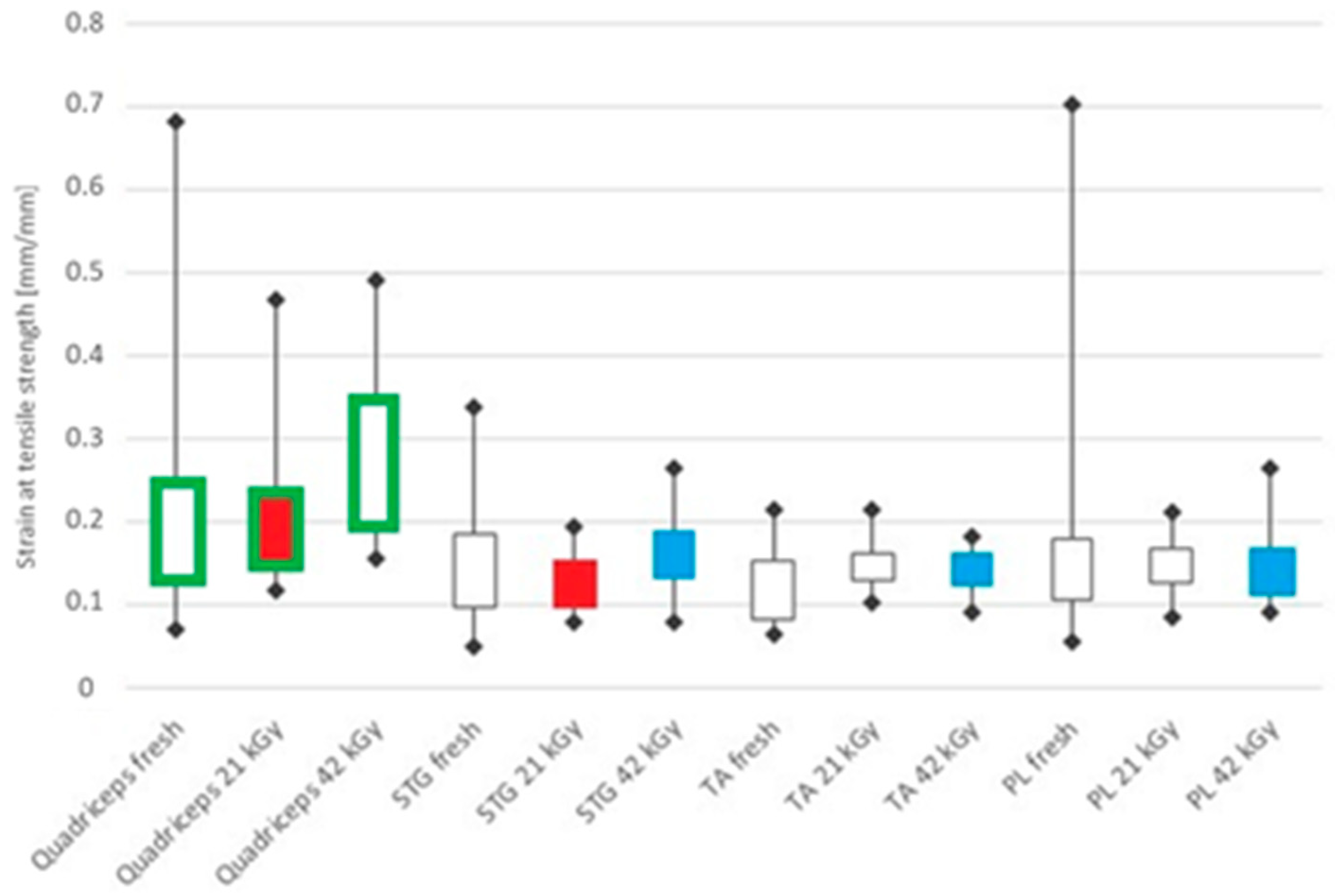
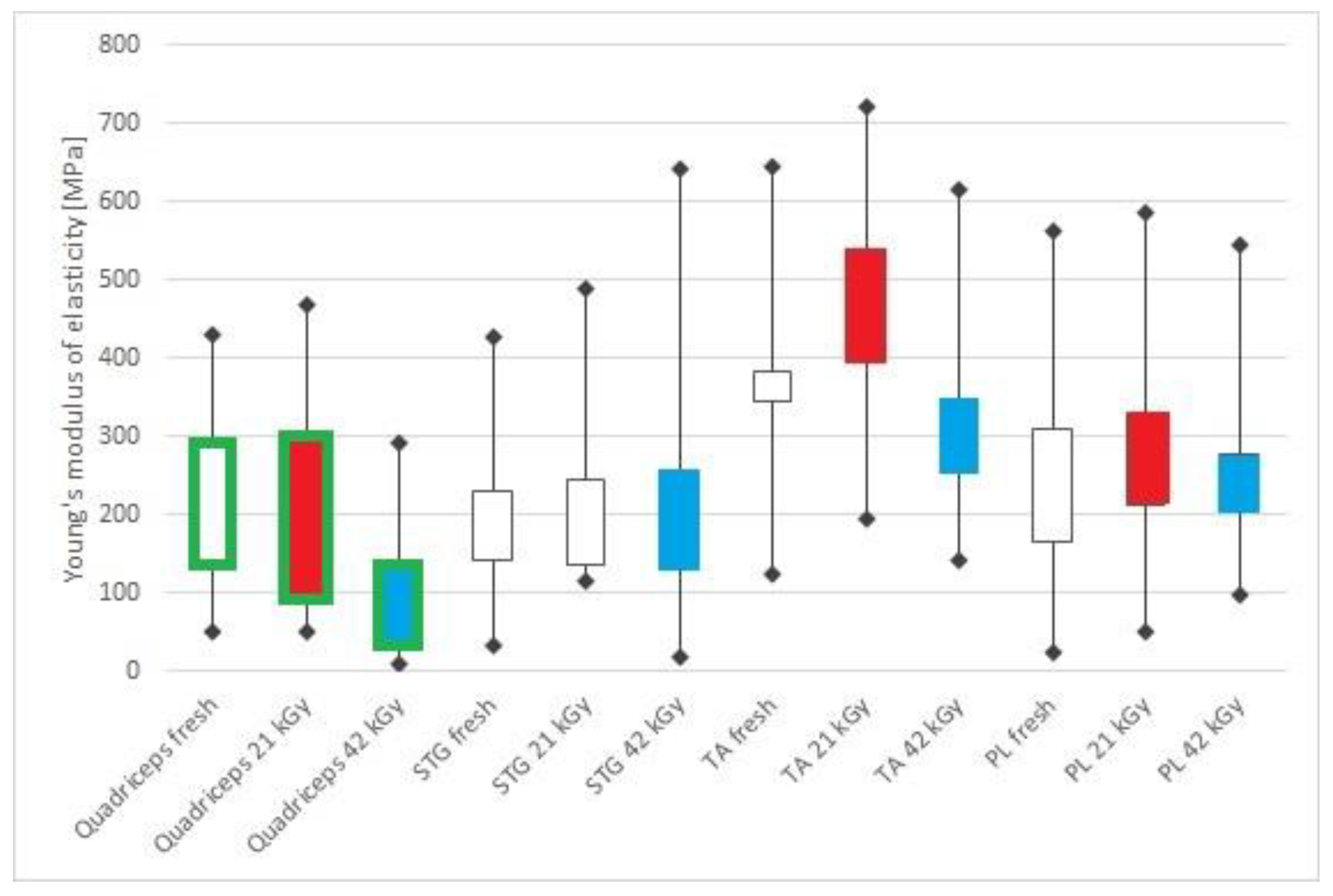
In the present study, 268 tendons from 35 human cadavers were examined. From every cadaver, the following types of tendon were harvested: 67–67 pieces quadriceps and peroneus longus (PL), 69 pieces semitendinosus + gracilis (STG) and 65 pieces tibialis anterior (TA) tendons. The types of tendon grafts were collected from human cadavers within 24 h post mortem. All tendons were visually inspected before mechanical testing, and the cortexes of the dead were also examined.
Each graft was soaked in a radioprotective solution for 4 h at 40 °C with agitation and then for 24 h at 4 °C, according to Grieb et al. [13], to help prevent the harmful effects of sterilization and storage on the tissue. The solution contained 2.7% D-mannitol, 3.8% D-trehalose, 16.7% 1,2-propanediol and 24.2% dimethyl-sulfoxide (all w/w, manufacturer: Sigma-Aldrich, Saint Louis, MO, USA). Each specimen was labeled in a separate container.
The grafts were then divided into three groups. Group A contained 63 fresh specimens (16 quadriceps, 17 semitendinosus + gracilis, 14 tibialis anterior, 16 peroneus longus, the average age of the donors was 78.4 ± 15.69 years). The other groups of tendons were frozen slowly to −80 °C. Group B (22 quadriceps, 22 semitendinosus + gracilis, 22 tibialis anterior, 22 peroneus longus, the average age of donors was 85.2 ± 15.31 years) were sterilized with a bactericidal dose (target dose: 21 kGy, dose range: 18–24 kGy). Group C contained the other 117 tendons (29 quadriceps, 30 semitendinosus + gracilis, 29 tibialis anterior, 29 peroneus longus, the average age of donors was 77.8 ± 20.61 years) sterilized with a virucidal dose (target dose: 42 kGy, dose range: 38–46 kGy). The exact numbers for the distribution between the groups were based on cadaver availability and the number of successfully harvested, intact tendons, bearing in mind having a statistically significant number of tendons in each tested group.
According to the results of a preliminary study [11], the freezing, the irradiation, and thawing were unified at all graft types. Irradiation was performed on frozen grafts, with the same method at all grafts. Before the test, the tendons were thawed at 37 °C for 20 min. Prior to the start of the test, the cross-section of the tendons and the length of the clamp were measured with micrometric calipers.
For the endurance tests, we used an Instron 8872 servohydraulic load frame (Instron Ltd., High Wycombe, UK) equipped with an Instron Dynacell load cell with a 25 kN load capacity and an Instron Fasttrack 8800 control unit and a freezer clamp structure at the accredited materials testing laboratory of the Budapest University of Technology and Economics Biomechanical Research Center. The strain characteristics of the specimens were also investigated by a Mercury Monet (Sobriety, Kurim, Czech Republic) optical digital image correlation (DIC) machine. During the preliminary tests, three minutes of freezing was used for proper fixation and for preventing the freezing of the grafts at the gauge length [12]. Based on our previous result [12], the effect of frost had a negligible effect on the measuring length because the thermal conductivity of the tissue is weak. The force–elongation curve was determined after 1000 cycles at a frequency of 2 Hz between 50 and 250 N. The method of the load-to-failure test is summarized in [12]. The cross-section of the specimen was measured by a digital caliper, and the strain (crosshead displacement) and force were measured by the tensile tester for the calculation of the presented results. The tests were authorized by the Research Ethics Committee of Uzsoki utcai and Péterffy Sándor Hospital (license number: 03/2009).
Comparing biological materials is not easy, as there is a big difference between humans, including their tissues. Therefore, for statistical comparison, a nonparametric procedure had to be used to see if the samples could be derived from the same distribution. The variables were compared in groups by using the Kruskal–Wallis test, which helps to decide if the observed tendencies are random or show a significant effect. The significant Kruskal-Wallis test shows whether one sample is stochastically more dominant than the other sample. Statistical analysis was performed with Statsoft Statistica 13.3 (Statsoft Inc., Tulsa, OK, USA). The median with the corresponding interquartile range (25 and 75% percentile) is displayed for each calculated parameter. Multiple comparisons of mean ranks for all groups were applied for post hoc analyses. As a statistical significance, a p-value of less than 0.05 was accepted.

3. Results

Firstly, we investigated the tendon’s fracture behavior. Optical images of a typical failure process are presented in Figure 1. The typical force–elongation curves of each type of tendon are presented in Figure 2.
The failure process is visible from the images presented in Figure 1. In most of the tests, longitudinal fractures formed in the tendons during the elongation, so the co-working of the adjacent fiber bundles was hindered, and the modulus of elasticity (slope of the curve) decreased after every fracture step. When comparing different tendons, the main failure process is the same; only the severity of each force-drop is different. Some differences can also be seen, which can be connected to crack propagation. While in the curve of STG presented in Figure 2, ductile failure with quasi-plastic deformation occurs, for example, in the case of the quadricep tendon’s curve, the cracks and partial fractures are more severe. In the case of the quadricep tendon, the process is also worth investigating: irreversible damage progresses even from relatively low elongation, the loss of connection between the fiber bundles is not so critical, and the force required to elongate the tendon further increases.
In Table 1, the numerical data of tensile modulus, maximum load, strain at maximum force and fracture strain are summarized and compared.
In group A (no freezing, no irradiation), we found no difference in maximum loads, strain at maximum force and tensile modulus among the tendons (Table 1, Figure 3, Figure 4 and Figure 5). Fracture strain of the quadricep tendons was significantly inferior to the TA (p = 0.0016) tendons in this respect (Table 1, Figure 6—green color).
In group B (frozen and irradiated with 21 kGy), the maximum loads of the STG tendons were significantly lower compared to the maximum loads of the quadricep (p = 0.0193) and TA tendons (p = 0.0272) (Table 1, Figure 3—red color). The strain at maximum force of the quadricep tendons was inferior compared to the STG tendons (p = 0.0022) (Table 1, Figure 4—red color). The tensile modulus of the quadricep tendons was significantly inferior to that of the TA (p = 0.0313) and PL tendons (p = 0.0201) (Table 1, Figure 5—red color). The fracture strain of the quadricep tendons was worse than that of the STG (p = 0.0001), the TA (p = 0.00001), and the PL tendons (p = 0.00001) (Table 1, Figure 6—red color).
In group C (frozen and irradiated with 42 kGy), we compared the maximum loads and found that TA tendons showed a significantly higher maximum load than the STG (p = 0.0014) and the PL tendons (p = 0.0122) (Table 1, Figure 3—blue color). The strain at maximum force of the quadricep tendons performed better than the STG (p = 0.00001), the TA (p = 0.0006), and the PL tendons (p = 0.0020). (Table 1, Figure 4—blue color). The tensile modulus of the quadricep tendons showed lower than the STG (p = 0.0046), the TA (p = 0.000001), and the PL tendons (p = 0.00001) (Table 1, Figure 5—blue color). The fracture strain of the quadricep tendons was significantly inferior to that of the STG (p = 0.0000), TA (p = 0.0034), and PL tendons (p = 0.0042) (Table 1, Figure 6—blue color).
A higher gamma irradiation dose reduced the strain at maximum force of the quadricep tendons (p = 0.0005–0.0237) more significantly than that of the other two groups (Table 1, Figure 4—green brackets). The strain at maximum force of the STG, the TA, and the PL tendons was similar in all groups (Table 1, Figure 4). In the case of the quadricep tendons (p = 0.0151), however, the higher dose of gamma irradiation significantly decreased the tensile modulus (group D) (Table 1, Figure 5—green line). Fracture strain was not affected by freezing and gamma irradiation; we found no difference in the results of any of the tendons (Table 1, Figure 6).

4. Discussion

The replacement of autografts with allografts from organ banks in ligament reconstruction is a hot topic in medicine [14,15]. The demand for ACL allografts has increased in the last decade [1], and postoperative results are promising. This is visible in the increase in allograft use from 2% (between 1986 and 1996) to 14% (between 1996 and2001), and it further increased around 2007 as its number reached 20–30% in the US [16]. Orthopedic surgeons do not have to struggle with the problems of the donor site, size and the number of the grafts [17]. In the case of multiple ligament reconstruction or ACL revision operations, the surgeons cannot remove enough tendons from the patients to replace the injured ligaments.
In our work, we have combined engineering and medical knowledge to check the feasibility of safe transplantation after freezing and irradiation treatment using different doses aimed for the disinfection of the ligaments, which are necessary for long term bank storage and transportation. Our method can provide better accuracy for strain measurement and fracture analysis of the ligaments, make the whole measurement more robust and give deeper insight into the behavior of and the differences between the tendon types, as it is based on digital image correlation.
Four types of allografts for potential ligamentous reconstruction were evaluated in this study. The allografts were subjected to either freezing and a 21 kGy dose of gamma irradiation or freezing and a 42 kGy dose of gamma irradiation. The four main mechanical parameters were evaluated and compared: tensile modulus, which represents the elasticity of the materials, maximum load, which is the limiting factor for each ligaments strength, strain at maximum force, which shows the specific strain at the maximum load and fracture strain which is the ultimate limiting strain. These parameters give a good overview of material characteristics, which is useful for the selection of the right replacement by doctors and also for simulation and modelling purposes for engineers.
In this study, the doubled tibialis anterior and peroneus longus tendons performed best in general. They were the stiffest, showing the highest tensile modulus (Table 1, Figure 5). Strain at maximum force (Table 1, Figure 4) and fracture strain (Table 1, Figure 6) were better than or equal to those of all other currently tested ACL grafts.
Previous studies [18,19] reported mixed effects of freezing. In the work of Chen et al., no difference was shown in endurance properties [18]. In contrast, Giannini S. et al. reported decreased load-bearing capacity and stiffness as a result of freezing [19]. Comparing the native and frozen tendon samples, no significant difference was found in endurance properties (Figure 3, Figure 4, Figure 5 and Figure 6). This could be the effect of the cryoprotectant solution. When comparing the reported results to the articles in the literature [1,7,8,10,20], the range of standard deviation is comparable to those reported by other research groups, which shows that considering the nature of biological samples, the results are reliable.
It was previously well-known that high doses of gamma irradiation hurt the biomechanical parameters of tendon grafts [21,22,23,24]. However, our measured values were significantly higher than those required for walking (303–355 N) [25] or aggressive early rehabilitation programs (450 N) [26].
The decrease in maximum load can be the result of gamma irradiation, which was visible in both irradiated groups. This means that the strength of the irradiated tendons decreased. This may be the reason behind the early failures of the grafts. The other evaluated TA’s parameters did not change (Table 1).
The results confirm the findings of our previous research [11]. The aggregate results of the present and prior studies [11,12] show that there is a difference in irradiation sensitivity of the examined tendons, which has to be considered before application. The tendons types which were the most affected by the treatments were quadricep grafts. When the donors are properly screened, a gamma irradiation treatment below 21 kGy does no severe damage but can destroy infectious bacteria on the samples without compromising the mechanical integrity of the tendons. We found that there is a large variation in most of the mechanical properties between the samples within the sample groups. This corresponds with findings already published in the literature [1,7,8,10,20,25], and may be attributed to the age and general health of the donors (mean age of the donors was around 80 years old) at their time of death. In order to determine the exact cause of the variation in mechanical properties between samples, further tests are needed. Finally, it can be concluded that the tibialis anterior and the peroneus longus are recommended for replacement in ACL reconstruction when gamma radiation is required.

Author Contributions

Conceptualization, G.S., R.M.K. and K.P.; methodology, G.S., R.M.K. and K.P.; investigation, D.F. and T.T.; writing—original draft preparation, D.F., G.S. and P.K.; writing—review and editing, R.M.K. and T.T.; All authors have read and agreed to the published version of the manuscript.

Funding

This work was supported by Hungarian National Research, Development and Innovation Office (NKFIH) through grant OTKA K116189 (Research project entitled “In vitro investigation of human tissues and definition of their mechanical materials models”), by the Higher Education Excellence Program of the Ministry of Human Capacities in the framework of the Biotechnology research area of the Budapest University of Technology and Economics (BME FIKP-BIO) and by the ÚNKP-18-3 New National Excellence Program of the Ministry of Human Capacities of Hungary under grant no. ÚNKP-18-3-I-BME-183.

Conflicts of Interest

The authors declare no conflict of interest.

References

  1. Jung, H.J.; Vangipuram, G.; Fisher, M.B.; Yang, G.; Hsu, S.; Bianchi, J.; Ronholdt, C.; Woo, S.L. The effects of multiple freeze-thaw cycles on the biomechanical properties of the human bone-patellar tendon-bone allograft. J. Orthop. Res. 2011, 8, 1193–1198. [Google Scholar] [CrossRef] [PubMed] [Green Version]
  2. Zaffagnini, S.; di Sarsina, T.R.; Bonanzinga, T.; Nitri, M.; Macchiarola, L.; Stefanelli, F.; Lucidi, G.; Grassi, A. Does donor age of nonirradiated achilles tendon allograft influence mid-term results of revision ACL reconstruction? Joints 2018, 6, 10–15. [Google Scholar] [CrossRef] [PubMed] [Green Version]
  3. Eagan, M.J.; McAllister, D.R. Biology of allograft incorporation. Clin. Sports Med. 2009, 28, 203–214. [Google Scholar] [CrossRef] [PubMed]
  4. Wilde, J.; Bedi, A.; Altchek, D.W. Graft selection for revision ACL reconstruction. In Revision ACL Reconstruction; Marx, R.G., Ed.; Springer: New York, NY, USA, 2014; pp. 75–86. [Google Scholar] [CrossRef]
  5. Buda, R.; Ruffilli, A.; Di Caprio, F.; Ferruzzi, A.; Faldini, C.; Cavallo, M.; Vannini, F.; Giannini, S. Allograft salvage procedure in multiple-revision anterior cruciate ligament reconstruction. Am. J. Sports Med. 2013, 41, 402–410. [Google Scholar] [CrossRef] [PubMed]
  6. Kainer, M.A.; Linden, J.V.; Whaley, D.N.; Holmes, H.T.; Jarvis, W.R.; Jernigan, D.B.; Archibald, L.K. Clostridium infections associated with musculoskeletal-tissue allografts. N. Engl. J. Med. 2004, 350, 2564–2571. [Google Scholar] [CrossRef] [PubMed]
  7. Scheffler, S.U.; Scherler, J.; Pruss, A.; von Versen, R.; Weiler, A. Biomechanical comparison of human bone-patellar tendon-bone grafts after sterilization with peracetic acid ethanol. Cell Tissue Bank. 2005, 6, 109–115. [Google Scholar] [CrossRef]
  8. Greaves, L.L.; Hecker, A.T.; Brown, C.H., Jr. The effect of donor age and low-dose gamma irradiation on the initial biomechanical properties of human tibialis tendon allografts. Am. J Sports Med. 2008, 36, 1358–1366. [Google Scholar] [CrossRef]
  9. Ng, K.W.; Wanivenhaus, F.; Chen, T.; Abrams, V.D.; Torzilli, P.A.; Warren, R.F.; Maher, S.A. Differential cross-linking and radio-protective effects of geninpin on mature bovine and patella tendons. Cell Tissue Bank. 2013, 14, 21–32. [Google Scholar] [CrossRef] [Green Version]
  10. Almqvist, K.F.; Jan, H.; Vercruysse, C.; Verbeeck, R.; Verdonk, R. The tibialis tendon as a valuable anterior cruciate ligament allograft substitute: Biomechanical properties. Knee Surg. Sports Traumatol. Arthrosc. 2007, 15, 1326–1330. [Google Scholar] [CrossRef]
  11. Hangody, G.; Szebényi, G.; Abonyi, B.; Kiss, R.; Hangody, L.; Pap, K. Does a different dose of gamma irradiation have the same effect on five different types of tendon allografts?—A biomechanical study. Int. Orthop. 2017, 41, 357–365. [Google Scholar] [CrossRef] [Green Version]
  12. Hangody, G.; Pánics, G.; Szebényi, G.; Kiss, R.; Hangody, L.; Pap, K. Pitfalls during biomechanical testing—Evaluation of different fixation methods for measuring tendons endurance properties. Acta Physiol. Int. 2016, 103, 86–93. [Google Scholar] [CrossRef] [PubMed] [Green Version]
  13. Grieb, T.A.; Forng, R.-Y.; Bogdansky, S.; Ronholdt, C.; Parks, B.; Drohan, W.N.; Burgess, W.H.; Lin, J. High-dose gamma irradiation for soft tissue allografts: High margin of safety with biomechanical integrity. J. Orthop. Res. 2006, 24, 1011–1018. [Google Scholar] [CrossRef] [PubMed]
  14. Vyas, D.; Rabuck, S.J.; Harner, C.D. Allograft anterior cruciate ligament reconstruction: Indications, techniques, and outcomes. J. Orthop. Sports Phys. Therap. 2012, 42, 196–207. [Google Scholar] [CrossRef] [PubMed] [Green Version]
  15. Zheng, X.; Li, T.; Wang, J.; Dong, J.; Gao, S. Medial collateral ligament reconstruction using bone-patellar tendon-bone allograft for chronic medial knee instability combined with multi-ligament injuries: A new technique. J. Orthop. Surg. Res. 2016, 11, 85–89. [Google Scholar] [CrossRef] [PubMed] [Green Version]
  16. Hoburg, A.; Keshlaf, S.; Schmidt, T.; Smith, M.; Gohs, U.; Perka, C.; Pruss, A.; Scheffler, S. High-dose electron beam sterilization of soft-tissue grafts maintains significantly improved biomechanical properties compared to standard gamma treatment. Cell Tissue Bank. 2015, 16, 219–226. [Google Scholar] [CrossRef]
  17. Pap, K. Dilemma of the orthopedic surgeon—What kind of graft should we use for ACL reconstruction? Orthop. Rheumatol. Open Access J. 2018, 10, 555786. [Google Scholar] [CrossRef]
  18. Chen, L.; Wu, Y.; Yu, J.; Jiao, Z.; Ao, Y.; Yu, C.; Wang, J.; Cui, G. Effect of repeated freezing-thawing on the Achilles tendon of rabbits. Knee Surg. Sports Traumatol. Arthrosc. 2011, 19, 1028–1034. [Google Scholar] [CrossRef]
  19. Giannini, S.; Buda, R.; Di Caprio, F.; Agati, P.; Bigi, A.; De Pasquale, V.; Ruggeri, A. Effects of freezing on the biomechanical and structural properties of human posterior tibial tendons. Int. Orthop. 2008, 32, 145–151. [Google Scholar] [CrossRef] [Green Version]
  20. Mabe, I.; Hunter, S. Quadriceps tendon allografts as an alternative to Achilles tendon allografts: A biomechanical comparison. Cell Tissue Bank. 2014, 15, 523–529. [Google Scholar] [CrossRef]
  21. Pearsall, A.W.; Hollis, M.J.; Russel, G.V., Jr.; Scheer, Z. A biomechanical comparison of three lower extremity tendons for ligamentous reconstruction about the knee. Arthroscopy 2003, 19, 1091–1096. [Google Scholar] [CrossRef]
  22. Di Matteo, B.; Loibl, M.; Andriolo, L.; Filardo, G.; Zellner, J.; Koch, M.; Angele, P. Biologic agents for anterior cruciate ligament healing: A systematic review. World J. Orthop. 2016, 7, 592–603. [Google Scholar] [CrossRef] [PubMed]
  23. Dong, S.; Huangfu, X.; Xie, G.; Zhang, Y.; Shen, P.; Li, X.; Qi, J.; Zhao, J. Decellularized versus fresh-frozen allografts in anterior cruciate ligament reconstruction: An in vitro study in a rabbit model. Am. J. Sports Med. 2015, 43, 1924–1934. [Google Scholar] [CrossRef] [PubMed]
  24. Conrad, B.P.; Rappé, M.; Horodyski, M.; Farmer, K.W.; Indelicato, P.A. The effect of sterilization on mechanical properties of soft tissue allografts. Cell Tissue Bank. 2013, 14, 359–366. [Google Scholar] [CrossRef] [PubMed]
  25. Nagura, T.; Matsumoto, H.; Kiriyama, Y.; Chaudhari, A.; Andriacchi, T.P. Tibiofemoral joint contact force in deep knee flexion and its consideration in knee osteoarthritis and joint replacement. J. Appl. Biomech. 2006, 22, 305–313. [Google Scholar] [CrossRef] [Green Version]
  26. Noyes, F.; Butler, D.; Grood, E.; Zernicke, R.F.; Hefzy, M.S. Biomechanical analysis of human ligament grafts used in knee-ligament repairs and reconstructions. J. Bone Jt. Surg. Am. 1985, 66, 344–352. [Google Scholar] [CrossRef]
Figure 1. Typical damage process of a tested tendon (optical images—left, force–elongation curve—right, note the formation of longitudinal cracks separating the fiber bundles).
Applsci 10 05369 g001
Figure 2. Typical force–elongation curves recorded during the tensile tests of the tendons.
Applsci 10 05369 g002
Figure 3. Minimum, maximum, median, 25% and 75% percentile values of maximum load. Red color: STG 21 kGy significantly lower than the quadriceps 21 kGy and TA 21 kGy. Blue color: TA 42 kGy significantly higher than the STG 42 kGy and PL 42 kGy.
Applsci 10 05369 g003
Figure 4. Minimum, maximum, median, 25% and 75% percentile values of strain at maximum force. Red color: quadriceps 21 kGy inferior to the STG 21 kGy. Blue color: quadriceps 42 kGy better than the STG 42 kGy, TA 42 kGy, and the PL 42 kGy. Green bracket: quadriceps 42 kGy significantly higher than the quadriceps fresh and quadriceps 21 kGy.
Applsci 10 05369 g004
Figure 5. Minimum, maximum, median, 25 and 75% percentile values of tensile moduli. Red color: Quadriceps 21 kGy significantly inferior to the TA 21 kGy and PL 21 kGy. Blue color: quadriceps 42 kGy lower than the STG 42 kGy, TA 42 kGy, and the PL 42 kGy. Green bracket: quadriceps 42 kGy significantly inferior to the quadriceps fresh and quadriceps 21 kGy.
Applsci 10 05369 g005
Figure 6. Minimum, maximum, median, 25 and 75% percentile values of fracture strain. Red color: Quadriceps 21 kGy worse than the STG 21 kGy, TA 21 kGy, and PL 21 kGy. Blue color: quadriceps 42 kGy significantly inferior to the STG 42 kGy, TA 42 kGy, and the PL 42 kGy. Green color: TA fresh significantly inferior to the quadriceps fresh.
Applsci 10 05369 g006
Table 1. Results of biomechanical tests of the tendons.
Tendon Types and Mechanical PropertiesGroup AGroup BGroup C
Median25%75%Median25%75%Median25%75%
Quadriceps
Tensile modulus (MPa)191.31138.13292.42120.2490.13299.2983.2030.65137.05
Maximum load (N)1939.751121.972727.152803.681653.263591.972564.771835.383265.92
Strain at maximum force (-)0.15400.13400.24850.18410.14670.22990.27290.19490.3472
Fracture strain (-)0.33700.27660.60900.32310.24810.48850.43920.35740.5909
Semitendinosus + gracilis (STG)
Tensile modulus (MPa)186.46142.82229.26210.28137.08244.11222.66128.59255.73
Maximum load (N)1922.961501.002374.842171.411330.372414.742357.611827.422670.83
Strain at maximum force (-)0.13000.09800.18640.13460.09850.15400.16310.13150.1895
Fracture strain (-)0.24600.15000.28500.19240.16850.22480.22200.19910.2815
Tibialis anterior (TA)
Tensile modulus (MPa)343.24308.25383.33432.55393.64538.25318.85257.63347.66
Maximum load (N)2582.222236.382784.232552.102322.613176.633063.902603.713416.14
Strain at maximum force (-)0.10410.08310.15430.14760.12850.16300.14820.12250.1618
Fracture strain (-)0.15800.10990.21310.16560.14730.20210.17810.16110.1922
Peroneus longus (PL)
Tensile modulus (MPa)250.33166.58309.85267.83212.43329.96256.55203.47277.46
Maximum load (N)2490.821657.513083.022398.302166.183092.632339.752111.682773.67
Strain at maximum force (-)0.14020.10490.17900.14890.12690.16900.14200.11080.1675
Fracture strain (-)0.19470.12660.44400.18270.14870.21860.16600.14170.1930

