Article

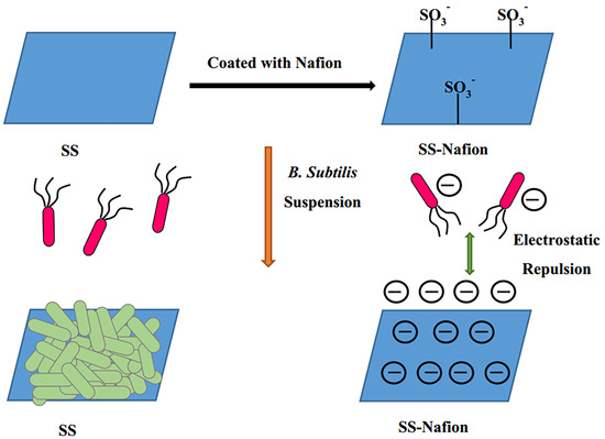
The Effectiveness of Nafion-Coated Stainless Steel Surfaces for Inhibiting Bacillus Subtilis Biofilm Formation

1
Department of Bioengineering and Biotechnology, College of Chemical Engineering, Huaqiao University, Xiamen 361021, China
2
Department of Chemical and Biochemical Engineering, College of Chemistry and Chemical Engineering, Xiamen University, Xiamen 361005, China
*
Author to whom correspondence should be addressed.
Appl. Sci. 2020, 10(14), 5001; https://doi.org/10.3390/app10145001
Submission received: 19 June 2020 / Revised: 13 July 2020 / Accepted: 16 July 2020 / Published: 21 July 2020
(This article belongs to the Section Chemical and Molecular Sciences)

Abstract

:
Stainless steel is one of most commonly used materials in the world; however, biofilms on the surfaces of stainless steel cause many serious problems. In order to find effective methods of reducing bacterial adhesion to stainless steel, and to investigate the role of electrostatic effects during the formation of biofilms, this study used a stainless steel surface that was negatively charged by being coated with Nafion which was terminated by sulfonic groups. The results showed that the roughness of stainless steel discs coated with 1% Nafion was similar to an uncoated surface; however the hydrophobicity increased, and the Nafion-coated surface reduced the adhesion of Bacillus subtilis by 75% compared with uncoated surfaces. Therefore, a facile way to acquire antibacterial stainless steel was found, and it is proved that electrostatic effects have a significant influence on the formation of biofilms.

Graphical Abstract

1. Introduction

In nature, bacteria attach to surfaces and interfaces in the form of microbial communities, and embed in a matrix of hydrated extracellular polymeric substances (EPS), referred to as a biofilm [1]. Bacteria in biofilm are up to 600 times less susceptible to disinfectants and 1000 times less vulnerable to antibiotics compared to planktonic bacteria, because the structure of the biofilm protects them from environmental insults [2,3]. Biofilms are ubiquitous and responsible for numerous problems, either in daily life or in industry [4,5]. Most bacterial infections and a variety of other diseases including some cancers are related to biofilms [6,7,8,9]. Therefore, biofilms are an urgent problem that needs to be solved. Numerous studies have been done to solve this problem; among them, developing antibacterial materials has proved one of the most effective ways [10,11,12,13,14]. As recently reported, current antibacterial materials can be classified into two categories: surfaces that resist bacterial attachment and surfaces that kill the bacteria. Bacteria-resistant surfaces normally possess certain properties that result in substrates resisting bacterial attachment, such as superhydrophobic surfaces. On the other hand, bactericidal surfaces will kill bacteria using biocides that are incorporated into the surface, e.g., antibacterial metals, peptides, and so on [15,16].
Bacterial adhesion is influenced by the chemical reactivity of the surface, which is controlled by the surface properties, including surface composition, structure and electronic properties [17,18]. Surface properties, such as hydrophobicity/hydrophilicity [19,20,21], roughness [22,23,24,25] and surface free energies [26,27] have been shown to influence bacterial adhesion. Numerous studies have investigated the correlations between bacterial adhesion and surface properties, but there are no consistent answers to date. For surface wettability, most researchers agree that bacteria tend to adhere to hydrophobic surfaces rather than hydrophilic ones [19,28,29,30]; in contrast, Boulange-Peterman et al. found that hydrophilic surfaces promote bacterial adhesion [31,32]. Many researchers have shown that surface roughness influences bacterial adhesion, in that the extent of adhesion increases in correlation with surface roughness [24,25,33,34]. However, some researchers have pointed out that surface roughness plays only a limited role in the initial adhesion; the influence of surface roughness is more important regarding biofilm detachment [35]. Flint et al. found that there was little correlation between adhesion and surface roughness [22,23,36]. Moreover, Ha et al. reported that the influence of surface roughness on biofilm formation was different with different bacteria [37]. According to the relationship between surface energy and bacterial adhesion found by Baier et al., surface free energies influence bacterial adhesion significantly; bacterial adhesion was shown to be minimized when the surface free energy was around 25 mN/m [26,38,39]. Jonsson et al. demonstrated that bacterial adhesion decreased as surface energy increased and vice versa [40,41]. However, some reports showed contrary results [42,43]. As the cell envelope of bacteria is charged, scholars propose that electrostatic interactions will also affect bacterial adhesion and the stability of biofilms [44,45,46]. Studies have proved that cations in solution (e.g., Ca2+, Mg2+) affect biofilm formation [47,48,49]. Put another way, a substratum with a charged surface will also influence the formation of biofilms. Normally, the cell envelopes of bacteria are negatively charged at neutral pH; therefore, surfaces with a negative charge will probably inhibit biofilm adhesion because of electrostatic interactions [44,50,51].
The surface topography is also an important factor in relation to bacterial adhesion. Surface topographies with microscale and nanoscale features have been shown to play an important role in a number of cellular responses; for example, in relation to cell adhesion, it is demonstrated that bacteria respond to nanostructured surfaces by modulating their attachment behavior [52,53,54]. Recent studies suggest that surface nanofeature parameters (e.g., height, diameter and spacing) correlate with the overall antibacterial qualities of surfaces [55,56,57,58]. It was established that the nano-/micro- scale characteristics of the substrate will affect the early stages of biofilm formation on metal surfaces, and influence the morphology of biofilms [58]. Regulation of surface nanotopography has been reported as a successful strategy to endow different materials with antibacterial properties [59,60,61,62]. Bruzaud et al. developed a new superhydrophobic stainless steel surface by controlling nanostructures which reduced pathogenic bacteria adhesion [59]. Hasan et al. obtained aluminum substrates with a multiscale surface architecture by wet etching. While smooth surfaces killed 7% of Escherichia coli and 3% of Staphylococcus aureus cells, aluminum with multiscale surfaces killed 97% and 28%, respectively [60]. Ludecke et al. reduced the adhesion of Escherichia coli and Staphylococcus aureuswas by altering the nanoroughness of nanostructured titanium surfaces, which were produced by physical vapor deposition [61]. Not limited to the above examples, plenty of studies have validated the hypothesis that antibacterial qualities correlate with nanostructured surfaces, which indicates that nanostructured surfaces are promising in their ability to reduce bacterial adhesion and biofilm formation [62,63,64].
Stainless steel (SS) is one of the most commonly used raw materials due to its physico-chemical stability, cleanability and economic benefits. However, biofilms on the surface of SS may lead to biofouling and biocorrosion, resulting in damage to SS structures in a wide variety of industrial sectors, including medical instruments, pipes, heat exchangers etc. [65,66,67,68]; this brings public health concerns and environmental worries, alongside the economic loss. Thus, effective strategies to control bacterial adhesion and biofilm formation on the surface of SS are urgently needed.
Nafion is a polymer of perfluoro-sulfonic acid developed from a variation of Teflon by Dupont Company, made from copolymerization groups of perfluoroalkyl sulfonyl fluoride with tetrafluoroethylene (TFE). The chemical structure of Nafion, as shown in Figure 1, has a hydrophobic tetrafluoroethylene backbone and perfluoronated vinyl-ethers terminated by sulfonic groups [69]. The hydrogen ion of the sulfonic acid groups of Nafion dissociates in solution, and the remaining polymers are negatively charged.
In our previous study [70], we found that negatively charged SS surfaces with Nafion coating could reduce the E. coli adhesion. However, this study only illustrated the efficiency of such surfaces for reducing the adhesion of E. coli, which is one kind of Gram-negative (G) bacteria. In general, bacteria are divided into two kinds, according to the color that appears after Gram staining: Gram negative (G) bacteria and Gram positive (G+) bacteria. Differences in bacterial cell envelopes contribute to color differences, and may also influence the antibacterial ability of antibacterial materials [71]. Therefore, the aim of this study is to investigate the antibacterial ability of a Nafion-coated SS surface on G+ bacteria, and the role of surface properties on bacterial adhesion. Bacillus subtilis (B. subtilis), which is a representative G+ bacteria, was selected as a test organism.
In this work, we report a facile way to modify the SS to be negatively charged via Nafion coating. The surface properties were characterized using a contact angle measuring instrument, laser scanning microscope (LSM) and scanning electron microscopy (SEM). Furthermore, the antibacterial properties of the Nafion-coated SS surfaces against G+ B. subtilis were also studied.

2. Materials and Methods

2.1. Materials

Nafion (DE520CS 5%) was obtained from DuPont company; 24-well-microtiter plates were purchased from Corning Costar Corp. (New York, NY, USA); Maximum Recovery Diluent (MRD), yeast extract powder, peptone and agar were obtained from Oxoid (Hampshire, UK). All other chemicals were purchased from Sigma-Aldrich unless specified otherwise.
B. subtilis purchased from China General Microbiological Culture Collection Center (CGMCC) was used for bacterial adhesion assays. The bacteria were kept in at −80 °C in a refrigerator (Thermo Fisher Scientific, Waltham, MA, USA) and were active overnight at 37 °C in Luria Bertani (LB) broth medium or LB-agar (1.5% agar) plates before use. The LB broth medium contained 0.5% (w/v) yeast extract powder, 1% (w/v) peptone and 1% (w/v) sodium chloride.

2.2. Toxcity Test of Nafion Solution to B. subtilis

Firstly, 5% (Wt/V) Nafion was diluted with absolute ethanol to different concentrations as follows: 0.5%, 1%, 1.5% and 2%. Secondly, 100 μL Nafion solution with different concentrations was evenly spread on the surface of the LB-agar plates. Finally, 50 μL B. subtilis suspension of 104 CFU × mL−1 was plated on the Nafion-coated LB-agar plates and cultured at 37 °C in an incubator for 24 h to count the colonies.

2.3. Preparation of Nafion-Coated Stainless Steel Discs

The SS (304) discs (8 mm diameter and 1 mm thick) were cleaned by 0.5% (wt%) NaOH, acetone and double-distilled water in sequence, with each step taking five minutes. Nafion (DE520CS 5%) was diluted to 1% (v/v) with absolute ethanol; then, the cleaned discs were coated with 1% (v/v) Nafion solution by dipping and then dried by nitrogen.

2.4. Surface Characterization

The surface energy and wetting properties of the SS surface were evaluated by contact angles, measured using a KRÜSS, DSA 30 (Hamburg, Germany). Distilled water was used as a probe during testing. The image was captured within 5 s of the water drop placement on the discs. Three different spots per substrate on three different discs were measured to provide an average contact angle.
The roughness of SS discs was characterized by a laser scanning microscope (LSM, Keyence, VK-X100/X200, Shanghai, China. The SS discs were placed on the table of the LSM microscope and linear and surface scanning was performed for a comprehensive surface analysis. Therefore, the roughness data of the surfaces were obtained by the computer directly. Each disc was measured thrice. The data presented are the means of the three values.

2.5. Biofilm Formation Assay and Quantification

The biofilm formation of B. subtilis and the quantification of bacteria adhesion were performed according to Zhong [70] with minor modifications. A total of 2 mL B. subtilis suspension of 107 CFU × mL−1 was added into each well of a 24-well-microtiter plate. Then, the SS discs with or without Nafion coating were placed in the wells and cultured at 37 °C at a rotational speed of 150 min−1 for 96 h to form biofilms. At least three microtiter plate wells were used for each sample.
Quantification of bacteria adhesion was carried out by a viable plate counting method. SS discs after biofilm formation were rinsed thoroughly with MRD in order to remove nonadherent bacteria (three times). Then, the discs were put into 5 mL centrifuge tubes with 2 mL MRD and sonicated (KUDOS, SK8200GT, Shanghai, China) for 5 min at 4 °C to transfer the embedded bacteria from SS discs to MRD. The MRD suspension with B. subtilis was continuously diluted ten times afterwards. The dilutions were plated on LB-agar plates and cultured at 37 °C for 24 h before counting the viable colonies. Three parallel plates for each sample were used to calculate the average number of adhering bacteria, and each experiment was performed at least twice.
Light microscopy (OLYPUS, BX41, Tokyo, Japan) and scanning electron microscopy (SEM) (Quanta 200 Environmental Scanning Electron Microscope, FEI, Hillsboro, USA) were used to assess the appearance of biofilms.

3. Results

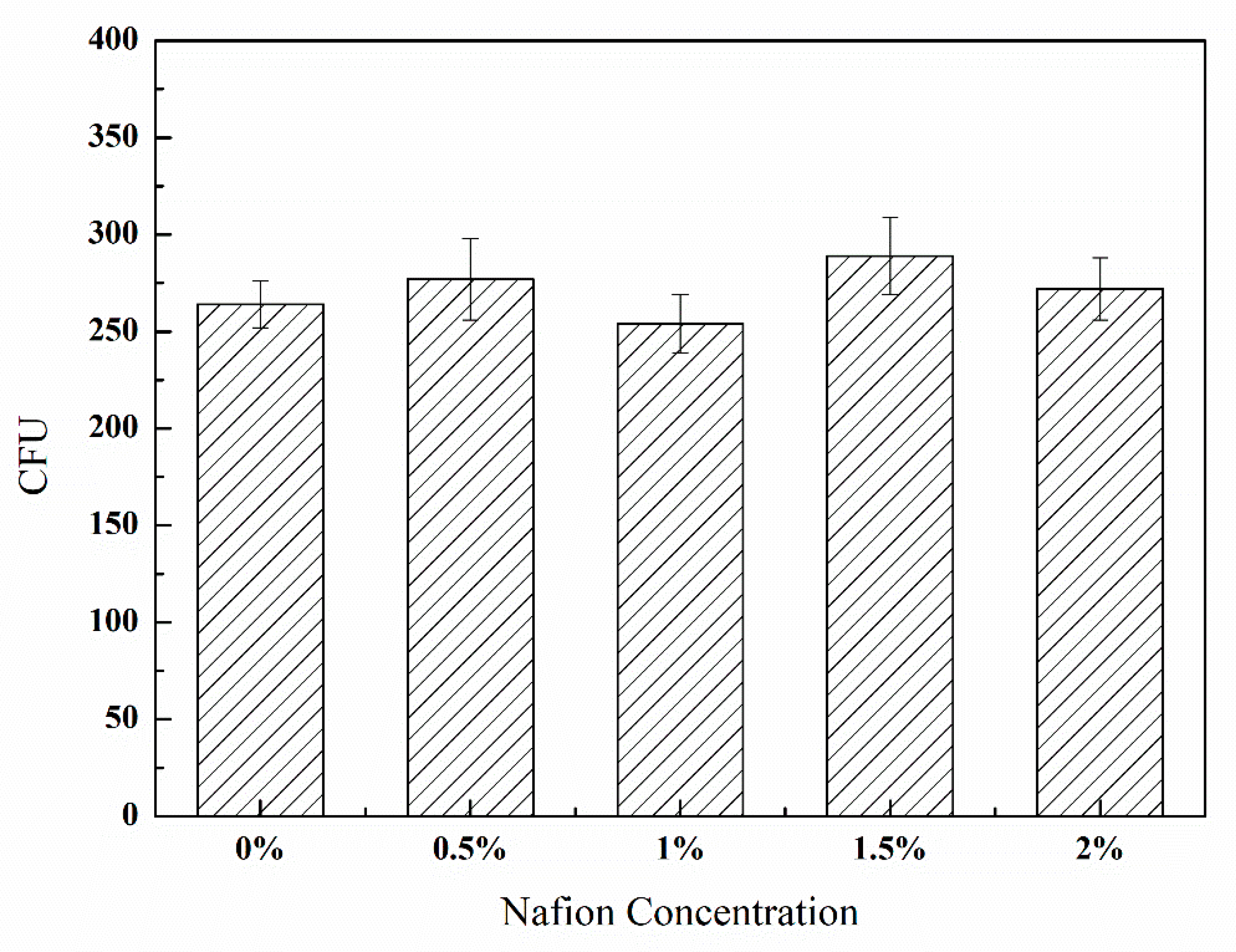
3.1. Toxicity of Nafion Solution to B. subtilis

The toxicity of the Nafion solution to B. subtilis was investigated as mentioned in Section 2.2. As shown in Figure 2, the Colony-Forming Units (CFU) of B. subtilis cells on the plates with different concentrations of Nafion were similar. The results indicate that Nafion has no obvious inhibition on the growth of Bacillus subtilis, and has good biocompatibility, which is consistent with the findings of Turner et al. [72,73].

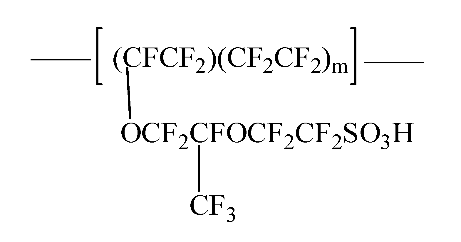
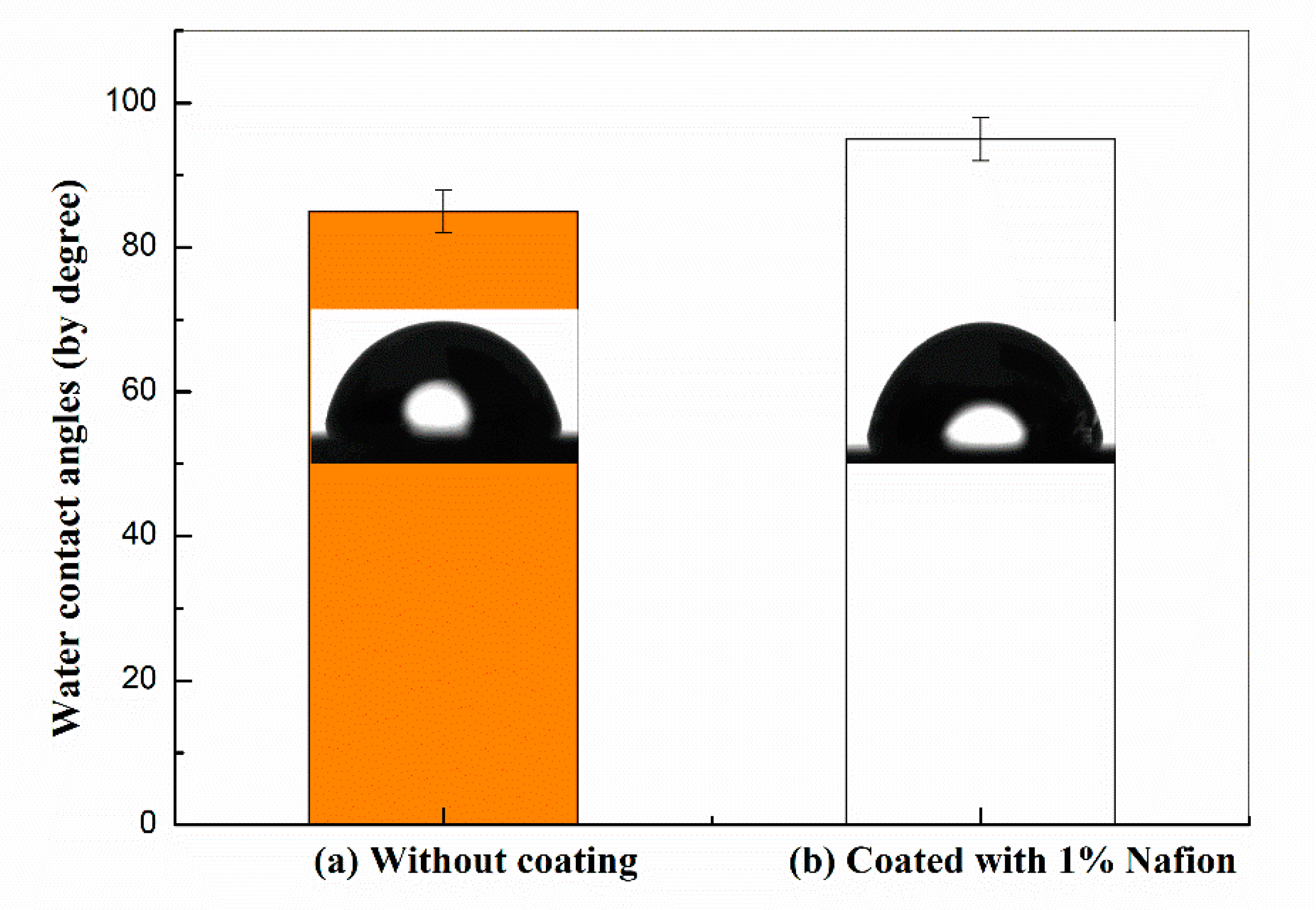
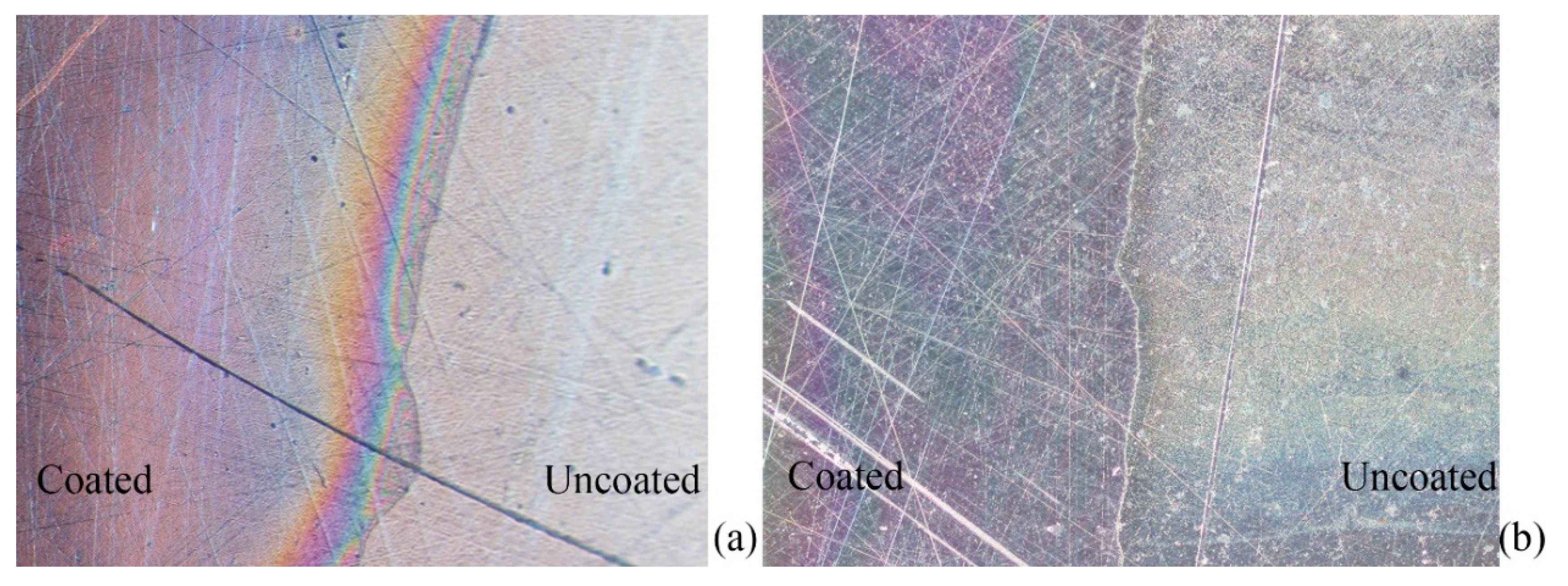
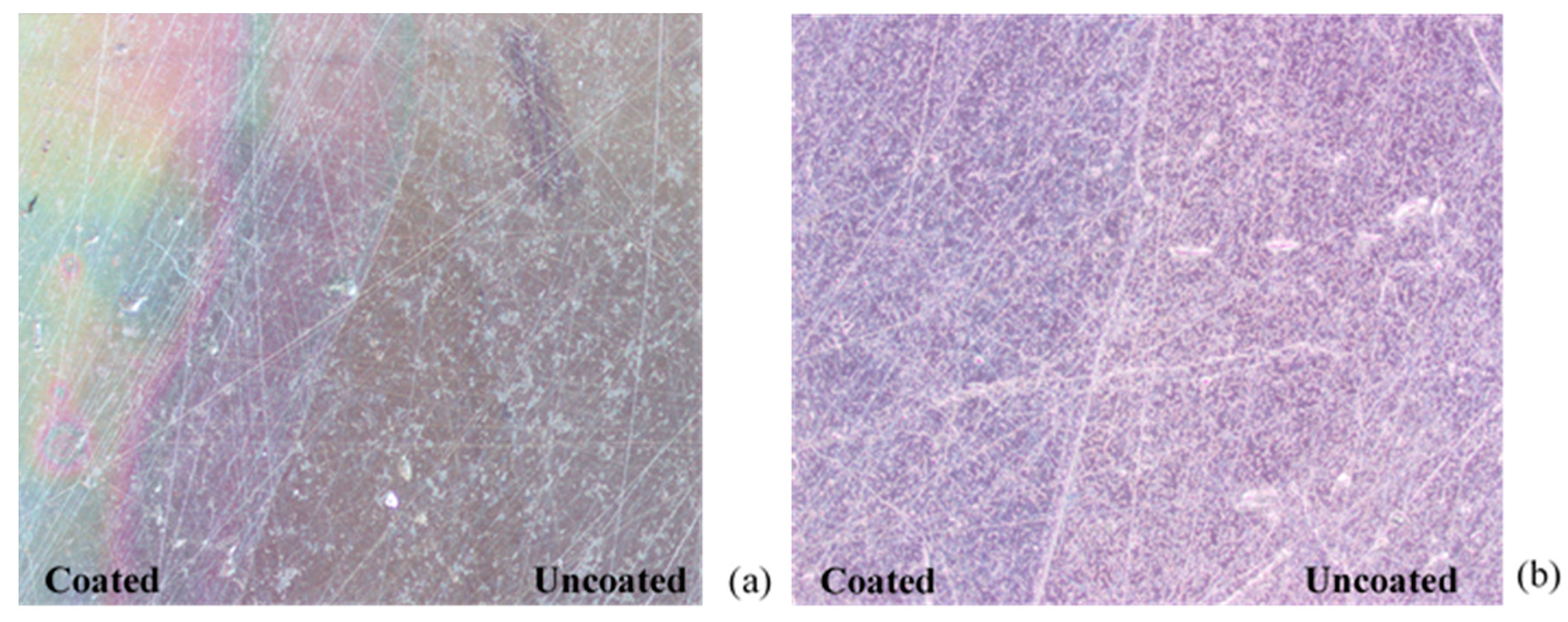
3.2. Surface Fabrication and Their Characterization of Nafion-Coated Stainless Steel

The roughness and contact angles of 1% Nafion-coated SS discs were measured and compared with uncoated SS discs. As shown in Figure 3, after coating with 1% Nafion, the water contact angles of the SS discs increased (Figure 3). The surface roughness remained unchanged compared with uncoated surfaces, as the arithmetical mean deviation of the profile (Ra) of both types of SS disc was about 0.27 μm.

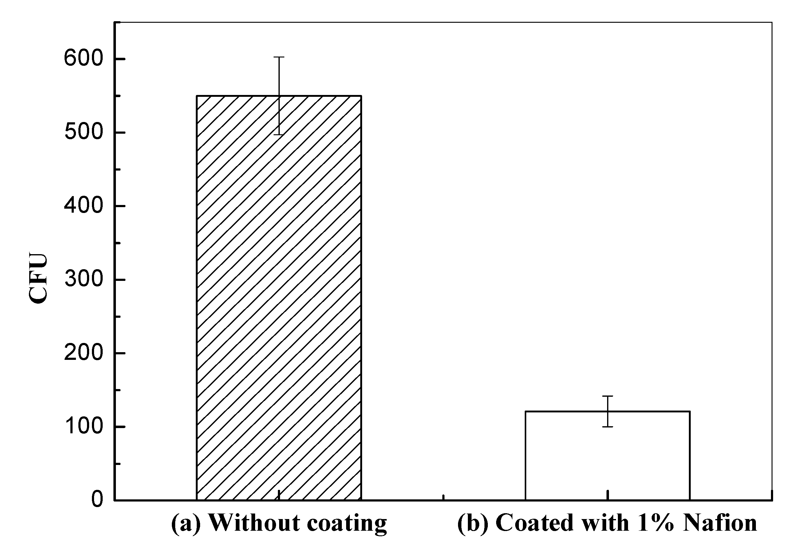
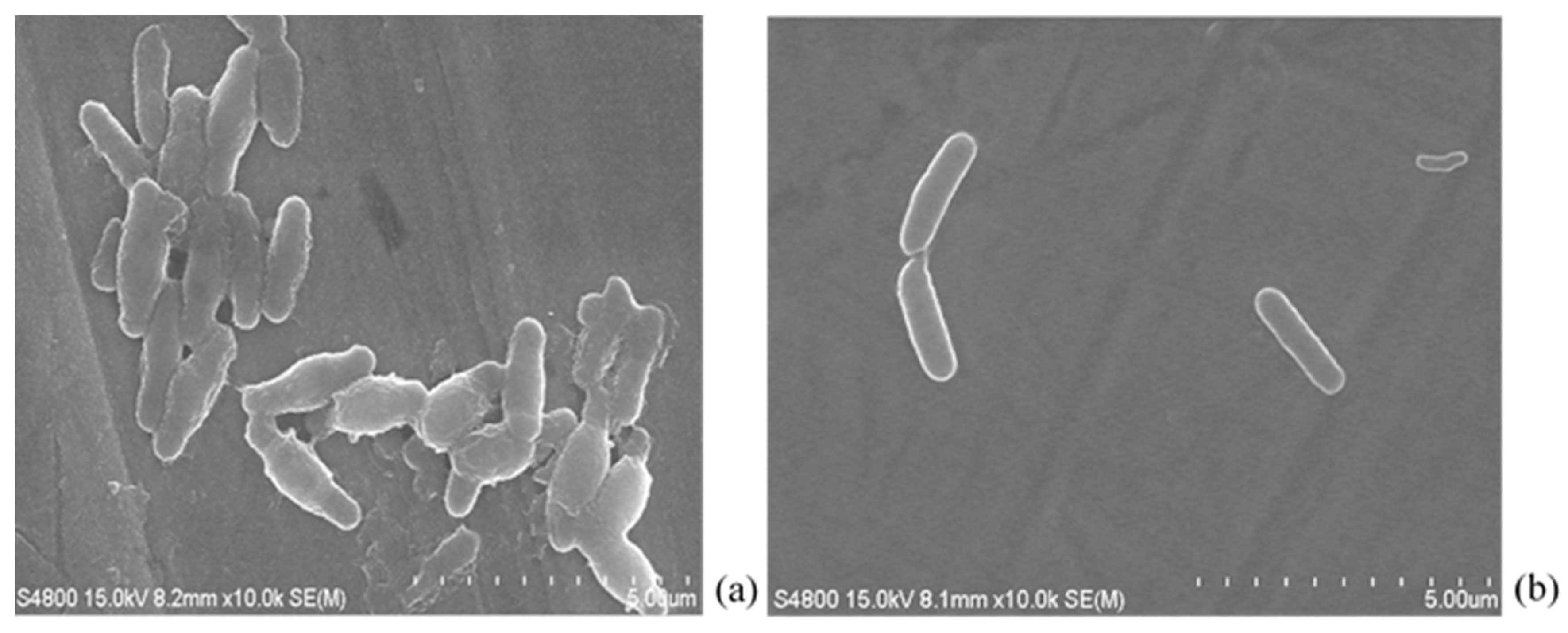
3.3. Antibacterial Adhesion of Nafion-Coated Stainless Steel Surfaces

To investigate the antibacterial properties of the Nafion-coated SS surface, a quantitative in vitro antibacterial assay was carried out by the viable plate counting method. Figure 4 and Figure 5 demonstrate the CFU of B. subtilis cells adhered to SS discs without coating or coated with 1% Nafion. As shown in the figures, compared with the SS discs without Nafion coating, an approximate 75% decrease in the adherence of bacteria was observed with 1% Nafion-coated SS discs.
Next, the SS discs were half-coated with 1% Nafion. Figure 5 shows microscopic images of a half-coated stainless steel disc before and after the biofilm forming steps. As shown, negligible differences were observed between the Nafion-coated half and the uncoated half before biofilm formation. After the biofilm forming steps, more substances adhered to the half without Nafion compared with the Nafion-coated half, indicating that the discs coated with 1% Nafion inhibit B. subtilis adhesion. The SEM images (Figure 6) also show fewer bacteria on the half coated with 1% Nafion than on the half without coating.

3.4. Durability of Antibacterial Properties

The anti-B. subtilis-biofilm ability of Nafion-coated stainless steel is verified by the above results. The antibacterial ability of a reused Nafion-coated surface was also investigated. The first part of the experiment was the same as the above biofilm formation steps; Figure 7a shows a B. subtilis-biofilm which formed on this SS disc. Subsequently, the SS disc was washed in distilled water and immersed into a newly prepared B. subtilis suspension for 96 h. The B. subtilis-biofilm that formed on this disc was then observed by a microscope (Figure 7b). As shown in Figure 7a,b, there were far more substances on the surface of the reused SS disc than on the surface when the disc was used for the first time. That is because the firmly adhered EPS and cells on the SS disc after first usage could not be cleaned completely by ultrapure water, and substances kept adhering to the reused disc. Figure 7b reveals that the number of substances attached on the Nafion-coated half of the reused disc was slightly less than on the uncoated half. The reason for this was that the Nafion-coated half disc inhibited bacteria adhesion, but when the surface was totally covered by biofilm, the antibacterial ability was suppressed.

4. Discussion

Many bacteria are known to form biofilms on material surfaces; this phenomenon is responsible for many serious problems. Thus, the elimination of bacteria from material surfaces is of great interest for both industry and daily life [1,2,3,4,5]. Therefore, the objectives of this study were to identify factors that influence, and find a facile way to reduce, bacterial adhesion on SS. Nafion, which is commonly used as a proton exchange membrane, was chosen because of the biocompability and its negatively charged polymeric blocks [69,72,73]. It has been proven that Nafion is able to hinder the formation of biofilms by E. coli, i.e., a typical G bacteria [70]; however, the antibacterial ability of Nafion against G+ bacteria has not yet been reported.
For this work, it was hypothesized that Nafion would reduce the adhesion of G+ bacteria. Herein, SS surfaces were coated with 1% Nafion, and the biofilms of the G+ bacteria B. subtilis that formed on both coated and uncoated SS surfaces were evaluated. According to the results of this study, the Nafion-coated surface reduced the adhesion of B. subtilis by 75% compared with the uncoated surface. As reported by several studies, the antibacterial efficacies of materials differ with G+ and G bacteria [56,61,62]. However, comparing the results of our previous study [70], Nafion-coated surfaces have been shown to have similar inhibitory efficacy on the formation of biofilms by E. coli and B. subtilis, which indicates that the differences in the bacterial cell envelopes of G+ and G bacteria do not significantly influence the antibacterial ability of Nafion-coated SS surfaces.
In order to identify factors that influence the adhesion of B. subtilis to Nafion-coated SS surfaces, the toxicity of Nafion against B. subtilis and the surface properties of SS were investigated. The biocompatibility of Nafion has been verified by many studies [72,73]. As shown by the results of this study, Nafion had no significant toxicity on B. subtilis. Therefore, we deduced that Nafion-coated SS surfaces inhibit biofilm formation by resisting bacterial attachment rather than killing the bacteria.
Furthermore, previous studies have indicated that antibacterial properties are related to surface characteristics [17,18]. This work focused on the effects of surface roughness, wettability and electrostatic interactions. The roughness of SS discs before and after coating with 1% Nafion showed that the average Ra for both was about 0.27 μm, which means that the surface roughness of the SS discs before and after coating was comparable. The hydrophilicity of SS surfaces was estimated by contact angles (Figure 3); after coating with 1% Nafion, the SS surfaces became more hydrophobic. As agreed by most researchers [19,28,29,30], bacteria prefer to adhere to hydrophobic surfaces, rather than hydrophilic surfaces; thus, Nafion-coated SS surfaces will attract more bacteria than uncoated surfaces because of their increased hydrophobicity. However, as mentioned, Nafion-coated SS surfaces showed dramatically reduced B. subtilis adhesion in the present study. As bacteria are almost always negatively charged in aqueous suspension, and Nafion-coated SS discs surface are negatively charged when exposed to solutions because of the sulfonic acid groups in Nafion [69,71], we propose that Nafion-coated surfaces resisted bacterial adhesion by electrostatic repulsion. These results suggest the importance of electrostatic interactions in overcoming the adhesion of B. subtilis to Nafion-coated SS.

5. Conclusions

In this work, we developed a facile way to develop SS surfaces with antibacterial adhesion properties using a Nafion coating; the ability of a Nafion-coated surface to inhibit B. subtilis adhesion was studied. The results showed that Nafion-coating on SS discs reduced the adhesion of G+ bacteria Bacillus subtilis by nearly 75% compared with uncoated SS discs, by increasing electrostatic repulsion between the SS surface and the bacterial cells. However, it should be noted that this study only investigated the adhesion behavior of Bacillus subtilis as a presentative G+ bacteria. There is great level of interest in finding approaches which prevent the adhesion of pathogenic G+ bacteria such as S. aureus or C. difficile, which pose significant challenges in the medical and the food industries. Therefore, further studies on the adhesion behavior of these bacteria on Nafion-coated SS surfaces would be useful.

Author Contributions

Conceptualization, L.Z.; Data curation, L.Z. and Y.S.; Formal analysis, L.Z.; Funding acquisition, L.Z. and S.Z.; Investigation, L.Z.; Methodology, L.Z. and Y.S.; Project administration, L.Z.; Resources, S.Z.; Supervision, L.Z.; Validation, Y.S.; Writing—original draft, L.Z.; Writing—review & editing, L.Z. and S.Z. All authors have read and agreed to the published version of the manuscript.

Funding

This research was funded by the National Natural Science Foundation of China, grant number 31901101, the funds of Huaqiao University, grant number 600005-Z17Y0074, and Quanzhou Science and Technology Bureau grant number 2018C005.

Conflicts of Interest

The authors declare no conflict of interest.

References

  1. Flemming, H.C.; Wingender, J.; Szewzyk, U.; Steinberg, P.; Rice, S.A.; Kjelleberg, S. Biofilms: An emergent form of bacterial life. Nat. Rev. Microbiol. 2016, 14, 563–575. [Google Scholar] [CrossRef]
  2. Arciola, C.R.; Campoccia, D.; Montanaro, L. Implant infections: Adhesion, biofilm formation and immune evasion. Nat. Rev. Microbiol. 2018, 18, 397–409. [Google Scholar] [CrossRef] [PubMed]
  3. Koo, H.; Allan, R.N.; Howlin, R.P.; Stoodley, P.; Stoodley, L.H. Targeting microbial biofilms: Current and prospective therapeutic strategies. Nat. Rev. Microbiol. 2017, 15, 740–755. [Google Scholar] [CrossRef] [PubMed]
  4. Kip, N.; Veen, J.A. The dual role of microbes in corrosion. ISME J. 2015, 9, 542–551. [Google Scholar] [CrossRef] [PubMed]
  5. Li, X.; Wang, H.; Hu, C.; Yang, M.; Hu, H.; Niu, J. Characteristics of biofilms and iron corrosion scales with ground and surface waters in drinking water distribution systems. Corros. Sci. 2015, 90, 331–339. [Google Scholar] [CrossRef]
  6. Torretta, S.; Drago, L.; Marchisio, P.; Ibba, T.; Pignataro, L. Role of biofilms in children with chronic adenoiditis and middle ear disease. J. Clin. Med. 2019, 8, 671. [Google Scholar] [CrossRef] [PubMed] [Green Version]
  7. Costerton, J.W.; Stewart, P.S.; Greenberg, E.P. Bacterial biofilms: A common course of persistent infections. Science 1999, 284, 1318–1322. [Google Scholar] [CrossRef] [Green Version]
  8. Dejea, C.M.; Wick, E.C.; Hechenbleikner, E.M.; White, J.R.; Welch, J.L.M.; Rossetti, B.J.; Peterson, S.N.; Snesrud, E.C.; Borisy, G.G.; Lazarev, M.; et al. Microbiota organization is a distinct feature of proximal colorectal cancers. Proc. Natl. Acad. Sci. USA 2014, 111, 18321–18326. [Google Scholar] [CrossRef] [Green Version]
  9. Henriques, P.C.; Borges, I.; Pinto, A.M.; Magalhaes, F.D.; Goncalves, I.C. Fabrication and antimicrobial performance of surfaces integrating graphene-based materials. Carbon 2018, 132, 709–732. [Google Scholar] [CrossRef]
  10. Vestby, L.K.; Grønseth, T.; Simm, R.; Nesse, L.L. Bacterial biofilm and its role in the pathogenesis of disease. Antibiotics 2020, 9, 59. [Google Scholar] [CrossRef] [Green Version]
  11. Alexandra, M.B.; Marta, F.G. Poly (ionic liquid)s as antimicrobial materials. Eur. Pol. J. 2018, 105, 135–149. [Google Scholar]
  12. Liao, C.; Li, Y.; Tjong, S.C. Anti-bacterial Activities of Aliphatic Polyester Nanocomposites with Silver Nanoparticles and/or Graphene Oxide Sheets. Nanomaterials 2019, 9, 1102. [Google Scholar] [CrossRef] [PubMed] [Green Version]
  13. Pallavicini, P.; Dacarro, G.; Diaz-Fernandez, Y.A.; Taglietti, A. Coordination chemistry of surface-grafted ligands for anti-bacterial materials. Coordin. Chem. Rev. 2014, 275, 37–53. [Google Scholar] [CrossRef]
  14. Li, L.; Molin, S.; Yang, L.; Ndoni, S. Sodium Dodecyl Sulfate (SDS)-Loaded Nanoporous Polymer as Anti-bacterial Surface Coating Material. Int. J. Mol. Sci. 2013, 14, 3050–3064. [Google Scholar] [CrossRef] [PubMed]
  15. Ahmed, W.; Zhai, Z.; Gao, C. Adaptive anti-bacterial biomaterial surfaces and their applications. Mater. Today Bio 2019, 2, 100017. [Google Scholar] [CrossRef] [PubMed]
  16. Kaur, R.; Liu, S. Anti-bacterial surface design–Contact kill. Prog. Surf. Sci. 2016, 91, 136–153. [Google Scholar] [CrossRef]
  17. Hochella, M.F.; Eggleston, C.M.; Elings, V.B.; Parks, G.A.; Brown, G.E.; Wu, C.M.; Kjoller, K. Mineralogy in two dimensions: Scanning tunneling microscopy of semiconducting minerals with implications on geochemical reactivity. Am. Mineral. 1989, 74, 1233–1246. [Google Scholar]
  18. Arnold, J.W.; Bailey, G.W. Surface finishes on stainless steel reduce bacterial attachment and early biofilm formation: Scanning electron and atomic force microscopy study. Poult. Sci. 2000, 79, 1839–1845. [Google Scholar] [CrossRef]
  19. Saeki, D.; Nagashima, Y.; Sawad, I.; Matsuyama, H. Effect of hydrophobicity of polymer materials used for water purification membranes on biofilm formation dynamics. Colloid Surf. A 2016, 506, 622–628. [Google Scholar] [CrossRef]
  20. Boudarel, H.; Mathias, J.; Blaysat, B.; Grédiac, M. Towards standardized mechanical characterization of microbial biofilms: Analysis and critical review. NPJ Biofilms Microbiomes 2018, 17, 1–15. [Google Scholar] [CrossRef]
  21. Rodríguez-Melcón, C.; Alonso-Calleja, C.; Capita, R. Architecture and viability of the biofilms formed by nine Listeria strains on various hydrophobic and hydrophilic materials. Appl. Sci. 2019, 9, 5256. [Google Scholar]
  22. Flint, S.H.; Brooks, J.D.; Bremer, P.J. Properties of the stainless steel substrate, influencing the adhesion of thermo-resistant streptococci. J. Food. Eng. 2000, 43, 235–242. [Google Scholar] [CrossRef]
  23. Ammar, Y.; Swailes, D.; Bridgens, B.; Chen, J. Influence of surface roughness on the initial formation of biofilm. Surf. Coat. Technol. 2015, 284, 410–416. [Google Scholar] [CrossRef] [Green Version]
  24. Preedy, E.; Perni, S.; Nipiĉ, D.; Bohinc, K.; Prokopovich, P. Surface roughness mediated adhesion forces between borosilicate glass and gram-positive bacteria. Langmuir 2014, 30, 9466–9476. [Google Scholar] [CrossRef] [Green Version]
  25. Zhang, Q.; Yu, Z.; Jin, S.; Liu, C.; Li, Y.; Guo, D.; Hu, M.; Ruan, R.; Liu, Y. Role of surface roughness in the algal short-term cell adhesion and long-term biofilm cultivation under dynamic flow condition. Algal Res. 2020, 46, 101787. [Google Scholar] [CrossRef]
  26. Pereni, C.I.; Zhao, Q.; Liu, Y.; Abel, E. Surface free energy effect on bacterial retention. Colloid Surf. B 2006, 48, 143–147. [Google Scholar] [CrossRef]
  27. Khan, M.M.T.; Chapman, T.; Cochran, K.; Schuler, A.J. Attachment surface energy effects on nitrification and estrogen removal rates by biofilms for improved wastewater treatment. Water Res. 2013, 7, 2190–2198. [Google Scholar] [CrossRef]
  28. Cerca, N.; Pier, G.B.; Vilanova, M.; Oliveira, R.; Azeredo, J. Quantitative analysis of adhesion and biofilm formation hydrophilic and hydrophobic surfaces of clinical isolates of Staphylococcus epidermidis. Res. Microbiol. 2005, 156, 506–514. [Google Scholar] [CrossRef] [Green Version]
  29. Song, D.; Zhang, W.; Cheng, W.; Jia, B.; Wang, P.; Sun, Z.; Ma, J.; Zhai, X.; Qi, J.; Liu, C. Micro fine particles deposition on gravity-driven ultrafiltration membrane to modify the surface properties and biofilm compositions: Water quality improvement and biofouling mitigation. Chem. Eng. J. 2020, 393, 123270. [Google Scholar] [CrossRef]
  30. Jindal, S.; Anand, S.; Huang, K.; Goddard, J.; Metzger, L.; Amamcharla, J. Evaluation of modified stainless steel surfaces targeted to reduce biofilm formation by common milk sporeformers. J. Dairy Sci. 2016, 12, 9502–9513. [Google Scholar] [CrossRef] [Green Version]
  31. Agarwall, S.V.; Ellepola, K.; Costa, M.C.F.; Fechine, G.J.M.; Morin, J.L.P.; Neto, A.H.C.; Seneviratne, C.J.; Rosa, V. Hydrophobicity of graphene as a driving force for inhibiting biofilm formation of pathogenic bacteria and fungi. Dent. Mater. 2019, 35, 403–413. [Google Scholar] [CrossRef]
  32. Boulange-Peterman, L. Processes of bioadhesion on stainless steel surfaces and cleanability: A review with special reference to the food industry. Biofouling 1996, 10, 275–300. [Google Scholar] [CrossRef]
  33. Huang, Y.; Zheng, Y.; Li, J.; Liao, Q.; Fu, Q.; Xia, A.; Fu, J.; Sun, Y. Enhancing microalgae biofilm formation and growth by fabricating microgrooves onto the substrate surface. Bioresour. Technol. 2018, 261, 36–43. [Google Scholar] [CrossRef] [Green Version]
  34. Guo, C.; Duan, D.; Sun, Y.; Han, Y.; Zhao, S. Enhancing Scenedesmus obliquus biofilm growth and CO2 fixation in a gas-permeable membrane photobioreactor integrated with additional rough surface. Alagal Res. 2019, 43, 101620. [Google Scholar] [CrossRef]
  35. Eginton, P.J.; Gibson, H.; Holah, J.; Handley, P.S.; Gilbert, P. The influence of substratum properties on the attachment of bacterial cells. Colloid Surf. B 1995, 5, 153–159. [Google Scholar] [CrossRef]
  36. Han, A.; Tsoi, J.K.H.; Matinlinna, J.P.; Chen, Z. Influence of grit-blasting and hydrofluoric acid etching treatment on surface characteristics and biofilm formation on zirconia. Coatings 2017, 7, 130. [Google Scholar] [CrossRef]
  37. Ha, K.Y.; Chung, Y.G.; Ryoo, S.J. Adherence and biofilm formation of Staphylococcus Epidermidis and Mycobacterium Tuberculosis on various spinal implants. Spine 2005, 30, 38–43. [Google Scholar] [CrossRef]
  38. Zhao, Q.; Liu, Y.; Wang, C.; Wang, S.; Müller-Steinhagen, H. Effect of surface free energy on the adhesion of biofouling and crystalline fouling. Chem. Eng. Sci. 2005, 60, 4858–4865. [Google Scholar] [CrossRef]
  39. Liu, Y.; Zhao, Q. Influence of surface energy of modified surfaces on bacterial adhesion. Biophys. Chem. 2005, 117, 39–45. [Google Scholar] [CrossRef]
  40. Jonssona, A.S.; Jonsson, B. The influence of nonionic and ionic surfactants on hydrophobic and hydrophilic ultrafiltration membranes. J. Membr. Sci. 1991, 56, 49–76. [Google Scholar] [CrossRef]
  41. Brink, L.E.S.; Elbers, S.J.G.; Robbertson, T.; Both, P. The anti-fouling action of polymers pre-adsorbed on ultrafiltration and microfiltration membranes. J. Membr. Sci. 1993, 76, 281–291. [Google Scholar] [CrossRef]
  42. Bakker, D.P.; Huijs, F.M.; de Vries, J.; Klijnstra, J.W.; Busscher, H.J.; van der Mei, H.C. Bacterial deposition to fluoridated and non-fluoridated polyurethane coatings with different elastic modulus and surface tension in a parallel plate and a stagnation point flow chamber. Colloids Surf. B 2003, 32, 179–190. [Google Scholar] [CrossRef]
  43. Boulange-Peterman, L.; Baroux, B.; Bellon-Fontaine, M.N. The influence of metallic surface wettability on bacterial adhesion. J. Adhes. Sci. Technol. 1993, 7, 221–230. [Google Scholar] [CrossRef] [Green Version]
  44. Terada, A.; Okuyama, K.; Nishikawa, M.; Tsuneda, S.; Hosomi, M. The effect of surface charge property on Escherichia coli initial adhesion and subsequent biofilm formation. Biotechnol. Bioeng. 2012, 109, 1745–1754. [Google Scholar] [CrossRef] [PubMed]
  45. Schwegmann, H.; Feitz, A.J.; Frimmel, F.H. Influence of the zeta potential on the sorption and toxicity of iron oxide nanoparticles on S. cerevisiae and E. coli. J. Colloid Interface Sci. 2010, 347, 43–48. [Google Scholar] [CrossRef]
  46. Zheng, Y.; Huang, Y.; Xia, A.; Qian, F.; Wei, C. A rapid inoculation method for microalgae biofilm cultivation based on microalgae-microalgae co-flocculation and zeta-potential adjustment. Bioresource Technol. 2019, 278, 272–278. [Google Scholar] [CrossRef]
  47. Kannan, A.; Karumanchi, S.L.; Ramalingam, S.; Gautam, P. Quantitative study on the effect of calcium and magnesium palmitate on the formation of Pseudomonas aeruginosa biofilm. J. Microbiol. Immunol. 2016, 49, 988–991. [Google Scholar] [CrossRef] [Green Version]
  48. Song, B.; Leff, L.G. Influence of magnesium ions on biofilm formation by Pseudomonas fluorescens. Microbiol. Res. 2006, 161, 355–361. [Google Scholar] [CrossRef]
  49. Kurniawan, A.; Tsuchiya, Y.; Eda, S.; Morisaki, H. Characterization of the internal ion environment of biofilms based on charge density and shape of ion. Colloids Surf. B 2015, 136, 22–26. [Google Scholar] [CrossRef]
  50. Kreve, S.; Reis, A.C. Influence of the electrostatic condition of the titanium surface on bacterial adhesion: A systematic review. J. Prosthet. Dent. 2020, (in press). [Google Scholar] [CrossRef]
  51. Graham, M.V.; Cady, N.C. Nano and microscale topographies for the prevention of bacterial surface fouling. Coatings 2014, 4, 37–59. [Google Scholar] [CrossRef] [Green Version]
  52. Skoog, S.A.; Kumar, G.; Narayan, R.J.; Goering, P.L. Biological responses to immobilized microscale and nanoscale surface topographies. Pharmacol. Ther. 2018, 182, 33–55. [Google Scholar] [CrossRef] [PubMed]
  53. Hochbaum, A.I.; Aizenberg, J. Bacteria pattern spontaneously on periodic nanostructure arrays. Nano Lett. 2010, 10, 3717–3721. [Google Scholar] [CrossRef]
  54. Crawford, R.J.; Webb, H.K.; Truong, V.K.; Hasan, J.; Ivanova, E.P. Surface topographical factors influencing bacterial attachment. Adv. Colloid Interface Sci. 2012, 179, 142–149. [Google Scholar] [CrossRef]
  55. Nguyen, D.H.K.; Pham, V.T.H.; Truong, V.K.; Sbarski, I.; Wang, J.; Balcytis, A.; Juodkazis, S.; Mainwaring, D.E.; Crawford, R.J.; Ivanova, E.P. Role of topological scale in the differential fouling of Pseudomonas aeruginosa and Staphylococcus aureus bacterial cells on wrinkled gold-coated polystyrene surfaces. Nanoscale 2018, 10, 5089–5096. [Google Scholar] [CrossRef]
  56. Tripathy, A.; Sen, P.; Su, B.; Briscoe, W.H. Natural and bioinspired nanostructured bactericidal surfaces. Adv. Colloid Interface Sci. 2017, 248, 85–104. [Google Scholar] [CrossRef]
  57. Elbourne, A.; Coyle, V.E.; Truong, V.K.; Sabri, Y.; Kandjani, A.E.; Bhargava, S.K.; Ivanova, E.P.; Crawford, R.J. Multi-directional electrodeposited gold nanospikes for anti-bacterial surface applications. Nanoscale Adv. 2018. [Google Scholar] [CrossRef] [Green Version]
  58. Díaz, C.; Schilardi, P.L.; Salvarezza, R.C.; Fernández Lorenzo de Mele, M. Nano/microscale order affects the early stages of biofilm formation on metal surfaces. Langmuir 2007, 23, 11206–11210. [Google Scholar] [CrossRef]
  59. Bruzaud, J.; Tarrade, J.; Celia, E.; Darmanin, T.; De Givenchy, E.T.; Guittard, F.; Herry, J.M.; Guilbaud, M.; Bellon-Fontaine, M.N. The design of superhydrophobic stainless steel surfaces by controlling nanostructures: A key parameter to reduce the implantation of pathogenic bacteria. Mater. Sci. Eng. C Mater. Biol. Appl. 2017, 73, 40–47. [Google Scholar] [CrossRef]
  60. Hasan, J.; Jain, S.; Padmarajan, R.; Purighalla, S.; Sambandamurthy, V.K.; Chatterjee, K. Multi-scale surface topography to minimize adherence and viability of nosocomial drug-resistant bacteria. Mater. Des. 2018, 140, 332–344. [Google Scholar] [CrossRef]
  61. Ludecke, C.; Roth, M.; Yu, W.; Horn, U.; Bossert, J.; Jandt, K.D. Nanorough titanium surfaces reduce adhesion of Escherichia coli and Staphylococcus aureus via nano adhesion points. Colloids Surf. B 2016, 145, 617–625. [Google Scholar] [CrossRef]
  62. Luan, Y.; Liu, S.; Pihl, M.; van der Mei, H.C.; Liu, J.; Hizal, F.; Choi, C.; Chen, H.; Ren, Y.; Busscher, H.J. Bacterial interactions with nanostructured surfaces. Curr. Opin. Colloid Interface Sci. 2018, 38, 170–189. [Google Scholar] [CrossRef]
  63. Modaresifar, K.; Azizian, S.; Ganjian, M.; Fratila-Apachitei, L.E.; Zadpoor, A.A. Bactericidal effects of nanopatterns: A systematic review. Acta Biomater. 2019, 83, 29–36. [Google Scholar] [CrossRef] [Green Version]
  64. Lin, N.; Berton, P.; Moraes, C.; Rogers, R.D.; Tufenkji, N. Nanodarts, nanoblades, and nanospikes: Mechano-bactericidal nanostructures and where to find them. Adv. Colloid Interface Sci. 2018, 252, 55–68. [Google Scholar] [CrossRef] [Green Version]
  65. Bohinc, K.; Drazic, G.; Abram, A.; Jevšnik, M.; Jeršek, B.; Nipič, D.; Kurinčič, M.; Raspor, P. Metal surface characteristics dictate bacterial adhesion capacity. Int. J. Adhes. Adhes. 2016, 68, 39–46. [Google Scholar] [CrossRef]
  66. Bekmurzayeva, A.; Duncanson, W.J.; Azevedo, H.S.; Kanayeva, D. Surface modification of stainless steel for biomedical applications: Revisiting a century-old material. Mater. Sci. Eng. 2018, 93, 1073–1089. [Google Scholar] [CrossRef]
  67. Ogawa, A.; Takakura, K.; Hirai, N.; Kanematsu, H.; Kuroda, D.; Kougo, T.; Sano, K.; Terada, S. Biofilm formation plays a crucial rule in the initial step of carbon steel corrosion in air and water environments. Materials 2020, 13, 923. [Google Scholar] [CrossRef] [Green Version]
  68. Zhang, J.; Li, W.; Chen, J.; Qi, W.; Wang, F.; Zhou, Y. Impact of biofilm formation and detachment on the transmission of bacterial antibiotic resistance in drinking water distribution systems. Chemosphere 2018, 203, 368–380. [Google Scholar] [CrossRef]
  69. Hoyer, B.; Florence, T.M.; Batley, G.E. Application of polymer-coated glassy carbon electrodes in anodic stripping voltammetry. Anal. Chem. 1987, 59, 1608–1614. [Google Scholar] [CrossRef]
  70. Zhong, L.J.; Pang, L.Q.; Che, L.M.; Wu, X.; Chen, X. Nafion coated stainless steel for anti-biofilm application. Colloids Surf. B 2013, 111, 252–256. [Google Scholar] [CrossRef]
  71. Madigan, M.T.; Martinko, J.M.; Bender, K.S.; Buckley, D.H.; Stahl, D.A. Chapter 2, Microbial Cell Structure and Function. In Brock Biology of Microorganisms, 14th ed.; Pearson Education: New York, NY, USA, 2014; pp. 25–67. [Google Scholar]
  72. Turner, R.F.B.; Harrison, D.J.; Rojotte, R.V. Preliminary in vivo biocompatibility studies on perfluorosulphonic acid polymer membranes for biosensor applications. Biomaterials 1991, 12, 361–368. [Google Scholar] [CrossRef]
  73. Dai, Z.; Mohwald, H. Highly stable and biocompatible Nafion-Based capsules with controlled permeability for low-molecular-weight species. Chem. Eur. J. 2002, 8, 4751–4755. [Google Scholar] [CrossRef]
Figure 1. Chemical structure of Nafion.
Applsci 10 05001 g001
Figure 2. CFU of B. subtilis cells on the LB-agar plates with different concentrations of Nafion.
Applsci 10 05001 g002
Figure 3. Water contact angles (by degree) of stainless steel discs before and after coating with 1% Nafion.
Applsci 10 05001 g003
Figure 4. Number of viable adherent cells of B. subtilis (CFU) on stainless steel discs coated with 1% Nafion (b) and without coating (a).
Applsci 10 05001 g004
Figure 5. Microscopic image of a SS disc, half of which had been coated with Nafion. (a) Surfaces of half-coated SS disc without biofilm. (b) Surfaces of half-coated SS disc with B. subtilis biofilm.
Applsci 10 05001 g005
Figure 6. SEM images of (a) B. subtilis biofilms on half of SS disc without coating, and (b) B. subtilis biofilms on the half of SS disc with 1% Nafion coating.
Applsci 10 05001 g006
Figure 7. Antibacterial ability of a reused Nafion-coated SS surface: (a) biofilm on a half Nafion-coated SS disc for the first antibacterial test, and (b) biofilm on the reused half of the Nafion-coated SS disc.
Applsci 10 05001 g007

