Case Report

A Novel Approach Using Customized Miniplates as Skeletal Anchorage Devices in Growing Class III Patients: A Case Report

1
Division of Pediatric dentistry, Hallym University Sacred Heart Hospital, Anyang 14066, Korea
2
Graduate School of Clinical Dentistry, Hallym University, Chuncheon 24252, Korea
3
Division of Oral and Maxillofacial Surgery, Hallym University Sacred Heart Hospital, Anyang 14066, Korea
4
Institute of Clinical Dentistry, Hallym University, Chuncheon 24252, Korea
5
Division of Orthodontics, Hallym University Sacred Heart Hospital, Anyang 14066, Korea
*
Author to whom correspondence should be addressed.
Both authors contributed equally to this work.
Appl. Sci. 2020, 10(12), 4067; https://doi.org/10.3390/app10124067
Submission received: 5 May 2020 / Revised: 10 June 2020 / Accepted: 11 June 2020 / Published: 12 June 2020
(This article belongs to the Special Issue Application of CAD/CAM and 3D Printing Technologies in Dentistry)

Abstract

:
Facemasks using tooth-borne anchorages have been used primarily for the treatment of Class III malocclusion with maxillary undergrowth. However, when using a tooth as an anchorage, if the stability of the tooth used as an anchor is weak, the anchoring function may fail as the tooth tilts. Meanwhile, the use of skeletal anchorages such as implants, mini-implants, and mini-plates has been claimed to minimize the side effects of using dental anchorage. This case report describes the treatment of a six-year-old male patient with Class III malocclusion, presenting maxillary undergrowth and mandibular prognathism. Due to the mobility of the anchoring primary teeth, a device using dental anchorage was replaced with that using customized skeletal anchorage for the treatment. Customized guides and miniplates for the surgery were fabricated in advance through a computer-assisted system, in order to avoid possible damage to the adjacent tooth buds. The customized plates were accurately and passively placed on the intended part, showing the desired outcome.

1. Introduction

Class III malocclusions with deficient or retrusive maxilla can be treated orthopedically in primary and mixed dentition. In general, it can be treated using a face mask with the use of tooth-borne anchorage devices, such as transpalatal arch or rapid palatal expander (RPE). Traditionally, maxillary teeth have been used as an anchorage to apply forces to the maxilla [1]. However, there are some problems with protracting the maxilla using teeth anchorage. These include the loss of teeth anchorage, which is of concern, especially in situations where preservation of arch length is necessary. Loss of anchorage occurs due to the mesial movement of the posterior teeth resulting from the tipping, and the increased vertical dimension can also be seen in this situation [2,3,4].
Skeletal anchorage using osseointegrated titanium implants, mini-implants and miniplates has been suggested to overcome the demerits [2,5,6,7]. In case reports on animals and humans, it has been demonstrated that osseointegrated implants and miniplates are biologically compatible and provide an excellent anchorage for orthodontic force [8]. In particular, Chen, Y. J. et al. claimed that the miniplate has greater stability compared with mini- or microscrews [9].
According to the previous study, the number and position of the miniplates affect the stability and stress values at the bone screw holes [10]. A customized miniplate has a design that may favor lower stress values, as it is manufactured in the shape and size precisely determined in the virtual surgery. Moreover, it can be positioned in an optimal position for each patient’s anatomical structure and the direction of the orthopedic force.
The development of virtual surgical planning with computer-aided design (CAD) and computer-aided manufacturing (CAM) technology has significantly contributed to dentistry. The adoption of this modern technology has been reported to be predictable and safe in diverse fields of dentistry, including surgical guides for various operations and dental molds for radiotherapy [11,12]. In particular, the effectiveness and applicability of surgical guides and customized miniplates based on virtual surgery have been demonstrated in many studies.
In clinics, the use of skeletal anchorage for the orthopedic treatment of growing children with mixed dentition is rarely considered a primary choice for several reasons. These reasons include sufficient anchorage force from the tooth, the reluctance to undergo surgery of the patient or his/her parent, and many delicate anatomical structures the clinicians should avoid, such as tooth buds.
The purpose of this report is to offer and propose a novel method of treatment for growing class III patients by introducing the use of three-dimensional virtual surgery, prefabricated surgical guides, and miniplates. To the best of our knowledge, it is the first report on a temporary skeletal anchorage device using virtual surgery with computer-aided manufacturing in a growing patient.

2. Case Report

2.1. Diagnosis and Etiology

A six-year-old male patient visited the university hospital with a chief complaint of protruded mandible. Written informed consent was obtained, and an exemption was granted from the Hallym University Sacred Heart Hospital institutional review board committee since it is a case report. Clinically, he had a concave facial profile and a protrusive mandible. Intraoral and dental cast examination revealed that he had an anterior crossbite and open bite. Congenital absence of all the second premolars and delayed development of upper first and second molars were noted on the panoramic radiograph. A thorough investigation using cephalometric analysis indicated a growth pattern of skeletal class III presenting maxillary undergrowth and mandibular prognathism. The treatment started with a conventional rapid palatal expander (RPE) combined with a facemask. However, after five months of treatment, the upper primary canines, the anchoring teeth, started to show severe mobility due to physiologic root resorption. Additionally, the first molars were not erupted yet due to their delayed development. The first primary molars also showed mobility due to the physiologic root resorption, although orthopedic treatment with a facemask was still needed (Figure 1).
Therefore, the tooth-borne anchorage device could no longer be used, and a surgical approach for skeletal anchorage devices was instead recommended in this circumstance.

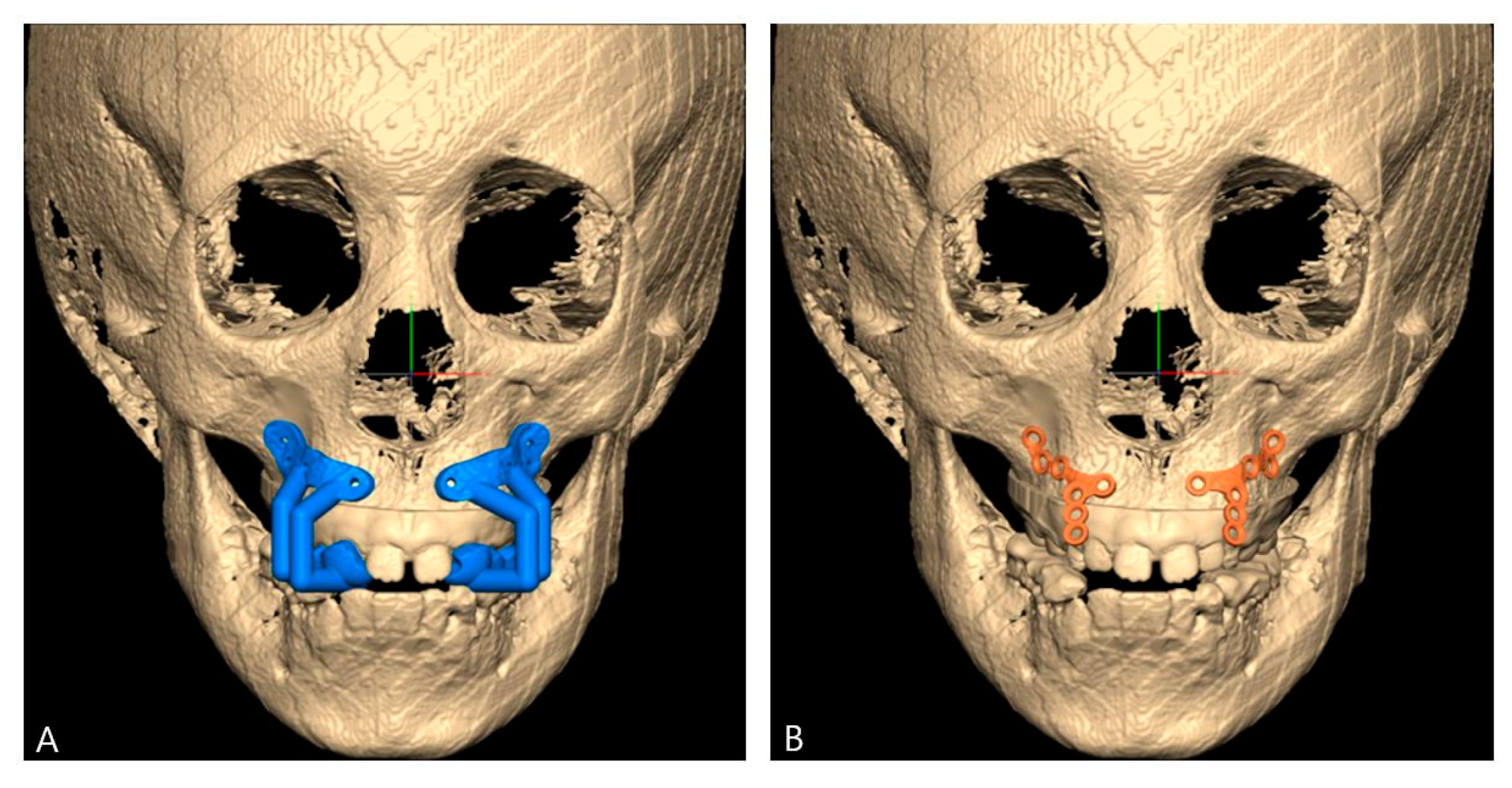
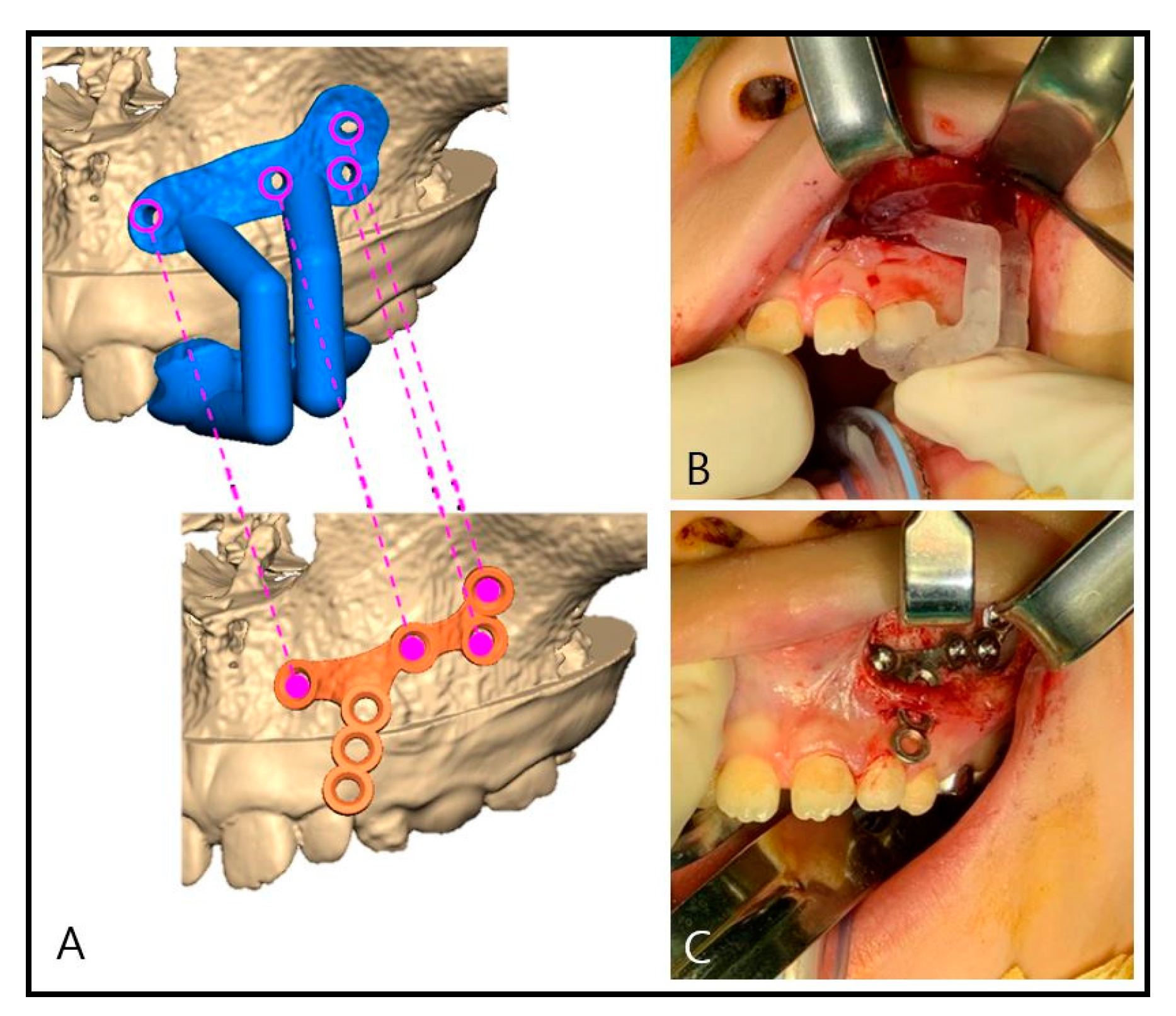
2.2. Computer-Assisted Surgical Planning

In order to determine the safe location of the miniplates, cone-beam computed tomography (CBCT; Alphard 3030, Asahi, Inc., Kyoto, Japan) was taken. Diagnostic models and bite registration were taken two weeks before the surgery and were scanned through a desktop model scanner (Freedom UHD®, Dof Inc., Seoul, Korea). All the saved files were merged into a three-dimensional image for the virtual surgery via a specific program (FaceGide®, MegaGen Co., Daegu, Korea) [11].
Afterward, the authors discussed the desired location of the miniplates considering both the orthodontic and the surgical aspects through a virtual simulation, which could be visualized on the monitor. After the final location of the miniplate reached an agreement, the plan was saved for manufacturing the patient-customized guides and patient-customized miniplates (Figure 2) [11].
After exporting the images as Standard Tessellation Language (STL) files, the actual materials were produced with a rapid prototyping machine (S30 3D printer, Rapid Shape GmbH, Heimsheim, Germany) and a computer numerical control machine (ARDEN, TPS Korea Ltd., Gwangju, Korea). The customized miniplates were made of titanium (Medical Grade 4, S-Tech corp. Tainan, Taiwan). The fabricated guides and miniplates were then sent to the operation room in advance for the sterilization.

2.3. Surgical Procedure
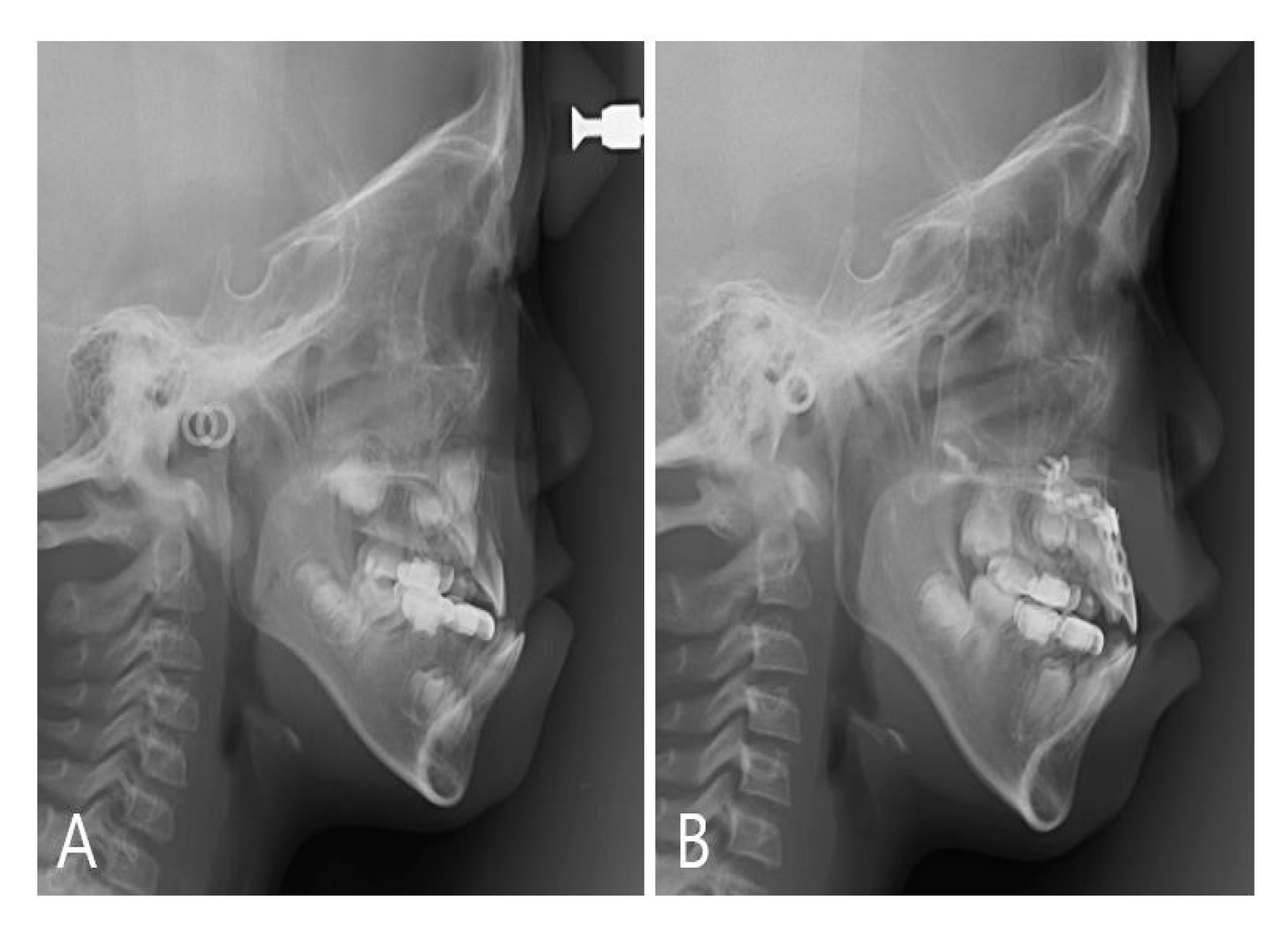
The surgery was performed under general anesthesia due to the patient’s fear. Does of 0.15 g of thiopental sodium, 0.03 mg of fentanyl and 15 mg of rocuronium were administered by intravenous injection. Upon intubation, anesthesia was maintained using sevoflurane and oxygen by inhalation. After the incision was made, the mucoperiosteal flap was elevated to expose the cortical bone of the surgical site. Then, the guide was adapted to the upper lateral incisor, primary canine, and the first primary molar (Figure 3B). The location of the planned drilling hole was marked with a pencil on the cortical bone. After the guide was removed, the customized miniplates were applied and fixed with 6-mm self-tapping screws at the planned position (Figure 3A). Four screws were used on each side to resist a protraction force. The end of the miniplate was exposed between the primary canine and lateral incisor area, resting over the attached gingiva to prevent gingival irritation (Figure 3C).

3. Results

Visible surgical simulation combined with customized guides and plates enabled more accurate communication between orthodontist and surgeon and, consequently, a satisfactory surgical result. The customized plates were seated on the planned position symmetrically while the tooth follicles and anterior teeth were intact (Figure 4).
The facemask was applied at a 30-degree angle to the occlusal plane, using a force of 16 oz per side, and was worn daily for about 10 h on average (Figure 5).
It has been a year and three months since the surgery, and the patient is still under orthodontic treatment using a facemask without any complications. No mobility, inflammation, or other problems associated with plates have been detected and the anchor still showed a high stability.
The cephalometric radiograph at each time point was taken in order to compare the results of the facemask treatment over two years and six months, showing differential forward growth of the maxilla (Figure 6).
Measurement on cephalometric radiograph revealed that overjet had been changed from −0.3 mm (Figure 6B and Figure 7A) to 1.6 mm (Figure 6D and Figure 7C). The pictures showed that it is presently within a normal range (Figure 7).

4. Discussion

In this case, tooth-borne anchorages, combined with an RPE appliance, were used for nine months in the early phase of the treatment. However, due to the mobility of the anchoring teeth, these were replaced with skeletal anchorage. In the studies of NK Lee et al., and Şar, Ç. et al., the facemask with miniplate allowed greater forward movement of the maxilla, less posterior repositioning and opening rotation of the mandible, and less undesired dentoalveolar effects than the facemask with a tooth-borne anchorage appliance [13,14]. Therefore, skeletal anchorage can be beneficial in growing children of mixed dentition requiring greater anchorage force.
The straight, conventional miniplates need to be bent according to the bone surface before fixation, resulting in longer surgical time, increased fatigue, and an incomplete fit [15]. Since the customized miniplate had been prefabricated as designed, no additional effort or time was required for bending the miniplate. This process also allowed the miniplate to be made more firm and rigid, reducing the possibility of fracture or deformation of the plates during protraction. In the study of Y Liu et al., the customized fixation system with a topologically optimized structure was claimed to be superior in the biomechanical aspect to the conventional system, presenting less stress, strain, and displacement inside the plate [16].
A use of temporarily placed miniscrews was first mentioned for orthodontic anchorage in 1997 [17] and has now become an established orthodontic anchorage aid used commonly in a variety of ways. For optimum patient safety and success of the miniscrew fixation, local anatomical structure, soft tissue around the plate, and patient home care are the essential factors to consider. According to several works of literature, orthodontic screws show a success rate of 70–90% [18]. Complications may include root contact, inflammation, insufficient stability, and perforation of the maxillary sinuses. S Kuroda et al. identified that a significant risk factor for their failure is root contact [19]. In growing patients of mixed dentition, it is also essential to avoid further damage to the developing buds of the permanent teeth. When inserting miniscrews near delicate anatomical structures, a surgical guide can be used to accurately locate the place and the direction to avoid potential damage to the adjacent structures. Furthermore, J Sugawara et al. demonstrated that infection infrequently occurs in about 10% of patients, and that plate loosening has occurred in only 1% of all cases [20]. It is thought that plate loosening or infection may occur because the ready-made plate does not adhere to the bone surface accurately. With the use of a customized plate, it is possible to overcome the problems of a ready-made plate because it can be precisely adhered to the curved surface of an individual patient.
For the success of fixation, a thorough understanding of bone density and shape is essential. When it comes to the location, the miniplate for maxilla protraction is usually placed in the infrazygomatic crest. Previous studies confirmed that the infrazygomatic crest generally shows a good quality of bone, which provides sufficient anchorage to keep the screw in load [21,22,23]. On the other hand, Şar, Ç. et al. placed miniplates laterally on the piriform aperture of the maxilla. The authors demonstrated the choice of a placement site was in order to apply protraction forces as close as possible to the center of resistance of the nasomaxillary complex [14]. Each method can generate different vector forces depending on the anatomical location of the skeletal anchorage, resulting in different effects on the craniofacial complex.
Furthermore, in young patients, lack of bone thickness and density can lead to a split of cortical bone and a loss of primary stability of the miniplate, requiring re-operation for replacement or modification of the treatment plan [21]. Using surgical simulation with the CBCT image, the authors determined the optimal fixation position considering the bone thickness when preparing guides and plates. Therefore, a year after surgery, customized miniplates still showed a high stability.
A proper placement technique is essential for the success of fixation. The long axis of the miniplate was positioned as parallel as possible to the direction of the orthopedic force to obtain stability and efficiency [10]. The left and the right anchorage devices were inserted symmetrically at the same height and position, which helped to transmit the same force to both sides of the maxilla when the facemask was used. When protracting the maxilla, forces of the same direction and magnitude were to be applied equally on both sides. Surgical guides and customized plates can make this possible. The surgical guide allowed the customized plate to be inserted accurately into the planned site. Therefore, both customized plates were placed in a symmetrical direction and vertical height, avoiding significant anatomical structures. With the advancement in technology, surgeons can enhance the precision and safety of the surgery.

5. Conclusions

For the treatment of a growing class III patient presenting severe maxillary undergrowth, customized guides and miniplates for skeletal anchorages were fabricated in advance through a computer-assisted system. The present technique is beneficial compared to the conventional approach. It helps avoid possible damage to the permanent teeth and surrounding follicles, allows symmetrical placement of the anchorages, and delivers the surgical plan to the actual surgery with high accuracy.

Author Contributions

Conceptualization, B.-E.Y. and I.-Y.P.; data collection and analysis, J.-I.R. and S.-W.C.; investigation, B.-E.Y., S.-H.O., J.-W.K., and S.-H.B.; writing—original draft preparation, J.-I.R., and S.-W.C.; writing—review and editing, B.-E.Y.; supervision, B.-E.Y. All authors have read and agreed to the published version of the manuscript.

Funding

This research received no external funding.

Acknowledgments

This work was supported by the Medical Device Technology Development Program (20006006, development of artificial intelligence-based augmented reality surgery system for oral and maxillofacial surgery) funded by the Ministry of Trade, Industry, and Energy (MOTIE, Sejong-si, Korea).

Conflicts of Interest

The authors declare no potential conflict of interest with respect to the authorship and/or publication of this article.

References

  1. Kambara, T. Dentofacial changes produced by extraoral forward force in the Macaca irus. Am. J. Orthod. 1977, 71, 249–277. [Google Scholar] [CrossRef]
  2. Cha, B.-K.; Ngan, P.W. Skeletal Anchorage for Orthopedic Correction of Growing Class III Patients. Semin. Orthod. 2011, 17, 124–137. [Google Scholar] [CrossRef]
  3. Kapust, A.J.; Sinclair, P.M.; Turley, P.K. Cephalometric effects of face mask/expansion therapy in Class III children: A comparison of three age groups. Am. J. Orthod. Dentofac. Orthop. 1998, 113, 204–212. [Google Scholar] [CrossRef]
  4. Hickham, J.H. Maxillary protraction therapy: Diagnosis and treatment. J. Clin. Orthod. 1991, 25, 102–113. [Google Scholar] [PubMed]
  5. Smalley, W.M.; Shapiro, P.A.; Hohl, T.H.; Kokich, V.G.; Branemark, P.I. Osseointegrated titanium implants for maxillofacial protraction in monkeys. Am. J. Orthod. Dentofac. Orthop. 1988, 94, 285–295. [Google Scholar] [CrossRef]
  6. Cha, B.K.; Choi, D.S.; Ngan, P.; Jost-Brinkmann, P.G.; Kim, S.M.; Jang, I.S. Maxillary protraction with miniplates providing skeletal anchorage in a growing Class III patient. Am. J. Orthod. Dentofac. Orthop. 2011, 139, 99–112. [Google Scholar] [CrossRef] [PubMed]
  7. Cope, J.B. Temporary anchorage devices in orthodontics: A paradigm shift. Semin. Orthod. 2005, 11, 3–9. [Google Scholar] [CrossRef]
  8. Branemark, P.I. Osseointegration and its experimental background. J. Prosthet Dent. 1983, 50, 399–410. [Google Scholar] [CrossRef]
  9. Chen, Y.J.; Chang, H.H.; Huang, C.Y.; Hung, H.C.; Lai, E.H.; Yao, C.C. A retrospective analysis of the failure rate of three different orthodontic skeletal anchorage systems. Clin. Oral Implant. Res. 2007, 18, 768–775. [Google Scholar] [CrossRef]
  10. Kimura, A.; Nagasan, T.; Kaneko, T.; Miyamoto, J.; Nakajima, T. A comparative study of most suitable miniplate fixation for mandibular symphysis fracture using a finite element model. Keio J. Med. 2006, 55, 1–8. [Google Scholar] [CrossRef] [Green Version]
  11. Kim, J.W.; Kim, J.C.; Jeong, C.G.; Cheon, K.J.; Cho, S.W.; Park, I.Y.; Yang, B.E. The accuracy and stability of the maxillary position after orthognathic surgery using a novel computer-aided surgical simulation system. BMC Oral Health 2019, 19, 18. [Google Scholar] [CrossRef] [PubMed]
  12. Lancellotta, V.; Pagano, S.; Tagliaferri, L.; Piergentini, M.; Ricci, A.; Montecchiani, S.; Saldi, S.; Chierchini, S.; Cianetti, S.; Valentini, V.; et al. Individual 3-dimensional printed mold for treating hard palate carcinoma with brachytherapy: A clinical report. J. Prosthet. Dent. 2019, 121, 690–693. [Google Scholar] [PubMed]
  13. Lee, N.K.; Yang, I.H.; Baek, S.H. The short-term treatment effects of face mask therapy in Class III patients based on the anchorage device: Miniplates vs rapid maxillary expansion. Angle Orthod. 2012, 82, 846–852. [Google Scholar] [PubMed]
  14. Sar, C.; Arman-Ozcirpici, A.; Uckan, S.; Yazici, A.C. Comparative evaluation of maxillary protraction with or without skeletal anchorage. Am. J. Orthod. Dentofac. Orthop. 2011, 139, 636–649. [Google Scholar]
  15. Woo, I.H.; Kim, J.W.; Kim, J.Y.; Yang, B.E. A New Method of Fixation with the Yang’s Keyhole Plate System for the Treatment of Mandible Fractures. J. Craniofacial Surg. 2017, 28, 508–512. [Google Scholar]
  16. Liu, Y.F.; Fan, Y.Y.; Jiang, X.F.; Baur, D.A. A customized fixation plate with novel structure designed by topological optimization for mandibular angle fracture based on finite element analysis. Biomed. Eng. Online 2017, 16, 131. [Google Scholar]
  17. Kanomi, R. Mini-implant for orthodontic anchorage. J. Clin. Orthod. 1997, 31, 763–767. [Google Scholar]
  18. Crismani, A.G.; Bertl, M.H.; Celar, A.G.; Bantleon, H.P.; Burstone, C.J. Miniscrews in orthodontic treatment: Review and analysis of published clinical trials. Am. J. Orthod. Dentofac. Orthop. 2010, 137, 108–113. [Google Scholar] [CrossRef] [PubMed]
  19. Kuroda, S.; Yamada, K.; Deguchi, T.; Hashimoto, T.; Kyung, H.M.; Takano-Yamamoto, T. Root proximity is a major factor for screw failure in orthodontic anchorage. Am. J. Orthod. Dentofac. Orthop. 2007, 131, S68–S73. [Google Scholar]
  20. Sugawara, J.; Nishimura, M. Minibone plates: The skeletal anchorage system. Semin. Orthod. 2005, 11, 47–56. [Google Scholar] [CrossRef]
  21. Lee, H.S.; Choi, H.M.; Choi, D.S.; Jang, I.; Cha, B.K. Bone thickness of the infrazygomatic crest area in skeletal Class III growing patients: A computed tomographic study. Imaging Sci. Dent. 2013, 43, 261–266. [Google Scholar] [CrossRef] [PubMed] [Green Version]
  22. De Clerck, H.; Geerinckx, V.; Siciliano, S. The Zygoma Anchorage System. J. Clin. Orthod. 2002, 36, 455–459. [Google Scholar] [PubMed]
  23. Kaya, B.; Arman, A.; Uckan, S.; Yazici, A.C. Comparison of the zygoma anchorage system with cervical headgear in buccal segment distalization. Eur. J. Orthod. 2009, 31, 417–424. [Google Scholar] [CrossRef] [PubMed] [Green Version]
Figure 1. Panoramic radiograph before using the skeletal anchorage. Exfoliation of the upper primary canines and first primary molars on both sides is imminent, resulting in increased mobility.
Applsci 10 04067 g001
Figure 2. The surgical guides and the customized miniplates in the virtual surgery. (A) The surgical guides. (B) The customized miniplates.
Applsci 10 04067 g002
Figure 3. Application of surgical guides and miniplates. (A) The predicted drilling holes are placed on the surgical guide. Each drilling hole on the guide corresponds to that on the customized miniplate. (B) The guide adapted to the surgical site during surgery. (C) The customized miniplate fixed with the screws during surgery.
Applsci 10 04067 g003
Figure 4. Panoramic radiograph after fixation of the miniplate. The upper canines and the first premolars on both sides, which are located close to the screws, continued eruption after the fixation of the miniplate. (A) Immediately after the surgery. (B) 10 months after the surgery.
Applsci 10 04067 g004aApplsci 10 04067 g004b
Figure 5. The patient wearing a facemask for maxillary protraction.
Applsci 10 04067 g005
Figure 6. Changes in cephalometric radiograph after the orthopedic treatment. (A) At the time of the first visit. (B) Immediately after the surgery. (C) 3 months after the surgery. (D) 8 months after the surgery.
Applsci 10 04067 g006aApplsci 10 04067 g006b
Figure 7. Changes in overbite and overjet in centric occlusion after the maxillary protraction treatment with the skeletal anchorage. (A) Immediately after the miniplate fixation surgery. (B) 3 months after the surgery. (C) 8 months after the surgery. (D) 1 year after the surgery.
Applsci 10 04067 g007

