Review

Mast Cells, Astrocytes, Arachidonic Acid: Do They Play a Role in Depression?

1
Dipartimento di Scienze Farmaceutiche, Università degli Studi di Perugia, Via Romana, 06126 Perugia, Italy
2
Dipartimento di Scienze Mediche Veterinarie, Università di Bologna, Via Tolara di Sopra, 50, 40064 Ozzano dell’Emilia (Bologna), Italy
*
Author to whom correspondence should be addressed.
Appl. Sci. 2020, 10(10), 3455; https://doi.org/10.3390/app10103455
Submission received: 23 April 2020 / Revised: 13 May 2020 / Accepted: 15 May 2020 / Published: 17 May 2020

Abstract

:
Evidence support that brain membrane fatty acids play a crucial role in psychopathologies such as depression and anxiety disorders. Although the pathogenesis of depression is not still defined, drugs commonly used to reduce arachidonic turnover in the brain can control mood disorders, such as depression. Both astrocytes and mast cells release arachidonic acid during silent inflammation. Here, we hypothesize that arachidonic acid freed from lipid droplets of mast cells, as well as the one released from activated astrocytes, could contribute to characterize a depressive condition, and the fatty acids profile of mast cells, astrocytes and microglia could also vary, reflecting the pathophysiological depressive state of the subject. Finally, there is evidence that gut microbiota is deeply implicated in mood and behavioral disorders. Human gut microbiota can control nervous system diseases through neuroimmune pathways.

1. Introduction

Major depression (MD), bipolar disorder (BD) and anxiety disorders are severe illnesses that represent a social problem, high prevalence rates, and are often comorbid. MD and BD present multiple combinations of somatic, social, and emotive symptoms, including anxiousness and lack of concentration. These disorders can cause disability, with important economic consequences. The rate of these psychopathologies increased beyond 18% between 2005 and 2015 (World Health Organization, 2017) [1]. In addition, depression significantly increases the mortality rate. Research supports the concept that in psychopathologies, there is a contemporary presence of chronic silent inflammation, a condition that can increase depression incidence [2].
The absence of reliable markers for the diagnosis of depression has thwarted the differential diagnosis of MD and BD; however, through the study of the fatty acids of the platelets, it has been possible to realize a mathematical function capable of discriminating the MD from the BD [3,4,5].
Literature shows that in psychopathological disorders, there are modifications of the cholinergic neurotransmission, the imbalance of excitatory/inhibitory signaling, the hyperactivity of the hypothalamic–pituitary axis, and abnormal patterns in neurogenesis [6].
In addition, neuronal proteins and activities are involved [7].
Some factors can influence the function of the membrane and cytoskeleton; among these, we note the lipid composition, the internal cell pressure, the density and distribution of the cytoskeleton, and the relationship between surface area and volume [8,9,10].
It is well documented that brain lipids define protein functionality. In the brain, neuronal membranes are formed by a wide variety of lipids, with highly dynamic regulation in their composition. Such lipid composition can influence mood, perception and emotional behavior, such as depression and anxiety disorders [3,11,12].
Preclinical data suggested a crucial role for polyunsaturated fatty acids (PUFAs), glycerolized phospholipids, and sphingolipids in the onset of depression and anxiety disorders [11]. There is also evidence that the gut microbiota is deeply involved in mood and behavioral disorders. In humans, conditions of the microbiota have been linked to anxiety and depression [13].
Although the pathogenesis of depression is not still defined, drugs commonly used to reduce arachidonic turnover in the brain can control depressive disorders [14].

2. Arachidonic Acid Sources

Recently, lines of evidence suggest that the membrane lipid component to Gsα-protein, tubulin, and state of consciousness are related in a complex way. Depending on the lipid concentration phase of the membrane, tubulin can serve as a regulator of the hydrolysis of membrane phospholipids as well as G-proteins [9]. In this context, the state of continuous renewal and exchange of fatty acids can be thought of in terms of a mechanical force on the cytoskeleton both in physiological and pathological conditions [10,15,16,17].
Arachidonic acid (AA) is an abundant PUFA of the membrane phospholipids. It is an integral component of cell membranes, conferring to the mammalian cell membranes their fluidity at physiological temperatures. A complex molecular sequence affects the entire molecular mechanism and may affect the production of regulatory molecules, including enzyme activities and metabolic reactions, which can influence the viscosity of the membrane.
The principal effect of AA is its conversion via cyclooxygenase (COX) and lipoxygenase (LOX) enzymes and, consequently, the stimulus to produce a variety of oxygenated PUFA bioactive derivatives known as eicosanoids (prostaglandins, PGE, thromboxanes, lipoxins, leukotrienes, hydroxyeicosatetraenoic, and epoxyeicosatetraenoic acids), and docosatrienes (neuroprotectins and resolvins) which are locally acting hormone-like compounds and are known modulate inflammation status (Figure 1). These molecules play roughly some role in depression. In particular, AA processing follows the release of some cytosolic and secretory enzymes belonging to the rate-limiting phospholipase A2 (PLA2) enzymes. In the brain, AA is specifically hydrolyzed by cytosolic PLA2 (c PLA2) in a Ca2+-dependent manner in response to a wide range of stimuli [18]. PLA2 hydrolyzes the sn-2 fatty acyl bond of membrane glycerophospholipids, including phosphatidylcholine (PC) and phosphatidylethanolamine (PE).
Arachidonic acid metabolites play crucial roles in maintaining health conditions by regulating physiological processes, development, and growth of skeletal muscle, resolution of the inflammation process, and production of angiogenic factors. AA and its metabolites are also implicated in various processes, including hypoxia, ischemia, and hypoglycemia [19]. The release of AA has been well characterized in both neural and non-neural cells.
The lipo-soluble metabolites of AA spread rapidly across membranes. Therefore, in addition to acting within the cell that produced them, these molecules can act on neighboring cells acting as transcellular messengers [20].
Patients suffering from major depression displayed a higher level of PLA2 activity [21].
An increased AA/omega-3 fatty acid ratio, especially docosahexaenoic acid (DHA), is associated with depression [11].
LOX generates anti-inflammatory docosahexaenoic acid(DHA)-derived metabolites and lipoxins derived from AA via the 5-LOX pathway. Studies on 5-LOX-deficient transgenic mice models suggested that 5-LOX activity may contribute to depression-like behaviors [22]. In addition, 5-LOX inhibitors, by increasing phosphorylation of glutamatergic receptor type 1, might normalize synaptic plasticity in depression state and produce antidepressant effects [23].
Drugs commonly used to treat BD, such as lithium, carbamazepine, and valproic acid, significantly reduce AA turnover in the brain. This evidence suggests the possibility of a role for the benefit observed from using omega-3 PUFA in mood disorders because omega-3 PUFA competitively inhibits the metabolism of AA to eicosanoids [24]. Lithium also influences downstream in the AA pathway by inhibiting the COX-2 and reducing the inflammation condition [24].
Eicosanoids families are modulated by neuroglia such as microglia and astrocyte. In particular, eicosanoids are produced by microglia [25].
COX-1 is constitutively expressed in the body and acts as a “house-keeping” enzyme. COX-2 is an inducible enzyme under stress conditions [26,27].
There are few studies on the clinical effects of COX-2 inhibitors in depression.
The add-on celecoxib treatment showed higher remission rates compared with the placebo group [28]. So, celecoxib can be considered as an effective add-on treatment for unipolar depressive patients [28]. However, a study has reported that the efficacy of COX-2 and non-selective COX inhibitors on depressive symptoms appears to be inconsistent [29].
Studies suggested that inflammatory processes may contribute to pathophysiological processes since increased levels of pro-inflammatory cytokines and PGE2 have been found in a subset of patients with MD [30].

3. Sphingolipids and Sphingomyelinase Role in Depression

There is evidence that the sphingolipids increase the levels of ceramides and acid sphingomyelinase in MD patients [31].
Sphingolipids are membrane structural components forming a physical barrier that influences the local membrane composition and mobility, as well as the activity of receptor-mediated signaling [32].
Sphingolipids are also implicated in various cell signaling pathways and physiological processes, including growth, differentiation, inflammatory responses, and apoptosis [32,33,34,35]. Sphingolipids form lipid rafts, membrane compartments rich in G-protein-coupled receptors. Rafts influence membrane fluidity [36]. Lipid rafts are sites for acid sphingomyelinase activation and ceramide formation in response to stressor agents.
Membrane viscosity can change the state of the Gsα-protein. The connection of the Gsα-protein to tubulin, according to the lipid concentration of the membrane, can regulate, in a positive or negative way, the hydrolysis of phosphatidylinositol diphosphate as well as the G-proteins. Tubulin forms complexes with high affinity with certain G-proteins. These complexes induce the activation of the Gsα-protein, resulting in further activation of the protein kinase C, and favoring a mechanism in which the cytoskeleton, in turn, influences the signal of the G-protein. According to Donati et al., the position of the Gsα-protein in the microdomain of the lipid membrane raft is relevant [37].
In pathophysiological conditions, the balance between ceramide and sphingosine-1-phosphate may be considered of relevant importance [38]. Sphingomyelin regulates cPLA2 -dependent release of AA, whereas ceramide stimulates cPLA2 and AA. In particular, sphingomyelin inhibits cPLA2. The cPLA2, via sphingolipid metabolic activity, can be regulated by pro-inflammatory cytokines, such as tumor necrosis factor-α (TNF-α) and interleukin-1 (IL-1) [35].
Recently, it has been observed that gut microbiota is involved in sphingolipid function [39]. In particular, some intestinal bacterial species are able to synthesize these lipids [40]. The conversion of ceramide into various sphingolipids, which are able to either improve or prevent the inflammatory status, depends on the signaling pathway and cell microenvironment. So, gut microbiota conditions could establish the fate of sphingolipids.
A growing body of literature has also shown that intestinal bacteria regulate basic physiologic processes. Gut microbiota is implicated in various disease states contributing to the regulation of mood disorders. Observational studies reported a bidirectional link between depression and the gut microbiome [41]. MD patients have an altered gut microbiome composition when compared to healthy controls. In particular, MD subjects tends to be lower all gut bacteria, with a higher abundance of bacterial phyla associated with inflammation, such as Bacteroidetes, and a reduction in phyla associated with a decrease in inflammation, such as Firmicutes. In addition, Lactobacillus and Bifidobacterium are reported as having a positive influence on depression conditions [42].

4. Mast Cells and Depression

MD and BD show a higher incidence in patients with chronic infections, and this suggests that chronic silent inflammation can influence depression incidence [2]. In addition, reports refer that the finding of mastocytosis, a pathology characterized by accumulation and activation of mast cells (MCs), is common in MD and other neurological and psychiatric symptoms such as anxiety, sleep disorders, and headaches [42,43].
Mast cells are classically known as “allergy cells”; they are involved in allergic reactions because they undergo a degranulation process and, as a consequence, they release vasoactive, inflammatory and nociceptive mediators [44,45,46]. However, MCs play also a crucial role under various pathophysiological conditions, in which a chronic silent inflammation condition is the common mediator. In particular, at low physiological levels, IL-1β, IL-6, and IL-8 are representative of chronic inflammation, and MCs are the producers and effectors of these cytokines. Evidence suggests that depressive and anxiogenic components, on the neuronal and microglial biological side, can be modulated by MCs and their substances (Figure 1) [44,46].
Mast cells are present in variable quantity according to the host immune status, residing in mucous and connective tissues and in lamina propria of the gastrointestinal tract. They circulate in the blood and occupy a strategic position in proximity of nerves and lymphatic vessels [45,47]. MCs can be degranulated by calcitonin-gene-related peptide (CGRP) released from nociceptors. MCs release histamine that, in turn, activates mechanosensitive C-fibers, releasing CGRP and substance P (SP) [47,48]. The peripheral neurogenic inflammation is sustained by MCs through the further release of SP and CGRP, perpetuating the release of inflammatory molecules via positive feedback loops [48]. Activated MCs also release serotonin (5-hydroxytriptamine, 5-HT), proteases, cytokines, and prostaglandin D2. Mast cells are involved in acute and chronic stress, and under inflammation conditions, an increase of the communication between MCs and enteric nervous fibers has been observed [46,49,50]. The cross-talk between MCs and the nervous system expresses a bidirectional pathway between the central nervous system and the gastrointestinal tract, where stress influences intestinal function through activation of MCs and mediators release, secondary to external stress factors.
Although not in a great number, MCs also reside in the brain and, in particular, in the area of postrema, parenchyma of thalamus and hypothalamus, leptomeninges, pineal organ, infundibulum, choroid plexus, and in the dura mater of the spinal cord [46]. Mast cells can release enough amount of pro-inflammatory mediators that can disrupt blood–brain barrier integrity and activate glia and neurons [44]. MCs can rapidly generate eicosanoids. Additionally, eicosanoids can, in turn, regulate MC activity by paracrine and autocrine manner, exerting pro-inflammatory actions. In particular, MCs serve as a source of AA by PLA2 mediation from membrane phospholipids (PLs) [51].
Inside MCs are cytoplasmic lipid droplets (LDs), a niche where lipid mediators can be biosynthesized. This observation has been studied in MCs from lung tissue, in MCs derived from peripheral or cord blood progenitors, and in MCs from bone-marrow-derived cells. However, it is plausible to believe that such LDs are also present in brain MCs [52].
LDs are the major storage of AA in human MCs [52]. In particular, AA can be esterified into triglycerides (TGs) or into PLs. PLs are constituents of the LD surface monolayer, and LDs have a high capacity of incorporating AA (Figure 1). Dvorak et al. observed that LDs in MCs just serve as source and reservoir for AA and its metabolites for eicosanoid generation [53]. In particular, MCs provide AA by a likely combination of different PLA2 enzyme mediation. In addition, it has been observed that the degranulation of activated MCs is not accompanied by a co-release of LDs. LDs are granule-independent bodies, and they are frequently present in MCs residing in inflammation regions [54]. As a consequence of MC maturation, LDs increase in number and size, being the TG the predominant lipid class. LDs in MCs are stimulated by oleic acid and AA, and they constitute an interesting tool to analyze the regulatory mechanisms of MCs and AA metabolism [53].
Therefore, an activated MC status could induce an increase in AA and, in turn, to be responsible for the likely regulation of cellular functions in a depressive condition. Finally, ceramide is central in defining the immune response since it is able to inhibit MC activation and degranulation [55].

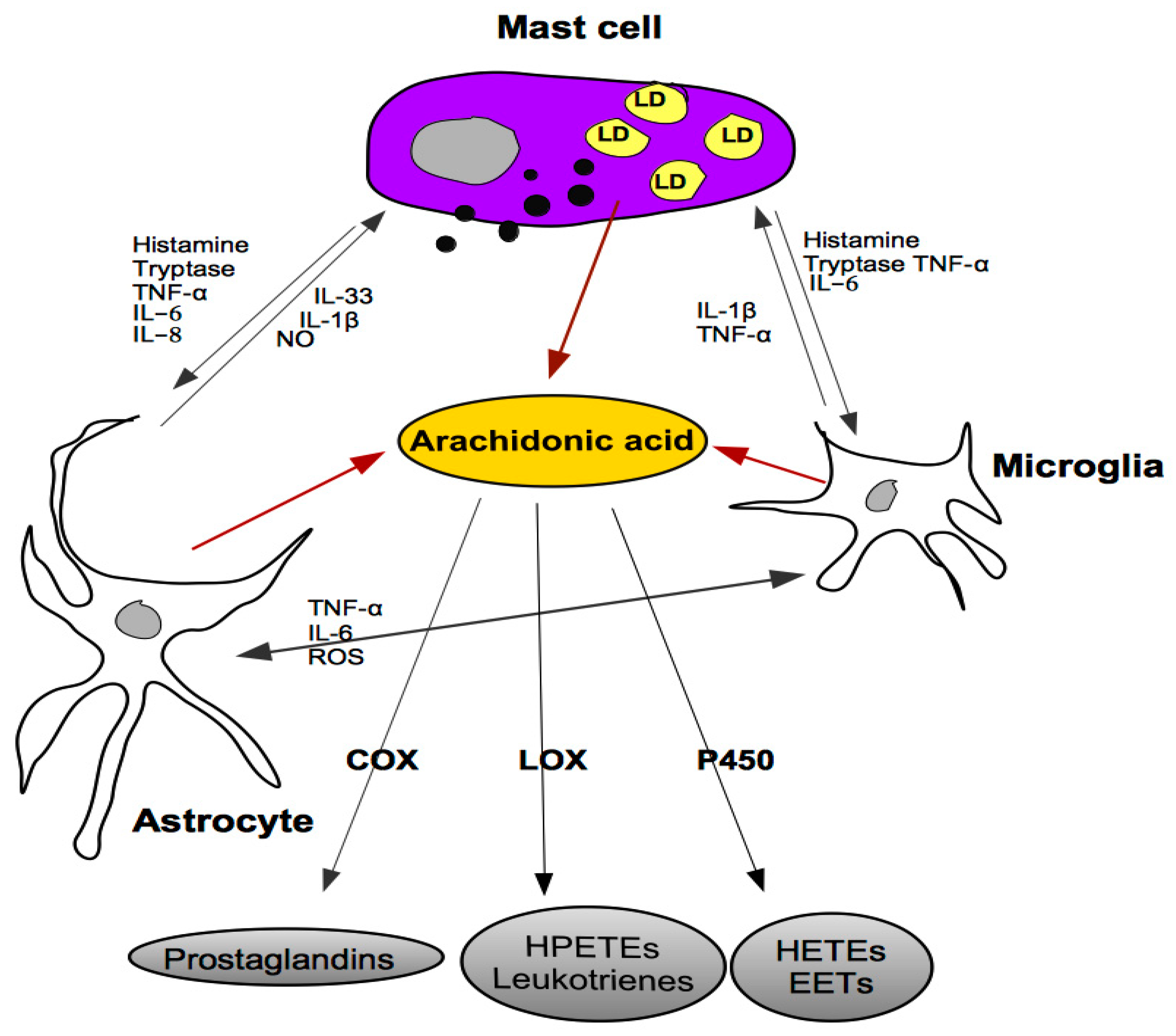
5. Cross-Talk between Mast Cells, Astrocytes and Microglia

In the brain, MCs surround the blood vessels, interacting with neurons, glia, and endothelial cells [44]. Astrocytes have a role of supporting cells and provide homeostatic control and trophic support within the brain [56]. Just one astrocyte may interact with up to 2,000,000 synapses in humans, and they are more than simple support cells [57]. In particular, astrocytes can influence synaptic activity and behavior and modulate neuronal circuits [58].
In patients suffering from major depression, there is an increased glial cell nuclei size in the prefrontal cortex [59]. Interestingly, the increase in the number and size of the astrocyte reflects brain size and cognitive capabilities [60].
In addition, microglia and astrocytes can release AA. Evidence suggests that astrocytes play a crucial role in elongation and desaturation of omega-6 and omega-3 essential fatty acid precursors to produce DHA and AA [61]. PUFAs accumulate in the brain. Astrocytes release PUFAs by lipopolysaccharide stimuli [18]. Therefore, astrocytes release DHA and AA under basal and stimulated conditions, including glutamate, bradykinin, ATP. In addition, IL-1 increases the ATP-induced release of AA from mouse astrocytes [62].
Under neuroinflammation, which is associated with microglial activation, the AA cascade is chronically upregulated through secretion of cytokines (e.g., interleukin (IL)-1β or tumor necrosis factor (TNF)-α) that stimulate astrocytic receptors that are coupled to activation of PLA2 and excess glutamatergic levels that stimulate their receptors neuronally and cause excitotoxicity [63].
Yet, in the brain, microglia and astrocytes cross-talk with MCs through a positive feedback loop, as much as MCs, once activated, are inclined to perpetuate inflammatory state [46] (Figure 1). Studies report that abnormalities in microglial activation can also be involved in the pathophysiology of epilepsy-associated depression and in multiple sclerosis. Microglia can be activated as a result of chronic peripheral inflammation, and its activation plays a crucial role in the pathogenesis and the pathophysiology of these disorders as a source of cytokines [64,65].
Finally, alteration in microglia observed in germ-free animals is solved with supplementation of short-chain fatty acids, coming from intestinal bacterial fermentation [66].

6. Serotonin and Depression

The best evidence of the role that 5-HT plays in the pathophysiology of depression is sustained by tryptophan depletion, the precursor of 5-HT. In particular, this observation suggests that low 5-HT activity can compromise the molecularity of the mechanisms which participate in the recovery from depressive status rather than exerting a primary role in depression [67]. In addition, pro-inflammatory cytokines can induce the activation of indoleamine 2,3-dioxygenase. This enzyme modulates the conversion of tryptophan to kynurenine. Higher levels of kynurenine are linked with a depression condition [68]. Therefore, inflammation could induce depression by reducing cerebral 5-HT activity [69]. Finally, in a model of depression, it has been evidenced that treatment with a tryptophan analog influences the stress response more than the behavior [7].
Intestinal endocrine cells (ECs) are major producers of 5-HT in humans. They are distributed along the intestinal mucosa, and this allows their function as a bridge of translators of molecular information between the intestinal lumen and the nervous system [70]. The ECs also secrete corticotropin-releasing hormone, cholecystokinin, and somatostatin under luminal stimulation by microbial factors derived from the gut microbiota and central stimuli. Among the others, the gut–brain connection seems attributable, in addition to the role that 5-HT plays as a central physiological mediator. Transport and/or use anomalies could favor MC accumulation. 5-HT influences biology, adhesion, and migration of MCs [71]. In this context, gut microbiota appears as a conductor in the cross-talk between inflammation and 5-HT imbalance, and the intestinal bacteria may be a potential therapeutic target for 5-HT-related disorders. In particular, tryptophan-regulating bacteria may have potential as antidepressants. An imbalance of gut microbiota may affect the level of neurotransmitters, including 5-HT [72].
5-HT stimulates the release of AA in hippocampal neurons co-cultured with glial cells but not in the case that glial cells are cultured alone, but through a type 2 5-HT receptor [73]. Furthermore, the release of AA stimulated by 5-HT is Ca2+-dependent [74]. Analysis of the eicosanoids produced in hippocampal cultures revealed that various prostaglandins, leukotrienes, and small amounts of hydroxyeicosatetraenoic acids (HETEs) were produced under basal conditions and that 5-HT stimulated an increase of all the metabolites of the cultures. AA and its lipoxygenase metabolite, 12-hydroperoxy-5,8,10,14-eicosatetraenoic acid, inhibit the release of neurotransmitters [20].
Finally, experimental and clinical evidence supports the involvement of 5-HT in learning processes and in both short-term memory and long-term memory [75,76,77,78,79]. The activating effect of 5-HT on memory function appears analogous to the action of serotonergic antidepressants, which, by preventing the presynaptic reuptake of 5-HT, prolong its permanence in the synaptic cleft and therefore facilitate its use by the post-synaptic neuron. At a biomolecular level, this would explain the reduction of cognitive functions, and particularly memory, which accompanies or precedes symptoms of depression [80].
In the absence of an effective reuptake and/or transport mechanism, 5-HT remains free in the neuronal domain of the brain, as well as in the enteric neuron domain and in the circulation.
Defect or excess of 5-HT, in depression but also in other psychiatric disorders, can have relationships with serious diseases such as scleroderma, intestinal inflammation, multiple sclerosis, coronary heart disease, or osteoporosis. For these pathologies, a high incidence of depressive disorder is recognized [81,82,83,84,85]. Intriguingly, the pro-inflammatory cytokines IL-1β, IL-6, and IL-8 can also influence the viscoelastic properties of the blood, increasing the hypercoagulability of whole blood. In fact, together with MCs, platelets are sensitive to systemic inflammatory changes, and all three cytokines can cause platelet hyper-activation, leading to pro-coagulant activity [86]. In this way, an increase in the activation of MC and platelet could be implicated as a potential pathophysiological pathway linking MD and cardiovascular diseases, including atherosclerosis and thrombosis.

7. Conclusions and Perspectives

In the brain, both glia and MCs release AA during a condition of silent inflammation to control the same and in order to contribute to the maintenance of homeostasis. In this manner, they perform as modulators of neuronal function under inflammation.
The same composition on lipids of MCs, astrocytes, and microglia could vary, reflecting the condition of the subject.
Between depressive and healthy subjects, a significant difference within the fatty acids composition of platelets has been found [87]. In particular, the relationships among palmitic acid (PA), linoleic acid (LA), and AA make the difference between MD and BD. Furthermore, LA, which is a metabolic precursor for AA synthesis, seems involved as a fine-tuning regulator of the membrane dynamic, both in platelets and neurons. This suggests that the membrane mobility and LA are crucial factors in the driving of the psychopathology and of the expression of quantum brain aspects, such as consciousness, and that 5-HT is still involved in a quantum consciousness [17].
There is strong evidence that each fatty acid combination of PA, LA, and AA, in platelets, is responsible for the membrane mobility and, therefore, of the molecular conditioning of the cellular structures (Gsα and tubulin) and that one of the main therapeutic targets is the reduction of the AA [88,89,90,91].
In conclusion, a silent inflammation status, as well as a depression condition, involves an alteration on 5-HT, prostaglandins, and AA levels; yet, a dysregulation of the intestinal microbiota composition can start but also exacerbate intestinal disorders, as well as can influence a depressive state; an inflammation condition is produced by and engages MCs but also the activation of platelets, which are particularly sensitive to a systemic inflammatory change.
These molecules are, therefore, excellent indicators and health tools and can provide suggestions for interventions.

Author Contributions

Conceptualization, G.T. and M.C.; writing—original draft preparation and editing, G.T.; writing—review, G.T. and M.C; supervision, G.T. All authors have read and agreed to the published version of the manuscript.

Funding

This research received no external funding.

Conflicts of Interest

The authors declare no conflict of interest.

Abbreviations

AAarachidonic acid
BDbipolar disorder
CNScentral nervous system
COXcyclo-oxygenase
DHAdocosahexaenoic acid
ECendocrine cell
5-HT5-hydroxytryptamine, serotonin
HETEhydroxyeicosatetraenoic acid
LAlinoleic acid
LDlipid droplet
LOXlipooxygenase
MCmast cell
MDmajor depression
PApalmitic acid
PCphosphatidylcholine
PEphosphatidylethanolamine
PGEprostaglandin
PLphospholipid
PLA2phospholipase A2
PUFApolyunsaturated fatty acid
SCFAsshort-chain fatty acids
SPsubstance
TGtriglyceride

References

  1. World Health Organization. World Health Statistics 2017: Monitoring Health for the SDGs, Sustainable Development Goals; World Health Organization: Geneva, Switzerland, 2017. [Google Scholar]
  2. Hauser, W.; Janke, K.H.; Klump, B.; Hinz, A. Anxiety and depression in patients with inflammatory bowel disease: Comparison with chronic liver disease patients and the general population. Inflamm. Bowel Dis. 2011, 17, 621–632. [Google Scholar] [CrossRef] [PubMed]
  3. Cocchi, M.; Tonello, L. Running the hypothesis of a bio molecular approach to psychiatric disorder characterization and fatty acids therapeutical choices. Ann. Gen. Psychiatry 2010, 9 (Suppl. S1). [Google Scholar] [CrossRef] [Green Version]
  4. Benedetti, S.; Bucciarelli, S.; Canestrari, F.; Catalani, S.; Mandolini, S.; Marconi, V.; Mastrogiacomo, A.; Silvestri, R.; Tagliamonte, M.; Venanzini, R.; et al. Platelet’s Fatty Acids and Differential Diagnosis of Major Depression and Bipolar Disorder through the Use of an Unsupervised Competitive- Learning Network Algorithm (SOM). Open J. Depress. 2014, 3, 52–73. [Google Scholar] [CrossRef] [Green Version]
  5. Cocchi, M.; Tonello, L.; Rasenick, M. Human depression: A new approach in quantitative psychiatry. Ann. Gen. Psychiatry 2010, 9, 25. [Google Scholar] [CrossRef] [PubMed] [Green Version]
  6. Milenkovic, V.M.; Stanton, E.H.; Nothdurfter, C.; Rupprecht, R.; Wetzel, C.H. The Role of Chemokines in the Pathophysiology of Major Depressive Disorder. Int. J. Mol. Sci. 2019, 20, 2283. [Google Scholar] [CrossRef] [PubMed] [Green Version]
  7. Biagini, G.; Pich, E.M.; Carani, C.; Marrama, P.; Gustafsson, J.A.; Fuxe, K.; Agnati, L.F. Indole-pyruvic acid, a tryptophan ketoanalogue, antagonizes the endocrine but not the behavioral effects of repeated stress in a model of depression. Biol. Psychiatry 1993, 33, 712–719. [Google Scholar] [CrossRef]
  8. Agnati, L.F.; Cortelli, P.; Biagini, G.; Bjelke, B.; Fuxe, K. Different classes of volume transmission signals exist in the central nervous system and are affected by metabolic signals, temperature gradients and pressure waves. Neuroreport 1994, 6, 9–12. [Google Scholar] [CrossRef]
  9. Sheetz, M.P.; Sable, J.E.; Döbereiner, H.-G. Continuous Membrane-Cytoskeleton Adhesion Requires Continuous Accommodation to Lipid and Cytoskeleton Dynamics. Annu. Rev. Biophys. Biomol. Struct. 2006, 35, 417–434. [Google Scholar] [CrossRef]
  10. Cocchi, M.; Tonello, L.; Gabrielli, F. Possible Roles of Cell Membrane & Cytoskeleton in Quantum Aspect of Psychiatry. J. Conscious. Explor. Res. 2012, 3, 1082–1100. [Google Scholar]
  11. Müller, C.P.; Reichel, M.; Muhle, C.; Rhein, C. Brain membrane lipids in major depression and anxiety disorders. Biochim. Biophys. Acta 2015, 1851, 1052–1065. [Google Scholar] [CrossRef] [Green Version]
  12. Cocchi, M.; Tonello, L. Bio molecular considerations in Major Depression and Ischemic Cardiovascular Disease. CNS Agents Med. Chem. 2010, 9, 2–11. [Google Scholar] [CrossRef]
  13. Goodhand, J.R.; Wahed, M.; Mawdsley, J.E.; Farmer, A.D.; Aziz, Q.; Rampton, D.S. Mood disorders in inflammatory bowel disease: Relation to diagnosis, disease activity, perceived stress, and other factors. Inflamm. Bowel Dis. 2012, 18, 2301–2309. [Google Scholar] [CrossRef] [PubMed]
  14. Rao, J.S.; Lee, H.-J.; Rapoport, S.I.; Bazinet, R.P. Mode of action of mood stabilizers: Is the arachidonic acid cascate a common target? Mol. Psychiatry 2008, 13, 585–596. [Google Scholar] [CrossRef] [PubMed] [Green Version]
  15. Heron, D.S.; Shinitzky, M.; Hershkowitz, M.; Samuel, D. Lipid Fluidity Markedly Modulates the Binding of Serotonin to Mouse Brain Membranes. Proc. Natl. Acad. Sci. USA 1980, 77, 7463–7467. [Google Scholar] [CrossRef] [PubMed] [Green Version]
  16. Cocchi, M.; Gabrielli, F.; Tonello, L.; Pregnolato, M. Interactome hypthesis of Depression. NeuroQuantology 2010, 4, 603–613. [Google Scholar]
  17. Cocchi, M.; Minuto, C.; Tonello, L.; Gabrielli, F.; Bernroider, G.; Tuszynski, J.A.; Cappello, F.; Rasenick, M. Linoleic acid: Is this the key that unlocks the quantum brain? Insights linking broken symmetries in molecular biology, mood disorders and personalistic emergentism. BMC Neurosci. 2017, 18, 38. [Google Scholar] [CrossRef] [Green Version]
  18. Aizawa, F.; Nishinaka, T.; Yamashita, T.; Nakamoto, K.; Koyama, Y.; Kasuya, F.; Tokuyama, S. Astrocytes release polyunsatured fatty acids by lipopolysaccharide stimuli. Biol. Pharm. Bull. 2016, 39, 1100–1106. [Google Scholar] [CrossRef] [Green Version]
  19. Wolfe, L.S. Eicosanoids: Prostaglandins, Thromboxanes, Leukotrienes, and Other Derivatives of Carbon-20 Unsaturated Fatty Acids. J. Neurochem. 1982, 38, 1–14. [Google Scholar] [CrossRef]
  20. Piomelli, D.; Volterra, A.; Dale, N.; Siegelbaum, S.A.; Kandel, E.R.; Schwartz, J.H.; Belardetti, F. Lipoxygenase metabolites of arachidonic acid as second messengers for presynaptic inhibition of Aplysia sensory cells. Nature 1987, 328, 38–43. [Google Scholar] [CrossRef]
  21. Su, K.-P.; Huang, S.-Y.; Peng, C.-Y.; Lai, H.-C.; Huang, C.-L.; Chen, Y.-C.; Aitchison, K.J.; Pariante, C.M. Phospholipase A2 and cyclooxygenase 2 genes influence the risk of interferon-alpha-induced depression by regulating polyunsatured fatty acids levels. Biol. Psychiatry 2010, 67, 550–557. [Google Scholar] [CrossRef] [Green Version]
  22. Manev, R.; Manev, H. 5-Lipoxygenase as a putative link between cardiovascular and psychiatric disorders. Critic Rev. Neurobiol. 2004, 16, 181–186. [Google Scholar] [CrossRef] [PubMed]
  23. Manev, R.; Mrazovac, D.; Manev, H. Possible role for interactions between 5-lipoxygenase (5-LOX) and AMPA GluR1 receptors in depression and in antidepressant therapy. Med. Hypotheses 2007, 69, 1076–1079. [Google Scholar] [CrossRef] [PubMed] [Green Version]
  24. Rapoport, S.I. Lithium and the other mood stabilizers effective in bipolar disorder target the rat brain arachidonic acid cascade. ACS Chem. Neurosci. 2014, 5, 459–467. [Google Scholar] [CrossRef]
  25. Matsuo, M.; Hamasaki, Y.; Fujiyama, F.; Miyazaki, S. Eicosanoids are produced by microglia, not by astrocytes, in rat glial cell cultures. Brain Res. 1995, 685, 201–204. [Google Scholar] [CrossRef]
  26. Farooqui, A.A.; Ong, W.Y.; Horrocks, L.A. Inhibitors of brain phospholipase A2 activity: Their neuropharmacological effects and therapeutic importance for the treatment of neu- rologic disorders. Pharmacol. Rev. 2006, 58, 591–620. [Google Scholar] [CrossRef] [Green Version]
  27. Phillis, J.W.; Horrocks, L.A.; Farooqui, A.A. Cyclooxygenases, lipoxygenases, and epoxygenases in CNS: Their role and involvement in neurological disorders. Brain Res. Rev. 2006, 52, 201–243. [Google Scholar] [CrossRef]
  28. Faridhosseini, F.; Sadeghi, R.; Farid, L.; Pourgholami, M. Celecoxib: A new augmentation strategy for depressive mood episodes. A systematic review and meta-analysis of randomized placebo-controlled trials. Hum. Psychopharmacol. 2014, 29, 216–223. [Google Scholar] [CrossRef]
  29. Eyre, H.A.; Air, T.; Proctor, S.; Rositano, S.; Baune, B.T. A critical review of the efficacy of non-steroidal anti-inflammatory drugs in depression. Prog. Neuropsychopharmacol. Biol. Psychiatry 2015, 57, 11–16. [Google Scholar] [CrossRef]
  30. Johansson, D.; Falk, A.; Marcus, M.M.; Svensson, T.H. Celecoxib enhances the effect of reboxetine and fluoxetine on cortical noradrenaline and serotonin output in the rat. Prog. Neuro Psychopharmacol. Biol. Psychiatry 2012, 39, 143–148. [Google Scholar] [CrossRef]
  31. Kornhuber, J.; Medlin, A.; Bleich, S.; Jendrossek, V.; Henkel, A.W.; Wiltfang, J.; Gulbins, E. High activity of acid sphingomyelinase in major depression. J. Neural Transm. 2005, 112, 1583–1590. [Google Scholar] [CrossRef]
  32. Hannun, Y.A.; Obeid, L.M. Principles of bioactive lipid signalling: Lessons from sphingolipids. Nat. Rev. Mol. Cell Biol. 2008, 9, 139–150. [Google Scholar] [CrossRef] [PubMed]
  33. Albi, E.; Cataldi, S.; Baldi, E.; Bucherelli, C.; Ferri, I.; Sidoni, A.; Codini, M.; Conte, C.; Beccari, T.; Traina, G. Vitamin D receptor expression and acid sphingomyelinase activity in prefrontal region of a learning anial model. Arch. Ital. Biol. 2019, 157, 120–128. [Google Scholar] [PubMed]
  34. Cataldi, S.; Arcuri, C.; Hunot, S.; Légeron, F.P.; Mecca, C.; Garcia-Gil, M.; Lazzarini, A.; Codini, M.; Beccari, T.; Tasegian, A.; et al. Neutral Sphingomyelinase Behaviour in Hippocampus Neuroinflammation of MPTP-Induced Mouse Model of Parkinson’s Disease and in Embryonic Hippocampal Cells. Mediators Inflamm. 2017, 2017, 2470950. [Google Scholar] [CrossRef] [Green Version]
  35. Nakamura, H.; Murayama, T. The role of sphingolipids in arachidonic acid metabolism. J. Pharmacol. Sci. 2014, 124, 307–312. [Google Scholar] [CrossRef] [PubMed] [Green Version]
  36. Veiga, M.P.; Arrondo, J.L.R.; Goni, F.M.; Alonso, A.; Marsh, D. Interaction of cholesterol with sphingomyelin in mixed membrane containing phosphatidylcholine, studied by spin-label ESR and IR spectroscopies. A possible stabilization of gel-phase sphingolipid domains by cholesterol. Biochemistry 2001, 40, 2614–2622. [Google Scholar] [CrossRef]
  37. Donati, R.J.; Dwivedi, Y.; Roberts, R.C.; Conley, R.R.; Pandey, G.N.; Rasenick, M.M. Postmortem brain tissue of depressed suicides reveals increased Gs localization in lipid raft domains where it is less likely to activate adenylyl cyclase. J. Neurosci. 2008, 28, 3042–3050. [Google Scholar] [CrossRef] [Green Version]
  38. Mühle, C.; Wagner, C.J.; Farber, K.; Richter-Schmidinger, T.; Gulbins, E.; Lenz, B.; Kornhuber, J. Secretory sphingomyelinase in the serum of medicated patients predicts the prospective course of depression. J. Clin. Med. 2019, 8, 846. [Google Scholar] [CrossRef] [Green Version]
  39. Bryan, P.F.; Karla, C.; Edgar Alejandro, M.T.; Sara Elva, E.P.; Gemma, F.; Luz, C. Sphingolipids as Mediators in the Crosstalk between Microbiota and Intestinal Cells: Implications for Inflammatory Bowel Disease [published correction appears in Mediators Inflamm. 2016;2016:7267956]. Mediat. Inflamm. 2016, 2016, 9890141. [Google Scholar]
  40. An, D.; Na, C.; Bielawski, J.; Hannun, Y.A.; Kasper, D.L. Membrane sphingolipids as essential molecular signals for Bacteroides survival in the intestine. Proc. Natl. Acad. Sci. USA 2011, 108 (Suppl. S1), 4666–4671. [Google Scholar] [CrossRef] [Green Version]
  41. Bastiaanssen, T.F.S.; Cussotto, S.; Claesson, M.J.; Clarke, G.; Dinan, T.G.; Cryan, J.F. Gutted! Unraveling the role of the microbiome in major depressive disorder. Harv. Rev. Psychiatry 2020, 28, 26–39. [Google Scholar]
  42. Kim, Y.K.; Shin, C. The microbiota-gut-brain axis in neuropsychiatric disorders: Pathophysiological and novel treatments. Curr. Neuropharmacol. 2018, 16, 559–573. [Google Scholar] [CrossRef] [PubMed]
  43. Moura, D.S.; Sultan, S.; Georgin-Lavialle, S.; Pillet, N.; Montestruc, F.; Gineste, P.; Barete, S.; Damaj, G.; Moussy, A.; Lortholary, O.; et al. Depression in patients with mastocytosis: Prevalence, features and effects of masitinib therapy. PLoS ONE 2011, 6, e26375. [Google Scholar] [CrossRef] [PubMed]
  44. Hendriksen, E.; van Brgeijk, D.; Oosting, R.S.; Redegeld, F.A. Mast cells in neuroinflammation and brain disorders. Neurosci. Biobehav. Rev. 2017, 79, 119–133. [Google Scholar] [CrossRef] [PubMed]
  45. Traina, G. Mast cells in the brain- Old cells, new target. J. Integr. Neurosci. 2017, 16, S69–S83. [Google Scholar] [CrossRef]
  46. Traina, G. Mast cells in gut and brain and their potential role as an emerging therapeutic target for neural diseases. Front. Cell. Neurosci. 2019, 13, 345. [Google Scholar] [CrossRef]
  47. Irmak, D.K.; Kilinc, E.; Tore, F. Shared fate of meningeal mast cells and sensory neurons in migraine. Front. Cell Neurosci. 2019, 13, 136. [Google Scholar] [CrossRef] [Green Version]
  48. Julius, D.; Basbaum, A.I. Molecular mechanisms of nociception. Nature 2001, 413, 203–210. [Google Scholar] [CrossRef]
  49. Barbara, G.; Wang, B.; Stanghellini, V.; de Giorgio, R.; Cremon, C.; Di Nardo, G.; Trevisani, M.; Campi, B.; Geppetti, P.; Tonini, M.; et al. Mast cell-dependent excitation of visceral-nociceptive sensory neurons in irritable bowel syndrome. Gastroenterology 2007, 132, 26–37. [Google Scholar] [CrossRef]
  50. Conte, C.; Sichetti, M.; Traina, G. Gut-brain axis: Focus on neurodegeneration and mast cells. Appl. Sci. 2020, 10, 1828. [Google Scholar] [CrossRef] [Green Version]
  51. Dichlberger, A.; Kovanen, P.T.; Schneider, J. Mast cells: From lipid droplets to lipid mediators. Clin. Sci. 2013, 125, 121–130. [Google Scholar] [CrossRef] [Green Version]
  52. Dichlberger, D.; Schlager, S.; Kovanen, P.T.; Schneider, W.J. Lipid droplets in activated mast cells—A significant source of triglyceride-derived arachidonic acid for eicosanoid production. Eur. J. Pharmacol. 2016, 785, 59–69. [Google Scholar] [CrossRef] [PubMed]
  53. Dvorak, A.M.; Dvorak, H.F.; Peters, S.P.; Shulman, E.S.; MacGlashan, D.W., Jr.; Pyne, K.; Harvey, V.S.; Galli, S.J.; Lichtenstein, L.M. Lipid bodies: Cytoplasmic organelles important to arachidonate metabolism in macrophages and mast cells. J. Immunol. 1983, 131, 2965–2976. [Google Scholar] [PubMed]
  54. Bozza, P.T.; Magalhães, K.G.; Weller, P.F. Leukocyte lipid bodies—Biogenesis and functions in inflammation. Biochim. Biophys. Acta 2009, 1791, 540–551. [Google Scholar] [CrossRef] [PubMed] [Green Version]
  55. Matsukawa, T.; Izawa, K.; Isobe, M.; Takahashi, M.; Maehara, A.; Yamanishi, Y.; Kaitani, A.; Okumura, K.; Teshima, T.; Kitamura, T.; et al. Ceramide-CD300f binding suppresses experimental colitis by inhibiting ATP-mediated mast cell activation. Gut 2016, 65, 777–787. [Google Scholar] [CrossRef] [PubMed]
  56. Lindsay, R.M. Adult rat brain astrocytes support survival of both NGF- dependent and NGF-insensitive neurones. Nature 1979, 282, 80–82. [Google Scholar] [CrossRef]
  57. Oberheim, N.A.; Takano, T.; Han, X.; He, W.; Lin, J.H.; Wang, F.; Xu, Q.; Wyatt, J.D.; Pilcher, W.; Ojemann, J.G.; et al. Uniquely hominid features of adult human astrocytes. J. Neurosci. 2009, 29, 3276–3287. [Google Scholar] [CrossRef]
  58. Zhou, X.; Xiao, Q.; Xie, L.; Yang, F.; Wang, L.; Tu, J. Astrocyte, a Promising Target for Mood Disorder Interventions. Front. Mol. Neurosci. 2019, 12, 136. [Google Scholar] [CrossRef]
  59. Rajkowska, G.; Miguel-Hidalgo, J.J.; Wei, J.; Dilley, G.; Pittman, S.D.; Meltzer, H.Y.; Overholser, J.C.; Roth, B.L.; Stockmeier, C.A. Morphometric evidence for neuronal and glial prefrontal cell pathology in major depression. Biol. Psychiatry 1999, 45, 1085–1098. [Google Scholar] [CrossRef]
  60. Stogsdill, J.A.; Eroglu, C. The interplay between neurons and glia in synapse development and plasticity. Curr. Opin. Neurobiol. 2017, 42, 1–8. [Google Scholar] [CrossRef] [Green Version]
  61. Moore, S.A.; Yoder, E.; Murphy, S.; Dutton, G.R.; Spector, A.A. Astrocytes, not neurons, produce docosahexaenoic acid (22:6ω-3) and Arachidonic Acid (20:4ω-6). J. Neurochem. 1991, 56, 518–524. [Google Scholar] [CrossRef]
  62. Stella, N.; Estelles, A.; Siciliano, J.; Tencé, M.; Desagher, S.; piomelli, D.; Glowinski, J.; Prémont, J. Interleukin-1 enhances the ATP-evoked release of arachidonic acid from mouse astrocytes. J. Neurosci. 1997, 17, 2939–2946. [Google Scholar] [CrossRef] [PubMed] [Green Version]
  63. Chang, Y.C.; Kim, H.W.; Rapoport, S.I.; Rao, J.S. Chronic NMDA administration increases neuroinflammatory markers in rat frontal cortex: Cross-talk between excitotoxicity and neuroinflammation. Neurochem. Res. 2008, 33, 2318–2323. [Google Scholar] [CrossRef] [Green Version]
  64. Morris, G.; Reiche, E.; Murru, A.; Carvalho, A.F.; Maes, M.; Berk, M.; Puri, B.K. Multiple Immune-Inflammatory and Oxidative and Nitrosative Stress Pathways Explain the Frequent Presence of Depression in Multiple Sclerosis. Mol. Neurobiol. 2018, 55, 6282–6306. [Google Scholar] [CrossRef] [PubMed] [Green Version]
  65. Peng, W.F.; Fa, F.; Li, X.; Zhang, Q.Q.; Ding, J.; Wang, X. Different behavioral and pathological changes between epilepsy-associated depression and primary depression models. Epilepsy Behav. 2018, 83, 212–218. [Google Scholar] [CrossRef] [PubMed]
  66. Fung, T.C.; Olson, C.A.; Hsiao, E.Y. Interactions between the microbiota, immune and nervous systems in health and disease. Nat. Neurosci. 2017, 20, 145–155. [Google Scholar] [CrossRef] [PubMed]
  67. Ruhé, H.G.; Mason, N.S.; Schene, A.H. Mood is indeirectly related to serotonin, norepinephrine and dopamine levels in humans: A meta-analysis of monoamine depletion studies. Mol. Psychiatry 2007, 12, 331–359. [Google Scholar] [CrossRef] [Green Version]
  68. Gabbay, V.; Ely, B.A.; Babb, J.; Liebes, L. The possible role of the kynurenine pathway in anhedonia in adolescents. J. Neural Transm. 2012, 119, 253–260. [Google Scholar] [CrossRef] [Green Version]
  69. Schiepers, O.J.G.; Wichers, M.C.; Maes, M. Cytokines and major depression. Prog. Neuropsychopharmacol. Biol. Psychiatry 2005, 29, 201–217. [Google Scholar] [CrossRef]
  70. Rhee, S.H.; Pothoulakis, C.; Mayer, E.A. Principles and clinical implications of the brain–gut–enteric microbiota axis. Nat. Rev. Gastroenter. Hepatol. 2009, 6, 306–314. [Google Scholar] [CrossRef] [Green Version]
  71. Kushnir-Sukhov, N.M.; Gilfillan, A.M.; Coleman, J.W.; Brown, J.M.; Bruening, S.; Toth, M.; Metcalfe, D.D. 5-ydroxytryptamine Induces Mast Cell Adhesion and Migration. J. Immunol. 2006, 177, 6422–6432. [Google Scholar] [CrossRef] [Green Version]
  72. Waclawiková, B.; El Aidy, S. Role of Microbiota and Tryptophan Metabolites in the Remote Effect of Intestinal Inflammation on Brain and Depression. Pharmaceuticals 2018, 11, 63. [Google Scholar] [CrossRef] [PubMed] [Green Version]
  73. Felder, C.C.; Kanterman, R.Y.; Ma, A.L.; Axelrod, J. Serotonin stimulates phospholipase A2 and the release of arachidonic acid in hippocampal neurons by a type 2 serotonin receptor that is independent of inositolphospholipid hydrolysis. Proc. Natl. Acad. Sci. USA 1990, 87, 2187–2191. [Google Scholar] [CrossRef] [PubMed] [Green Version]
  74. Irvine, R.F. How is the level of free arachidonic acid controlled in mammalian cells? Biochem. J. 1982, 204, 3–16. [Google Scholar] [CrossRef] [PubMed] [Green Version]
  75. Catarsi, S.; GarciaGil, M.; Traina, G.; Brunelli, M. Seasonal-variation of serotonin content and nonassociative learning of swim induction in the leech Hirudo medicinalis. J. Comp. Physiol. A 1990, 167, 469–474. [Google Scholar] [CrossRef] [PubMed]
  76. Bailey, C.H.; Chen, M.; Keller, F.; Kandel, E.R. Serotonin-mediated endocytosis of apCAM: An early step of learning-related synaptic growth in Aplysia. Science 1992, 256, 645–649. [Google Scholar] [CrossRef]
  77. Zaccardi, M.L.; Traina, G.; Cataldo, E.; Brunelli, M. Sensitization and dishabituation of swim induction in the leech Hirudo medicinalis: Role of serotonin and cyclic AMP. Behav. Brain Res. 2004, 153, 317–326. [Google Scholar] [CrossRef]
  78. Zaccardi, M.L.; Mozzachiodi, R.; Traina, G.; Brunelli, M.; Scuri, R. Molecular mechanisms of short-term habituation in the leech Hirudo medicinalis. Behav. Brain Res. 2012, 229, 235–243. [Google Scholar] [CrossRef]
  79. Traina, G.; Ristori, C.; Brunelli, M.; Scuri, R. Acetyl-l-carnitine prevents serotonin-induced behavioural sensitization and dishabituation in Hirudo medicinalis. Behav. Brain Res. 2013, 253, 323–328. [Google Scholar] [CrossRef]
  80. Kraus, C.; Castrén, E.; Kasper, S.; Lanzenberger, R. Serotonin and neuroplasticity- Links beetween molecular, functional and structural pathphysiology in depression. Neurosci. Behav. Res. 2017, 77, 317–326. [Google Scholar] [CrossRef] [Green Version]
  81. Ostojic, P.; Zivojinovic, S.; Reza, T.; Milivojevic, D.; Damjanov, N. Symptoms of depression and anxiety in Serbian patients with systemic sclerosis: Impact of disease severity and socioeconomic factors. Mod. Rheumatol. 2010, 20, 353–357. [Google Scholar] [CrossRef]
  82. Kurina, L.; Goldacre, M.; Yeates, D.; Gill, L. Depression and anxiety in people with inflammatory bowel disease. J. Epidemiol. Comm. Health 2001, 55, 716–720. [Google Scholar] [CrossRef] [PubMed] [Green Version]
  83. Graff, L.A.; Walker, J.R.; Bernstein, C.N. Depression and anxiety in inflammatory bowel disease: A review of comorbidity and management. Inflamm. Bowel Dis. 2009, 15, 1105–1118. [Google Scholar] [CrossRef] [PubMed]
  84. Cocchi, M.; Tonello, L.; Lercker, G. Fatty acids, membrane viscosity, serotonin and ischemic heart disease. Lipids Health Dis. 2010, 9, 97. [Google Scholar] [CrossRef] [PubMed] [Green Version]
  85. Bab, I.A.; Yirmiya, R. Depression and bone mass. Ann. N. Y. Acad. Sci. 2010, 1192, 170–175. [Google Scholar] [CrossRef]
  86. Bester, J.; Pretorious, E. Effects of IL-1β, IL-6 and IL-8 on erythrocytes, platelets and clot viscoelasticity. Sci. Rep. 2016, 6, 32188. [Google Scholar] [CrossRef] [PubMed]
  87. Cocchi, M.; Tonello, L.; Amato, P.; De Lucia, A. Platelet and Brain Fatty acid transfer: Hypothesis on Arachidonic Acid and its relationship to Major Depression. J. Biol. Res. 2009, 82, 47–53. [Google Scholar] [CrossRef]
  88. Tonello, L.; Cocchi, M. The Cell Membrane: Is it a bridge from psychiatry to quantum consciousness? NeuroQuantology 2010, 1, 54–60. [Google Scholar] [CrossRef] [Green Version]
  89. Tonello, L.; Cocchi, M.; Gabrielli, F.; Tuszynski, J.A. On the possible quantum role of serotonin in consciousness. J. Integr. Neurosci. 2015, 14, 295. [Google Scholar] [CrossRef]
  90. Cocchi, M.; Bernroider, G.; Rasenick, M.; Tonello, L.; Gabrielli, F.; Tuszynski, J.A. Document of Trapani on animal consciousness and quantum brain function: A hypothesis. J. Integr. Neurosci. 2017, 16, 1–5. [Google Scholar] [CrossRef]
  91. Cocchi, M.; Tonello, L.; Gabrielli, F. Linoleic Acid: Fine tuning regulator of mood disorders? In Proceedings of the 13th Conference of Italian Researchers in the World, Dallas, TX, USA, 1 December 2018. [Google Scholar]
Figure 1. Cross-talk between mast cells, astrocytes, and microglia as arachidonic acid sources. The black dots in mast cells are the typical granules containing preformed molecules. COX, cyclo-oxygenase; EET, epoxyeicosatrienoic acid; HETE, hydroxyeicosatetraenoic acid; HPETE, hydroperoxyeisosatetraenoic acid; IL-, interleukin-; LOX, lipoxygenase; NO, nitric oxide; LD, lipid droplet; P450, cytochrome P450 epoxygenase; ROS, reactive oxygen species; TNF-α, tumor nerve factor-α.
Applsci 10 03455 g001

