A Customized VGG19 Network with Concatenation of Deep and Handcrafted Features for Brain Tumor Detection

 ,
,  , and
, and 
Abstract
:1. Introduction
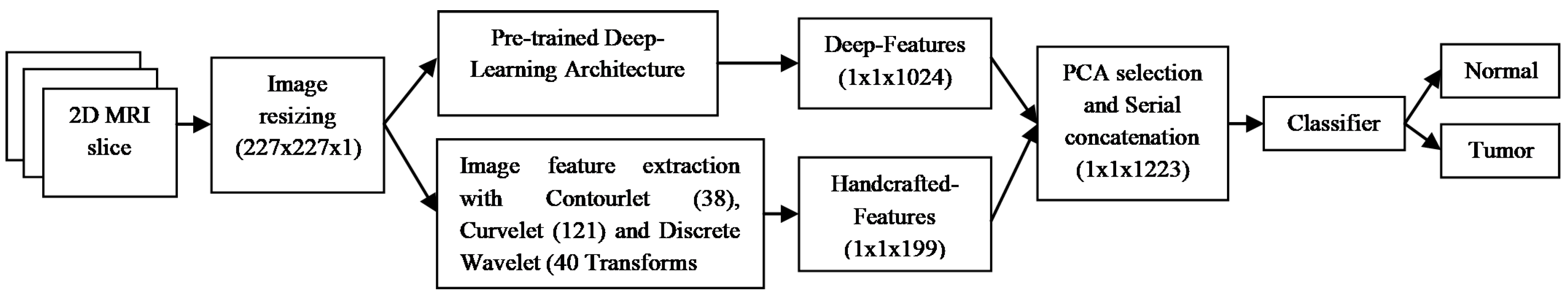
2. Materials and Methods
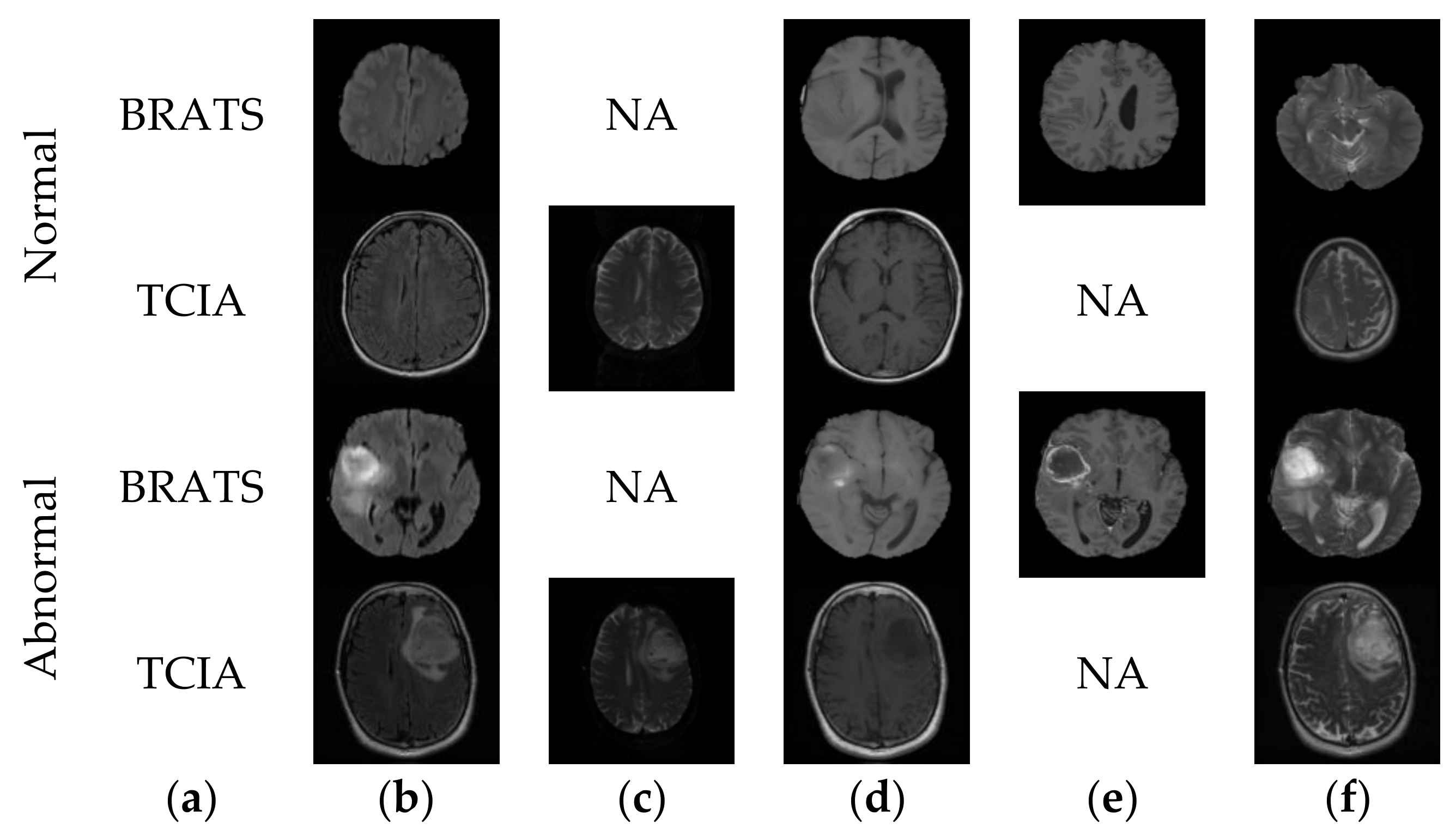
2.1. Image Collection and Processing
2.2. Handcrafted Feature Extraction
2.3. Feature Selection and Concatenation
2.4. Classification
- Decision tree: DT is one of the more famous methodologies used to categorize the linear and non-linear information with a sequence of testing methods, which expands like a tree. The DT utilizes a quality exploration situation as the root and internal nodes, and the class label forms terminal nodes. Once a DT has been shaped, categorization is accomplished by the conclusions taken in each branch of the tree. Other particulars of DT can be found in [43,44,45,46].
- K-nearest neighbor: KNN is a well-known technique often considered to classify medical images based on an existing feature set. In this work, KNN is considered to classify the brain MRIs of varied modality. During the classification task, the KNN evaluates the space among new features to each training feature and discovers the best neighbor. The earlier works on the KNN can be found in [43,44,45,46].
- Support vector machine: SVM categorizer uses a hyperplane for labeling of dataset based on features gathered throughout the training stage. SVM is one of the most frequently used to categorize MRI images. Radial basis function-based SVM (SVM-RBF) is used to sort the 2D MRI with the elected features. In SVM-RBF, the kernel value is controlled by a scaling parameter “σ”; and this value is varied from 0.2 to 1.9 with a step size of 0.1. Furthermore, the SVM with linear polynomial kernel (SVM-Linear) is also adopted to grade the MRI database [43].
2.5. Performance Measures and Validation
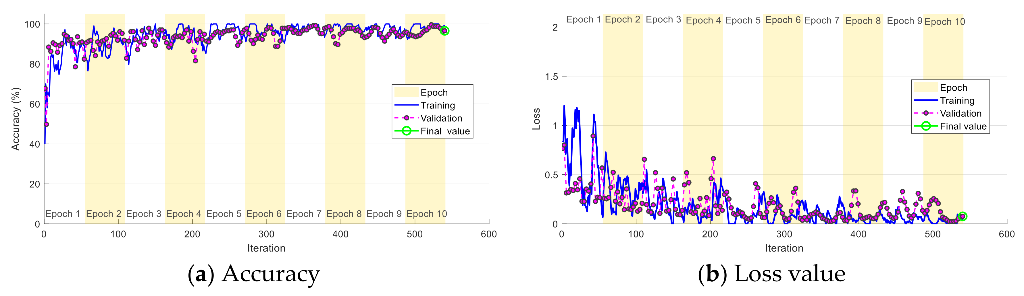
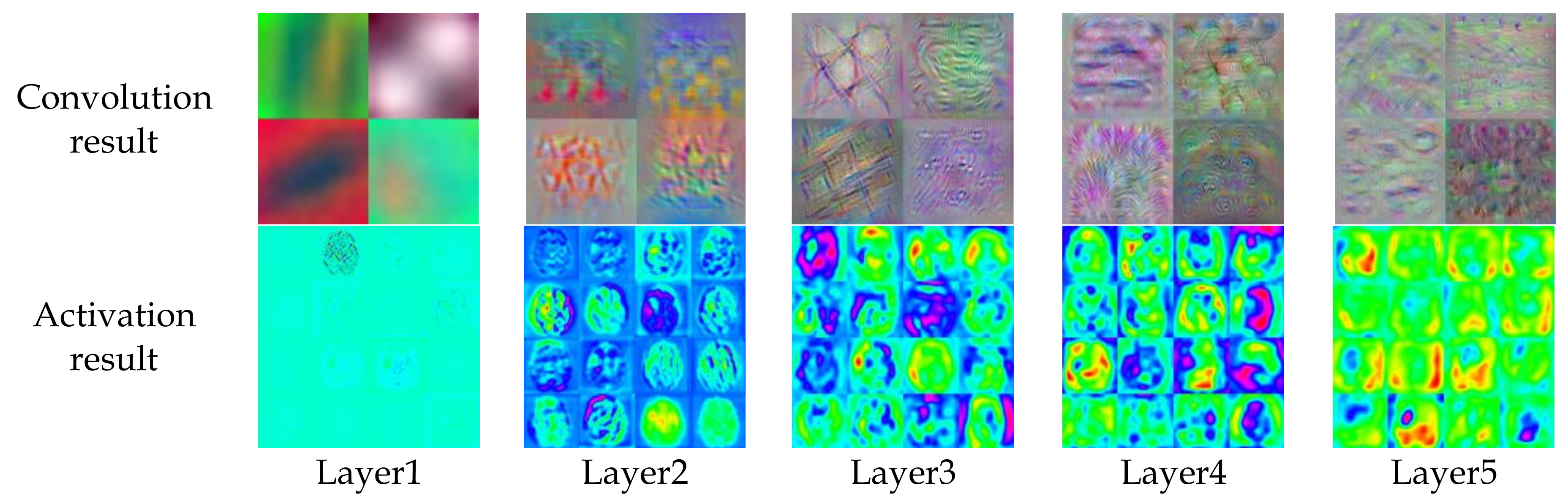
3. Experimental Outcome and Discussions
- (i)
- Enhancing the handcrafted feature vector by considering the additional texture and shape features.
- (ii)
- Adjusting the fully-connected and drop-out layers to improve the categorization accuracy.
- (iii)
- Improving the feature-concatenation technique to attain better results.
- (iv)
- Implementing the proposed VGG19 DLA to classify the tumors into low/high grade gliomas.
- (v)
- Developing a neural-network model for ependymal tumor dissemination.
4. Conclusions
Author Contributions
Funding
Acknowledgments
Conflicts of Interest
References
- Bhandary, A. Deep-learning framework to detect lung abnormality–A study with chest X-Ray and lung CT scan images. Pattern Recogn. Lett. 2020, 129, 271–278. [Google Scholar] [CrossRef]
- Pugalenthi, R.; Rajakumar, M.P.; Ramya, J.; Rajinikanth, V. Evaluation and classification of the brain tumor MRI using machine learning technique. Control Eng. Appl. Inf. 2019, 21, 12–21. [Google Scholar]
- Fernandes, S.L.; Tanik, U.J.; Rajinikanth, V.; Karthik, K.A. A reliable framework for accurate brain image examination and treatment planning based on early diagnosis support for clinicians. Neural Comput. Appl. 2019, 1–12. [Google Scholar] [CrossRef]
- Dey, N. Social-group-optimization based tumor evaluation tool for clinical brain mri of flair/diffusion-weighted modality. Biocybern. Biomed. Eng. 2019, 39, 843–856. [Google Scholar] [CrossRef] [Green Version]
- Louis, D.N. The 2016 world health organization classification of tumors of the central nervous system: A summary. Acta Neuropathol. 2016, 131, 803–820. [Google Scholar] [CrossRef] [Green Version]
- Rajinikanth, V.; Satapathy, S.C.; Fernandes, S.L.; Nachiappan, S. Entropy based segmentation of tumor from brain MR images—A study with teaching learning based optimization. Pattern Recogn. Lett. 2017, 94, 87–95. [Google Scholar] [CrossRef]
- Bauer, S.; Wiest, R.; Nolte, L.P. A survey of MRI-based medical image analysis for brain tumor studies. Phys. Med. Biol. 2013, 58, R97. [Google Scholar] [CrossRef]
- El-Dahshan, E.S.A.; Mohsen, H.M.; Revett, K. Computer-aided diagnosis of human brain tumor through MRI: A survey and a new algorithm. Expert Syst. Appl. 2014, 41, 5526–5545. [Google Scholar] [CrossRef]
- Palani, T.K.; Parvathavarthini, B.; Chitra, K. Segmentation of brain regions by integrating meta heuristic multilevel threshold with markov random field. Curr. Med. Imaging Rev. 2016, 12, 4–12. [Google Scholar] [CrossRef]
- Rajinikanth, V.; Raja, N.S.M.; Kamalanand, K. Firefly algorithm assisted segmentation of tumor from brain MRI using Tsallis function and Markov random field. Control Eng. Appl. Inform. 2017, 19, 97–106. [Google Scholar]
- Amin, J.; Sharif, M.; Yasmin, M. Big data analysis for brain tumor detection: Deep convolutional neural networks. Future Gener. Comput. Syst. 2018, 87, 290–297. [Google Scholar] [CrossRef]
- Thanaraj, P.; Parvathavarthini, B. Multichannel interictal spike activity detection using time–frequency entropy measure. Australas. Phys. Eng. Sci. Med. 2017, 40, 413–425. [Google Scholar] [CrossRef] [PubMed]
- Raja, N.S.M.; Fernandes, S.L.; Dey, N.; Satapathy, S.C. Contrast enhanced medical MRI evaluation using Tsallis entropy and region growing segmentation. J. Ambient. Intell. Hum. Comput. 2018, 1–12. [Google Scholar] [CrossRef]
- Liu, M.; Zhang, J.; Nie, D. Anatomical landmark based deep feature representation for MR images in brain disease diagnosis. IEEE J. Biomed. Health Inform. 2018, 22, 1476–1485. [Google Scholar] [CrossRef] [PubMed]
- Kanmani, P.; Marikkannu, P. MRI brain images classification: A multi-level threshold based region optimization technique. J. Med. Syst. 2018, 42, 62. [Google Scholar] [CrossRef]
- Wang, G.; Li, W.; Zuluaga, M.A. Interactive medical image segmentation using deep learning with image-specific fine-tuning. IEEE Trans. Med. Imaging 2018, 37, 1562–1573. [Google Scholar] [CrossRef] [PubMed]
- Gudigar, A.; Raghavendra, U.; San, T.R.; Ciaccio, E.J.; Acharya, U.R. Application of multiresolution analysis for automated detection of brain abnormality using MR images: A comparative study. Future Gener. Comput. Syst. 2019, 90, 359–367. [Google Scholar] [CrossRef]
- Buda, M. Association of genomic subtypes of lower-grade gliomas with shape features automatically extracted by a deep learning algorithm. Comput. Biol. Med. 2019, 109, 218–225. [Google Scholar] [CrossRef] [Green Version]
- Talo, M.; Baloglu, U.B.; Yıldırım, O.; Acharya, U.R. Application of deep transfer learning for automated brain abnormality classification using MR images. Cognit. Syst. Res. 2019, 24, 176–188. [Google Scholar] [CrossRef]
- Talo, M.; Yildirim, O.; Baloglu, U.B.; Aydin, G.; Acharya, U.R. Convolutional neural networks for multi-class brain disease detection using MRI image. Comput. Med. Imag. Grap. 2019, 78, 101673. [Google Scholar] [CrossRef]
- Amin, J. Brain tumor detection using statistical and machine learning method. Comput. Methods. Progr. Biomed. 2019, 177, 69–79. [Google Scholar] [CrossRef] [PubMed]
- Sharif, M.; Tanvir, U.; Munir, E.U.; Khan, M.A.; Yasmin, M. Brain tumor segmentation and classification by improved binomial thresholding and multi-features selection. J. Ambient. Intell. Hum. Comput. 2018, 1–20. [Google Scholar] [CrossRef]
- Fabelo, H. Deep learning-based framework for ln vivo identification of glioblastoma tumor using hyperspectral images of human brain. Sensors 2019, 19, 920. [Google Scholar] [CrossRef] [PubMed] [Green Version]
- Sajid, S.; Hussain, S.; Sarwar, A. Brain tumor detection and segmentation in MR images using deep learning. Arab. J. Sci. Eng. 2019, 44, 9249–9261. [Google Scholar] [CrossRef]
- Acharya, U.R. Automatic detection of ischemic stroke using higher order spectra features in brain MRI images. Cognit. Syst. Res. 2019, 58, 134–142. [Google Scholar] [CrossRef]
- Bakator, M.; Radosav, D. Deep learning and medical diagnosis: A review of literature. Multimodal Technol. Interact. 2018, 2, 47. [Google Scholar] [CrossRef] [Green Version]
- Havaei, M. Brain tumor segmentation with deep neural networks. Med. Image Anal. 2017, 35, 18–31. [Google Scholar] [CrossRef] [Green Version]
- Mallick, P.K. Brain MRI image classification for cancer detection using deep wavelet autoencoder-based deep neural network. IEEE Access 2019, 7, 46278–46287. [Google Scholar] [CrossRef]
- Tiwari, A.; Srivastava, S.; Pant, M. Brain tumor segmentation and classification from magnetic resonance images: Review of selected methods from 2014 to 2019. Pattern Recogn. Lett. 2019, 131, 244–260. [Google Scholar] [CrossRef]
- Sharif, M.I.; Li, J.P.; Khan, M.A.; Saleem, M.A. Active deep neural network features selection for segmentation and recognition of brain tumors using MRI images. Pattern Recogn. Lett. 2020, 129, 181–189. [Google Scholar] [CrossRef]
- Mohsen, H. Classification using deep learning neural networks for brain tumors. Future Comput. Inform. J. 2018, 3, 68–71. [Google Scholar] [CrossRef]
- Menze, B.H.; Jakab, A.; Bauer, S. The multimodal brain tumor image segmentation benchmark (BRATS). IEEE Trans. Med. Imaging 2015, 34, 1993–2024. [Google Scholar] [CrossRef] [PubMed]
- Brain Tumour Database (BraTS-MICCAI). Available online: http://hal.inria.fr/hal-00935640 (accessed on 15 February 2020).
- Clark, K.; Vendt, B.; Smith, K.; Freymann, J.; Kirby, J.; Koppel, P.; Moore, S.; Phillips, S.; Maffitt, D.; Pringle, M.; et al. The cancer imaging archive (TCIA): Maintaining and operating a public information repository. J. Digit. Imaging 2013, 26, 1045–1057. [Google Scholar] [CrossRef] [PubMed] [Green Version]
- Pedano, N.; Flanders, A.E.; Scarpace, L.; Mikkelsen, T.; Eschbacher, J.M.; Hermes, B.; Ostrom, Q. Radiology data from the cancer genome atlas low grade glioma [TCGA-LGG] collection. Cancer Imaging Arch. 2016. [Google Scholar] [CrossRef]
- Chang, K. Automatic assessment of glioma burden: A deep learning algorithm for fully automated volumetric and bi-dimensional measurement. Neuro Oncol. 2019, 21, 1412–1422. [Google Scholar] [CrossRef]
- Chang, P. Deep-learning convolutional neural networks accurately classify genetic mutations in gliomas. Am. J. Neuroradiol. 2018, 39, 1201–1207. [Google Scholar] [CrossRef] [Green Version]
- Proscans Diagnostics PVT. LTD. Homepage. Available online: https://proscans.in (accessed on 1 November 2019).
- Munir, K. Cancer diagnosis using deep learning: A bibliographic review. Cancers 2019, 11, 1235. [Google Scholar] [CrossRef] [Green Version]
- Tandel, G.S. A review on a deep learning perspective in brain cancer classification. Cancers 2019, 11, 111. [Google Scholar] [CrossRef] [Green Version]
- Nadeem, M.W. Brain tumor analysis empowered with deep learning: A review, taxonomy, and future challenges. Brain Sci. 2020, 10, E118. [Google Scholar] [CrossRef] [Green Version]
- Khawaldeh, S. Noninvasive grading of glioma tumor using magnetic resonance imaging with convolutional neural networks. Appl. Sci. 2018, 8, 27. [Google Scholar] [CrossRef] [Green Version]
- Acharya, U.R.; Fernandes, S.L.; WeiKoh, J.E.; Ciaccio, E.J.; Fabell, M.K.M.; Tanik, U.J.; Rajinikanth, V.; Yeong, C.H. Automated detection of alzheimer’s disease using brain MRI images—A study with various feature extraction techniques. J. Med. Syst. 2019, 43, 302. [Google Scholar] [CrossRef] [PubMed]
- Acharya, U.R.; Sree, S.V.; Ang, P.C.A.; Yanti, R.; Suri, J.S. Application of non-linear and wavelet based features for the automated identification of epileptic EEG signals. Int. J. Neural Syst. 2012, 22, 1250002. [Google Scholar] [CrossRef] [PubMed]
- Acharya, U.R.; Sudarshan, V.K.; Adeli, H.; Santhosh, J.; Koh, J.E.W. A novel depression diagnosis index using nonlinear features in EEG signals. Eur. Neurol 2015, 74, 79–83. [Google Scholar] [CrossRef]
- Acharya, U.R.; Faust, O.; Sree, S.V.; Molinari, F.; Garberoglio, R.; Suri, J.S. Cost-effective and non-invasive automated benign & malignant thyroid lesion classification in 3D contrast-enhanced ultrasound using combination of wavelets and textures: A class of thyroscan™ algorithms. Technol. Cancer Res. Treat 2011, 10, 371–380. [Google Scholar] [PubMed] [Green Version]
- Acharya, U.R.; Fujita, H.; Lih, O.S.; Adam, M.; Tan, J.H.; Chua, C.K. Automated detection of coronary artery disease using different durations of ECG segments with convolutional neural network. Knowl. Based Syst. 2017, 132, 62–71. [Google Scholar] [CrossRef]
- Raghavendra, U.; Bhat, N.S.; Gudigar, A.; Acharya, U.R. Automated system for the detection of thoracolumbar fractures using a CNN architecture. Future Gener. Comput. Syst. 2018, 85, 184–189. [Google Scholar] [CrossRef]
- Usharani, T.; Snekhalatha, U.; Palani, T.K.; Kumar, J. Human tongue thermography could be a prognostic tool for prescreening the type II diabetes mellitus. Evid. Based Complement. Altern. 2020, 2020, 3186208. [Google Scholar]
- Adapa, D.; Raj, A.N.J.; Alisetti, S.N.; Zhuang, Z.; Naik, G. A supervised blood vessel segmentation technique for digital fundus images using Zernike Moment based features. PLoS ONE 2020, 15, e0229831. [Google Scholar] [CrossRef]
- Zhuang, Z.; Fan, G.; Yuan, Y.; Raj, A.N.J.; Qiu, S. A fuzzy clustering based color-coded diagram for effective illustration of blood perfusion parameters in contrast-enhanced ultrasound videos. Comput. Methods. Progr. Biomed. 2019, 105233. [Google Scholar] [CrossRef]
- Noe, J.R.A. A multi-sensor system for silkworm cocoon gender classification via image processing and support vector machine. Sensors 2019, 19, 2056. [Google Scholar]
- Zhuang, Z. Nipple segmentation and localization using modified u-net on breast ultrasound images. J. Med. Imaging. Health Inform 2019, 9, 1827–1837. [Google Scholar] [CrossRef]
- Satapathy, S.C.; Rajinikanth, V. Jaya algorithm guided procedure to segment tumor from brain MRI. J. Optim. 2018, 2018, 3738049. [Google Scholar] [CrossRef] [Green Version]


| Image Class | Modality | Number of Images for Training | Number of Images for Testing |
|---|---|---|---|
| Normal (BRATS+TCIA) | Mixed (Flair+T2) | 1000 | 400 |
| Abnormal (BRATS) | Flair | 1500 | 600 |
| T2 | 1500 | 600 | |
| T1C | 1500 | 600 | |
| Abnormal (TCIA) | T2 | 1000 | 400 |
| Abnormal (Clinical) | T2 | 200 | 200 |
| Network | Modality | TP | FN | TN | FP | ACC | PRE | SEN | SPE | F1S | NPV |
|---|---|---|---|---|---|---|---|---|---|---|---|
| AlexNet | Flair | 377 | 23 | 581 | 19 | 95.80 | 95.20 | 94.25 | 96.83 | 94.72 | 96.19 |
| T2 | 368 | 32 | 573 | 27 | 94.10 | 93.16 | 92.00 | 95.50 | 92.58 | 94.71 | |
| T1C | 361 | 39 | 568 | 32 | 92.90 | 91.86 | 90.25 | 94.67 | 91.05 | 93.57 | |
| VGG16 | Flair | 379 | 21 | 585 | 15 | 96.40 | 96.19 | 94.75 | 97.50 | 95.47 | 96.53 |
| T2 | 364 | 36 | 570 | 30 | 93.40 | 92.39 | 91.00 | 95.00 | 91.69 | 94.06 | |
| T1C | 368 | 32 | 564 | 36 | 93.20 | 91.09 | 92.00 | 04.00 | 91.54 | 94.63 | |
| VGG19 | Flair | 381 | 19 | 584 | 16 | 96.50 | 95.97 | 95.25 | 97.33 | 95.61 | 96.85 |
| T2 | 378 | 22 | 581 | 19 | 95.90 | 95.21 | 94.50 | 96.83 | 94.86 | 96.35 | |
| T1C | 370 | 30 | 574 | 26 | 94.40 | 93.43 | 92.50 | 95.67 | 92.96 | 95.03 | |
| ResNet50 | Flair | 371 | 29 | 567 | 33 | 93.80 | 91.83 | 92.75 | 94.50 | 92.29 | 95.13 |
| T2 | 358 | 42 | 565 | 35 | 92.30 | 91.09 | 89.50 | 94.17 | 90.29 | 93.08 | |
| T1C | 362 | 38 | 566 | 34 | 92.80 | 91.41 | 90.50 | 94.33 | 90.95 | 93.71 | |
| ResNet101 | Flair | 374 | 26 | 583 | 17 | 95.70 | 95.65 | 93.50 | 97.17 | 94.56 | 95.73 |
| T2 | 361 | 39 | 568 | 32 | 92.90 | 91.85 | 90.25 | 94.67 | 91.05 | 93.57 | |
| T1C | 366 | 34 | 565 | 35 | 93.10 | 91.27 | 91.50 | 94.17 | 91.39 | 94.32 |
| Classifier | Modality | TP | FN | TN | FP | ACC | PRE | SEN | SPE | F1S | NPV |
|---|---|---|---|---|---|---|---|---|---|---|---|
| DT | Flair | 379 | 21 | 587 | 13 | 96.60 | 96.68 | 94.75 | 97.83 | 95.71 | 96.55 |
| T2 | 377 | 23 | 584 | 16 | 96.10 | 95.93 | 94.25 | 97.33 | 95.08 | 96.21 | |
| T1C | 368 | 32 | 571 | 29 | 93.90 | 92.69 | 92.00 | 95.17 | 92.35 | 94.69 | |
| KNN | Flair | 369 | 31 | 581 | 19 | 95.00 | 95.10 | 92.25 | 96.83 | 93.65 | 94.93 |
| T2 | 371 | 29 | 580 | 20 | 95.10 | 94.88 | 92.75 | 96.67 | 93.80 | 95.24 | |
| T1C | 368 | 32 | 578 | 22 | 94.60 | 94.36 | 92.00 | 96.33 | 93.16 | 94.75 | |
| SVM-Linear | Flair | 370 | 30 | 583 | 17 | 95.30 | 95.61 | 92.50 | 97.17 | 94.03 | 95.11 |
| T2 | 374 | 26 | 579 | 21 | 95.30 | 94.68 | 93.50 | 96.50 | 94.09 | 95.70 | |
| T1C | 372 | 28 | 571 | 29 | 94.30 | 92.77 | 93.00 | 95.17 | 92.88 | 95.33 | |
| SVM-RBF | Flair | 378 | 22 | 589 | 11 | 96.70 | 97.17 | 94.50 | 98.17 | 95.82 | 96.40 |
| T2 | 374 | 26 | 582 | 18 | 95.60 | 95.41 | 93.50 | 97.00 | 94.44 | 95.72 | |
| T1C | 369 | 31 | 574 | 26 | 94.30 | 93.42 | 92.25 | 95.67 | 92.83 | 94.88 |
| Dataset | Classifier | Modality | TP | FN | TN | FP | ACC | PRE | SEN | SPE | F1S | NPV |
|---|---|---|---|---|---|---|---|---|---|---|---|---|
| BRATS | DT | Flair | 388 | 12 | 589 | 11 | 97.70 | 97.24 | 97.00 | 98.17 | 97.12 | 98.00 |
| T2 | 385 | 15 | 586 | 14 | 97.10 | 96.49 | 96.25 | 97.67 | 96.37 | 97.50 | ||
| T1C | 383 | 17 | 581 | 19 | 96.40 | 95.27 | 95.75 | 96.83 | 95.51 | 97.16 | ||
| KNN | Flair | 387 | 13 | 591 | 9 | 97.80 | 97.73 | 96.75 | 98.50 | 97.24 | 97.85 | |
| T2 | 384 | 16 | 588 | 12 | 97.20 | 96.96 | 96.00 | 98.00 | 96.48 | 97.35 | ||
| T1C | 385 | 15 | 589 | 11 | 97.40 | 97.22 | 96.25 | 98.17 | 96.73 | 97.52 | ||
| SVM-Linear | Flair | 392 | 8 | 592 | 8 | 98.40 | 98.00 | 98.00 | 98.67 | 98.00 | 98.67 | |
| T2 | 391 | 9 | 590 | 10 | 98.10 | 97.50 | 97.75 | 98.33 | 97.63 | 98.50 | ||
| T1C | 384 | 16 | 587 | 13 | 97.10 | 96.72 | 96.00 | 97.83 | 96.36 | 97.34 | ||
| SVM-RBF | Flair | 395 | 5 | 596 | 4 | 99.10 | 99.00 | 98.75 | 99.33 | 98.87 | 99.17 | |
| T2 | 394 | 6 | 595 | 5 | 98.90 | 98.74 | 98.50 | 99.17 | 98.62 | 99.00 | ||
| T1C | 389 | 11 | 588 | 12 | 97.70 | 97.01 | 97.25 | 98.00 | 97.13 | 98.16 | ||
| TCIA | SVM-RBF | T2 | 393 | 7 | 391 | 9 | 98.00 | 97.76 | 98.25 | 97.75 | 98.01 | 98.24 |
| Clinical | SVM-RBF | T2+Flair | 395 | 5 | 194 | 6 | 98.17 | 98.50 | 98.75 | 97.00 | 98.63 | 97.49 |
| Reference | Approach | Accuracy (%) |
|---|---|---|
| Amin et al. [11] | Machine learning with fused features | 97 (SVM) |
| 98 (Naïve-Bayes) | ||
| 86 (Ensemble) | ||
| 97 (DT) | ||
| 97 (KNN) | ||
| Mallick et al. [28] | Deep neural network | 89 |
| Sharif et al. [30] | Deep learning with feature fusion | 97.8 (SoftMax) |
| 93.6 (MSVM) | ||
| 92.2 (KNN) | ||
| 94.4 (Ensemble) | ||
| Gudigar et al. [17] | Shearlet transform + texture + PSO SVM | 97.38 |
| Khawaldeh et al. [42] | Convolutional neural networks | 91.16 |
© 2020 by the authors. Licensee MDPI, Basel, Switzerland. This article is an open access article distributed under the terms and conditions of the Creative Commons Attribution (CC BY) license (http://creativecommons.org/licenses/by/4.0/).
Share and Cite
Rajinikanth, V.; Joseph Raj, A.N.; Thanaraj, K.P.; Naik, G.R. A Customized VGG19 Network with Concatenation of Deep and Handcrafted Features for Brain Tumor Detection. Appl. Sci. 2020, 10, 3429. https://doi.org/10.3390/app10103429
Rajinikanth V, Joseph Raj AN, Thanaraj KP, Naik GR. A Customized VGG19 Network with Concatenation of Deep and Handcrafted Features for Brain Tumor Detection. Applied Sciences. 2020; 10(10):3429. https://doi.org/10.3390/app10103429
Chicago/Turabian StyleRajinikanth, Venkatesan, Alex Noel Joseph Raj, Krishnan Palani Thanaraj, and Ganesh R. Naik. 2020. "A Customized VGG19 Network with Concatenation of Deep and Handcrafted Features for Brain Tumor Detection" Applied Sciences 10, no. 10: 3429. https://doi.org/10.3390/app10103429
APA StyleRajinikanth, V., Joseph Raj, A. N., Thanaraj, K. P., & Naik, G. R. (2020). A Customized VGG19 Network with Concatenation of Deep and Handcrafted Features for Brain Tumor Detection. Applied Sciences, 10(10), 3429. https://doi.org/10.3390/app10103429