Article

Effects of Microplastic on Human Gut Microbiome: Detection of Plastic-Degrading Genes in Human Gut Exposed to Microplastics—Preliminary Study

1
University Center of Excellence for Nutraceuticals, Bioscience and Biotechnology Research Center, Institut Teknologi Bandung, Bandung 40132, Indonesia
2
School of Life Sciences and Technology, Institut Teknologi Bandung, Bandung 40132, Indonesia
3
Department of Biology, Faculty of Science and Technology, Airlangga University, Surabaya 60115, Indonesia
4
University CoE Research Center for Bio-Molecule Engineering, Universitas Airlangga, Surabaya 60115, Indonesia
5
Department of Preventive Veterinary Medicine, College of Animal and Veterinary Science, Southwest Minzu University, Chengdu 610093, China
6
Microbial Genetics Department, Eberhard Karls University of Tuebingen, 72076 Tuebingen, Germany
7
Generasi Biologi Indonesia (Genbinesia) Foundation, Gresik 61171, Indonesia
8
Biology Department, Institut Teknologi Sepuluh Nopember, Surabaya 60111, Indonesia
*
Authors to whom correspondence should be addressed.
Environments 2022, 9(11), 140; https://doi.org/10.3390/environments9110140
Submission received: 14 September 2022 / Revised: 3 November 2022 / Accepted: 7 November 2022 / Published: 9 November 2022

Abstract

:
Microplastics are major pollutants in the environment, and it is currently established that microplastics have already entered human food chains and been incorporated into the human body through ingestion and inhalation. Several works in animal models have already reported the adverse effect of microplastic exposure on biological systems; however, the effect of microplastic contamination on human health is still far from understood. In previous work, we reported microplastic contamination in the digestive tract of the Indonesian coastal and highland population. Using the same stool samples, we extended our previous work by investigating gut microbial composition in samples with and without microplastic contamination using next-generation sequencing. We found that microplastic contamination does not affect overall gut microbial composition and the abundance of gut-health-related genes. However, we found a negative and positive correlation between specific types of microplastics with certain bacterial taxa, especially from the genera Roseburia, Clostridium, and Prevotella. Despite the lack of a profound effect on the gut microbiome, we detected for the first time the presence of genes encoding plastic-degrading enzymes in the human gut microbiome, suggesting an adaptation of gut microbiome to microplastic contamination.

Graphical Abstract

1. Introduction

Plastic is the biggest pollutant in the ocean ecosystem, and around 12.7 million tons of plastic enters the ocean every year [1]. Among the polluting countries, Indonesia is the second biggest contributor, with around 3.2 million tons of plastic waste produced in Indonesia every year, 1.29 million tons of which ended up in the ocean [1,2]. Due to mechanical and physicochemical processes, in nature, plastics are fragmented into small particles with a size of less than 5 mm in length called microplastics. As a pollutant, microplastics are extremely durable and degrade slowly, often for hundreds or even thousands of years [3]. Because of its abundance, durability, and size, microplastics are often ingested and incorporated in the tissues, organs, and bodies of various sea organisms. Various types of microplastics were found to be accumulated in zooplankton [4], seagrass and algae [5], many Crustaceans [6], and Echinodermata species [7].
Microplastics are also found in different fish species, including species consumed by humans, such as tuna (Scombridae) [8], swordfish (Xiphias gladius) [9], and parrot fish (Scaridae) [10]. In Indonesia, microplastics are detected in marine and freshwater fish, such as in killifish (Aplocheilus sp.) from Ciliwung River, Jakarta [11]; cutlass fishes (Trichiurus sp.) and croaker fish from Pangandaran Bay, West Java [12]; and Gambusia affinis from Brantas River, East Java [13]. Microplastic can be found, not only in sea organisms, but also in salt and bottled water consumed by humans [14,15,16,17]. Consequently, various works have reported the detection of microplastic in human bodies. It has been found in human saliva [16], placenta [17,18], lung tissue [19,20], and stool samples, including stool samples from the coastal and highland populations of Indonesia [21,22].
Depending on age and sex, it is estimated that around 74,000 to 113,000 microplastic particles enter the human body annually [23]. Nevertheless, whether microplastic exposure poses a substantial effect on human health is less understood. The lack of physiological data on the effect of microplastic contamination on human health represents a pivotal gap of information that need to be addressed. Microplastics contain various toxic chemicals [24] that might be harmful for human cells and the microbial community in our digestive tract (gut microbiome). Gut microbiota play pivotal roles in the regulation of the immune system by metabolizing proteins and complex carbohydrates. It produces metabolic products that facilitate cross-talk between gut epithelium and immune cells [25]. Gut microbiota are also involved in the regulation of the nervous system by communicating via the vagus nerve, tryptophan, and short-chain fatty acids’ metabolisms, as well as by contributing to neural development [26,27]. In the digestive system, gut microbiota modulate fat digestion and adsorption, as well as complex carbohydrates’ catabolism [28,29]; they are essential for human health [30]. Several works have reported that exposure to microplastics can lead to gut microbiota dysbiosis in crayfish [23], zebrafish [31], and mice [32]. However, there is still no data regarding the effect of microplastic on the human gut microbiome. In our previous work, we reported microplastic contamination in the Indonesian digestive tract [21,22]. Using the same stool samples, we extended the previous studies by investigating the effect of microplastic contamination on the gut microbiome.
In this study, for the first time, we analyzed the correlation between microplastic contamination and the gut microbiome in human samples. We found that microplastic does not affect the overall profile of the human gut microbiome, but it is both positively and negatively correlated with an abundance of several microbial taxa. We also reported for the first time the detection of genes encoding plastic-degrading enzymes in the human gut, inferring gut microbiota adaptation to microplastic exposure.

2. Materials and Methods

2.1. Study Participants, Stool Sample Collection and DNA Extraction

The stool samples used in this study were the same samples used in previous studies regarding microplastic contamination in the Indonesian digestive tract [21,22]. Stool samples were collected once per individual from 22 healthy participants from 2 study populations: the coastal population of Kenjeran, Surabaya, Indonesia (9 male and 2 female) and highland population of Pacet, Mojokerto, Indonesia (5 male and 6 female) [21,22]. The participants were selected based on the following criteria: in a healthy condition; aged 20–50 years old; and did not consume any antibiotics for 2 months before sample collection. To minimize microbial contamination during sample collection and processing, the participants were provided with sterile glass containers with lids and spoons made of steel. We also used sterile glass and steel utensils during sample preparation and microbiome analysis. The DNA was then extracted directly following stool sample collection using a Zymbiomic DNA Miniprep Kit (Zymo Research). Additionally, some parts of each sample were used for microplastic contamination measurements using Raman spectroscopy [21,22].
The collection of human stool samples was approved by the Health Research Ethic Committee of Universitas Surabaya (No. 005-OL/KE/III/2021). All samples were anonymized and obtained with written consent from the participants.

2.2. Metagenomic Analysis

2.2.1. Library Preparation and Sequencing

Isolated genomic DNA was quality controlled and then subjected for the preparation of sequencing libraries. Libraries were prepared using the Nextera XT DNA Library Preparation Kit (Illumina, San Diego, CA, USA). After library preparation, all libraries were pooled and sequenced with a read length of 2 × 100 bp on an Ilumina NovaseqTM 6000 system (San Diego, CA, USA). The raw data obtained were subjected to bioinformatics, statistical analyses, and deposited to the DNA Data Bank of Japan (DDJP) with submission number SSUB023028.

2.2.2. Taxonomic and Functional Data Analysis

A total of 10 million of the adapter-trimmed raw forward reads were aligned to the RefSeq protein database (version 94) using Diamond in BLASTX mode [33]. Taxonomic placement was performed using the lowest common ancestor (LCA) algorithm, implemented in MEGAN6 Ultimate Edition (version 6.15.2) [34]. Only taxa with relative sequence abundances above 0.001% were considered. Functional classification was carried out in MEGAN6 Ultimate Edition (version 6.15.2) [34] by assigning the reads to KEGG, SEED, VFDB, and Interpro identifiers.
To assess the diversity of the samples, we computed the Shannon–Wiener Index (Hs) and evenness. The Hs is a measure of the total number of species present in each sample (the richness) and how often they occur (relative sequence abundance). To evaluate the similarity between the samples, Bray–Curtis distances were calculated using the relative sequence abundances of the detected species (>0.01%), and a principal coordinates analysis (PCoA) was conducted to assign each sample a location in the 2D space. Using this analysis, groups of very similar samples can be identified, as well as outliers.

2.2.3. Normalized Read Counts

The normalized genes read count was analyzed as previously performed [35]. Sequences of the reference genes (Table S1) were downloaded from the NCBI database. All reads of all samples were mapped against these sequences using BWA [36] and the number of mapped reads extracted from the resulting samfiles using SAMtools [37]. Read counts were then normalized for the number of sequenced reads and the length of the respective genes to make them comparable between samples and between genes.

2.3. Statistical Analysis

Spearman’s correlation analysis was used to determine the relationship between microbial diversity, microbial abundance, and microplastic composition. The microplastic contamination data were taken from previous publications [21,22]. We only performed correlation analysis for the microplastic type with a contamination prevalence value ≥ 3 (out of 22) (Table S2). After sorting, the level of HDPE (high-density polyethylene), PP (polypropylene), and PS (polystyrene) contamination in the stool samples were correlated with microbial diversity and richness value. The contamination levels of these microplastics were also correlated with the relative abundance of microbial taxa at the phylum, family, and genus level. In addition, we also performed correlation analysis between microplastic contamination with the read count of the gut-health-related genes and genes encoding plastic-degrading enzymes. Gut-health-related genes are genes belonging to the gut microbiome that are known to have a positive effect on human gut health.

3. Results

3.1. Coastal and Highland Populations Showed Similar Gut Microbiome Profiles despite Different Microplastic Contamination between the Two Populations

In previous work, we showed that high-density polyethylene (HDPE) was the most prevalent type of microplastic contaminant in the gut of the coastal population, while polypropylene (PP) was the most prevalent microplastic contaminating the gut of the highland population [21,22]. As different populations were exposed to different types of microplastic, we carried out a follow-up study by performing microbiome profiling to investigate the possible correlation between microplastic contamination and gut microbiome composition.
Gut microbiome analyses showed that the microbiota composition between the coastal and highland populations were quite similar. In both populations, the most abundant taxa at the phylum level were Bacteroidetes, Firmicutes, and Proteobacteria; at the family level, the most abundant were Prevotellaceae, Bacteroidaceae, Enterobacteriaceae, and Ruminococcaceae; and at the genus level, they were Prevotella, Bacteroides, Escherichia, and Faecalibacterium. These compositions are comparable to the normal human gut microbiome profiles in generalback [38]. There was also no noticeable difference in gut microbial composition between individuals with and without microplastic contamination in their gastrointestinal tract (Figure 1A; Figures S1A and S2A). In individuals H1, H3, and C3, there were changes in bacterial dominance. In these three individuals, Bacteroides was detected as the most abundant bacteria, while in other samples Prevotella was more abundant. It is unclear whether these microbial changes are related to microplastic contamination as no microplastics were detected in the stool samples collected from individual C3.
In accordance with microbial composition and abundance data, the principal component analyses at the phylum, family, and genus level failed to separate the highland and coastal sample. In addition, there was no visible separation between the samples with and without microplastic contamination (Figure 1B, Figures S1B and S2B). The high abundance of Bacteroides in individuals H1, H3, and C3 was also reflected in the PCoA analysis where these three samples were separated from the rest of the samples.
Although microplastic contamination seems to not have a profound effect on the human gut microbiome, we found significant correlations between microplastic contamination level and the relative abundance of some gut bacteria at the genus and species level (Table 1). HDPE contamination was negatively correlated with the relative abundance of Bacteroides, while PP and PS showed the opposite effect on microbial composition. The PP contamination level in stool was positively correlated with the relative abundance of Roseburia and Clostridium, but negatively correlated with Prevotella copri. On the other hand, PS was negatively correlated with Roseburia and Clostridium but had a positive correlation with P. copri.

3.2. Microplastic Contamination in the Human Gut Showed No Correlation with the Gut Microbiome Diversity and Richness

We further analyzed the microbiome data by performing microbial diversity, richness, and evenness analyses. Sample C3 from the coastal population showed the highest diversity and richness value, clearly separating it from the other samples. This result indicates microbial dysbiosis; however, there was no microplastic contamination detected in the stool samples collected from individual C3, suggesting that the observed microbial dysbiosis was not caused by microplastic contamination (Figure 2). We performed correlation analyses by comparing the level of HDPE, PP, and PS contamination in the stool samples with microbial diversity, richness, and evenness values. We found no significant correlation between these parameters, again indicating that microplastic contamination had no substantial effect on the composition of human gut microbiota. Note that in this study, we only assessed microplastic contamination and gut microbiome composition at one time point. Further studies involving measurements at different time points are required to fully elucidate the effect of microplastics on gut microbiota.

3.3. Microplastic Contamination Did Not Correlate with Gut-Health-Related Gene Abundance

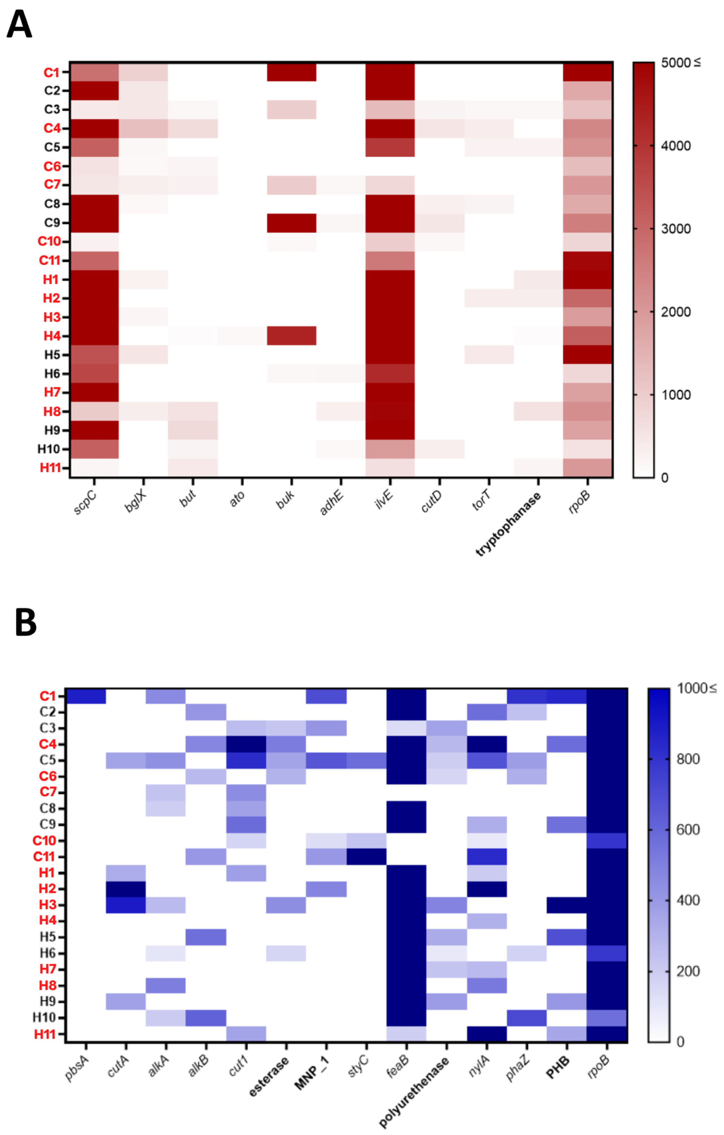
Previous works using human cell cultures and mice experiments showed the adverse effect of microplastic contamination on the immune [39], reproductive [40], and digestive systems [41]. To evaluate the effect of microplastic exposure on the human digestive system, we performed correlation analysis between microplastic contamination with the read count of the gut-health-related genes, such as short-chain fatty acid metabolisms-related genes and aromatic amino acid metabolisms-related genes (Figure 3A and Table S1). Housekeeping gene rpoB (a housekeeping gene, encoding RNA polymerase subunit B) was used as a reference control to analyze the gene reads obtained from metagenome sequencing. The normalized read count of the gut-health-related genes seems to be varied among the samples. scpC, a gene-encoding enzyme related to propionate production [42,43], and ilvE, a gene-encoding branched-chain amino acid aminotransferase [44,45], were found to abundant in the stool samples. In most samples, the read count of these two genes was higher than the housekeeping gene. Other gut-health-related genes, in general, had lower read counts than the housekeeping gene. To investigate whether the variation in the abundance of gut-health-related genes between individuals was correlated with the level of microplastic contaminations, we performed correlation analysis between the two parameters. However, none of the genes were significantly correlated with the level of microplastic contamination, suggesting that the abundance of gut-health-related genes are not influenced by the level of microplastic exposure in the gastrointestinal tract.

3.4. Genes Encoding Plastic-Degrading Enzymes Were Detected in the Human Gut

Although microplastic contamination seems to not affect the overall gut microbiome profile, interestingly, we found that the human gut microbiome harbors various genes that encode plastic-degrading enzymes. We then analyzed the reads by normalizing it, with rpoB as a comparison. Our results show that the samples collected from Indonesian coastal and highland populations contained various plastic-degrading enzyme-encoding genes with a different abundance and prevalence (Figure 3B and Table S1). The gene with the highest read count and also the most prevalent gene was feaB. This gene encodes phenylacetaldehyde dehydrogenase, which is reported to be found in styrene-degrading bacteria [46]. The other plastic-degrading genes were found at a lower abundance and were less prevalent compared to feaB and rpoB. pbsA, a gene-encoding polyester-degrading enzyme, was found only in sample C1 with a relatively high read count. However, the read counts of this gene, or any other plastic-degrading genes, were not correlated with the level of microplastic contamination in the stool samples. This is the first report of the existence of plastic-degrading genes in human stool samples, indicating the adaptation of the human gut ecosystem to constant microplastic exposure.

4. Discussion

Several studies have reported the adverse effect of microplastic exposure in human tissue cultures and in various animal models [41,47,48,49]. However, there is still no data regarding the direct effects of microplastic contamination on human health or physiological condition. Microplastic contamination and accumulation inside the human body have been extensively reported in recent years, such as in skin [16], hair [16], breastmilk [50], placenta [17,18], lung [19,20], and feces [21,22,51]. The detection of microplastics in feces implies that the human digestive tract has been exposed to microplastics. The presence of microplastics in the human digestive tract might cause gut microbiota dysbiosis. To test this hypothesis, we performed metagenome sequencing in stool samples with and without microplastic contamination to obtain an insight into the possible effects of microplastic exposure on the gut microbiome. In this study, microplastic and gut microbial analyses were performed at a single time point, whereas microplastic contamination in an individual might change over time. To fully understand the effect of microplastic on human gut microbiota, measurements at different time points are required. This work is intended to be a preliminary study because the effect of microplastic contamination in human samples has never been evaluated.
Our analyses revealed that there is no significant correlation between the level of HDPE, PP, and PS contamination and the gut microbiota overall composition, richness, diversity, and evenness. A possible explanation for this is that the microplastic contamination level in this study is still too low from the required threshold in which the microplastics can have a substantial effect on the gut microbiome. Our results are in agreement with an in vivo study in mice, which reported that daily exposure to polystyrene and polyethylene microplastics at a contamination level of 266 µg/kg body weight for 6 weeks and 100 mg/kg for 30 days, respectively, did not have a significant effect on the overall gut microbial composition and diversity level [52,53]. However, the reported effect of microplastic exposure on gut microbiota in mice varies. For example, studies by Jin et al. (2019) and Lu et al. (2018) showed that, despite not affecting overall diversity, polystyrene exposure decreased the abundance of α−Proteobacteria, γ-Proteobacteria, Firmicutes, and Actinobacteria [32,54]; and a study by Li et al. (2020) showed that a daily ingestion of 0.4 µg/kg body weight polyethylene increased the abundance of Firmicutes, Melainabacteria, and Bacteroidetes, causing gut microbiota dysbiosis [55]. Similar effects were observed in our study, where polystyrene exposure caused a decrease in Clostridium and Roseburia relative abundance, and HDPE, a derivative of polyethylene, caused a decrease in Bacteroides relative abundance (Table 1).
Gut microbiome plays a crucial role in gut health by metabolizing important compounds such as fatty acids [56,57], branched-chain amino acids [58,59], complex polysaccharides [60], and aromatic amino acids [61,62]. It also produces compounds that functions as activators for some signaling cascades related to immune [63,64], neural [65,66,67], and other physiological responses [56,68,69]. These metabolic capabilities of the gut microbiota are modulated by the controlled expression of the associated genes. However, the abundance of gut-health-related genes in the collected stool samples in this study showed no correlation with the level of microplastic contamination, possibly due to the same reason why the gut microbiome profiles were not affected by microplastic exposure. The level of microplastic contamination detected in this study was not high enough to induce significant changes in gut microbiota composition and gene abundance.
Despite the lack of a significant correlation between microplastic contamination and the microbial profile and gene abundance, surprisingly, genes encoding plastic-degrading enzymes were detected in the stool samples. The occurrence of these genes in the stool samples most probably came from the ingestion of the harboring bacteria. feaB, a gene that encodes phenylacetaldehyde dehydrogenase, which is an enzyme that contributes to styrene degradation [46], was found to be highly prevalent and abundant in the studied human stool samples. On the other hand, pbsA, a gene that encodes poly(tetramethylene succinate) depolymerase, an enzyme that functions in polyester family degradation [70], are the least prevalent plastic-degrading gene, as it was only detected in 1 out of 22 stool samples. The feaB gene was reported to be found in several strains of Eschericia coli [71,72] which is possibly the reason why feaB is the most prevalent and most abundant plastic-degrading gene in this study.
The presence of genes encoding plastic-degrading enzymes in the human gut microbiome imply gut microbiota adaptation against constant microplastic exposure due to human ingestion and inhalation. We hypothesize that the occurrence of microbes with plastic-degrading genes in the human gut is not due to microplastic contamination. It might be acquired from consumed water and food. Microbes that possess plastic-degrading enzyme-encoding genes might already exist in human consumables because plastic is ubiquitous in the environment. These microbes then enter the human digestive tract and colonize the human gut. As microplastic ingestion has been reported to influence the gut bacterial composition [48], it might drive the selective effect that favors the spreading of these genes in the human gut microbiome.

5. Conclusions

In this study, we showed that the level of microplastic contamination in two different Indonesian populations was not correlated with overall gut microbiota composition, richness, and diversity. Microplastic exposure level was also not significantly correlated with the abundance of genes related to gut health. Despite the absence of a correlation, we detected for the first time the existence of genes encoding plastic-degrading enzymes in human stool samples, in human stool both with and without microplastic contamination. Microbes with genes encoding plastic-degrading enzymes might already exist in human consumables, entering the human gut through ingestion. We hypothesized that to have a significant and observable effect on human gut microbiome composition, a certain microplastic contamination threshold is required. In this study, no significant correlation was observed between microplastic contamination and microbiome composition, probably because the threshold had not been met yet. Note that the number of the samples in this study was relatively small for a strong correlation analysis. Studies with larger numbers of samples covering measurement at different time points are required to evaluate the full extent of microplastic effect on human gut microbiota.

Supplementary Materials

The following supporting information can be downloaded at: https://www.mdpi.com/article/10.3390/environments9110140/s1, Figure S1: Gut microbiome profiles at the family level of Indonesian coastal and highland population exposed to microplastics contamination. (A) Relative abundance of microbia associated with the stool samples of coastal and highland individuals at the family level. (B) The principal component analysis of the samples based on the gut microbiota composition at the genus level. Abbreviations of samples: C—Coastal, H—Highland, sample code in red represents individuals with microplastic contamination, while sample code in black represents individuals without microplastic contamination.; Figure S2: Gut microbiome profiles at the phylum level of Indonesian coastal and highland population exposed to microplastic contamination. (A) Relative abundance of microbia associated with the stool samples of coastal and highland individuals at the phylum level. (B) The principal component analysis of the samples based on the gut microbiota composition at the genus level. Abbreviations of samples: C—Coastal, H—Highland, sample code in red represents individuals with microplastic contamination, while sample code in black represents individuals without microplastic contamination. Table S1: Accession numbers of the reference genes for read count analyses.; Table S2: Microplastic contamination level in human stool samples of coastal and highland populations in Indonesia. Data obtained from previous studies [21,22].

Author Contributions

Conceptualization, H.N., A.T.W. and A.L.; methodology, A.T.W. and A.L.; software, H.N.; validation, H.N., A.T.W., A.L., F., N., L.H., N.H. and F.G.; formal analysis, H.N., A.T.W., A.L., F., N., L.H., N.H. and F.G.; investigation, P.W.W.S., B.Q.P. and H.S.; resources, H.N., A.T.W., A.L. and F.G.; data curation, H.N., A.T.W. and A.L.; writing—original draft preparation, A.T.W. and A.L.; writing—review and editing, A.T.W. and A.L.; visualization, A.L. and B.Q.P.; supervision, H.N., A.T.W. and A.L.; project administration, H.N., A.T.W. and A.L.; funding acquisition, H.N., A.T.W. and A.L. All authors have read and agreed to the published version of the manuscript.

Funding

This research was funded by the PPKI program from the Indonesian World Class University Research Scheme (1325/PKS/ITS/2021; 177/UN3.15/PT/2021; 542/IT1.B07.1/TA.00/2021).

Data Availability Statement

The raw data obtained from NGS analyses were deposited to DNA Data Bank of Japan (DDJP) with the submission number SSUB023028.

Acknowledgments

We thank Aparna Viswanathan Ammanath, Yohanes Kartijo Putro, Fahmi Ikhlasul Amalludin, Faiza Nur Fauzia, Elsalisa Ainur Rofiqa, Yasri Fardiansyah, and Izzatul Islami, for their technical assistance during the research.

Conflicts of Interest

The authors declare no conflict of interest.

References

  1. Jambeck, J.R.; Geyer, R.; Wilcox, C.; Siegler, T.R.; Perryman, M.; Andrady, A.; Narayan, R.; Law, K.L. Marine pollution. Plastic waste inputs from land into the ocean. Science 2015, 347, 768–771. [Google Scholar] [CrossRef] [PubMed]
  2. Forum, W.E. The New Plastics Economy: Rethinking the Future of Plastics; Ellen MacArthur Foundation: Geneva, Switzerland, 2016. [Google Scholar]
  3. Hartmann, N.B.; Huffer, T.; Thompson, R.C.; Hassellov, M.; Verschoor, A.; Daugaard, A.E.; Rist, S.; Karlsson, T.; Brennholt, N.; Cole, M.; et al. Are We Speaking the Same Language? Recommendations for a Definition and Categorization Framework for Plastic Debris. Environ. Sci. Technol. 2019, 53, 1039–1047. [Google Scholar] [CrossRef] [PubMed] [Green Version]
  4. Cole, M.; Lindeque, P.; Fileman, E.; Halsband, C.; Goodhead, R.; Moger, J.; Galloway, T.S. Microplastic ingestion by zooplankton. Environ. Sci. Technol. 2013, 47, 6646–6655. [Google Scholar] [CrossRef] [PubMed]
  5. Seng, N.; Lai, S.; Fong, J.; Saleh, M.F.; Cheng, C.; Cheok, Z.Y.; Todd, P.A. Early evidence of microplastics on seagrass and macroalgae. Mar. Freshw. Res. 2020, 71, 922–928. [Google Scholar] [CrossRef]
  6. McGoran, A.R.; Clark, P.F.; Smith, B.D.; Morritt, D. High prevalence of plastic ingestion by Eriocheir sinensis and Carcinus maenas (Crustacea: Decapoda: Brachyura) in the Thames Estuary. Environ. Pollut. 2020, 265, 114972. [Google Scholar] [CrossRef]
  7. Plee, T.A.; Pomory, C.M. Microplastics in sandy environments in the Florida Keys and the panhandle of Florida, and the ingestion by sea cucumbers (Echinodermata: Holothuroidea) and sand dollars (Echinodermata: Echinoidea). Mar. Pollut. Bull. 2020, 158, 111437. [Google Scholar] [CrossRef]
  8. Neto, J.G.B.; Rodrigues, F.L.; Ortega, I.; Rodrigues, L.D.S.; Lacerda, A.; Coletto, J.L.; Kessler, F.; Cardoso, L.G.; Madureira, L.; Proietti, M.C. Ingestion of plastic debris by commercially important marine fish in southeast-south Brazil. Environ. Pollut. 2020, 267, 115508. [Google Scholar] [CrossRef]
  9. Romeo, T.; Pietro, B.; Peda, C.; Consoli, P.; Andaloro, F.; Fossi, M.C. First evidence of presence of plastic debris in stomach of large pelagic fish in the Mediterranean Sea. Mar. Pollut. Bull. 2015, 95, 358–361. [Google Scholar] [CrossRef]
  10. Goss, H.; Jaskiel, J.; Rotjan, R. Thalassia testudinum as a potential vector for incorporating microplastics into benthic marine food webs. Mar. Pollut. Bull. 2018, 135, 1085–1089. [Google Scholar] [CrossRef]
  11. Cordova, M.R.; Riani, E.; Shiomoto, A. Microplastics ingestion by blue panchax fish (Aplocheilus sp.) from Ciliwung Estuary, Jakarta, Indonesia. Mar. Pollut. Bull. 2020, 161, 111763. [Google Scholar] [CrossRef]
  12. Ismail, M.R.; Lewaru, M.W.; Prihadi, D.J. Microplastics Ingestion by Fish in The Pangandaran Bay, Indonesia. 2019. World News Nat. Sci 2019, 23. [Google Scholar]
  13. Buwono, N.R.; Risjani, Y.; Soegianto, A. Spatio-temporal patterns of occurrence of microplastics in the freshwater fish Gambusia affinis from the Brantas River, Indonesia. Environ. Pollut. 2022, 311, 119958. [Google Scholar] [CrossRef]
  14. Mason, S.A.; Welch, V.G.; Neratko, J. Synthetic Polymer Contamination in Bottled Water. Front. Chem. 2018, 6, 407. [Google Scholar] [CrossRef] [Green Version]
  15. Zhang, Q.; Xu, E.G.; Li, J.; Chen, Q.; Ma, L.; Zeng, E.Y.; Shi, H. A Review of Microplastics in Table Salt, Drinking Water, and Air: Direct Human Exposure. Environ. Sci. Technol. 2020, 54, 3740–3751. [Google Scholar] [CrossRef]
  16. Abbasi, S.; Turner, A. Human exposure to microplastics: A study in Iran. J. Hazard. Mater. 2021, 403, 123799. [Google Scholar] [CrossRef]
  17. Ragusa, A.; Svelato, A.; Santacroce, C.; Catalano, P.; Notarstefano, V.; Carnevali, O.; Papa, F.; Rongioletti, M.C.A.; Baiocco, F.; Draghi, S.; et al. Plasticenta: First evidence of microplastics in human placenta. Environ. Int. 2021, 146, 106274. [Google Scholar] [CrossRef]
  18. Braun, T.; Ehrlich, L.; Henrich, W.; Koeppel, S.; Lomako, I.; Schwabl, P.; Liebmann, B. Detection of Microplastic in Human Placenta and Meconium in a Clinical Setting. Pharmaceutics 2021, 13, 921. [Google Scholar] [CrossRef]
  19. Amato-Lourenco, L.F.; Carvalho-Oliveira, R.; Junior, G.R.; Dos Santos Galvao, L.; Ando, R.A.; Mauad, T. Presence of airborne microplastics in human lung tissue. J. Hazard. Mater. 2021, 416, 126124. [Google Scholar] [CrossRef]
  20. Jenner, L.C.; Rotchell, J.M.; Bennett, R.T.; Cowen, M.; Tentzeris, V.; Sadofsky, L.R. Detection of microplastics in human lung tissue using muFTIR spectroscopy. Sci. Total Environ. 2022, 831, 154907. [Google Scholar] [CrossRef]
  21. Luqman, A.; Nugrahapraja, H.; Wahyuono, R.A.; Islami, I.; Haekal, M.H.; Fardiansyah, Y.; Putri, B.Q.; Amalludin, F.I.; Rofiqa, E.A.; Götz, F.; et al. Microplastic Contamination in Human Stools, Foods, and Drinking Water Associated with Indonesian Coastal Population. Environments 2021, 8, 138. [Google Scholar] [CrossRef]
  22. Wibowo, A.T.; Nugrahapraja, H.; Wahyuono, R.A.; Islami, I.; Haekal, M.H.; Fardiansyah, Y.; Sugiyo, P.W.W.; Putro, Y.K.; Fauzia, F.N.; Santoso, H.; et al. Microplastic Contamination in the Human Gastrointestinal Tract and Daily Consumables Associated with an Indonesian Farming Community. Sustainability 2021, 13, 12840. [Google Scholar] [CrossRef]
  23. Cox, K.D.; Covernton, G.A.; Davies, H.L.; Dower, J.F.; Juanes, F.; Dudas, S.E. Correction to Human Consumption of Microplastics. Environ. Sci. Technol. 2020, 54, 10974. [Google Scholar] [CrossRef] [PubMed]
  24. Prata, J.C.; da Costa, J.P.; Lopes, I.; Duarte, A.C.; Rocha-Santos, T. Environmental exposure to microplastics: An overview on possible human health effects. Sci. Total Environ. 2020, 702, 134455. [Google Scholar] [CrossRef] [PubMed]
  25. Yoo, J.Y.; Groer, M.; Dutra, S.V.O.; Sarkar, A.; McSkimming, D.I. Gut Microbiota and Immune System Interactions. Microorganisms 2020, 8, 1587. [Google Scholar] [CrossRef] [PubMed]
  26. Sharon, G.; Sampson, T.R.; Geschwind, D.H.; Mazmanian, S.K. The Central Nervous System and the Gut Microbiome. Cell 2016, 167, 915–932. [Google Scholar] [CrossRef] [Green Version]
  27. Ojeda, J.; Avila, A.; Vidal, P.M. Gut Microbiota Interaction with the Central Nervous System throughout Life. J. Clin. Med. 2021, 10, 1299. [Google Scholar] [CrossRef]
  28. Martinez-Guryn, K.; Hubert, N.; Frazier, K.; Urlass, S.; Musch, M.W.; Ojeda, P.; Pierre, J.F.; Miyoshi, J.; Sontag, T.J.; Cham, C.M.; et al. Small Intestine Microbiota Regulate Host Digestive and Absorptive Adaptive Responses to Dietary Lipids. Cell Host Microbe 2018, 23, 458–469. [Google Scholar] [CrossRef] [Green Version]
  29. Cantarel, B.L.; Lombard, V.; Henrissat, B. Complex carbohydrate utilization by the healthy human microbiome. PLoS ONE 2012, 7, e28742. [Google Scholar] [CrossRef] [Green Version]
  30. Lu, L.; Luo, T.; Zhao, Y.; Cai, C.; Fu, Z.; Jin, Y. Interaction between microplastics and microorganism as well as gut microbiota: A consideration on environmental animal and human health. Sci. Total Environ. 2019, 667, 94–100. [Google Scholar] [CrossRef]
  31. Qiao, R.; Deng, Y.; Zhang, S.; Wolosker, M.B.; Zhu, Q.; Ren, H.; Zhang, Y. Accumulation of different shapes of microplastics initiates intestinal injury and gut microbiota dysbiosis in the gut of zebrafish. Chemosphere 2019, 236, 124334. [Google Scholar] [CrossRef]
  32. Lu, L.; Wan, Z.; Luo, T.; Fu, Z.; Jin, Y. Polystyrene microplastics induce gut microbiota dysbiosis and hepatic lipid metabolism disorder in mice. Sci. Total Environ. 2018, 631–632, 449–458. [Google Scholar] [CrossRef]
  33. Buchfink, B.; Xie, C.; Huson, D.H. Fast and sensitive protein alignment using DIAMOND. Nat. Methods 2015, 12, 59–60. [Google Scholar] [CrossRef]
  34. Huson, D.H.; Beier, S.; Flade, I.; Gorska, A.; El-Hadidi, M.; Mitra, S.; Ruscheweyh, H.J.; Tappu, R. MEGAN Community Edition—Interactive Exploration and Analysis of Large-Scale Microbiome Sequencing Data. PLoS Comput. Biol. 2016, 12, e1004957. [Google Scholar] [CrossRef] [Green Version]
  35. Luqman, A.; Zabel, S.; Rahmdel, S.; Merz, B.; Gruenheit, N.; Harter, J.; Nieselt, K.; Gotz, F. The Neuromodulator-Encoding sadA Gene Is Widely Distributed in the Human Skin Microbiome. Front. Microbiol. 2020, 11, 573679. [Google Scholar] [CrossRef]
  36. Li, H.; Durbin, R. Fast and accurate short read alignment with Burrows-Wheeler transform. Bioinformatics 2009, 25, 1754–1760. [Google Scholar] [CrossRef] [Green Version]
  37. Li, H.; Handsaker, B.; Wysoker, A.; Fennell, T.; Ruan, J.; Homer, N.; Marth, G.; Abecasis, G.; Durbin, R.; Genome Project Data Processing, S. The Sequence Alignment/Map format and SAMtools. Bioinformatics 2009, 25, 2078–2079. [Google Scholar] [CrossRef] [Green Version]
  38. Backhed, F.; Fraser, C.M.; Ringel, Y.; Sanders, M.E.; Sartor, R.B.; Sherman, P.M.; Versalovic, J.; Young, V.; Finlay, B.B. Defining a healthy human gut microbiome: Current concepts, future directions, and clinical applications. Cell Host Microbe 2012, 12, 611–622. [Google Scholar] [CrossRef] [Green Version]
  39. Djouina, M.; Vignal, C.; Dehaut, A.; Caboche, S.; Hirt, N.; Waxin, C.; Himber, C.; Beury, D.; Hot, D.; Dubuquoy, L.; et al. Oral exposure to polyethylene microplastics alters gut morphology, immune response, and microbiota composition in mice. Environ. Res. 2022, 212, 113230. [Google Scholar] [CrossRef]
  40. Jin, H.; Ma, T.; Sha, X.; Liu, Z.; Zhou, Y.; Meng, X.; Chen, Y.; Han, X.; Ding, J. Polystyrene microplastics induced male reproductive toxicity in mice. J. Hazard. Mater. 2021, 401, 123430. [Google Scholar] [CrossRef]
  41. Stock, V.; Bohmert, L.; Lisicki, E.; Block, R.; Cara-Carmona, J.; Pack, L.K.; Selb, R.; Lichtenstein, D.; Voss, L.; Henderson, C.J.; et al. Uptake and effects of orally ingested polystyrene microplastic particles in vitro and in vivo. Arch. Toxicol. 2019, 93, 1817–1833. [Google Scholar] [CrossRef]
  42. Chen, Q.; Wang, Q.; Wei, G.; Liang, Q.; Qi, Q. Production in Escherichia coli of poly(3-hydroxybutyrate-co-3-hydroxyvalerate) with differing monomer compositions from unrelated carbon sources. Appl. Environ. Microbiol. 2011, 77, 4886–4893. [Google Scholar] [CrossRef] [PubMed]
  43. Dellomonaco, C.; Rivera, C.; Campbell, P.; Gonzalez, R. Engineered respiro-fermentative metabolism for the production of biofuels and biochemicals from fatty acid-rich feedstocks. Appl. Environ. Microbiol. 2010, 76, 5067–5078. [Google Scholar] [CrossRef] [PubMed] [Green Version]
  44. Santiago, B.; MacGilvray, M.; Faustoferri, R.C.; Quivey, R.G., Jr. The branched-chain amino acid aminotransferase encoded by ilvE is involved in acid tolerance in Streptococcus mutans. J. Bacteriol. 2012, 194, 2010–2019. [Google Scholar] [CrossRef] [PubMed] [Green Version]
  45. Madsen, S.M.; Beck, H.C.; Ravn, P.; Vrang, A.; Hansen, A.M.; Israelsen, H. Cloning and inactivation of a branched-chain-amino-acid aminotransferase gene from Staphylococcus carnosus and characterization of the enzyme. Appl. Environ. Microbiol. 2002, 68, 4007–4014. [Google Scholar] [CrossRef] [PubMed] [Green Version]
  46. O’Leary, N.D.; O’Connor, K.E.; Dobson, A.D. Biochemistry, genetics and physiology of microbial styrene degradation. FEMS Microbiol. Rev. 2002, 26, 403–417. [Google Scholar] [CrossRef] [Green Version]
  47. Danopoulos, E.; Twiddy, M.; West, R.; Rotchell, J.M. A rapid review and meta-regression analyses of the toxicological impacts of microplastic exposure in human cells. J. Hazard. Mater. 2022, 427, 127861. [Google Scholar] [CrossRef]
  48. Hirt, N.; Body-Malapel, M. Immunotoxicity and intestinal effects of nano- and microplastics: A review of the literature. Part Fibre Toxicol. 2020, 17, 57. [Google Scholar] [CrossRef]
  49. Espinosa, C.; Garcia Beltran, J.M.; Esteban, M.A.; Cuesta, A. In vitro effects of virgin microplastics on fish head-kidney leucocyte activities. Environ. Pollut. 2018, 235, 30–38. [Google Scholar] [CrossRef]
  50. Ragusa, A.; Notarstefano, V.; Svelato, A.; Belloni, A.; Gioacchini, G.; Blondeel, C.; Zucchelli, E.; De Luca, C.; D’Avino, S.; Gulotta, A.; et al. Raman Microspectroscopy Detection and Characterisation of Microplastics in Human Breastmilk. Polymers 2022, 14, 2700. [Google Scholar] [CrossRef]
  51. Schwabl, P.; Koppel, S.; Konigshofer, P.; Bucsics, T.; Trauner, M.; Reiberger, T.; Liebmann, B. Detection of Various Microplastics in Human Stool: A Prospective Case Series. Ann. Intern. Med. 2019, 171, 453–457. [Google Scholar] [CrossRef]
  52. Luo, T.; Wang, C.; Pan, Z.; Jin, C.; Fu, Z.; Jin, Y. Maternal Polystyrene Microplastic Exposure during Gestation and Lactation Altered Metabolic Homeostasis in the Dams and Their F1 and F2 Offspring. Environ. Sci. Technol. 2019, 53, 10978–10992. [Google Scholar] [CrossRef]
  53. Deng, Y.; Yan, Z.; Shen, R.; Wang, M.; Huang, Y.; Ren, H.; Zhang, Y.; Lemos, B. Microplastics release phthalate esters and cause aggravated adverse effects in the mouse gut. Environ. Int. 2020, 143, 105916. [Google Scholar] [CrossRef]
  54. Jin, Y.; Lu, L.; Tu, W.; Luo, T.; Fu, Z. Impacts of polystyrene microplastic on the gut barrier, microbiota and metabolism of mice. Sci. Total Environ. 2019, 649, 308–317. [Google Scholar] [CrossRef]
  55. Li, B.; Ding, Y.; Cheng, X.; Sheng, D.; Xu, Z.; Rong, Q.; Wu, Y.; Zhao, H.; Ji, X.; Zhang, Y. Polyethylene microplastics affect the distribution of gut microbiota and inflammation development in mice. Chemosphere 2020, 244, 125492. [Google Scholar] [CrossRef]
  56. Blaak, E.E.; Canfora, E.E.; Theis, S.; Frost, G.; Groen, A.K.; Mithieux, G.; Nauta, A.; Scott, K.; Stahl, B.; van Harsselaar, J.; et al. Short chain fatty acids in human gut and metabolic health. Benef. Microbes 2020, 11, 411–455. [Google Scholar] [CrossRef]
  57. Morrison, D.J.; Preston, T. Formation of short chain fatty acids by the gut microbiota and their impact on human metabolism. Gut Microbes 2016, 7, 189–200. [Google Scholar] [CrossRef] [Green Version]
  58. Zhang, L.; Yue, Y.; Shi, M.; Tian, M.; Ji, J.; Liao, X.; Hu, X.; Chen, F. Dietary Luffa cylindrica (L.) Roem promotes branched-chain amino acid catabolism in the circulation system via gut microbiota in diet-induced obese mice. Food Chem. 2020, 320, 126648. [Google Scholar] [CrossRef]
  59. Wang, F.; Wan, Y.; Yin, K.; Wei, Y.; Wang, B.; Yu, X.; Ni, Y.; Zheng, J.; Huang, T.; Song, M.; et al. Lower Circulating Branched-Chain Amino Acid Concentrations Among Vegetarians are Associated with Changes in Gut Microbial Composition and Function. Mol. Nutr. Food Res. 2019, 63, e1900612. [Google Scholar] [CrossRef]
  60. El Kaoutari, A.; Armougom, F.; Gordon, J.I.; Raoult, D.; Henrissat, B. The abundance and variety of carbohydrate-active enzymes in the human gut microbiota. Nat. Rev. Microbiol. 2013, 11, 497–504. [Google Scholar] [CrossRef]
  61. Williams, B.B.; Van Benschoten, A.H.; Cimermancic, P.; Donia, M.S.; Zimmermann, M.; Taketani, M.; Ishihara, A.; Kashyap, P.C.; Fraser, J.S.; Fischbach, M.A. Discovery and characterization of gut microbiota decarboxylases that can produce the neurotransmitter tryptamine. Cell Host Microbe 2014, 16, 495–503. [Google Scholar] [CrossRef] [Green Version]
  62. Luqman, A.; Nega, M.; Nguyen, M.T.; Ebner, P.; Gotz, F. SadA-Expressing Staphylococci in the Human Gut Show Increased Cell Adherence and Internalization. Cell Rep. 2018, 22, 535–545. [Google Scholar] [CrossRef] [PubMed]
  63. Kelly, D.; Conway, S.; Aminov, R. Commensal gut bacteria: Mechanisms of immune modulation. Trends Immunol. 2005, 26, 326–333. [Google Scholar] [CrossRef] [PubMed]
  64. Round, J.L.; Mazmanian, S.K. The gut microbiota shapes intestinal immune responses during health and disease. Nat. Rev. Immunol. 2009, 9, 313–323. [Google Scholar] [CrossRef] [PubMed]
  65. Darch, H.; McCafferty, C.P. Gut microbiome effects on neuronal excitability & activity: Implications for epilepsy. Neurobiol. Dis. 2022, 165, 105629. [Google Scholar] [CrossRef] [PubMed]
  66. Yu, K.B.; Hsiao, E.Y. Roles for the gut microbiota in regulating neuronal feeding circuits. J. Clin. Investig. 2021, 131, e143772. [Google Scholar] [CrossRef]
  67. Muller, P.A.; Schneeberger, M.; Matheis, F.; Wang, P.; Kerner, Z.; Ilanges, A.; Pellegrino, K.; Del Marmol, J.; Castro, T.B.R.; Furuichi, M.; et al. Microbiota modulate sympathetic neurons via a gut-brain circuit. Nature 2020, 583, 441–446. [Google Scholar] [CrossRef]
  68. Andoh, A. Physiological Role of Gut Microbiota for Maintaining Human Health. Digestion 2016, 93, 176–181. [Google Scholar] [CrossRef]
  69. Schroeder, B.O.; Backhed, F. Signals from the gut microbiota to distant organs in physiology and disease. Nat. Med. 2016, 22, 1079–1089. [Google Scholar] [CrossRef]
  70. Uchida, H.; Shigeno-Akutsu, Y.; Nomura, N.; Nakahara, T.; Nakajima-Kambe, T. Cloning and sequence analysis of poly(tetramethylene succinate) depolymerase from Acidovorax delafieldii strain BS-3. J. Biosci. Bioeng. 2002, 93, 245–247. [Google Scholar] [CrossRef]
  71. Hanlon, S.P.; Hill, T.K.; Flavell, M.A.; Stringfellow, J.M.; Cooper, R.A. 2-phenylethylamine catabolism by Escherichia coli K-12: Gene organization and expression. Microbiology 1997, 143 Pt 2, 513–518. [Google Scholar] [CrossRef] [Green Version]
  72. Oelschlagel, M.; Heiland, C.; Schlomann, M.; Tischler, D. Production of a recombinant membrane protein in an Escherichia coli strain for the whole cell biosynthesis of phenylacetic acids. Biotechnol. Rep. 2015, 7, 38–43. [Google Scholar] [CrossRef]
Figure 1. Gut microbiome profiles of Indonesian coastal and highland population exposed to microplastics contamination. (A) Relative abundance of microbia associated with the stool samples of coastal and highland individuals at the genus level. (B) The principal component analysis of the samples based on the gut microbiota composition at the genus level. Abbreviations of samples: C—Coastal, H—Highland, sample code in red represents individuals with microplastic contamination, while sample code in black represents individuals without microplastic contamination.
Environments 09 00140 g001
Figure 2. Gut microbiota richness, diversity, and evenness value of Indonesian coastal and highland populations at the phylum, family, and genus level. Abbreviations of samples: C—Coastal, H—Highland, sample code in red represents individuals with microplastic contamination, while sample code in black represents individuals without microplastic contamination.
Environments 09 00140 g002
Figure 3. Target genes abundance in the populations contaminated with microplastics. Normalized read counts for (A) genes related to gut health and (B) genes encoding plastic-degrading enzymes with rpoB as housekeeping gene. Abbreviations of samples: C—Coastal, H—Highland, sample code in red represents individuals with microplastic contamination, while sample code in black represents individuals without microplastic contamination.
Environments 09 00140 g003
Table 1. Correlation between the level of contamination of microplastic types and the relative abundance of gut bacteria.
Type of MicroplasticSignificant Correlated TaxaSpearman’s Correlation Coefficient
High-density Polyethylene (Hdpe)Genus
Bacteroides−0.821
Polypropylene (Pp)Genus
Roseburia0.900
Clostridium1.000
Prevotellamassilia0.900
Species
Prevotella copri−1.000
Prevotellamassilia timonensis0.900
Polystyrene (Ps)Genus
Roseburia−1.000
Clostridium−1.000
Species
Prevotella copri1.000
Publisher’s Note: MDPI stays neutral with regard to jurisdictional claims in published maps and institutional affiliations.