Article

Effects of Dietary Exposure to Polystyrene Microplastics on the Thyroid Gland in Xenopus laevis

by
María Victoria Pablos
1,*,†,
María de los Ángeles Jiménez
2,†,
Eulalia María Beltrán
1,
Pilar García-Hortigüela
1,
María Luisa de Saint-Germain
1 and
Miguel González-Doncel
1
1
Laboratory for Ecotoxicology, Department of Environment and Agronomy, Spanish National Institute for Agricultural and Food Research and Technology (INIA-CSIC), 28040 Madrid, Spain
2
Histology Laboratory, Faculty of Veterinary Medicine, The Complutense University, Avda. Puerta de Hierro, s/n, Ciudad Universitaria, 28040 Madrid, Spain
*
Author to whom correspondence should be addressed.
These authors contributed equally to this work.
Environments 2025, 12(8), 252; https://doi.org/10.3390/environments12080252
Submission received: 30 May 2025 / Revised: 14 July 2025 / Accepted: 15 July 2025 / Published: 22 July 2025
(This article belongs to the Special Issue Ecotoxicity of Microplastics)

Abstract

Plastic manufacturing involves using compounds that could be considered endocrine disruptors. Consequently, concern about the effect of these particles on the hormonal regulation of various systems, including the hypothalamic–pituitary–thyroid axis, has been increasing. By applying the Amphibian Metamorphosis Assay (AMA), the effects of irregular polystyrene microplastics (PS) MPs on the thyroid gland of Xenopus laevis were investigated. The histological effects on other organs of tadpoles were also studied. Tadpoles were exposed to 500 and 50 µg of virgin PS MP particles, (200 µm range)/L, administered by diet for 21 days. PS dietary exposure revealed statistically significant effects for the snout to vent length and the whole body length apical endpoints on day 21. The histological survey of both treatment groups revealed no noteworthy effects on the thyroid gland, digestive tract, or kidneys, but slight modifications to the liver. Mild ultrastructural modifications were detected in tadpoles’ enterocytes and hepatocytes in both treatment groups, but were likely to be reversible. Overall, our results contrast with previous research results in which effects were observed, but using different types, concentrations, and numbers of MPs. All this suggests the need for standardized methods for the environmental risk assessment of MPs/NPs (nanoplastics). Concern about the risk of NPs seems to be greater, and more studies with NP particles should be conducted.

1. Introduction

In the last few years, the pervasive presence of microplastics (MPs) in nature has been paid increasing attention by the scientific community and the general public [1]. Accordingly, these particles have been included in the group of emerging contaminants [2]. The National Oceanic and Atmospheric Administration (NOAA) defines MPs as synthetic polymers with a maximum diameter of 5 mm [3]. Depending on their origin, they can be classified into primary plastics, which are produced mainly for personal care products, and secondary plastics, which result from the natural degradation of larger plastic items during use or after being disposed of through photolysis, biological degradation, or mechanical transformation, and are becoming more abundant than primary plastics [3,4,5]. Primary MPs enter the aquatic compartment mainly by wastewater treatment plants (WWTPs) [2,6,7]. Furthermore, secondary MPs appear in the freshwater compartment by landfill leachates and the direct freshwater contamination of large household plastic items that have later been weathered, among other ways [1,2,6,8].
A range of laboratory studies that have resorted to aquatic organisms report a number of toxic effects from MP or NP exposure. These effects consist of mortality [9], reproductive effects [10], neurotoxicity [11,12], decrease in population fitness [10], oxidative stress [11,13,14], mutagenic and cytotoxic changes [15,16], and changes in infection susceptibility [14,17], among others. In addition, the manufacture of plastics involves using substances with potential endocrine-disrupting effects [18,19] that act on different hypothalamic axes [20]. Therefore, studies to assess the direct effect of MP/NP exposure on endocrine regulation are considered necessary [20]. Amphibians are a good model to study potential endocrine-disrupting effects on the hypothalamic–pituitary–thyroid axis as a result of exposure to pollutants [21,22,23]. In addition, information about the effects of MPs/NPs on this taxonomic group is scarce compared to others [19,24]. Amphibians are considered a vulnerable group due to multiple causes, including anthropogenic pollution, and they have the ability to transfer pollutants from aquatic to terrestrial compartments [9,19]. Aquatic free-living amphibian larvae, some of which are indiscriminate omnivores or filter feeders, may ingest and, to a certain extent, eliminate MPs of various sizes, shapes, colors, and chemical compositions [25,26]. Frogs also play an important role in the food web and in controlling insect pest populations [19,24]. For all these reasons, there is a need for more information on the potential effects of MPs/NPs on this taxonomic group [19].
Therefore, this work aims to evaluate the effects of artificially generated polystyrene (PS) MP particles (≤200 µm, in the range of tadpole’s food, spirulina) on the hypothalamic–pituitary–thyroid axis of Xenopus laevis tadpoles using the Amphibian Metamorphosis assay (AMA) [21]. To our knowledge, no research has been conducted into the effects of microplastics on the thyroid axis by the AMA. This assay will provide the opportunity to study the potential impact of MPs and, indirectly, the substances used in their manufacture on hormone regulation, in particular on the hypothalamic–pituitary–thyroid axis. This study also examines the impact of the irregular MPs produced in our laboratory, which mimic the particles found in the environment, rather than using spherical commercial microbeads. PS was selected because of its wide applications and ubiquity in freshwater ecosystems once discarded from use [27,28,29]. The effects on tadpole development, apical endpoints, and histological alterations in the thyroid glands of exposed tadpoles were evaluated. Additionally, the potential effects on other organs following dietary exposure to MPs were investigated.

2. Materials and Methods

2.1. Animals and Husbandry

The experimental design was developed with Xenopus laevis tadpoles obtained from adults of an in-house breeding colony maintained under controlled environmental conditions of temperature (22 ± 1 °C) and photoperiod (12 h light:12 h dark). Laying was induced with two injections of human chorionic gonadotropin (hCG, Veterin Corion® 3000 UI, Divasa-Farmavic, Barcelona, Spain) into the dorsal lymph sac, following the pattern described elsewhere [30].
Active tadpoles that were in good condition and showed no signs of malformation were selected and transferred to four different aquaria containing dechlorinated tap water with a maximum density of 150 tadpoles per 50 L. They were allowed to reach stage N&F 51 (based on Niewkoop and Faber, (N&F) Table [31]). During this period, tadpoles were fed increasing quantities of Sera micron® (see Table S1, Supplementary Materials).
All the procedures complied with the institutional guidelines for the care and use of animals according to the Ethics Committee for Animal Research of the Spanish National Institute for Agricultural and Food Research and Technology, INIA-CSIC.

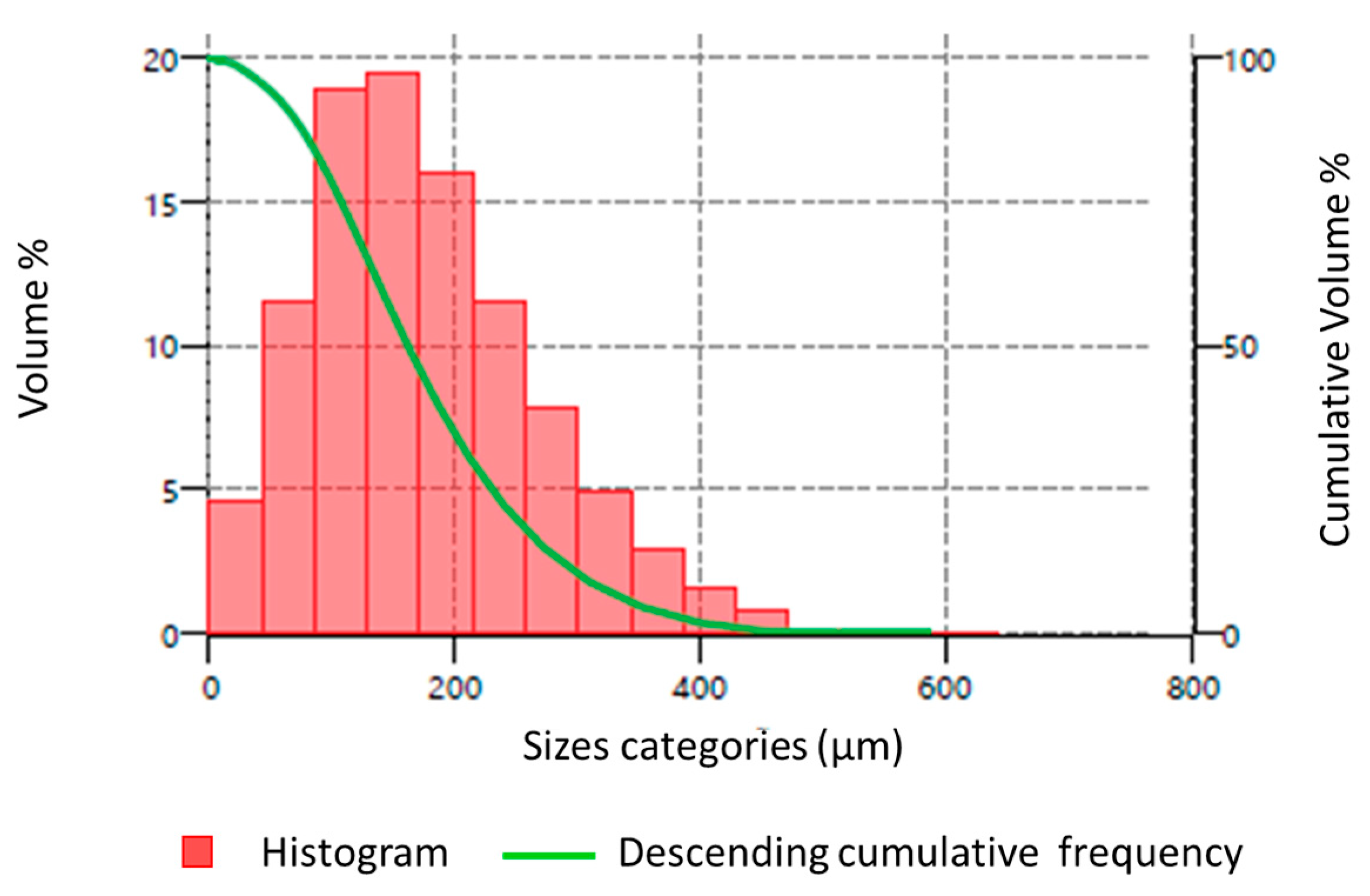
2.2. PS MP Preparation

The MP PS particles (200 µm range) used in this experimental design were the same as those employed in the study by González-Doncel et al. [32]. Briefly, the procedure for obtaining the particles is as follows: PS laboratory test tubes (Deltalab, Barcelona, Spain) were first shattered into small fragments, ground with a mixer mill (Retsch GMBH, Haan, Germany), and passed through a ≤200 µm sieve. These particles were characterized with a particle size analyser by laser diffraction (Mastersizer 3000, Malvern Analytical, Malvern, UK).

2.3. Food Contamination

The experimental design involved exposing the tadpoles to a control diet (uncontaminated food, Sera Micron®) and to two diets containing PS MPs mixed in food to obtain two treatment diets: 10X (8.4 mg PS MPs/g food/day) and 1X (0.84 mg PS MPs/g food/day). These MP concentrations corresponded to 2500 µg PS-MP/aquarium/day (500 µg PS-MP/L × 5L) and to 250 µg PS-MP/aquarium/day (50 µg PS-MP/L × 5L), respectively, incorporated into a basal diet of 300 mg food/aquarium/day.
The preparation of the PS 10X contaminated baseline diet was made by mixing 2 g of PS MPs with 2 g of the uncontaminated food (Sera Micron®) to thereafter incorporate amounts of uncontaminated food, which ensured a homogeneous mixture. The PS 1X baseline diet was achieved by diluting the PS 10X food with uncontaminated food at a ratio of 1:10, following the same mixing procedure described above. Both contaminated diets were administered at a constant level throughout the study to maintain the corresponding waterborne concentrations of PS-MPs in water. However, to meet the tadpoles’ increased nutritional requirements as they grew, increasing amounts of uncontaminated food were added to each aquarium at the same time as the basal diets containing PS-MPs (see Table S2, Supplementary Materials).

2.4. Exposure Design

The study was conducted following the OECD Test Guideline 231 [21], with some modifications. Groups of 22 tadpoles (stage N&F 51) were randomly transferred to four replicates per treatment (PS 10X and 1X) and control groups. Twelve aquaria, each containing 5 L of dechlorinated tap water, were used. Diets were administered daily for 21 days. The experimental conditions were monitored throughout the assay: room temperature was recorded daily. Water temperature, dissolved oxygen, pH, conductivity, and luminous intensity were randomly recorded weekly in one of the replicated groups. The study was performed under semi-static conditions, under which 100% water was renewed in each aquarium 3 times weekly.

2.4.1. Apical Endpoints

Mortality and morphological malformations in tadpoles were monitored daily throughout the experimental design. On day 7, five tadpoles per replicate (n = 20 per treatment) were randomly collected and anesthetized with 100 mg/L of tricaine methanesulfonate (MS-222, Argent Laboratories, Redmond, WA, USA), which was buffered with sodium bicarbonate. Individuals were rinsed with milli-Q water and gently blotted dry before recording the five apical endpoints: whole body length (WBL), left hind limb length (HLL), snout to vent length (SVL), wet body weight (BW), and developmental stage. For the WBL endpoint, the animals were photographed with a digital camera (mod.E-410, Olympus, Tokyo, Japan). For the HLL, SVL, and developmental stages, a digital camera coupled to a dissecting microscope (mod. SZX12, Olympus, Tokyo, Japan) was used. Measures were taken with the help of a computer-aided image analysis, Image-Pro Plus 4.0 (Media Cybernetics, Bethesda, MD, USA). Subsequently, they were humanely euthanized with an overdose of MS-222 (500 mg/L).
The same procedures were run for the remaining tadpoles on day 21 (15 animals per replicate, n = 60 per treatment). Five of these 15 tadpoles per replicate (n = 20 per treatment) were also randomly employed for the histological analyses of thyroid glands and to screen potential target organ systems and tissues (see below).

2.4.2. Histological Analysis

The tadpoles selected per treatment for histopathological examination were randomly divided to assess representative areas of thyroid glands, potential effects on coelomic organs, and for electron microscopy. The thyroid histology procedures followed the protocol described in the Guidance Document on Amphibian Thyroid Gland Histology [33], adapted for the AMA guideline [21]. After the anesthesia overdose, tadpoles were fixed in 10% neutral buffered formalin (40% formaldehyde). Fixed specimens were decapitated by transecting the carcass rostral to the heart with a razor blade. The head and cervical region were placed in a cassette and embedded in paraffin, with the ventral aspect facing the cutting surface of the block. Serial sections 50 microns apart were taken and examined under a magnifying scope until thyroid glands were detected. The two-step sections containing representative thyroid gland areas were selected and stained with hematoxylin–eosin for evaluation by a trained pathologist. Considering the inherent morphology variability in tadpole thyroid glands [33], the following features were considered: nuclei with vesicular chromatin, mild anisokaryosis, and foamy cytoplasm. The other histological features considered to be relevant signs of cellular adaptations and/or hyperplasia included follicular size variation, papillary projections with pseudostratified epithelium, and loss of nuclear polarity. Follicular changes were scored based on a scheme involving the previous features: Score 0: no relevant or only expected changes in follicles and follicular cells; Score 1: a single feature of cellular adaptation, hypertrophy, or hyperplasia; Score 2: papillary proliferation OR two features of score 1; Score 3: papillary proliferation AND one or more features of score 1.
Serial transverse sections of the remaining tadpoles were taken from each specimen and routinely processed to examine the remaining organs, i.e., the lungs, heart, kidney, liver, spleen, stomach, intestines, gonads, skin, skeletal muscle, and central nervous system.

2.4.3. Electron Microscopy

Formalin-fixed samples of liver and intestine of two randomly selected tadpoles from each treatment group were surveyed, sectioned into 1 mm pieces, and processed for electron microscopy as follows: first, the formalin-fixed tissue samples were thoroughly washed in distilled water and then placed in Karnowski solution for 2 h, which was renewed hourly. The samples were fixed with osmium and then gradually embedded in resin at increasing acetone and resin concentrations. Resin blocks were sectioned for ultrastructural visualization.

2.5. Statistical Data Analysis

The developmental and morphological endpoints were assessed for normality by the Shapiro–Wilk test and for homogeneity of variance by the Levene test. If these conditions were fulfilled, a one-way ANOVA test with post hoc tests was performed. Otherwise, non-parametric tests were conducted. Differences related to the control group were considered significant at p < 0.05. Statistical analyses were run with the IBM SPSS Statistics vs. 30 software. Following the AMA recommendation [21], the statistical evaluation for the HLL measurements was made by normalizing this value with the corresponding SVL value for each individual (i.e., the HLL to SVL ratio).

3. Results

3.1. PS MP Characterization, Experimental Conditions, Mortality, Developmental Assessment, and Morphometric Endpoints

The employed particles were identical to those used in the study by González-Doncel et al. [32]. The frequency histogram is shown in Figure 1, and the frequencies and percentiles of particle distribution are summarized in Table S3 of the Supplementary Materials. The characterization of the PS MP particles showed a size distribution with 10th, 50th, and 90th percentiles of 67.4, 161, and 302 µm, respectively, and a mode value of 180 µm.
The experimental conditions monitored throughout the assay complied with the acceptable levels for what is considered normal tadpole development (see Table S4, Supplemental Materials). No mortalities were detected in any of the replicated aquaria from the control group. The aquaria from the PS 10X MP and PS 1X MP treatment groups showed mortalities less than 20%, except in one replicated aquarium from the PS 1X MPs, where mortality reached 33%. This was assumed to be the result of occasional air pump failures at night. Table 1 summarizes the tadpole mortality in each replicate during the assay.
Tadpoles showed no relevant malformations in the control or the treatment group PS 1X MPs. These observations contrast with those from the PS 10X MP treatment in which occasional malformations were seen, mainly scoliosis. However, the final percentage of tadpoles affected by this alteration did not exceed 15% in each replicate aquarium.
The developmental stage distribution pattern on days 7 and 21 are presented in Table 2. No statistically significant differences for developmental stage were detected between the control and the PS 10X MP and PS 1x MP treatments, on either day 7 or day 21.
The morphometric values for the tadpoles assessed on days 7 and 21 are presented in Figure 2 and Figure 3, respectively.
None of the morphometric endpoints obtained from the PS 10X MP and the PS 1X MP groups on day 7 showed statistically significant differences compared to the control group. However, on day 21, both treatments, PS MPs 1X and 10X, resulted in a statistically significant reduction, and with a dose-related pattern, for the SVL endpoint versus the control group (Figure 3C). In addition, the PS 10X MP treatment showed a statistically significant increase for the WBL endpoint (Figure 3D).

3.2. Histological and Electron Microscopy Results

Significant histological features are presented in Figure 4A,F.

3.2.1. Histopathology of Thyroid Gland

In the control group, only one specimen scored 1, meaning there were minor cellular adaptations and slight evidence for hyperplasia. The remaining control individuals showed cellular changes within the normal limits. In the PS 1X MP treatment group, 70% of the specimens had cellular adaptations and signs of hyperplasia in the thyroid gland. Specifically at this concentration, 40% of the specimens scored 1 (contained only a single feature of cellular adaptation or hyperplasia), 10% scored 2, and 20% contained more evident features of proliferation and were, thus, scored as 3 (Figure 4A).
In the group exposed to concentrations of PS 10X MPs, 80% of the specimens contained at least 1 feature of adaptation/hyperplasia in the examined thyroids (scored 1), and 20% of specimens scored 3.

3.2.2. Histopathology of Gonads and Remaining Organs

The digestive tract was within the normal limits and contained ingested material in all the groups (Figure 4B). Three random specimens in the replicates from the controls and PS MPs 1X showed small amounts of calcium salts within renal tubules (Figure 4C). Extramedullary hematopoiesis was observed in all the livers regardless of treatment and as expected for the developmental stage. The PS 1X MP treatment revealed 50% of the livers with hepatocytes slightly decreased in size compared to the controls. A similar situation occurred in 20% of the livers from PS 10X MP treatment (Figure 4D). Both the testes and ovaries had adequately developed for the metamorphosis stage in the controls and treatment groups (Figure 4E,F).

3.2.3. Electron Microscopy

In the intestine, fragments of filamentous and polygonal, moderately electron-dense material, were identified within the lumen and between enterocyte microvilli in the specimens subjected to both PS MPs 1X and 10X concentrations (Figure 5B,C), but not in the control specimen (Figure 5A,D). In the enterocytes from the PS 1X MP concentration, the cytoskeleton revealed signs of edema, while desmosomes and mitochondria were within the normal limits (Figure 5E). In the specimen from the PS 10X MP replicates, in addition to edema, the cytoskeleton also contained numerous lysosomes and vesicles digesting membranous fragments and filamentous material with a similar density to the material within the lumen (Figure 5F).
In the liver (Figure 6A–D), both treatment group tadpoles contained cumuli of protein within bile canaliculi, suggesting bile stasis (Figure 6B,D). Hepatocytes from specimen PS 1X MPs also had mild evidence of degeneration (Figure 6A).

4. Discussion

In this research, we extended studies into the effects of PS-MPs on amphibians, including the potential effects of endocrine disruption on the hypothalamic–pituitary–thyroid axis. To the best of our knowledge, this is the first study to explore the effects of PS-MPs on the thyroid axis of tadpoles using the AMA test. Morphological, histological, and ultrastructural changes were also studied in other organs. To this end, Xenopus laevis tadpoles were exposed to irregularly shaped, heterogeneous virgin PS MPs via their diet. In summary, based on our experimental design, dietary MP PS exposure did not reveal any statistically significant morphological effects, except for the SVL and WBL endpoints observed on day 21. However, these effects could be related to general toxicity [21]. At the histological level, a mild tendency for follicular changes associated with hyperplasia or increased functionality was observed in the treatment groups, but these were considered minimal and could not be corroborated with the apical endpoints. In addition, the hepatocytes from the treatment groups were slightly smaller than those in the control group, particularly in the PS 1X MP group, but were not considered pathological in any case. No notable effects on the digestive tract or kidneys were observed. However, the electron microscopy evaluation conducted on a few tadpoles showed evident ultrastructural modifications in the enterocytes and hepatocytes of the tadpoles exposed to 1X and 10X PS MPs, but these were likely to be reversible.
The concentrations selected in our experimental design, in terms of mass of MP per volume (500 and 50 µg MP/L), fell within the range of those reported in other studies. Examples are the studies on amphibians [34,35], fish [11,32,36,37,38,39,40,41] or microinvertebrates [42,43]. Nevertheless, when considering the number of particles per unit volume of water, exposure to the irregular PS MPs used in our study was two to five orders of magnitude lower than in other published studies. These studies demonstrated toxic effects in aquatic organisms at concentrations far higher than those typically found in natural aquatic environments, often by several orders of magnitude [14,44]. Other examples include the study by Boyero and colleagues [9], in which tadpoles were exposed to a maximum concentration of 1800 particles/mL, and the study by De Felice and colleagues [34], in which the maximum concentration was 8.666 × 10⁵ particles/mL. However, our previous research with medaka fish, which resorted to the same size of PS MP particles and the same waterborne concentrations administered through diet, resulted in daily mean numbers of 3087 and 247 particles/L, for PS10X and 1X, respectively [32]. We, therefore, assumed a comparable number of particles present in each tank daily, which were bioavailable to tadpoles. It should be noted that particle size determines the number of particles that can exist at a given concentration [3]. As a result, comparisons between the results of experimental studies that report a wide range of MP abundances, sizes, and types are very difficult [3,45]. Hence, there is a need to harmonize MP exposure methods to achieve more realistic exposure approaches [4,14,32,46,47].
On the other hand, the goal of our study was not to quantify the precise dose of MPs ingested by individual tadpoles, but rather to evaluate the biological effects of microplastics that were made bioavailable through their incorporation into food, so that they would ingest MPs while eating. Some studies show that fish can spit out MPs if they are not ingested with food because they can detect them. However, if the MPs are incorporated into food, it is more likely that fish will ingest them [48]. A similar pattern can be expected for tadpoles. Additionally, the density of the used plastic, PS, causes it to float, meaning it remains with the food. Therefore, when food is placed in water, plastic particles are also present and bioavailable to tadpoles. For these reasons, exposure to MPs mixed with food can be considered a realistic worst-case scenario.
In line with previous laboratory studies [15,34], our experimental approach did not show any significant incidence of tadpole mortality. Contrarily, Boyero and co-workers [9] reported significant mortality in Alytes obstetricans tadpoles, which were exposed to comparatively higher concentrations of PS fluorescent microspheres (1800 particles/mL) and of a smaller size than the particles herein used (10 µm diameter). Small-sized plastic particles represent a higher risk for freshwater organisms than bigger ones [6,28,49,50] due to their potential to penetrate passively through cell membranes [14], whereas large-sized particles, like those in our study (200 µm), would require active transport, such as endocytosis [6].
Although our study detected a number of muscular–skeletal malformations in the tadpoles exposed to the PS MPs10X dietary concentration, they could not be considered statistically significant. Some authors have reported 10 to 30% of spontaneous malformations from an entire spawn of tadpoles with no known cause [51]. However, Araújo and co-workers [15] observed asymmetric deviation during tadpoles’ tail development based on the caudal insertion scores of individuals exposed to 60 mg of polyethylene MPs/L for 7 days. Other anatomical changes induced by MPs/NPs have been described in other studies; for example, Venâncio and co-workers [35] pointed out a significant percentage of malformations in tadpoles after dietary exposure to 1 mg of polymethylmethacrylate NPs/L for 48 h; these malformations consisted of gut externalization and affected 62% of the animals. In all these studies, tadpoles were exposed to MPs for considerably shorter periods than in our 21-day study, but the applied concentrations were much higher. Once more, the presence of anatomical malformations could be affected by the different exposure conditions and particle sizes used in these studies [29].
Considering the AMA apical endpoints, our study showed no effects on tadpole development, but a statistically significant dose–response reduction at the SVL endpoint and an increase in WBL from the 21-day PS 10X MP treatment. Other studies have observed similar responses; Tussellino et al. [27] showed a reduction in body length in Xenopus embryos injected with 50 nm PS NPs. Boyero and co-workers [9] and Balestrieri and co-workers [25] reported effects on amphibian growth after being exposed to MPs, while Venâncio and co-workers [35] showed the same effects for exposing NPs. However, these surveys had different hypotheses to explain these effects; the first two studies [9,25] attributed these effects to a decrease in the feeding rates, which could alter the energy used for growth. Venâncio and co-workers [35] related the negative effect on growth to an energy reduction as a result of the detoxification mechanisms involved in NP ingestion and the interference in the absorption of nutrients provoked by NP aggregates in the gut [35]. These hypotheses have also been formulated for fish exposure [28] and other aquatic organisms [7,14,52]. In contrast to these studies, effects of MP/NPs on WBL and SVL have also been observed in an opposite way to our study. Araújo and co-workers [15] reported a decrease in the WBL and an increase in the SVL of tadpoles exposed to PE MPs. In this aforementioned study, the size of the used MPs was smaller than that exposed in our study. Finally, other research works did not report any significant effects on growth from exposures to microspheres of PS [34,53]. These contradictory effects provoked by MP/NP exposure have also been noted in other animals, such as fish, where some works have depicted effects on growth [38] versus others that have not [32,40].
Once again, the variable response of toxic effects of MPs/NPs may be explained by the different tadpole stages exposed during the assays, the size of MPs/NPs, and the exposure time [19], which makes comparing the results between studies difficult. Additionally, the mechanisms of MPs' toxicity are poorly understood and require investigation [14].
This study focused on the histopathological effects of MPs on the thyroid and other organs. This assessment is considered a valuable tool for estimating the damage caused by xenobiotics, including MPs/NPs [29]. In the case of MP exposure, the effects are conditioned to particle size and shape [29]. In our research, MPs were apparently absent from different tissues at a histological level; the same results have been reported after exposure in other aquatic organisms [41].
The minimal changes in thyroid gland size and follicular proliferation, together with the absence of morphological and developmental markers, mean that it is unlikely that the clear endocrine disruption of the thyroid could be detected in this study, whether caused by the MPs or the substances used in their manufacture. In this case, further research correlating the histological results with molecular changes in the expression of thyroid-related genes and measurements of thyroid hormones has been suggested [32,50]. The previous study in medaka fish revealed the apparent attenuation of thyroid follicular epithelium and increased follicle diameter after the 150-day exposure to 10× concentration of PS MPs, which was suggestive of thyroid hypofunction [32]. In addition, studies with smaller plastic particles, in the range of NPs, should be carried out to understand the extent of the effects of these particles on thyroid axis functionality.
Broken down plastics in the gut lumen were not discriminated from those ingested within the gut lumen with light microscopy, but their presence was confirmed with the ultrastructural study. However, some reports have described adverse cellular changes in certain tissues such as gonads, kidneys, and eyes at increasing concentrations, generally higher than those used in our research [15,16,41]. The gonads in this study were well developed and lacked adaptive or pathological alterations. However, this does not disregard functional alterations that may occur later in the growth cycle or with persistent exposure to MPs. Chisada and co-workers [41] revealed negative effects on reproduction due to oxidative stress after exposure to adverse levels of MPs in aquatic organisms. However, the study of González-Doncel and co-workers [32] did not evidence any significant effects of PS MP exposure on offspring success, although the initial periods of the reproductive phase revealed a fall in the gravid females, fecundity, and fertilization rates in the animal exposed to PS MPs, values that were recovered to normal rates on the succeeding days.
The reported changes in kidneys include the sclerosis or thickening of glomerular basement membranes [41]. Our previous study with medaka fish showed signs of glomerular degeneration, congestive glomeruli, glomerulogenesis, and tubular degeneration in the PS 10X-treated fish. However, these changes could not be attributed entirely to PS exposure because similar changes were randomly present in the control group [32]. In the present study, such changes were not observed in any of the tadpoles. Only a minor presence of calcium salts within a few renal tubules was randomly observed and could not be strictly attributed to MPs, since this finding was also reported in the controls.
Finally, some studies have reported cases of liver tissue damage in fish following MP ingestion. This damage has been linked to oxidative stress and inflammatory responses, which can result in the death of hepatocytes in the liver and have been suggested as a potential mechanism of MPs' toxicity [54,55]. In our study, slight modifications in hepatocyte size were observed and occurred more frequently in the PS 1X treatment group.
The ultrastructural analysis revealed protein aggregates within bile canaliculi in both treatment groups, suggesting bile stasis and mild hepatocyte degeneration in the PS 1X MP group. The intestinal ultrastructure was examined to determine if there were any alterations that could provide insights into how and if MPs interacted at the cellular level. Fragments of fibrillar and polygonal material compatible with plastic were found in lumens and between enterocyte microvilli, confirming the presence of MPs in the digestive tract. At lower concentrations, enterocytes were within the normal limits, and no evidence for excessive pinocytosis or secondary lysosomes was present. However, at higher concentrations, the cytoskeleton evidenced a mild degree of degeneration and excessive lysosome digestion. This could imply some degree of reaction of the enterocyte to MP remnants at this level, or even absorption despite the lack of consequences at a systemic level.
In summary, and similarly to what has been reported in previous studies and reviews [56], our histological findings show that the impact of PS MPs at the given concentrations and size has little or no impact on tissues, and at the very least, injuries are mild at the ultrastructural level and likely reversible. This also agrees with our previous studies in medaka fish [32], in which histological injuries were mild and a direct association with PS exposure was elusive. However, due to the type of plastic used in our study (PS laboratory test tube ground and of different sizes and shapes), smaller PS particle sizes could be present in the mixture, albeit at low percentages. Additionally, the hypothesis proposed by some authors that MPs could degrade into smaller pieces once in the tadpole’s gut [52] could explain the presence of smaller PS particles. These particles may therefore be responsible for the slight effects observed in tadpoles at the cellular level.

5. Conclusions

This research extends the information about the effects of MPs on amphibians, a taxonomic class whose populations are declining because of multiple causes, including the anthropogenic pollution of emerging contaminants. To this end, the impact of PS-MPs on the thyroid axis of tadpoles and other organs was investigated. In conclusion, dietary exposure of PS MPs ≤ 200 µm, in the circumstances of this study, did not seem to affect normal Xenopus laevis tadpole development, but growth was slightly affected. In addition, further studies related to the effects of MPs on thyroid function should be conducted, and should include the correlation between the histological results with molecular changes in the expression of thyroid-related genes and measurements of thyroid hormones.
The contradictory results between studies about exposure to MPs/NPs could be explained according to the kind of plastic, its size and shape, exposure period, and the concentrations of the used plastic particles. This reinforces the idea that standardized methods for the environmental risk assessment of MPs/NPs are needed. Hence, there is an increasing necessity to carry out experimental designs that consider environmentally relevant concentrations for MPs by taking into account exposure to weathered plastics instead of virgin ones, and that include sharp-shaped particles rather than spherical microbeads. All this will provide more realistic information about the toxic effects of MPs. Furthermore, as concern about the risk of NPs seems greater than that for larger particles, more studies with smaller plastic particles within the NP range should be conducted. Finally, the histopathological and electron microscopy diagnoses provide valuable information about the effects of MPs/NPs at the cellular level. Thus, these techniques should be routinely incorporated to assess the toxic effects of plastics.

Supplementary Materials

The following supporting information can be downloaded at https://www.mdpi.com/article/10.3390/environments12080252/s1, Table S1, Food regime (mg per animal per day) administered throughout the development of the Xenopus laevis tadpoles in the pre-exposure period, based on the N&F stage of the animals. Table S2, Distribution of the food quantities according to tadpole stages and days of the assay. Table S3, Particle-size distribution frequencies (in percentage), 10th, 50th 90th percentiles and mode of the PS MP particles exposed to tadpoles of Xenopus laevis. Table S4, Physicochemical parameters in the test media during the assay.

Author Contributions

The authors have contributed to the following tasks: conceptualization: M.V.P. and M.G.-D. Methodology: M.V.P., M.d.l.Á.J., E.M.B. and M.G.-D. Investigation: M.V.P., M.d.l.Á.J., E.M.B., M.L.d.S.-G., P.G.-H. and M.G.-D. Writing—original draft: M.V.P., M.d.l.Á.J., E.M.B. and M.G.-D. Writing—review and editing: M.V.P., M.d.l.Á.J., E.M.B. and M.G.-D. Funding acquisition: M.V.P. and M.G.-D. All authors have read and agreed to the published version of the manuscript.

Funding

This work is part of the Grant RTI2018-096046-B-C21, funded by MCIN/AEI/10.13039/501100011033 and by “ERDF A way of making Europe”.

Data Availability Statement

The original contributions presented in this study are included in the article/Supplementary Material. Further inquiries can be directed to the corresponding author.

Acknowledgments

The authors wish to thank the company Instrumentación Específica de Materiales, S.A, IESMAT, and especially J.C. Puebla for his invaluable help in the characterization of the polystyrene particles.

Conflicts of Interest

The authors declare no conflict of interest.

References

  1. Duis, K.; Coors, A. Microplastics in the aquatic and terrestrial environment: Sources (with a specific focus on personal care products), fate and effects. Environ. Sci. Eur. 2016, 28, 2. [Google Scholar] [CrossRef] [PubMed]
  2. Eerkes-Medrano, D.; Thompson, R.C.; Aldridge, D.C. Microplastics in freshwater systems: A review of the emerging threats, identification of knowledge gaps and prioritisation of research needs. Water Res. 2015, 75, 63–82. [Google Scholar] [CrossRef] [PubMed]
  3. Ricciardi, M.; Pironti, C.; Motta, O.; Miele, Y.; Proto, A.; Montano, L. Microplastics in the Aquatic Environment: Occurrence, Persistence, Analysis, and Human Exposure. Water 2021, 13, 973. [Google Scholar] [CrossRef]
  4. Kögel, T.; Bjorøy, Ø.; Toto, B.; Bienfait, A.M.; Sanden, M. Micro- and nanoplastic toxicity on aquatic life: Determining factors. Sci. Total Environ. 2020, 709, 136050. [Google Scholar] [CrossRef] [PubMed]
  5. Araújo, A.P.C.; Lopes Rocha, T.; de Melo e Silva, D.; Malafaia, G. Micro(nano)plastics as an emerging risk factor to the health of amphibian: A scientometric and systematic review. Chemosphere 2021, 283, 131090. [Google Scholar] [CrossRef] [PubMed]
  6. Triebskorn, R.; Braunbeck, T.; Grummt, T.; Hanslik, L.; Huppertsberg, S.; Jekel, M.; Knepper, T.P.; Krais, S.K.; Müller, Y.K.; Pittroff, M.; et al. Relevance of nano- and microplastics for freshwater ecosystems: A critical review. TrAC 2019, 110, 375–392. [Google Scholar] [CrossRef]
  7. Qaiser, Z.; Aqeel, M.; Sarfraz, W.; Rizvi, Z.F.; Noman, A.; Naeem, S.; Khalid, N. Microplastics in wastewaters and their potential effects on aquatic and terrestrial biota. Case Stud. Chem. Environ. Eng. 2023, 8, 100536. [Google Scholar] [CrossRef]
  8. Chiu, M.C.; Ao, S.; Ling, C.; He, F.; Luo, Q.; Wen, Z.; Cai, Q.; Resh, V.H. Meta-ecosystem Frameworks Can Enhance Control of the Biotic Transport of Microplastics. Environ. Sci. Technol. 2024, 58, 12846–12852. [Google Scholar] [CrossRef] [PubMed]
  9. Boyero, L.; López-Rojo, N.; Bosch, J.; Alonso, A.; Correa-Araneda, F.; Pérez, J. Microplastics impair amphibian survival, body condition and function. Chemosphere 2020, 244, 125500. [Google Scholar] [CrossRef] [PubMed]
  10. Lee, K.W.; Shim, W.J.; Kwon, O.W.; Kang, J.H. Size-dependent effects of micro-polystyrene particles in the marine copepod Tigriopus japonicas. Environ. Sci. Technol. 2013, 47, 11278–11283. [Google Scholar] [CrossRef] [PubMed]
  11. Barboza, L.G.A.; Vieira, L.R.; Branco, V.; Figueiredo, N.; Carvalho, F.; Carvalho, C.; Guilhermino, L. Microplastics cause neurotoxicity, oxidative damage and energy-related changes and interact with the bioaccumulation of mercury in the European seabass, Dicentrarchus labrax (Linnaeus, 1758). Aquat. Toxicol. 2018, 195, 49–57. [Google Scholar] [CrossRef] [PubMed]
  12. Prüst, M.; Meijer, J.; Westerink, R.H.S. The plastic brain: Neurotoxicity of micro- and nanoplastics. Part. Fibre Toxicol. 2020, 17, 24. [Google Scholar] [CrossRef] [PubMed]
  13. Brandts, I.; Teles, M.; Gonçalves, A.P.; Barreto, A.; Franco-Martínez, L.; Tvarijonaviciute, A.; Martins, M.A.; Soares, A.M.V.M.; Tort, L.; Oliveira, M. Effects of nanoplastics on Mytilus galloprovincialis after individual and combined exposure with carbamazepine. Sci. Total Environ. 2018, 643, 775–784. [Google Scholar] [CrossRef] [PubMed]
  14. Ma, H.; Pu, S.; Liu, S.; Bai, Y.; Mandal, S.; Xing, B. Microplastics in aquatic environments: Toxicity to trigger ecological consequences. Environ. Pollut. 2020, 261, 114089. [Google Scholar] [CrossRef] [PubMed]
  15. Araújo, A.P.C.; Ferreira Silva de Melo, N.; Gonçalves de Oliveira Junior, A.; Postalli Rodriguesa, F.; Fernandes, T.; de Andrade Vieira, J.E.; Lopes Rochad, T.; Malafaia, G. How much are microplastics harmful to the health of amphibians? A study with pristine polyethylene microplastics and Physalaemus cuvieri. J. Hazard. Mater. 2020, 382, 121066. [Google Scholar] [CrossRef] [PubMed]
  16. Araújo, A.P.C.; Rodrigues Gomes, A.; Malafaia, G. Hepatotoxicity of pristine polyethylene microplastics in neotropical Physalaemus cuvieri tadpoles (Fitzinger, 1826). J. Hazard. Mater. 2020, 386, 121992. [Google Scholar] [CrossRef] [PubMed]
  17. Bosch, J.; Thumsová, B.; López-Rojo, N.; Pérez, J.; Alonso, A.; Fisher, M.C.; Boyero, L. Microplastics increase susceptibility of amphibian larvae to the chytrid fungus Batrachochytrium dendrobatidis. Sci. Rep. 2021, 11, 22438. [Google Scholar] [CrossRef] [PubMed]
  18. Rochman, C.M.; Browne, M.A.; Halpern, B.S.; Hentschel, B.T.; Hoh, E.; Karapanagioti, H.K.; Rios-Mendoza, L.M.; Takada, H.; Teh, S.; Thompson, R.C. Classify plastic waste as hazardous. Nature 2013, 494, 169–171. [Google Scholar] [CrossRef] [PubMed]
  19. Rahman, M.M.; Kim, E.-S.; Sung, H.-C. Microplastics as an emerging threat to amphibians: Current status and future perspectives. Heliyon 2024, 10, e28220. [Google Scholar] [CrossRef] [PubMed]
  20. Ullah, S.; Ahmad, S.; Guo, X.; Ullah, S.; Ullah, S.; Nabi, G.; Wanghe, K. A review of the endocrine disrupting effects of micro and nano plastic and their associated chemicals in mammals. Front. Endocrinol. Lausanne 2023, 13, 1084236. [Google Scholar] [CrossRef] [PubMed]
  21. OECD. Test No. 231: Amphibian Metamorphosis Assay, OECD Guidelines for the Testing of Chemicals, Section 2; OECD Publishing: Paris, France, 2009. [Google Scholar] [CrossRef]
  22. Ruthsatz, K.; Dausmann, K.H.; Paesler, K.; Babos, P.; Sabatino, N.M.; Peck, M.A.; Glos, J. Shifts in sensitivity of amphibian metamorphosis to endocrine disruption: The common frog (Rana temporaria) as a case study. Conserv. Physiol. 2020, 8, coaa100. [Google Scholar] [CrossRef] [PubMed]
  23. Kloas, W.; Stöck, M.; Lutz, I.; Ziková-Kloas, A. Endocrine disruption in teleosts and amphibians is mediated by anthropogenic and natural environmental factors: Implications for risk assessment. Phil. Trans. R. Soc. B 2024, 379, 20220505. [Google Scholar] [CrossRef] [PubMed]
  24. Shetu, M.H.; Parvin, F.; Tareq, S.M. Identifying the presence of microplastics in frogs from the largest delta of the world. Environ. Adv. 2023, 11, 100355. [Google Scholar] [CrossRef]
  25. Balestrieri, A.; Winkler, A.; Scribano, G.; Gazzola, A.; Lastrico, G.; Grioni, A.; Pellitteri-Rosa, D.; Tremolada, P. Differential effects of microplastic exposure on anuran tadpoles: A still underrated threat to amphibian conservation? Environ. Pollut. 2022, 303, 119137. [Google Scholar] [CrossRef] [PubMed]
  26. Szkudlarek, M.; Najbar, B.; Jankowiak, L. Variation in microplastic characteristics among amphibian larvae: A comparative study across different species and the influence of human activity. Sci. Rep. 2024, 14, 13574. [Google Scholar] [CrossRef] [PubMed]
  27. Tussellino, M.; Ronca, R.; Formiggini, F.; De Marco, N.; Fusco, S.; Netti, P.A.; Carotenuto, R. Polystyrene nanoparticles affect Xenopus laevis development. J. Nanopart. Res. 2015, 17, 70. [Google Scholar] [CrossRef]
  28. Lu, Y.; Zhang, Y.; Deng, Y.; Jiang, W.; Zhao, Y.; Geng, J.; Ding, L.; Ren, H. Uptake and Accumulation of Polystyrene Microplastics in Zebrafish (Danio rerio) and Toxic Effects in Liver. Environ. Sci. Technol. 2016, 50, 4054–4060. [Google Scholar] [CrossRef] [PubMed]
  29. Patra, I.; Huy, D.T.N.; Alsaikhan, F.; Opulencia, M.J.C.; Tuan, P.V.; Nurmatova, K.C.; Majdi, A.; Shoukat, S.; Yasin, G.; Margiana, R.; et al. Toxic effects on enzymatic activity, gene expression and histopathological biomarkers in organisms exposed to microplastics and nanoplastics: A review. Environ. Sci. Eur. 2022, 34, 80. [Google Scholar] [CrossRef]
  30. Pablos, M.V.; Beltrán, E.M.; Jiménez, M.A.; García-Hortigüela, P.; Fernández, A.; González-Doncel, M.; Fernández, C. Effect assessment of reclaimed water and carbamazepine exposure on the thyroid axis of X. laevis: Apical and histological effects. Sci. Total Environ. 2020, 723, 138023. [Google Scholar] [CrossRef] [PubMed]
  31. Nieuwkoop, P.D.; Faber, J. Normal Tables of Xenopus Laevis (Daudin); Garland Publishing, Inc.: New York, NY, USA, 1994. [Google Scholar]
  32. González-Doncel, M.; García-Mauriño, J.E.; Beltrán, E.M.; Fernández-Torija, C.; Andreu-Sánchez, O.; Pablos, M.V. Effects of life cycle exposure to polystyrene microplastics on medaka fish (Oryzias latipes). Environ. Pollut. 2022, 311, 120001. [Google Scholar] [CrossRef] [PubMed]
  33. OECD. Guidance Document on Amphibian Histology, OECD Series on Testing and Assessment, No. 82; OECD Publishing: Paris, France, 2007. [Google Scholar] [CrossRef]
  34. De FeLice, B.; Bacchetta, R.; Santo, N.; Tremolada, P.; Parolini, M. Polystyrene microplastics did not affect body growth and swimming activity in Xenopus laevis tadpoles. Environ. Sci. Pollut. Res. 2018, 25, 34644–34651. [Google Scholar] [CrossRef] [PubMed]
  35. Venâncio, C.; Melnic, I.; Tamayo-Belda, M.; Oliveira, M.; Martins, M.A.; Lopes, I. Polymethylmethacrylate nanoplastics can cause developmental malformations in early life stages of Xenopus laevis. Sci. Total Environ. 2022, 806, 150491. [Google Scholar] [CrossRef] [PubMed]
  36. Lechner, A.; Keckeis, H.; Lumesberger-Loisl, F.; Zens, B.; Krusch, R.; Tritthart, M.; Glas, M.; Schludermann, E. The Danube so colourful: A potpourri of plastic litter outnumbers fish larvae in Europe’s second largest river. Environ. Pollut. 2014, 188, 177–181. [Google Scholar] [CrossRef] [PubMed]
  37. Jin, Y.; Xia, J.; Pan, Z.; Yang, J.; Wang, W.; Fu, Z. Polystyrene microplastics induce microbiota dysbiosis and inflammation in the gut of adult zebrafish. Environ. Pollut. 2018, 235, 322–329. [Google Scholar] [CrossRef] [PubMed]
  38. Wang, J.; Li, Y.; Lu, L.; Zheng, M.; Zhang, X.; Tian, H.; Wang, W.; Ru, S. Polystyrene microplastics cause tissue damages, sex-specific reproductive disruption and transgenerational effects in marine medaka (Oryzias melastigma). Environ. Pollut. 2019, 254, 113024. [Google Scholar] [CrossRef] [PubMed]
  39. Wang, J.; Zheng, M.; Lu, L.; Li, X.; Zhang, Z.; Ru, S. Adaptation of life-history traits and trade-offs in marine medaka (Oryzias melastigma) after whole life-cycle exposure to polystyrene microplastics. J. Hazard. Mater. 2021, 414, 125537. [Google Scholar] [CrossRef] [PubMed]
  40. Qiang, L.; Lo, L.S.H.; Gao, Y.; Cheng, J. Parental exposure to polystyrene microplastics at environmentally relevant concentrations has negligible transgenerational effects on zebrafish (Danio rerio). Ecotoxicol. Environ. Saf. 2020, 206, 111382. [Google Scholar] [CrossRef] [PubMed]
  41. Chisada, S.; Yoshida, M.; Karita, K. Polyethylene microbeads are more critically toxic to the eyes and reproduction than the kidneys or growth in medaka, Oryzias latipes. Environ. Pollut. 2021, 268, 115957. [Google Scholar] [CrossRef] [PubMed]
  42. Goldstein, M.C.; Rosenberg, M.; Cheng, L. Increased oceanic microplastic debris enhances oviposition in an endemic pelagic insect. Biol. Lett. 2012, 8, 817–820. [Google Scholar] [CrossRef] [PubMed]
  43. De Felice, B.; Sabatini, V.; Antenucci, S.; Gattoni, G.; Santo, N.; Bacchetta, R.; Ortenzi, M.A.; Parolini, M. Polystyrene microplastics ingestion induced behavioral effects to the cladoceran Daphnia magna. Chemosphere 2019, 231, 423–431. [Google Scholar] [CrossRef] [PubMed]
  44. Edo, C.; González-Pleiter, M.; Leganés, F.; Fernández-Piñas, F.; Rosal, R. Fate of microplastics in wastewater treatment plants and their environmental dispersion with effluent and sludge. Environ. Pollut. 2020, 259, 113837. [Google Scholar] [CrossRef] [PubMed]
  45. Rossatto, A.; Zimmer Ferreira Arlindo, M.; Saraiva de Morais, M.; Denardi de Souza, T.; Saraiva Ogrodowski, C. Microplastics in aquatic systems: A review of occurrence, monitoring and potential environmental risks. Environ. Adv. 2023, 13, 100396. [Google Scholar] [CrossRef]
  46. SAPEA. Science Advice for Policy by European Academies. A Scientific Perspective on Microplastics in Nature and Society; SAPEA: Berlin, Germany, 2019. [Google Scholar] [CrossRef]
  47. Leusch, F.D.L.; Ziajahromi, S. Converting mg/L to particles/L: Reconciling the occurrence and toxicity literature on microplastics. Environ. Sci. Technol. 2021, 55, 11470–11472. [Google Scholar] [CrossRef] [PubMed]
  48. Ory, N.C.; Gallardo, C.; Lenz, M.; Thiel, M. Capture, swallowing, and egestion of microplastics by a planktivorous juvenile fish. Environ. Poll. 2018, 240, 566–573. [Google Scholar] [CrossRef] [PubMed]
  49. Abarghouei, S.; Hedayati, A.; Raeisi, M.; Hadavand, B.S.; Rezaei, H.; Abed-Elmdoust, A. Size-dependent effects of microplastic on uptake, immune system, related gene expression and histopathology of goldfish (Carassius auratus). Chemosphere 2021, 276, 129977. [Google Scholar] [CrossRef] [PubMed]
  50. Lin, W.; Ling, L.; Ling, L.; Luo, H.; Jiang, Y.; Li, X.; Yao, Y.; Yang, P. Combined effects of co-exposure to microcystin-LR and polystyrene microplastics on growth, brain pathology and thyroid hormone homeostasis in adult zebrafish. Ecotoxicol. Environ. Saf. 2025, 291, 117855. [Google Scholar] [CrossRef] [PubMed]
  51. Coady, K.K.; Lehman, C.M.; Currie, R.J.; Marino, T.A. Challenges and approaches to conducting and interpreting the amphibian metamorphosis assay and the fish short-term reproduction assay. Birth Defects Res. 2014, 101, 80–89. [Google Scholar] [CrossRef] [PubMed]
  52. Hu, L.; Chernick, M.; Hinton, D.E.; Shi, H. Microplastics in Small Waterbodies and Tadpoles from Yangtze River Delta, China. Environ. Sci. Technol. 2018, 52, 8885–8893. [Google Scholar] [CrossRef] [PubMed]
  53. Hu, L.; Su, L.; Xue, Y.; Mu, J.; Zhu, J.; Xu, J.; Shi, H. Uptake, accumulation and elimination of polystyrene microspheres in tadpoles of Xenopus tropicalis. Chemosphere 2016, 164, 611–617. [Google Scholar] [CrossRef] [PubMed]
  54. Sun, Y.; Qingfang, D.; Zhanga, Q.; Zhoua, X.; Chena, R.; Li, S.; Wu, Q.; Chen, H. Hazards of microplastics exposure to liver function in fishes: A systematic review and meta-analysis. Mar. Environ. Res. 2024, 196, 106423. [Google Scholar] [CrossRef] [PubMed]
  55. Li, S.; Shi, M.; Wang, Y.; Xiao, Y.; Cai, D.; Xiao, F. Keap1-Nrf2 pathway up- regulation via hydrogen sulfide mitigates polystyrene microplastics induced–hepatotoxic effects. J. Hazard Mater. 2021, 402, 123933. [Google Scholar] [CrossRef] [PubMed]
  56. De Sales-Ribeiro, C.; Brito-Casillas, Y.; Fernandez, A.; Caballero, M.J. An end to the controversy over the microscopic detection and effects of pristine microplastics in fish organs. Sci. Rep. 2020, 10, 12434. [Google Scholar] [CrossRef] [PubMed]
Figure 1. Frequency histogram and descending cumulative frequency of PS-MPs.
Environments 12 00252 g001
Figure 2. Results from the (A) hind limb length (HLL) normalized by SVL; (B) wet body weight (BW); (C) snout to vent length (SVL); and (D) whole body length (WBL) measurements on day 7 of the assay. p < 0.05 based on an ANOVA test, followed by an LSD test (for the SVL, weight, and WBL endpoints) or a two independent samples test (Mann–Whitney test) (for HLL normalized by the SVL endpoint).
Environments 12 00252 g002
Figure 3. Results from the (A) hind limb length (HLL) normalized by SVL; (B) wet body weight (BW); (C) snout to vent length (SVL); and (D) whole body length (WBL) measurements on day 21 of the assay. * asterisks denote statistically significant differences from the control groups. p < 0.05 based on an ANOVA test, followed by an LSD test (for HLL normalized by the SVL endpoint) or Dunnett T3 (for weight) or a two independent samples test (Mann–Whitney test) (for the WBL and SVL endpoints).
Environments 12 00252 g003
Figure 4. Selection of the main histopathological features: (A) Thyroid gland at ×40 magnification. Thyroid follicle with small papillary projection (arrow) and follicular cells with loss of polarity and segmental pseudostratification. Inset: low magnification (×4) of the thyroid glands with variation in follicle size. (B) Section of the coelomic cavity (at ×4 magnification) containing the stomach, pancreas, kidney, intestine, and partial fragment of the liver. (C) Kidney at ×40 magnification. Detail of slightly dilated renal tubules with low cuboidal epithelium and intraluminal deeply basophilic concretions, consistent with calcium salts (*). (D) Liver at ×10 magnification. Atrophied hepatocytes, lacking lipid vacuoles. (E) Detail of testes (T) (at ×10 magnification) in a male Xenopus laevis tadpole. (F) Detail of the ovary (Ov) (at ×10 magnification) in a female Xenopus laevis tadpole.
Environments 12 00252 g004
Figure 5. Electron microscopy of the intestine: (A) Control group. Three enterocytes with even-length microvilli, tight junctions in the apical region, and numerous mitochondria within the cytoskeleton. (B) PS 1X MP group: enterocyte microvilli separated by filamentous, heterogeneously electron-dense, 100–500 nm fragmented material. Inset: similar material consistent with the MPs within the lumen. (C) PS 10X MP group. Seven enterocytes and larger filamentous MP fragments in the lumen. Inset: detail of similar MP rests separating microvilli. (D) Control group: two enterocytes delimited by the plasmatic membrane and joined by desmosomes. (E) PS 1X MP group. Two enterocytes with abundant glycogen granules, mitochondria, and cytoskeleton separated by electron-lucent areas, consistent with edema. (F) PS 10X MP group. Disorganized enterocyte cytoskeleton with electronlucent spaces, and many secondary lysosomes digesting membrane fragments. Inset: detail of lysosome containing filamentous material consistent with MP electron density.
Environments 12 00252 g005
Figure 6. Electron microscopy of the liver. (A) PS 1X MP group. Hepatocyte with six lipid vacuoles, perinuclear rough endoplasmic reticulum, and mitochondria. The cytoskeleton is fragmented and variably electron-lucent (degeneration). (B) PS 1X MP group. Bile canaliculus with membrane and protein rests. (C) PS 10X MP group. Hepatocyte with cholesterol clefts, mitochondria, and lipid vacuoles within the cytoskeleton. (D) PS 10X MP group. Detail of cholesterol clefts and mitochondria.
Environments 12 00252 g006
Table 1. Tadpole mortality per replicate during the test.
ReplicatesMortality to Day 7 a% Mortality to Day 7Mortality from Day 7 to Day 21 b% Mortality from Day 7 to Day 21
Control A00%00%
Control B00%00%
Control C00%00%
Control D00%00%
PS1XA4 *20%5 *33.3%
PS1XB00%16.7%
PS1XC210%213.3%
PS1XD00%00%
PS10XA15%16.7%
PS10XB15%16.7%
PS10XC00%00%
PS10XD00%00%
a from day 1 to day 7, the total number of animals per replicate was 20. b from day 7 to day 21, the number of animals per replicate was 15. * this replicate underwent occasional air pump failures at night.
Table 2. Individual number of animals achieving different developmental stages on days 7 and 21.
N&F Stage Development on Day 7 a (n = 60)N&F Stage Development on Day 21 a (n = 170)
GroupsN&F 53N&F 54GroupsN&F 56N&F 57N&F 58N&F 59N&F 60
Control (n = 20)713Control (n = 60)0218391
PS 1X MPs (n = 20)911PS 1X MPs (n = 52)1113334
PS 10X MPs (n = 20)713PS 10X MPs (n = 58)0312376
a developmental stage based on the normal table of [31]. The statistical study is based on a Jonckheere–Terpstra test, followed by a Mann–Whitney test (p < 0.05) compared to the control group.
Disclaimer/Publisher’s Note: The statements, opinions and data contained in all publications are solely those of the individual author(s) and contributor(s) and not of MDPI and/or the editor(s). MDPI and/or the editor(s) disclaim responsibility for any injury to people or property resulting from any ideas, methods, instructions or products referred to in the content.

