Article

Microplastic Ingestion from Contaminated Prey in the Bearded Fireworm Hermodice carunculata (Pallas, 1766): Evidence for Rapid Excretion and Low Degradation

1
Department of Life Sciences, University of Modena and Reggio Emilia, Via Campi 213/D, 41125 Modena, Italy
2
Department of Chemical and Geological Sciences, University of Modena and Reggio Emilia, Via Campi 103, 41125 Modena, Italy
3
Department of Integrative Marine Ecology, Stazione Zoologica Anton Dohrn, Brindisi Marine Centre, 72100 Brindisi, Italy
4
NBFC—National Biodiversity Future Center, 90133 Palermo, Italy
*
Author to whom correspondence should be addressed.
Environments 2025, 12(10), 365; https://doi.org/10.3390/environments12100365
Submission received: 29 August 2025 / Revised: 25 September 2025 / Accepted: 2 October 2025 / Published: 7 October 2025
(This article belongs to the Special Issue Editorial Board Members’ Collection Series: Plastic Contamination)

Abstract

Microplastics (MPs) are widespread contaminants in seabeds, where they are bioavailable to benthic organisms including polychaetes. Among them, the bearded fireworm represents a potential target for MP, given its opportunistic predatory and scavenging habits, reaching high densities and displaying a wide expansion range in the Mediterranean Sea. In this pilot bench-scale study, we investigated MP ingestion and egestion in this species through a simplified two-level trophic chain, using mussels as prey. Mediterranean mussels were first exposed to fluorescently labelled polystyrene microspheres (micro-PS, nominal size of 10 µm) and offered to fireworms. Within three days, fireworm faecal pellets, intestines, and body fluids were collected and digested to quantify MP. In-depth microscopy analyses were carried out to evaluate potential chemical and physical alterations of MPs during gut passage. Minimal retention of MPs in fireworm tissues was observed, while faecal pellets contained substantial quantities of micro-PS. Despite most MPs exhibiting negligible chemical changes, they were covered by faecal matter and colonised by bacteria, with minor surface alterations. Our findings provide the first evidence of MP trophic transfer from a filter feeder to a carnivorous polychaete. The rapid excretion of MPs by bearded fireworms gives insights into polychaete-mediated MP fluxes and MP fate in benthic ecosystems.

1. Introduction

Plastic pollution is among the major global environmental threats of our time. Between 4.8 and 12.7 million tons of plastic waste enter the seas and oceans every year [1], breaking down into micro-sized plastic particles (MP, <5 mm) of different polymers and shapes [2,3]. At sea, MPs distribute along the water column and accumulate on the seabed [4,5,6], reaching high densities, such as those documented in the Mediterranean seafloor [4].
Due to their small size and heterogeneous nature, MPs are bioavailable to a wide range of benthic organisms, causing both physical damages, such as digestive tract injuries and obstruction, false satiation, and impaired feeding [7]. In addition, MPs have been associated with toxicity due to the release of hazardous plasticisers or adsorbed contaminants [8,9]. As a result, MPs might negatively affect marine benthic fauna, causing a significant decrease in survival and cumulative effects on feeding and growth [10]. Most toxicokinetic and toxicodynamic studies on benthos are confined to a few taxa, such as molluscs and fish [10], with a serious discrepancy between the high accumulation of MPs in sediments and data from environmental risk assessment [11].
MP–biological interactions are mainly a consequence of MP ingestion, which can occur by passive filtration [12,13], mistaking MPs as food [14,15], or indirectly, through contaminated prey [16]. Available ecotoxicity data on benthos have been acquired mainly through direct MP exposure in the water column or contaminated sediments, while indirect ingestion is overlooked, hampering relevant predictions of MP biomagnification along marine food webs [17,18].
In benthic communities, bivalves such as the Mediterranean mussel Mytilus galloprovincialis (Lamarck, 1819) are potentially the most common bioindicators to monitor MPs [19]. They reach high densities in both rocky and sandy seafloor habitats, and they display an efficient filter-feeding behaviour, potentially accumulating MPs [20] and transferring them to detritivores [21]. Other benthic organisms with different feeding strategies have been proposed as MP bioindicators, including polychaetes [22,23,24,25]. This taxon is a dominant component of the macrozoobenthos [26], occupying various ecological niches [26,27] and being a primary food source for fish, crustaceans, and seabirds [25]. Moreover, polychaetes are sensitive to a range of contaminants [25,28], making them ideal for both monitoring and laboratory studies.
Hermodice carunculata (Pallas, 1766), commonly known as the bearded fireworm, is an amphinomid polychaete, thermophilic and endemic to the Southern Mediterranean Sea, whose range has considerably expanded to Northern regions in recent years due to global warming [29,30]. This species is a voracious generalist predator and scavenger, feeding on several marine invertebrates, including cnidarians, sponges, and bryozoans [29,31]. Moreover, it frequently consumes commercially relevant fish species entangled in fishing nets, causing economic losses, particularly to artisanal fisheries [32].
Given its population growth, expansion range, diet flexibility, and adaptability, the bearded fireworm may serve as an effective bioindicator of MP contamination in benthic ecosystems. However, only a pilot study investigated MP content in this species [33], and no data are available regarding its ability to retain and potentially accumulate ingested MP.
This study aimed to assess MP ingestion in the bearded fireworm through contaminated prey (Mediterranean mussel) under controlled laboratory conditions. As model MPs, fluorescently labelled polystyrene microspheres (micro-PS) were used, allowing us to easily estimate MP (bio)availability, their internalisation in the fireworm tissues, as well as their excretion. PS is among the most common polymers used in marine ecotoxicology as a model for plastic particles [34,35]. Here, in-depth microscopy analyses were conducted to determine if MP physico-chemical properties were altered due to passage in the fireworm digestive tract, as previously observed for other marine organisms, such as fish, crustaceans, and echinoderms [36,37,38,39]. Our goal is to improve current knowledge about the trophic transfer of MPs from filter feeders to carnivorous polychaetes and their bioavailability within the marine benthic environment.

2. Materials and Methods

2.1. Collection and Acclimation of Bearded Fireworms

Adult specimens of the bearded fireworm were collected in shallow waters along the Ionian coasts of Apulia (Santa Maria al Bagno, Lecce, Italy) using baited traps according to [40] in September 2023. The organisms were transported to the Ecology and Ecotoxicology Laboratory of the University of Modena and Reggio Emilia (Modena, Italy), acclimated for approximately 3 weeks under controlled laboratory conditions, and raised for approximately 10 months prior to the experiments. Animals were kept in aquaria with a temperature-controlled water circulation system (temperature 22 ± 1 °C; salinity 32–36; photoperiod regime: 16 h light/8 h dark; total volume: 570 L) and fed ad libitum every two weeks with mussels (shell length of 5–6 cm) purchased from a local retailer.

2.2. Test Microplastics

Micro-PS, having a diameter of 10 ± 1.5 µm and labelled with Fluoresbrite Yellow Green (ex/em 441/485), were supplied by Polysciences (Warrington, PA, USA, Product number: 18140). Stock suspension (2.5% w/v, corresponding to 4.55 × 10⁷ particles/mL) was received in ultrapure water with no stabilisers or surfactants, according to the supplier. The fluorophore was embedded in the PS polymer matrix, thus considered stable over time, preventing leaching during the test [41,42]. Stock suspension was stored in the dark at 4 °C until use.
For the experiments, test suspensions were freshly prepared according to [41] and readily used. The size and shape of micro-PS were confirmed under optical fluorescence microscopy (Nikon Eclipse Ni-E coupled with the NIS-Elements BR 5.42.04 software, New York, NY, USA). Furthermore, micro-PS concentration in the experimental containers was verified. To this aim, suspensions of micro-PS (1 L, prepared with a nominal concentration of 100 MP/mL) were filtered at 1.2 µm (on a glass fibre filter, with a diameter of 25 mm), washing with 99.9% ethanol to ensure particle recovery and prevent aggregation on filter edges. This procedure was run in triplicate to calculate the micro-PS actual initial concentration. The filters were imaged at the optical fluorescence microscope at 4× magnification, exploiting the fluorescent signal of the micro-PS at 1% intensity, 50 ms exposure. Maps of the whole filter areas were acquired and analysed using ImageJ (version 1.53e, National Institutes of Health, Bethesda, MD, USA) to count micro-PS deposited onto the filters.

2.3. Experimental Set-Up

A schematic representation of the two-level experimental design and relevant time-points is shown in Figure S1. For the incubation, aerated artificial seawater (ASW) (35‰ Instant Ocean, Blacksburg, VA, USA, 0.45 µm filtered) was used to ensure oxygenation and prevent micro-PS settling.

2.3.1. Exposure of Mussels to Micro-PS

To obtain MP-contaminated prey, commercially available Mediterranean mussels were individually exposed to micro-PS at a nominal concentration of 100 MP/mL in 1 L glass beakers filled with aerated ASW. A preliminary experiment (Figure S2) was run to set optimal exposure conditions, as reported in the SI. Before the experiment, the wet weight (WW, including the shell) and total length (TL) of the mussels were recorded. At the end of the exposure (3 h), mussels (n = 7) were rinsed in ASW, removing the byssus, and offered to the bearded fireworms. The exposure medium (ASW) was filtered on glass fibre filters (1.2 µm pore size) for observation using optical fluorescence microscopy to determine the number of micro-PS filtered by each mussel during incubation. This was obtained by difference, considering the number of micro-PS left in ASW suspensions at the end of the exposure and the amount initially added to the beakers as reference. As a negative control, mussels kept in ASW at the same experimental conditions but not exposed to micro-PS were considered.

2.3.2. Exposure of Bearded Fireworms to Contaminated Prey

Bearded fireworms (n = 7) were individually placed in plastic tanks containing aerated ASW and small rocks (i.e., dead coral fragments) for environmental enrichment and acclimated for over one week at room temperature (~21 °C) and a 16:8 light/dark photoperiod. During this time, they were monitored daily to confirm good health status (i.e., display a resting position on the tank bottom and not develop morphological alterations such as swelling in the body wall).
Experiments started soon after mussels (with the posterior adductor muscle cut) were added to the tanks with bearded fireworms (Figure S3). After 6 h, residual mussels (or their valves, if fully consumed) were removed to avoid excessive nitrogen load, and bearded fireworms were monitored every 6–12 h. Water was renewed daily, and faecal pellets (Figure S4) and regurgitated mussels, if present, were promptly removed to ensure animal well-being. Once collected, faecal pellets were washed three times in filtered ASW and kept refrigerated with 70% ethanol. As a negative control, a group of bearded fireworms was fed with mussels not exposed to micro-PS.

2.4. Processing of Biological Samples

All specimens of bearded fireworms (n = 7) were processed within 3 d of mussel consumption following the protocol reported by [43]. Briefly, specimens were anaesthetised in 7% magnesium chloride (MgCl2) solution and ASW (1:1), and after 3 h, they were dissected to isolate their intestine (see SI for details). Considering that the intestine epithelium wall is quite simple [44], body fluids were also collected to account for micro-PS potentially leaked from the intestine during dissection. MP counts in intestine and body fluid samples are thus presented aggregated (as intestine samples).
All biological samples were processed to remove the organic matter and retrieve micro-PS. Samples of body fluids were processed by treatment with 30% hydrogen peroxide (1:1), incubated in a shaking water bath (at 60 rpm) for 48 h at 50 °C, and filtered using glass fibre filters (1.2 µm pore size). Faecal pellets were first rehydrated in distilled water and treated using 5 mL of 20% hydrogen peroxide (48 h at 50 °C) before filtration. Intestine samples underwent oxidative digestion with 50 mL of 30% hydrogen peroxide at the same experimental conditions, followed by a flotation step with Canola oil and deposition of micro-PS onto the filter surface. All processed samples were kept in Petri dishes, dried at room temperature, and observed under an optical fluorescence microscope to quantify micro-PS (Figure S5), as described in Section 2.2.

2.5. MP Characterisation

Scanning electron microscopy (SEM, Fei-Bruker Nova NanoSEM 450, FEI Co., Hillsboro, OR, USA) was used to determine potential alterations in micro-PS shape and surface roughness following sample processing (with 30% hydrogen peroxide) and digestive degradation [39]. Samples of micro-PS stock were sputter-coated (Emitech K550, Quorum Technologies Ltd., East Grinstead, West Sussex, UK) at 25 mA for 1 min and observed using SEM at the following operational conditions: high vacuum, 5 kV, 6.2–6.8 mm working, and 30 s live time acquisition.
Further examination using confocal Raman microscopy (µ-Raman) was carried out with a LabRAM HR Evolution microscope (Horiba Scientific, Kyoto, Japan) equipped with a CCD detector cooled at −60.1 °C. Spectra were acquired in the range of 500–1800 cm−1 using a 785 nm laser, 50× objective, and a 600 gr/mm grating, applying an integration time of 5 s and 10 accumulations per spectrum. Spectra were processed using Spectragryph software (v.1.2.16.1, Oberstdorf, Germany) [45] for baseline correction and noise subtraction.

2.6. Data Analysis

Due to the small number of specimens considered, data on MP content per individual are reported using descriptive statistics (mean and standard deviation) for the different samples examined and no further statistical analysis was conducted. Polymer identification based on µ-Raman analysis was carried out by the best match rate (based on Pearson’s correlation coefficient, ρ) between acquired spectra or with reference ones from open-source polymer libraries [46,47] available on Open-Specy [48].

3. Results and Discussion

3.1. MP Uptake in Marine Mussels

In this study, fluorescently labelled micro-PS were chosen to investigate indirect MP ingestion in bearded fireworms through a simplified two-level food chain. The initial MP concentration obtained from the analysis of optical fluorescence microscopy images corresponded to 38 ± 1 MP/mL, lower than the nominal concentration of 100 MP/mL tested (mean deviation of 62%). This high discrepancy may be attributed to the tendency of these particles to quickly settle, resulting in low stability suspensions.
After 3 h of incubation with mussels, the average micro-PS concentration in ASW corresponded to 9 ± 7 MP/mL. By difference with the actual concentration of micro-PS calculated above, we estimate that mussels filtered 77 ± 5% of the initial MP load, internalising approximately 10,220 ± 615 micro-PS/ind (Figure 1). Notably, specimen M9 (M-mytilus in Latin) showed the highest filtration rate, removing approximately 99% of the micro-PS (over 13,000 particles). No micro-PS were found in control specimens upon observation at the optical fluorescence microscope.
Considering that small mussels (WW of 12 ± 3 g, TL of 4.8 ± 0.3 cm, see SI for details, Table S1) were used for the two-level experiments, our findings are in line with previous studies regarding internalisation rates of these micro-PS in marine mussels [34]. With a nominal diameter of 10 µm, micro-PS fall within the optimal size range of particles taken up by mussels, which were chosen as bearded fireworm prey for the following reasons: (i) their high filtration rate (corresponding to approximately 4.5 L/h for a mussel with a 4.9 cm shell length, referred to Mytilus edulis (Linnaeus, 1758), [49]), making them highly susceptible to MP uptake [34]; (ii) documented evidence of MP ingestion [50,51], making them a valuable bioindicator for MP levels in marine coastal environments [20]; (iii) being high-energy content prey for marine organisms including the bearded fireworm, to the extent that they constitute the standard diet of laboratory-reared fireworms [29].

3.2. MP Transfer to Bearded Fireworms and Rapid Excretion

The choice of bearded fireworms of homogeneous length (see Table S2) was aimed at obtaining comparable times in the transit of the MPs in their intestinal tract. All bearded fireworms consumed the mussels within 6 h of incubation. Analysis of the biological samples confirmed the trophic transfer of micro-PS following the ingestion of contaminated mussels, while no particle was detected in the control specimens. Nevertheless, our findings show extremely low retention of MPs in the fireworm body (Figure 1): few micro-PS (n = 9) were detected in the intestine of specimen V8 (V-vermis in Latin) only, and likewise, few particles were found in the body fluids (3 ± 2.5 MP/ind), with a minimum of one MP found in two specimens (V7 and V9) and a maximum of seven MPs in V6, thus, four orders of magnitude lower than the number of micro-PS filtered by mussels. The presence of a few 10 µm sized micro-PS in the body fluids was associated with leakage from the intestine during the dissection; thus, these particles were considered part of the intestinal content as aggregated samples.
Differently, faecal pellets contained an average of 960 ± 440 micro-PS (Figure 1). In faecal pellets from the individual V6, only 448 MPs were recovered due to sample loss during processing; thus, this sample was not further considered. In some fireworm specimens, faecal pellets were released on different days post-feeding. However, no clear pattern was noted in the number of excreted micro-PS particles during incubation (Figure S6).
The higher micro-PS content found in faecal pellets compared to the intestinal tract supports the hypothesis of a rapid excretion of ingested MPs of 10 µm by the bearded fireworm. However, we cannot exclude the release of some micro-PS from contaminated mussels into the seawater medium upon transferring them to the fireworm tanks or a loss of particles during sample processing, as intestine samples underwent two-step processing (i.e., oxidative digestion and oil floatation) compared to other biological samples. Therefore, the difference in MP content between intestines and faecal pellets may be influenced by the MP extraction method.
Our findings are in line with the literature, as the absence of MP retention in the intestinal tract was previously reported in other polychaetes. In Hediste diversicolor (OF Müller, 1776), for example, [24,52] found no MPs in the anterior, middle, or posterior intestine, suggesting that MPs may not be retained in the worm gut and may be rapidly egested without accumulation. Our preliminary results suggest that MPs isolated from wild specimens in biomonitoring studies (e.g., [53]) are associated with continuous ingestion of MPs through their diet, leading to a dynamic balance with the surrounding environment rather than accumulation in their bodies [50,51]. Such ingestion–egestion dynamics could contribute to ensuring the bioavailability of MPs within benthic communities and may significantly influence the fate and distribution of MPs on the seafloor. Additionally, MP ingestion and potential accumulation are strongly influenced by several organism-specific ecological traits, with feeding mode playing a major role [54]. Filter-feeders are considered to ingest more quantities of a wide range of MP sizes compared to other taxa with different feeding strategies, while predators and deposit feeders are ultimately affected by different routes of MP ingestion [51,55,56]. Animal physiological traits, such as intestinal MP retention time, as well as clearance rates, can further regulate MP content in benthic invertebrates [57,58]. Within this framework, since polychaetes have been recently proposed as bioindicators for MP contamination in benthic environments [22,23,24,25], the observation that MPs may not accumulate but rather be rapidly egested in worms highlights the importance of carefully selecting sentinel species, considering their ecological and physiological traits, and of interpreting biomonitoring data with caution.

3.3. Limited MP Digestive Degradation

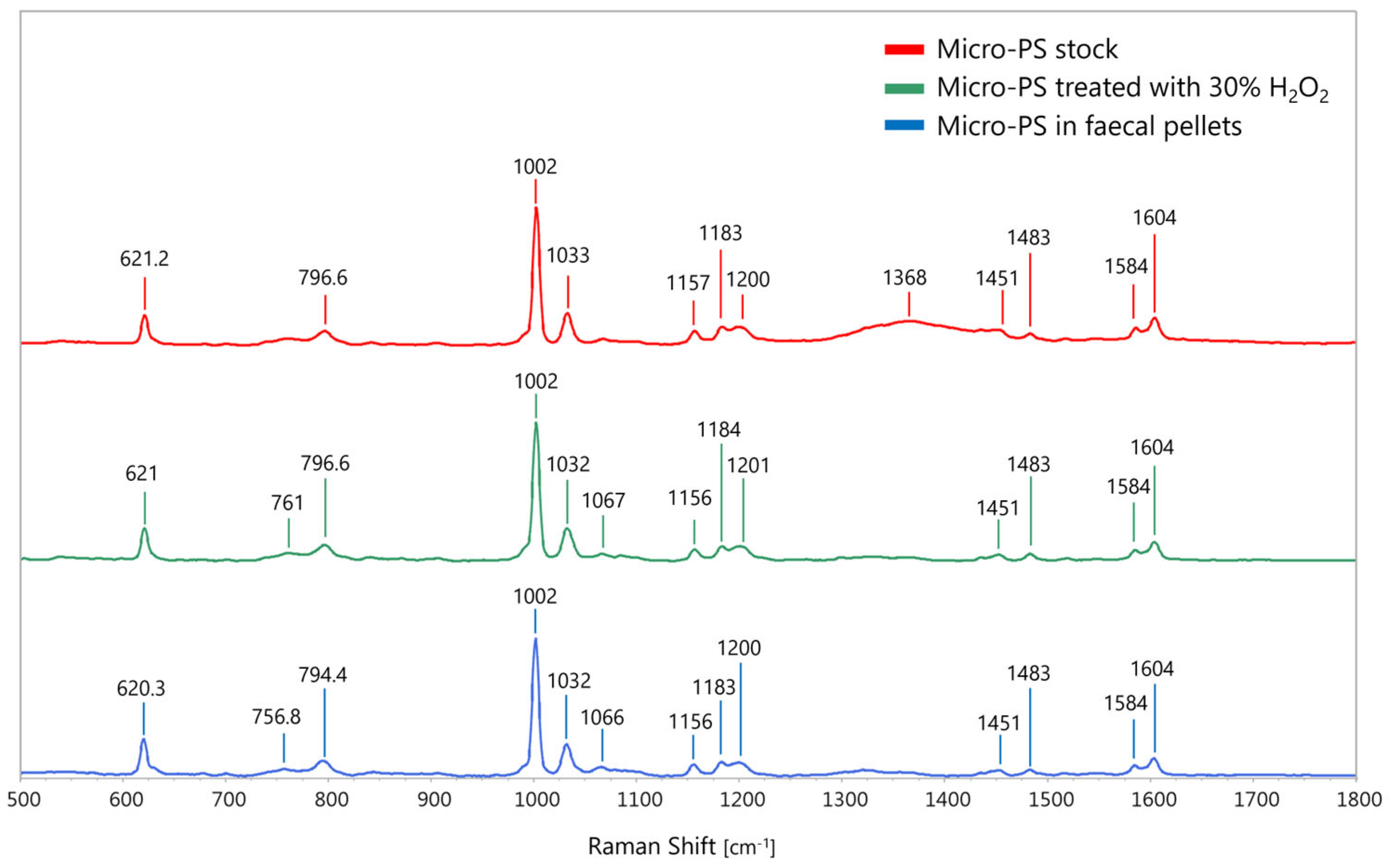
In-depth analyses further showed that the quick egestion of micro-PS in the bearded fireworm was associated with limited alteration of the particles. µ-Raman measurements of micro-PS stock suspensions confirmed PS composition (ρ = 0.91 with PS reference spectra). Indeed, PS characteristic spectral bands were identified (Figure 2), such as at ~621 cm−1, ~796 cm−1, ~1002 cm−1 (respiratory vibration of the benzene ring), ~1031 cm−1 (C–H deformation in-plane, [59]), 1449 cm−1, and ~1604 cm−1 (ring-skeletal stretch), as previously reported for PS nano- and microspheres in aqueous suspensions [60,61]. The strongest peak signal observed at ~1001 cm−1 is largely used as an indicator peak to identify PS, often in association with the peak at ~1031 cm–1 [62,63].
After treatment with hydrogen peroxide (30%), no relevant changes in the PS chemical fingerprint with respect to reference stock particles were observed (Figure S7), as shown by their high match rate (ρ = 0.87), demonstrating that the oxidative treatment did not affect polymer identification. Likewise, for micro-PS retrieved from faecal pellets, the overall polymer identification was not impaired, although the match rate slightly decreased compared to the reference stock (ρ = 0.86), suggesting negligible chemical changes on the micro-PS surface.
In the study [60], the authors reported that sample cleanup is required to identify plastic particles by µ-Raman spectroscopy in complex matrices. Our results underline the suitability of the extraction method adopted for the quantification of MPs in whole biological samples. Although micro-PS are not fully representative of the heterogeneous MP contamination in the marine environment, they are useful standard materials to set reference exposure conditions and, when fluorescently labelled, they are easily detected and mapped within biological matrices, thus providing major insights into toxicokinetic studies. To this aim, strict control procedures are necessary to confirm the stability of the fluorophore associated with the particles during incubation [64], enabling single-particle detection through fluorescence imaging. As for micro-PS, the stability of their fluorophore following uptake by marine organisms (i.e., sea urchins) and specimen processing via organic matter digestion was previously assessed by [41,42]. To further elucidate the tissue translocation of micro-PS, advanced techniques such as 3D mapping by volumetric Raman chemical imaging would be necessary [63].
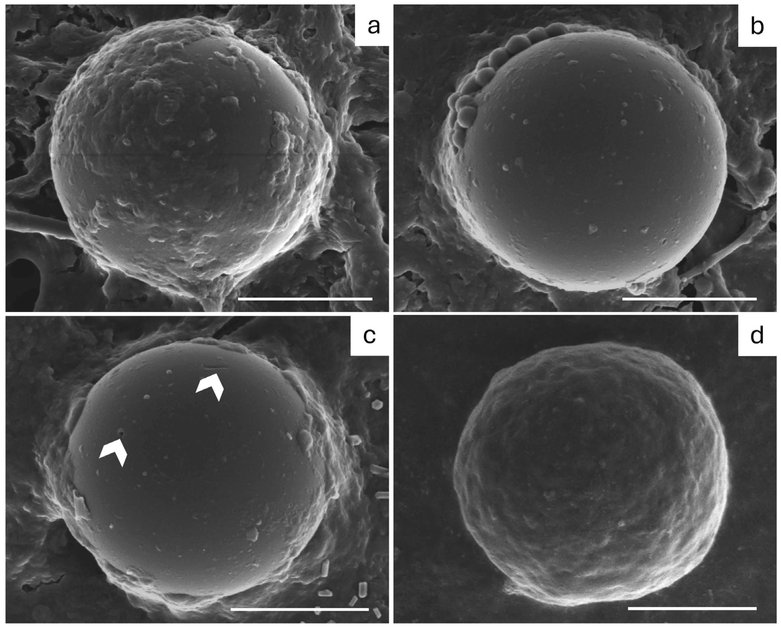
In addition, SEM confirmed that micro-PS extracted from fireworm faecal pellets (Figure 3) maintained the morphology of untreated pristine particles from the stock suspension. Most of the particles showed a high degree of coverage by faecal pellet matter (Figure 3a) and with the presence of bacteria colonising their surface (Figure 3b). In a few particles, some surface alterations were observed, such as randomly distributed pinpricks and small cracks (Figure 3c), potentially due to their suction and passage in the fireworm pharynx and gut. Only a few particles (2% of the particles observed) presented high alterations in surface roughness (Figure 3d).
Several studies have previously investigated the effect of the passage through the digestive tract in fish, crustaceans, and echinoderms on the degradation of MP particles [36,37,38,39]. Digestive fragmentation has been associated with MP mechanical breakdown, with surface abrasions being ideal microenvironments for bacterial colonisation [39] and accelerated degradation with the release of smaller MPs and nanoplastics in the environment [36]. Our findings align with previous reports of high MP incorporation in faecal pellets (e.g., [37,65]), suggesting a continuous biodistribution of plastic particles, accumulating in sediments, to benthic communities and coprophagous organisms.

4. Conclusions

Mediterranean benthic ecosystems are heavily impacted by MP pollution, with organisms exposed to high levels of MPs, leading to bioaccumulation within trophic levels and potentially biomagnification, although the latter is not supported by current field observations [18]. Despite the well-documented presence of MPs in some benthic taxa, such as bivalves and crustaceans, only a limited number of studies have explored ingestion pathways, such as indirect uptake through contaminated prey. To elucidate MP fate within benthic communities, studies investigating MP ingestion, retention, and depuration rates, as well as multi-level trophic transfer, are urgently needed. In this study, trophic transfer of model micro-PS was experimentally proven for the first time from a model filter feeder (Mediterranean mussel) to a carnivorous polychaete (bearded fireworm) in a simple two-level laboratory setting. Our findings confirmed the trophic transfer of micro-PS but also showed rapid excretion in the fireworm specimens. High levels of micro-PS, with negligible signs of digestive degradation, were found in their faecal pellets within 3 d after internalisation.
These preliminary results support the hypothesis of a dynamic balance of MP levels between these organisms and the surrounding environment, as previously suggested for other benthic species. Nevertheless, more toxicokinetic data on different MPs (i.e., varying polymer, size, and shape) are needed to elucidate clear patterns of ingestion and excretion for this heterogeneous group of particulate contaminants. Future research should also explore the dietary diversity of the bearded fireworm, considering the contribution of different prey to the MP intake in this polychaete species.

Supplementary Materials

The following supporting information can be downloaded at https://www.mdpi.com/article/10.3390/environments12100365/s1, Figure S1: Schematic representation of the two-level experimental set-up, sample processing and analysis; Figure S2: Preliminary experiment with individuals of the Mediterranean mussel (Mytilus galloprovincialis) exposed to suspensions of polystyrene microspheres (micro-PS) in aerated seawater; Figure S3: Positioning of a mussel with open valves in the tank containing an individual of the bearded fireworm Hermodice carunculata and predation of a bearded fireworm during incubation; Figure S4: Representative faecal pellets of the bearded fireworm; Figure S5: Optical fluorescent images of Yellow Green labelled polystyrene microspheres (micro-PS) extracted from the bearded fireworm intestine (a) and faecal pellets (b) and deposited on filters; Figure S6: Number of polystyrene microspheres (micro-PS) retrieved in faecal pellets of bearded fireworm specimens (V-vermis) (FP) released at different days post-feeding; Figure S7: Sanning Electron Microscopy (SEM) images of polystyrene microspheres (micro-PS) as received from the supplier (a) and after oxidative treatment (30% hydrogen peroxide); Table S1: Morphometric traits of mussels contaminated with polystyrene microspheres (micro-PS) and offered to fireworms; Table S2: Morphometric traits of the bearded fireworm specimens used in trophic transfer experiments as total length, number of chaetigers, and wet weight (WW).

Author Contributions

Conceptualization, V.F., R.S., D.P. and E.B.; methodology, All authors; formal analysis, V.F. and E.B.; investigation, V.F. and E.B.; resources, R.S., D.P. and C.M.; data curation, V.F. and E.B.; writing—original draft preparation, V.F. and E.B.; writing—review and editing, All authors; visualisation, V.F. and E.B.; supervision, E.B.; funding acquisition, R.S. and E.B. All authors have read and agreed to the published version of the manuscript.

Funding

This research was supported by the project PRIN 2022 “NANOplastics Toxicity Evaluation and Risk (management) in teRrestrial Agro-Ecosystems” (NanoTERRAE), PRIN 2022 call funded by MIUR—Ministero dell’Istruzione, dell’Università e della Ricerca and European Union–NextGenerationEU [CUP: E53C24003070006] for particle characterisation and by the University of Modena and Reggio Emilia (Public Engagement call) for animal sampling.

Data Availability Statement

The raw data supporting the conclusions of this article will be made available by the authors on request.

Acknowledgments

The authors acknowledge the Citizen Science project “Monitoraggio Vermocane” of the University of Modena and Reggio Emilia for the support during sampling. Raman and SEM analyses were carried out at the Centro Interdipartimentale Grandi Strumenti (CIGS) facilities of the University of Modena and Reggio Emilia: https://www.cigs.unimore.it/index.php.

Conflicts of Interest

The authors declare no conflicts of interest.

References

  1. Jambeck, J.R.; Geyer, R.; Wilcox, C.; Siegler, T.R.; Perryman, M.; Andrady, A.; Narayan, R.; Law, K.L. Plastic Waste Inputs from Land into the Ocean. Science 2015, 347, 768–771. [Google Scholar] [CrossRef]
  2. Compa, M.; Alomar, C.; Wilcox, C.; van Sebille, E.; Lebreton, L.; Hardesty, B.D.; Deudero, S. Risk Assessment of Plastic Pollution on Marine Diversity in the Mediterranean Sea. Sci. Tot. Environ. 2019, 678, 188–196. [Google Scholar] [CrossRef]
  3. Landrigan, P.J.; Stegeman, J.J.; Fleming, L.E.; Allemand, D.; Anderson, D.M.; Backer, L.C.; Brucker-Davis, F.; Chevalier, N.; Corra, L.; Czerucka, D.; et al. Human Health and Ocean Pollution. Ann. Glob. Health 2020, 86, 151. [Google Scholar] [CrossRef]
  4. Sanchez-Vidal, A.; Thompson, R.C.; Canals, M.; De Haan, W.P. The Imprint of Microfibres in Southern European Deep Seas. PLoS ONE 2018, 13, e0207033. [Google Scholar] [CrossRef]
  5. Harris, P.T. The Fate of Microplastic in Marine Sedimentary Environments: A Review and Synthesis. Mar. Pollut. Bull. 2020, 158, 111398. [Google Scholar] [CrossRef] [PubMed]
  6. Kane, I.A.; Clare, M.A.; Miramontes, E.; Wogelius, R.; Rothwell, J.J.; Garreau, P.; Pohl, F. Seafloor Microplastic Hotspots Controlle by Deep-Sea Circulation. Science 2020, 368, 1140–1145. [Google Scholar] [CrossRef]
  7. Issac, M.N.; Kandasubramanian, B. Effect of Microplastics in Water and Aquatic Systems. Environ. Sci. Pollut. Res. 2021, 28, 19544–19562. [Google Scholar] [CrossRef] [PubMed]
  8. Thushari, G.G.N.; Senevirathna, J.D.M. Plastic Pollution in the Marine Environment. Heliyon 2020, 6, e04709. [Google Scholar] [CrossRef] [PubMed]
  9. Cássio, F.; Batista, D.; Pradhan, A. Plastic Interactions with Pollutants and Consequences to Aquatic Ecosystems: What We Know and What We Do Not Know. Biomolecules 2022, 12, 798. [Google Scholar] [CrossRef]
  10. Mason, V.G.; Skov, M.W.; Hiddink, J.G.; Walton, M. Microplastics Alter Multiple Biological Processes of Marine Benthic Fauna. Sci. Tot. Environ. 2022, 845, 157362. [Google Scholar] [CrossRef]
  11. Sandgaard, M.H.; Palmqvist, A.; Bour, A.; Grønlund, S.N.; Hooge, A.; Selck, H.; Thit, A.; Syberg, K. Sediment Matters as a Route of Microplastic Exposure: A Call for More Research on the Benthic Compartment. Front. Mar. Sci. 2023, 9, 1100567. [Google Scholar] [CrossRef]
  12. Bonello, G.; Varrella, P.; Pane, L. First Evaluation of Microplastic Content in Benthic Filter-Feeders of the Gulf of La Spezia (Ligurian Sea). J. Aquat. Food Prod. Technol. 2018, 27, 284–291. [Google Scholar] [CrossRef]
  13. Rubini, S.; Munari, M.; Baldini, E.; Barsi, F.; Meloni, D.; Pussini, N.; Barchiesi, F.; Di Francesco, G.; Losasso, C.; Cocumelli, C.; et al. Microplastics in Mussels (Mytilus galloprovincialis): Understanding Pollution in Italian Seas. Toxics 2025, 13, 144. [Google Scholar] [CrossRef]
  14. Savoca, M.S.; Tyson, C.W.; McGill, M.; Slager, C.J. Odours from Marine Plastic Debris Induce Food Search Behaviours in a Forage Fish. Proc. R. Soc. B Biol. Sci. 2017, 284, 20171000. [Google Scholar] [CrossRef]
  15. Horie, Y.; Mitsunaga, K.; Yamaji, K.; Hirokawa, S.; Uaciquete, D.; Ríos, J.M.; Yap, C.K.; Okamura, H. Variability in Microplastic Color Preference and Intake among Selected Marine and Freshwater Fish and Crustaceans. Discov. Oceans 2024, 1, 5. [Google Scholar] [CrossRef]
  16. Zantis, L.J.; Bosker, T.; Lawler, F.; Nelms, S.E.; O’Rorke, R.; Constantine, R.; Sewell, M.; Carroll, E.L. Assessing Microplastic Exposure of Large Marine Filter-Feeders. Sci. Tot. Environ. 2022, 818, 151815. [Google Scholar] [CrossRef] [PubMed]
  17. Diepens, N.J.; Koelmans, A.A. Accumulation of Plastic Debris and Associated Contaminants in Aquatic Food Webs. Environ. Sci. Technol. 2018, 52, 8510–8520. [Google Scholar] [CrossRef]
  18. Miller, M.E.; Hamann, M.; Kroon, F.J. Bioaccumulation and Biomagnification of Microplastics in Marine Organisms: A Review and Meta-Analysis of Current Data. PLoS ONE 2020, 15, e0240792. [Google Scholar] [CrossRef]
  19. Ding, J.; Sun, C.; He, C.; Li, J.; Ju, P.; Li, F. Microplastics in Four Bivalve Species and Basis for Using Bivalves as Bioindicators of Microplastic Pollution. Sci. Tot. Environ. 2021, 782, 146830. [Google Scholar] [CrossRef]
  20. Li, J.; Lusher, A.L.; Rotchell, J.M.; Deudero, S.; Turra, A.; Bråte, I.L.N.; Sun, C.; Shahadat Hossain, M.; Li, Q.; Kolandhasamy, P.; et al. Using Mussel as a Global Bioindicator of Coastal Microplastic Pollution. Environ. Pollut. 2019, 244, 522–533. [Google Scholar] [CrossRef]
  21. Piarulli, S.; Airoldi, L. Mussels Facilitate the Sinking of Microplastics to Bottom Sediments and Their Subsequent Uptake by Detritus-Feeders. Environ. Pollut. 2020, 266, 115151. [Google Scholar] [CrossRef]
  22. Albignac, M.; Ghiglione, J.F.; Labrune, C.; Ter Halle, A. Determination of the Microplastic Content in Mediterranean Benthic Macrofauna by Pyrolysis-Gas Chromatography-Tandem Mass Spectrometry. Mar. Pollut. Bull. 2022, 181, 113882. [Google Scholar] [CrossRef] [PubMed]
  23. De Benedetto, G.E.; Fraissinet, S.; Tardio, N.; Rossi, S.; Malitesta, C. Microplastics Determination and Quantification in Two Benthic Filter Feeders Sabella spallanzanii, Polychaeta and Paraleucilla magna, Porifera. Heliyon 2024, 10, e31796. [Google Scholar] [CrossRef]
  24. Calmão, M.; Blasco, N.; Benito, A.; Thoppil, R.; Torre-Fernandez, I.; Castro, K.; Izagirre, U.; Garcia-Velasco, N.; Soto, M. Time-Course Distribution of Fluorescent Microplastics in Target Tissues of Mussels and Polychaetes. Chemosphere 2023, 311, 137087. [Google Scholar] [CrossRef]
  25. Pires, A.; Cuccaro, A.; Sole, M.; Freitas, R. Micro(Nano)Plastics and Plastic Additives Effects in Marine Annelids: A Literature Review. Environ. Res. 2022, 214, 113642. [Google Scholar] [CrossRef] [PubMed]
  26. Díaz-Castañeda, V.; Reish, D.J. Polychaetes in environmental studies. In Annelids in Modern Biology; Shain, D.H., Ed.; John Wiley & Sons, Inc.: Hoboken, NJ, USA, 2009; pp. 203–227. [Google Scholar]
  27. Rouse, G.; Pleijel, F. Polychaetes; Oxford University Press: Oxford, UK, 2001. [Google Scholar]
  28. Giangrande, A.; Licciano, M.; Musco, L. Polychaetes as Environmental Indicators Revisited. Mar. Pollut. Bull. 2005, 50, 1153–1162. [Google Scholar] [CrossRef] [PubMed]
  29. Righi, S.; Prevedelli, D.; Simonini, R. Ecology, Distribution and Expansion of a Mediterranean Native Invader, the Fireworm Hermodice carunculata (Annelida). Mediterr. Mar. Sci. 2020, 21, 575–591. [Google Scholar] [CrossRef]
  30. Krželj, M.; Cerrano, C.; Di Camillo, C.G. Enhancing Diversity Knowledge through Marine Citizen Science and Social Platforms: The Case of Hermodice carunculata (Annelida, Polycheta). Diversity 2020, 12, 311. [Google Scholar] [CrossRef]
  31. Simonini, R.; Maletti, I.; Righi, S.; Fai, S.; Prevedelli, D. Laboratory Observations on Predator–Prey Interactions between the Bearded Fireworm (Hermodice carunculata) and Mediterranean Benthic Invertebrates. Mar. Freshw. Behav. Physiol. 2018, 51, 145–158. [Google Scholar] [CrossRef]
  32. Tiralongo, F.; Marino, S.; Ignoto, S.; Martellucci, R.; Lombardo, B.M.; Mancini, E.; Scacco, U. Impact of Hermodice carunculata (Pallas, 1766) (Polychaeta: Amphinomidae) on Artisanal Fishery: A Case Study from the Mediterranean Sea. Mar. Environ. Res. 2023, 192, 106227. [Google Scholar] [CrossRef]
  33. Vecchi, S.; Bianchi, J.; Scalici, M.; Fabroni, F.; Tomassetti, P. Field Evidence for Microplastic Interactions in Marine Benthic Invertebrates. Sci. Rep. 2021, 11, 292. [Google Scholar] [CrossRef]
  34. Gonçalves, C.; Martins, M.; Sobral, P.; Costa, P.M.; Costa, M.H. An Assessment of the Ability to Ingest and Excrete Microplastics by Filter-Feeders: A Case Study with the Mediterranean Mussel. Environ. Pollut. 2019, 245, 600–606. [Google Scholar] [CrossRef] [PubMed]
  35. Corsi, I.; Bellingeri, A.; Bergami, E. Progress in Selecting Marine Bioindicators for Nanoplastics Ecological Risk Assessment. Ecol. Indic. 2023, 154, 110836. [Google Scholar] [CrossRef]
  36. Dawson, A.L.; Kawaguchi, S.; King, C.K.; Townsend, K.A.; King, R.; Huston, W.M.; Bengtson Nash, S.M. Turning Microplastics into Nanoplastics through Digestive Fragmentation by Antarctic Krill. Nat. Commun. 2018, 9, 1001. [Google Scholar] [CrossRef] [PubMed]
  37. Porter, A.; Smith, K.E.; Lewis, C. The Sea Urchin Paracentrotus lividus as a Bioeroder of Plastic. Sci. Tot. Environ. 2019, 693, 133621. [Google Scholar] [CrossRef] [PubMed]
  38. Cau, A.; Avio, C.G.; Dessì, C.; Moccia, D.; Pusceddu, A.; Regoli, F.; Cannas, R.; Follesa, M.C. Benthic Crustacean Digestion Can Modulate the Environmental Fate of Microplastics in the Deep Sea. Environ. Sci. Technol. 2020, 54, 4886–4892. [Google Scholar] [CrossRef]
  39. Babkiewicz, E.; Nowakowska, J.; Zebrowski, M.L.; Kunijappan, S.; Jarosińska, K.; Maciaszek, R.; Zebrowski, J.; Jurek, K.; Maszczyk, P. Microplastic Passage through the Fish and Crayfish Digestive Tract Alters Particle Surface Properties. Environ. Sci. Technol. 2025, 59, 5693–5703. [Google Scholar] [CrossRef]
  40. Simonini, R.; Righi, S.; Zanetti, F.; Fai, S.; Prevedelli, D. Development and Catch Efficiency of an Attracting Device to Collect and Monitor the Invasive Fireworm Hermodice carunculata in the Mediterranean Sea. Mediterr. Mar. Sci. 2021, 22, 706–714. [Google Scholar] [CrossRef]
  41. Murano, C.; Agnisola, C.; Caramiello, D.; Castellano, I.; Casotti, R.; Corsi, I.; Palumbo, A. How Sea Urchins Face Microplastics: Uptake, Tissue Distribution and Immune System Response. Environ. Pollut. 2020, 264, 114685. [Google Scholar] [CrossRef]
  42. Murano, C.; Donnarumma, V.; Corsi, I.; Casotti, R.; Palumbo, A. Impact of Microbial Colonization of Polystyrene Microbeads on the Toxicological Responses in the Sea Urchin Paracentrotus lividus. Environ. Sci. Technol. 2021, 55, 7990–8000. [Google Scholar] [CrossRef] [PubMed]
  43. Righi, S.; Forti, L.; Simonini, R.; Ferrari, V.; Prevedelli, D.; Mucci, A. Novel Natural Compounds and Their Anatomical Distribution in the Stinging Fireworm Hermodice carunculata (Annelida). Mar. Drugs 2022, 20, 585. [Google Scholar] [CrossRef]
  44. Marsden, J.R. The digestive tract of Hermodice carunculata (Pallas). Polychaeta: Amphinomidae. Can. J. Zool. 1963, 41, 165–184. [Google Scholar] [CrossRef]
  45. Menges, F. SpectraGryph-Optical Spectroscopy Software, Version 1.2.16.1. 2023. Available online: https://www.effemm2.de/spectragryph/ (accessed on 12 November 2023).
  46. Munno, K.; De Frond, H.; O’Donnell, B.; Rochman, C.M. Increasing the Accessibility for Characterizing Microplastics: Introducing New Application-Based and Spectral Libraries of Plastic Particles (SLoPP and SLoPP-E). Anal. Chem. 2020, 92, 2443–2451. [Google Scholar] [CrossRef]
  47. Miller, E.A.; Yamahara, K.M.; French, C.; Spingarn, N.; Birch, J.M.; Van Houtan, K.S. A Raman Spectral Reference Library of Potential Anthropogenic and Biological Ocean Polymers. Sci. Data 2022, 9, 183. [Google Scholar] [CrossRef]
  48. Cowger, W.; Steinmetz, Z.; Gray, A.; Munno, K.; Lynch, J.; Hapich, H.; Primpke, S.; De Frond, H.; Rochman, C.; Herodotou, O. Microplastic Spectral Classification Needs an Open Source Community: Open Specy to the Rescue! Anal. Chem. 2021, 93, 7543–7548. [Google Scholar] [CrossRef] [PubMed]
  49. Riisgård, H.U.; Larsen, P.S.; Pleissner, D. Allometric Equations for Maximum Filtration Rate in Blue Mussels Mytilus edulis and Importance of Condition Index. Helgol. Mar. Res. 2014, 68, 193–198. [Google Scholar] [CrossRef]
  50. Li, J.; Qu, X.; Su, L.; Zhang, W.; Yang, D.; Kolandhasamy, P.; Li, D.; Shi, H. Microplastics in Mussels along the Coastal Waters of China. Environ. Pollut. 2016, 214, 177–184. [Google Scholar] [CrossRef] [PubMed]
  51. Setälä, O.; Norkko, J.; Lehtiniemi, M. Feeding Type Affects Microplastic Ingestion in a Coastal Invertebrate Community. Mar. Pollut. Bull. 2016, 102, 95–101. [Google Scholar] [CrossRef]
  52. Revel, M.; Yakovenko, N.; Caley, T.; Guillet, C.; Châtel, A.; Mouneyrac, C. Accumulation and Immunotoxicity of Microplastics in the Estuarine Worm Hediste diversicolor in Environmentally Relevant Conditions of Exposure. Environ. Sci. Pollut. Res. 2020, 27, 3574–3583. [Google Scholar] [CrossRef]
  53. Bour, A.; Sturve, J.; Höjesjö, J.; Carney Almroth, B. Microplastic Vector Effects: Are Fish at Risk When Exposed via the Trophic Chain? Front. Environ. Sci. 2020, 8, 90. [Google Scholar] [CrossRef]
  54. Piarulli, S.; Vanhove, B.; Comandini, P.; Scapinello, S.; Moens, T.; Vrielinck, H.; Sciutto, G.; Prati, S.; Mazzeo, R.; Booth, A.M.; et al. Do different habits affect microplastics contents in organisms? A trait-based analysis on salt marsh species. Mar. Pollut. Bull. 2020, 153, 110983. [Google Scholar] [CrossRef]
  55. Keerthika, K.; Padmavathy, P.; Rani, V.; Jeyashakila, R.; Aanand, S.; Kutty, R. Evidence of microplastics in the polychaete worm (capitellids—Capitella capitata) (Fabricicus, 1780) along Thoothukudi region. Environ. Monit. Assess. 2024, 196, 556. [Google Scholar] [CrossRef]
  56. James, K.; Kripa, V.; Vineetha, G.; Padua, S.; Parvathy, R.; Lavanya, R.; Joseph, R.V.; Abhilash, K.S.; Babu, A.; John, S. Microplastic ingestion by the polychaete community in the coastal waters of Kochi, Southwest coast of India. Reg. Stud. Mar. Sci. 2023, 62, 102948. [Google Scholar] [CrossRef]
  57. Blasco, N.; Ibeas, M.; Aramendia, J.; Castro, K.; Soto, M.; Izagirre, U.; Garcia-Velasco, N. Depuration kinetics and accumulation of microplastics in tissues of mussel Mytilus galloprovincialis. Mar. Environ. Res. 2024, 202, 106731. [Google Scholar] [CrossRef] [PubMed]
  58. Hatzonikolakis, Y.; Raitsos, D.E.; Sailley, S.F.; Digka, N.; Theodorou, I.; Tsiaras, K.; Tsangaris, C.; Skia, G.; Ntzouvaras, A.; Triantafyllou, G. Assessing the physiological effects of microplastics on cultured mussels in the Mediterranean Sea. Environ. Pollut. 2024, 363, 125052. [Google Scholar] [CrossRef]
  59. Musto, P.; Borriello, A.; Agoretti, P.; Napolitano, T.; Di Florio, G.; Mensitieri, G. Selective Surface Modification of Syndiotactic Polystyrene Films: A Study by Fourier Transform- and Confocal-Raman Spectroscopy. Eur. Polym. J. 2010, 46, 1004–1015. [Google Scholar] [CrossRef]
  60. Gao, H.; Liu, C.; Wang, H.; Shen, H. Raman Spectra Characterization of Size-Dependent Aggregation and Dispersion of Polystyrene Particles in Aquatic Environments. Chemosphere 2023, 333, 138939. [Google Scholar] [CrossRef] [PubMed]
  61. Mayorga, C.; Athalye, S.M.; Boodaghidizaji, M.; Sarathy, N.; Hosseini, M.; Ardekani, A.; Verma, M.S. Limit of Detection of Raman Spectroscopy Using Polystyrene Particles from 25 to 1000 Nm in Aqueous Suspensions. Anal. Chem. 2025, 97, 8908–8914. [Google Scholar] [CrossRef]
  62. Tian, M.; Morais, C.L.M.; Shen, H.; Pang, W.; Xu, L.; Huang, Q.; Martin, F.L. Direct Identification and Visualisation of Real-World Contaminating Microplastics Using Raman Spectral Mapping with Multivariate Curve Resolution-Alternating Least Squares. J. Hazard. Mater. 2022, 422, 126892. [Google Scholar] [CrossRef] [PubMed]
  63. Benito-Kaesbach, A.; Amigo, J.M.; Izagirre, U.; Garcia-Velasco, N.; Arévalo, L.; Seifert, A.; Castro, K. Misinterpretation in Microplastic Detection in Biological Tissues: When 2D Imaging Is Not Enough. Sci. Tot. Environ. 2023, 876, 162810. [Google Scholar] [CrossRef]
  64. Malafaia, G.; da Luz, T.M.; Ahmed, M.A.I.; Karthi, S.; da Costa Araújo, A.P. When Toxicity of Plastic Particles Comes from Their Fluorescent Dye: A Preliminary Study Involving Neotropical Physalaemus cuvieri Tadpoles and Polyethylene Microplastics. J. Hazard. Mater. Adv. 2022, 6, 100054. [Google Scholar] [CrossRef]
  65. Bergami, E.; Manno, C.; Cappello, S.; Vannuccini, M.L.; Corsi, I. Nanoplastics affect moulting and faecal pellet sinking in Antarctic krill (Euphausia superba) juveniles. Environ. Int. 2020, 143, 105999. [Google Scholar] [CrossRef] [PubMed]
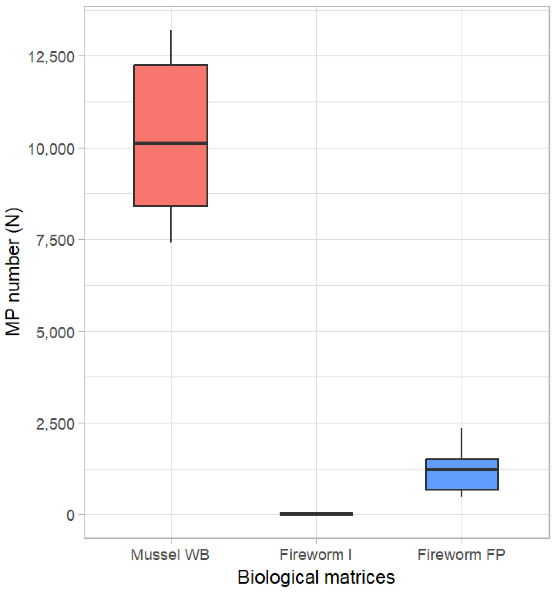
Figure 1. Number of polystyrene microspheres (micro-PS) in mussel whole body (WB), fireworm intestine and body fluids (I), and faecal pellets (FP), based on quantification at optical fluorescence microscopy.
Environments 12 00365 g001
Figure 2. Raman spectra of micro-PS from stock suspension (diluted in ultrapure water, red line), treated with 30% hydrogen peroxide (green line), and retrieved from fireworm faecal pellets (blue line).
Environments 12 00365 g002
Figure 3. Scanning electron micrographs of micro-PS extracted from fireworm faecal pellets, displaying (a) high coverage of faecal pellet matter; (b) microbial colonisation; (c) physical alterations as the mark and pinprick shown by white arrow heads; (d) severely altered surface. Scale bar: 5 µm.
Environments 12 00365 g003
Disclaimer/Publisher’s Note: The statements, opinions and data contained in all publications are solely those of the individual author(s) and contributor(s) and not of MDPI and/or the editor(s). MDPI and/or the editor(s) disclaim responsibility for any injury to people or property resulting from any ideas, methods, instructions or products referred to in the content.