Article

Toxicological Effects of Poly(Methyl Methacrylate) Microplastics in Caenorhabditis elegans: Impairment of Development, Reproduction, and Stress Responses

1
Department of Biology and Biotechnologies Charles Darwin, Sapienza University of Rome, Piazzale Aldo Moro 5, 00185 Rome, Italy
2
Department of Chemical Engineering Materials and Environment, Sapienza University of Rome, Via Eudossiana 18, 00184 Rome, Italy
*
Authors to whom correspondence should be addressed.
These authors contributed equally to this work.
Environments 2025, 12(10), 353; https://doi.org/10.3390/environments12100353
Submission received: 18 August 2025 / Revised: 23 September 2025 / Accepted: 25 September 2025 / Published: 30 September 2025
(This article belongs to the Special Issue Ecotoxicity of Microplastics)

Abstract

Microplastics (MPs) are plastic particles smaller than 5 mm that accumulate in ecosystems and can cause toxicity in organisms by affecting multiple biological processes. This study investigates the effects of poly(methyl methacrylate) microplastic microspheres (MPs, 200 µm diameter) on Caenorhabditis elegans, a widely used model in ecotoxicology. Nematodes were exposed to MPs at concentrations of 0.01, 0.1, 1, and 10 mg/mL, and various toxicological endpoints were assessed. The uptake of MPs was evaluated by µFT-IR analysis. The results indicate that MPs induce a concentration-dependent reduction in body length and alterations in the reproduction rate. Lifespan was also significantly reduced, with a 20% decrease at the highest concentration. Intestinal permeability assays revealed disruption of gut integrity at higher concentrations, and oxidative stress analysis showed a 1.8-fold increase in reactive oxygen species (ROS) levels at 10 mg/mL. Gene expression analysis via real-time qPCR indicated the upregulation of genes involved in oxidative stress and in DNA repair mechanisms. Additionally, the longevity-related transcription factors daf-16 and skn-1 were modulated, suggesting an adaptive stress response. These findings suggest that MPs impair growth, reproduction, and oxidative stress response in C. elegans, emphasizing the potential risks associated with microplastic exposure.

1. Introduction

In recent years, the pervasive presence of microplastics (MPs) in diverse ecosystems has emerged as a pressing environmental and public health concern. Defined as plastic particles smaller than 5 mm, MPs originate from the degradation of larger plastic debris or are intentionally manufactured as microbeads for use in industrial processes and personal care products [1]. Due to their small size, chemical stability, and persistence, MPs can be easily ingested by a broad spectrum of organisms, potentially leading to bioaccumulation, trophic transfer, and a wide range of toxicological effects [2,3]. While most of the research has focused on the impacts of MPs in aquatic environments, recent evidence highlights their growing presence and ecological risk in terrestrial ecosystems, particularly in soils [4]. MPs have been detected in agricultural fields, floodplains, and industrial soils, with concentrations ranging from 55.5 mg/kg to over 67,000 mg/kg [5,6]. These particles originate from various sources such as sewage sludge, fertilizers, and plastic mulching films [7]. Once in the soil, MPs can migrate to deeper layers and be ingested by soil-dwelling organisms including nematodes, earthworms, and insects, potentially disrupting soil biodiversity and ecosystem function [8]. Most ecotoxicological studies to date have focused on polystyrene (PS) micro- and nanoplastics, due to their commercial availability and ease of fluorescent labeling. In contrast, little is known about the biological effects of poly(methyl methacrylate) (PMMA) particles, despite their widespread use in medical devices, dentistry, coatings, and plastics manufacturing [9,10]. Different studies reported that PMMA particles can induce sublethal and behavioral effects in marine invertebrates, particularly in earlier life stages, depending on particle size, exposure route, and organismal physiology [11,12].
Given the increasing environmental occurrence of PMMA and the limited knowledge on its potential ecotoxicological effects, studies employing model organisms such as Caenorhabditis elegans are essential to dissect its impact on development, reproduction, and stress responses.
The nematode C. elegans [13] represents a robust model for investigating the biological effects of environmental pollutants, including MPs, due to its conserved molecular pathways, genetic tractability, short lifespan, and transparent body that facilitates real-time, in vivo imaging. Worm serves as a well-established model organism for investigating toxicological responses across various levels of biological organization, ranging from the molecular scale to the whole organism [14,15]. A major advantage of this model lies in the availability of mutant strains, which enable detailed exploration of the molecular pathways involved in both cellular and organismal responses [16]. It is particularly suited to assess sublethal toxicological endpoints such as oxidative stress, apoptosis, reproductive capacity, and behavioral responses [17]. Moreover, C. elegans shares a high degree of genetic homology with humans and possesses key conserved signaling pathways such as insulin/IGF-1 and p38 MAPK, which are known to mediate responses to environmental stressors, including plastic particles [18].
In this study, we systematically assessed the effects of poly(methyl methacrylate) MPs at varying concentrations on multiple physiological, molecular, and cellular endpoints in C. elegans. We investigated lifespan, aging-related parameters (e.g., pharyngeal pumping, locomotion), oxidative stress markers, mitochondrial health, DNA damage, and gene expression changes in stress response pathways.

2. Materials and Methods

2.1. Microplastic Suspension Preparation and C. elegans Growth Conditions

To prepare MPs suspensions, poly(methyl methacrylate) (PMMA) beads with a diameter of 200 μm were used. Poly(methyl methacrylate) (PMMA) particles were purchased from Polysciences, Inc., Warrington, PY, USA (Product Number 04553; CAS #9011-14-7). According to the manufacturer’s specifications, this PMMA has an intrinsic viscosity of 0.40 dL/g, which corresponds to an approximate molecular weight of about 75,000 Da.
Next, 10 mg/mL suspensions of PMMA microspheres were prepared in sterile distilled, deionized water (H2Odd) at a concentration of 10 mg/mL. To ensure homogeneity, the suspensions were vortexed for 2 min and sonicated in a water bath for 20 min immediately prior to use. Concentrations of 1 mg/mL, 0.1 mg/mL, and 0.01 mg/mL were obtained by serial dilution of the stock suspension, with vortexing performed before each transfer step. Sonication and all dilutions were performed on the day of exposure to minimize particle aggregation and settling. The stock suspension was found to remain stable when stored at 4 °C for up to 6 months. For the experiments, a wide a range of doses was chosen to better investigate the underlying mechanisms of toxicity.
The C. elegans strains used were wild-type N2, CL2166 (dvIs19 [(pAF15)gst-4p::GFP::NLS] III) and TJ356 (zIs356 [daf-16p::daf-16a/b::GFP + rol-6 (su1006)]) transgenic strains and QV225 skn-1 (zj15) mutant strain. All the strains were obtained from the Caenorhabditis Genetics Center (CGC), University of Minnesota, USA.
For nematode exposure, Nematode Growth Medium (NGM) plates were prepared by spreading 30 μL of E. coli OP50 (killed at 65 °C for 90 min) and 100 μL of each MP suspension onto the surface. Once the plates were dry, 10 fertile adult C. elegans were transferred to each plate and allowed to lay embryos for 8 h at 20 °C. After this period, the adults were removed, and the plates were incubated at 16 °C. Within 3 days, synchronized populations developed to the L4 or young adult stage and were subsequently used in the experimental assays. For control plates, only heat killed E. coli OP50 was applied without MPs (untreated). A total of 40 synchronized L4/adult nematodes were transferred to each treatment or control plate with a platinum wire. All plates were maintained at 16 °C.

2.2. Lifespan and Healthspan Analysis

Lifespan analysis was performed at 16 °C and synchronous nematodes, prepared as previously described, were transferred daily to new plates seeded with fresh lawns. They were scored as dead when they no longer responded to gentle touch with a platinum wire. At least 80 nematodes per condition were used in each experiment. For healthspan analysis, pharyngeal pumping activity and locomotion rate were assessed in 2-day- and 10-day-old adult C. elegans exposed to MPs at different concentrations (0.01 mg/mL, 0.1 mg/mL, 1 mg/mL,10 mg/mL) from embryos hatching. Measurements were conducted over a 30 s interval under a stereomicroscope SZ30 (Leica, Wetzlar, Germany). All experiments were performed in triplicate.

2.3. Body Length Measurement

To measure body length, C. elegans individuals at day 2, 3, 4, and 7 post-hatching, previously exposed to different concentrations of MPs, were mounted on agarose pads (3% agarose in M9 buffer supplemented with 20 mM sodium azide) and observed under a Leica MZ10F stereomicroscope equipped with an Axiocam 208 Color camera. Body length was determined using Zen 3.1 software. Measurements of nematodes treated with MPs were compared to untreated controls. Ten nematodes were analyzed for each condition.

2.4. Intestinal Permeability Assay Using Brilliant Blue FCF Staining

Intestinal permeability was assessed in 10-day-old adult C. elegans grown on NGM plates seeded with heat-killed E. coli OP50 and supplemented with 10 mg/mL MPs. Age-matched worms grown under the same conditions but without MP exposure were used as controls. For the assay, 15 worms per condition were transferred to a microscope slide with 50 μL of M9 buffer and washed several times with fresh M9 to remove residual bacteria. After the excess buffer was removed, worms were incubated in the dark for 3 h in 100 μL of a staining solution consisting of M9 buffer and 5% Brilliant Blue FCF (80717–100MG, Sigma-Aldrich, St. Louis, MO, USA). Following incubation, worms were washed again with M9 and mounted on 3% agarose pads containing 20 mM sodium azide for immobilization. Samples were imaged using a Zeiss Axiovert 25 microscope, Oberkochen, Germany. Lumen diameter was determined using ZEISS ZEN Microscopy Software 2011.

2.5. Fertility Assay

Fresh NGM plates were seeded with 30 μL of heat-killed E. coli OP50 and supplemented with 100 μL of each MP suspension (0.01, 0.1, 1 or 10 mg/mL). Control plates were prepared by adding only E. coli OP50 without any MPs.
A single L4-stage worm, obtained from synchronized populations pre-exposed to the same respective MP concentrations, was transferred onto each experimental or control plate. Worms were transferred daily to freshly prepared plates of the same treatment group to prevent mixing of progeny between days. The number of embryos laid each day was counted under a stereomicroscope SZ30 (Leica) and recorded. This procedure was repeated until egg laying ceased. Total brood size was calculated for each condition, and comparisons were made between MP-treated groups and the untreated control.

2.6. Measurement of Cytosolic and Mitochondrial Reactive Oxygen Species (ROS)

After larval development in the presence of MPs at different concentrations (0.01, 0.1, 1 or 10 mg/mL), cytosolic ROS production was assessed in 1-day-old and 4-day-old adult nematodes using the fluorescent probe 2′,7′-dichlorodihydrofluorescein diacetate (H2DCF-DA, 287810–100MG, Sigma-Aldrich). Worms were washed with M9 buffer and analysis was performed as reported in [13]. Following a 1 h incubation at 20 °C on a shaker in the dark, fluorescence was measured using a multiwell plate reader (Promega GloMax® Multidetection System, Madison, WI, USA) with excitation/emission wavelengths of 485/520 nm.
To determine mitochondrial ROS levels, N2 nematodes in 1- or 4-day adults were incubated for 1 h with 5 μM MitoTracker® Red CMXRos (ThermoFisher Scientific, Schwerte, Germany) that accumulates in mitochondria of live cells in a membrane potential-dependent manner.

2.7. Quantitative Real Time PCR

Total RNA was extracted from approximately 200 4-day-old adult nematodes per condition and the expression levels of the genes daf-16, daf-2, gst-4, sod-3, and skn-1, involved in oxidative stress response, and rad-51, msh-2, hus-1 and mre-11, involved in DNA damage response, were analyzed by real-time q-PCR, according to [19]. The sequences of the primers used for real time qPCR analysis are listed in Table S1. All experiments were performed in triplicate.

2.8. Fluorescence Analysis of Transgenic Strains

At the stage of 4 days of adulthood, synchronized gst-4::GFP and daf-16::GFP transgenic worms supplemented with different concentrations of MPs from embryo hatching were anesthetized with sodium azide (20 mmol L−1) (Sigma-Aldrich, St. Louis, MO, USA) and observed by Zeiss Axiovert 25 microscope. The experiments were repeated three times and 15 worms per group were used in each experiment. Images were taken at the time of exposure of 0.2 s and fluorescence was analyzed using ImageJ version 1.53e software. Scale bars were inserted by Zeiss ZEN Microscopy Software 2011.

2.9. FT-IR Microanalysis

Adult worms were fed from embryos hatching as described above with MPs (10 mg/mL). After the treatment, 4-day adult animals were washed several times with H2Odd to eliminate the external MPs and residues from the nematode cuticle. Single worms were deposited in 10 μL of H2Odd onto a silver mirror slide laying directly on the support for FT-IR analysis and dried at room temperature. For Fourier transformer infrared (FT-IR) micro spectroscopy, a Bruker HYPERION 2000 IR microscope, connected to a FT-IR spectrometer (Vertex 70, Bruker Optik GmbH, Ettlingen, Germany), equipped with a moving stage, liquid nitrogen cooled MCT detector, globar IR source, with infrared imaging capabilities was used. The microscope is equipped with a 15×IR Schwarzschild objective with 24 mm working distance. The absorption spectra between 2000 and 700 cm−1, acquired through OPUS/3D package, were collected with 3 cm−1 spectral resolution, 20 kHz speed and 128 scan coadditions for single point spectra. The area under analysis was selected through a variable diaphragm slot at approximately 20 μm × 30 μm, over a 540 μm × 930 μm total mapping area (837 analysis points). The OPUS 8.7 software was also used to mathematically subtract the average reflectance spectrum obtained on the control worm from each map point derived by MPs fed worm. Two- and three-dimensional representations of the spectral data were generated from space-resolved mapping measurements, with individual spectra assembled into line traces (2D) and contour plots (3D). Three independent experiments were performed to verify the reproducibility.

2.10. Statistical Analysis

All experiments were performed in at least three independent replicates. Data are presented as mean ± standard deviation (SD). Statistical significance was assessed using Student’s t-test or one-way ANOVA followed by Bonferroni’s post hoc test, performed with GraphPad Prism 9.0 software (GraphPad Software Inc., La Jolla, CA, USA). Differences were considered statistically significant at p < 0.05 and are indicated as follows: * p < 0.05, ** p < 0.01, and *** p < 0.001.

3. Results

3.1. MPs Affect Larval Development and Lifespan

To assess the effects of MPs on C. elegans larval development, body length was measured at 2, 3, 4, and 7 days post-hatching. As shown in Figure 1A, on day 2, no significant differences in body length were observed among nematodes exposed to MPs at concentrations of 0.01 mg/mL, 0.1 mg/mL, 1 mg/mL or 10 mg/mL, and compared to untreated controls. However, by day 4, a slight reduction, of about 5%, in body length was recorded in worms exposed to 10 mg/mL MPs, as compared to untreated population. On day 7, fully developed adult nematodes exposed to lower concentrations of MPs (0.01 and 0.1 mg/mL) showed body lengths comparable to controls. In contrast, worms treated with 10 mg/mL MPs remained significantly shorter than controls. Notably, animals exposed to 1 mg/mL also exhibited a significant reduction in length, of about 20% as compared to controls.
The use of C. elegans as a model system proved highly effective in evaluating the impact of MPs on organismal survival. The Kaplan–Meier survival analysis (Figure 1B) revealed a significant reduction in lifespan in worms exposed to MPs compared to untreated controls. The results showed a clear dose-dependent effect, with reduced median survival times in worms exposed to 1 and 10 mg/mL MPs compared to controls. Specifically, worms treated with 1 mg/mL reached 50% survival between day 8 and day 9, while those exposed to 10 mg/mL MPs reached 50% survival between day 7 and day 8. No significant reduction in longevity was observed at lower concentrations (0.01 mg/mL and 0.1 mg/mL). These findings indicate that MPs at 1 mg/mL and 10 mg/mL negatively affect C. elegans lifespan in a dose-dependent manner.

3.2. MP Supplementation Negatively Impacts Aging Markers

C. elegans represents an ideal model for studying aging-related mechanisms [20]. For this reason, in the present study, specific aging markers such as pharyngeal pumping rate and body bending were assessed.
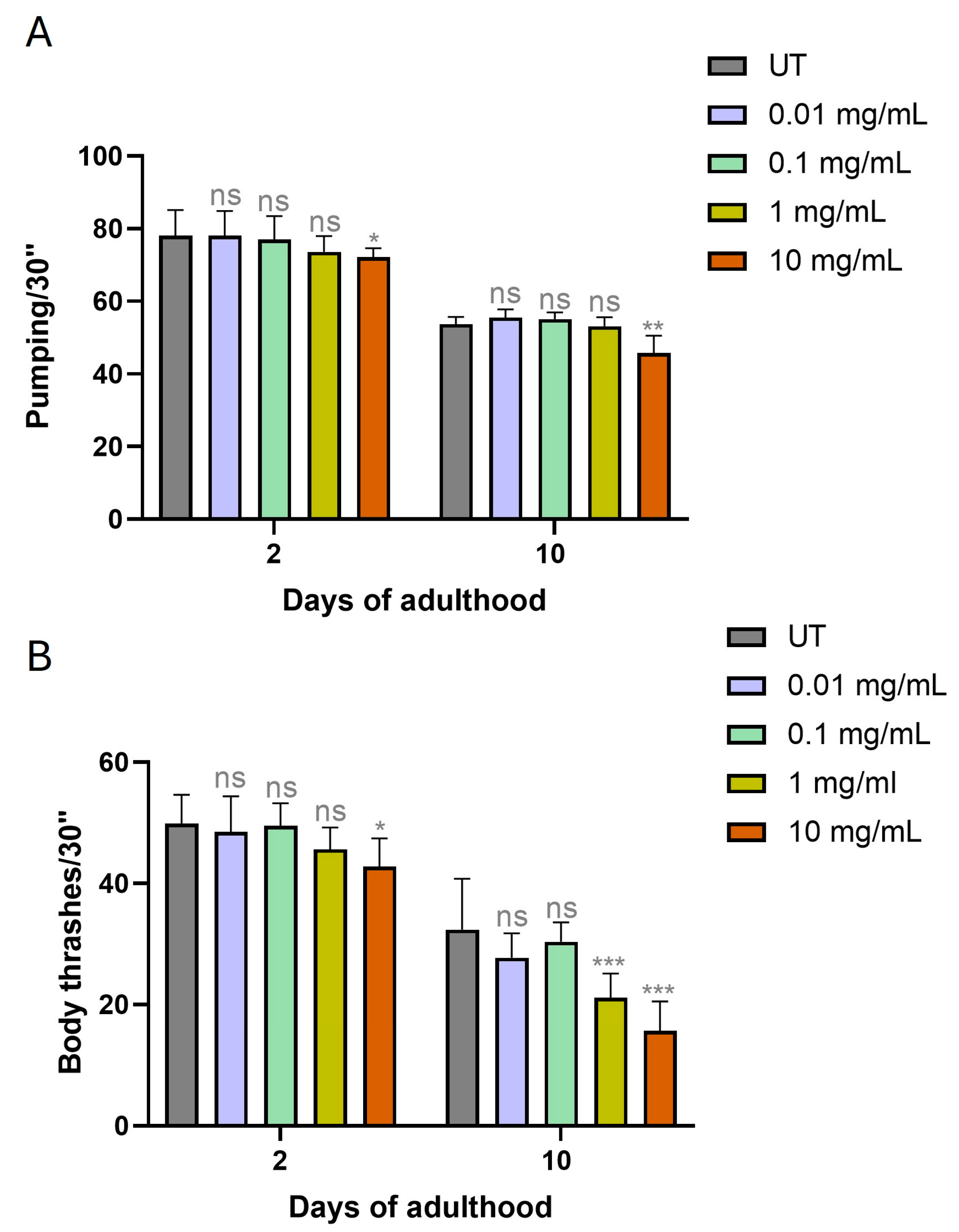
Pumping rate, defined as the frequency of pharyngeal contractions, reflecting feeding efficiency, typically declines with age. As shown in Figure 2A, no significant differences in pumping rate were observed in 2-day-old and 10-day-old adult worms exposed to 0.01 mg/mL, 0.1 mg/mL, and 1 mg/mL MPs. However, worms treated with the highest concentration (10 mg/mL) exhibited a significantly reduced pumping rate compared to controls, with a decrease of 8% on day 2 and 16% on day 10.
Body bending, which reflects nematode motility, also tends to decline with age and was therefore used as a senescence marker [21]. As shown in Figure 2B, no significant reduction in body bending was observed in 2-day-old worms treated with 0.01 mg/mL, 0.1 mg/mL, or 1 mg/mL MPs. In contrast, exposure to 10 mg/mL resulted in a 17% decrease in mobility. When the experiment was repeated on 10-day-old adult worms, no significant differences were found for the 0.01 and 0.1 mg/mL groups, while exposure to 1 mg/mL and 10 mg/mL MPs led to a reduction in mobility of about 40% and over 50%, respectively, compared to control animals.

3.3. MPs Desegregate Intestinal Integrity in C. elegans

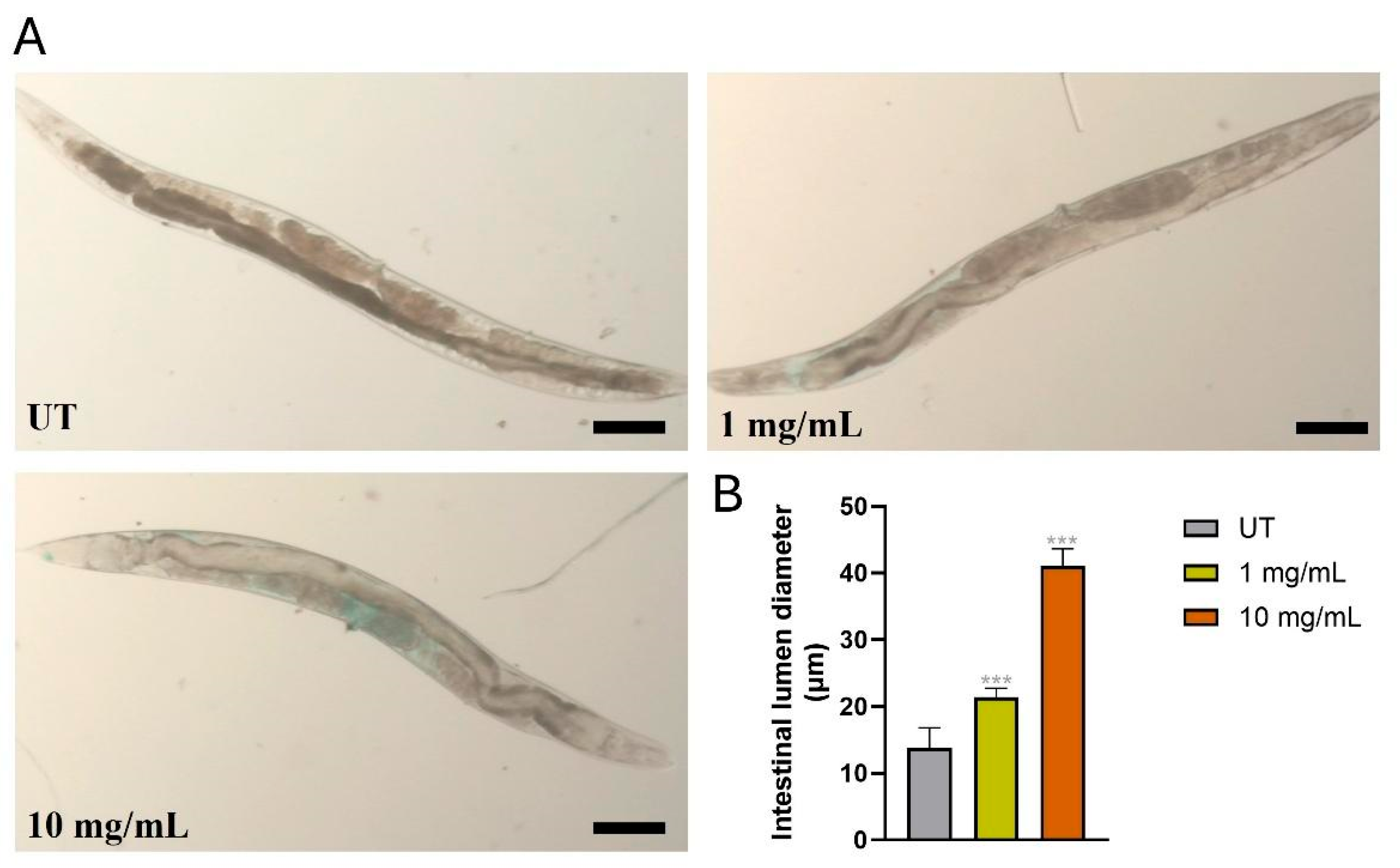
The effects of MPs on intestinal integrity were evaluated in adult C. elegans at day 10 of adulthood. For this purpose, the synthetic dye Brilliant Blue FCF was used, which binds to damaged tissues and enables the detection of structural and functional alterations in the intestinal tract. Treatment with MPs at a concentration of 10 mg/mL led to increased intestinal permeability, evidenced by the diffusion of the dye into areas surrounding the intestine (Figure 3A). This diffusion was exclusively observed in worms exposed to MPs, whereas in the control group, the dye remained confined within the intestinal lumen. Moreover, a quantitative analysis was conducted to measure the intestinal lumen diameter in adult worms at the same timepoints. As shown in Figure 3B, 10-day-old worms treated with 1 or 10 mg/mL MPs exhibited a marked expansion of the intestinal lumen, with increases of approximately 40% and 2-fold, respectively, compared to untreated controls. No significant alterations in intestinal morphology were observed in worms treated with 0.01 or 0.1 mg/mL MPs.

3.4. MPs Altered Fertility Rate and Embryos Formation

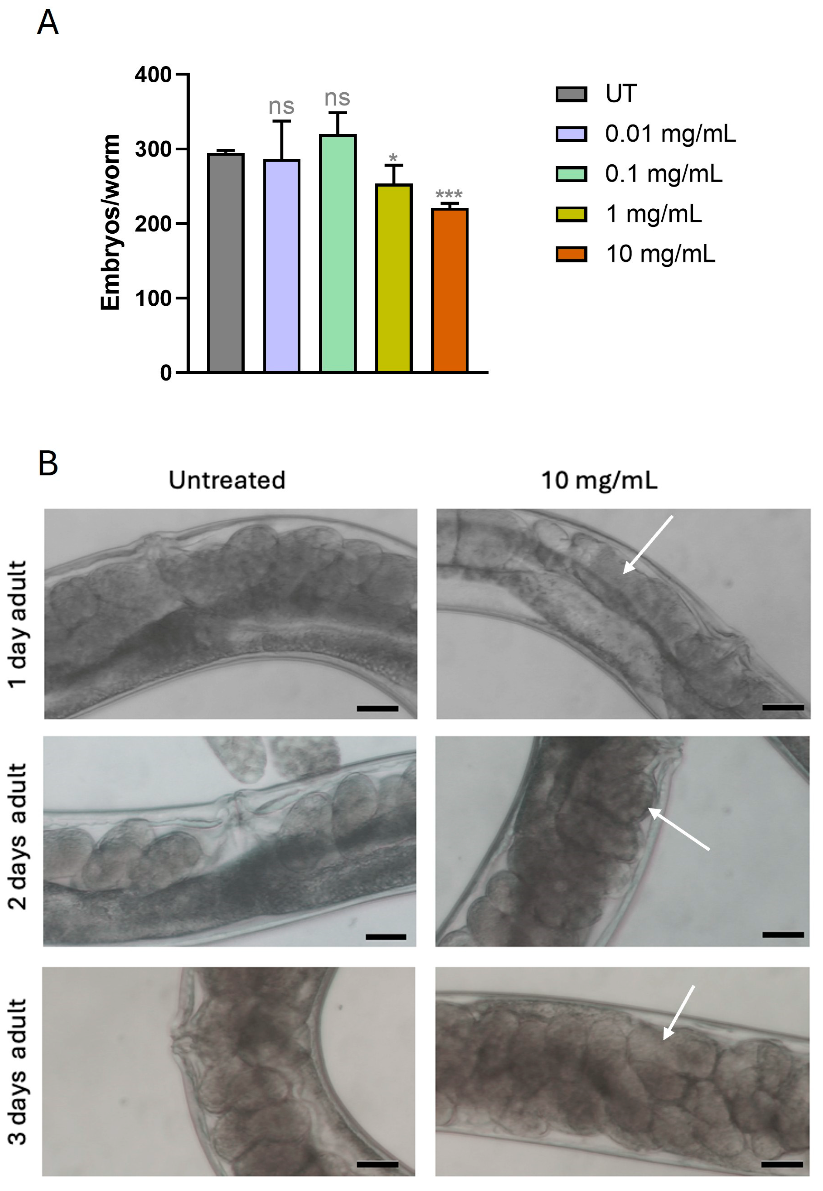
The fertility of C. elegans exposed to different concentrations of MPs was evaluated by analyzing brood size, defined as the total number of embryos produced per individual. This parameter provides a direct measure of reproductive capacity, enabling the comparison of fertility rates between treated groups and controls. As shown in Figure 4A, all worms exposed to MPs produced significantly less embryos than the untreated control group. Specifically, nematodes treated with 0.01, 0.1, and 1 mg/mL MPs exhibited reductions in embryo production of 20%, 25%, and 20%, respectively. The most pronounced decrease, of about 30%, was observed in the group exposed to 10 mg/mL MPs as compared to controls. In addition to brood size analysis, a morphological examination of embryos within 1-, 2-, and 3-day-old adult nematodes was performed using brightfield microscopy (Figure 4B). This analysis aimed to identify potential structural alterations in embryos following exposure to 10 mg/mL MPs. As illustrated in Figure 4B, treated embryos displayed marked morphological changes compared to those from control animals. Notably, embryos from exposed nematodes appeared less rounded and more heterogeneous in shape and size, suggesting that MPs exposure may interfere with proper embryonic development and potentially impair reproductive success.

3.5. MP Supplementation Increase Reactive Oxygen Species (ROS) Production

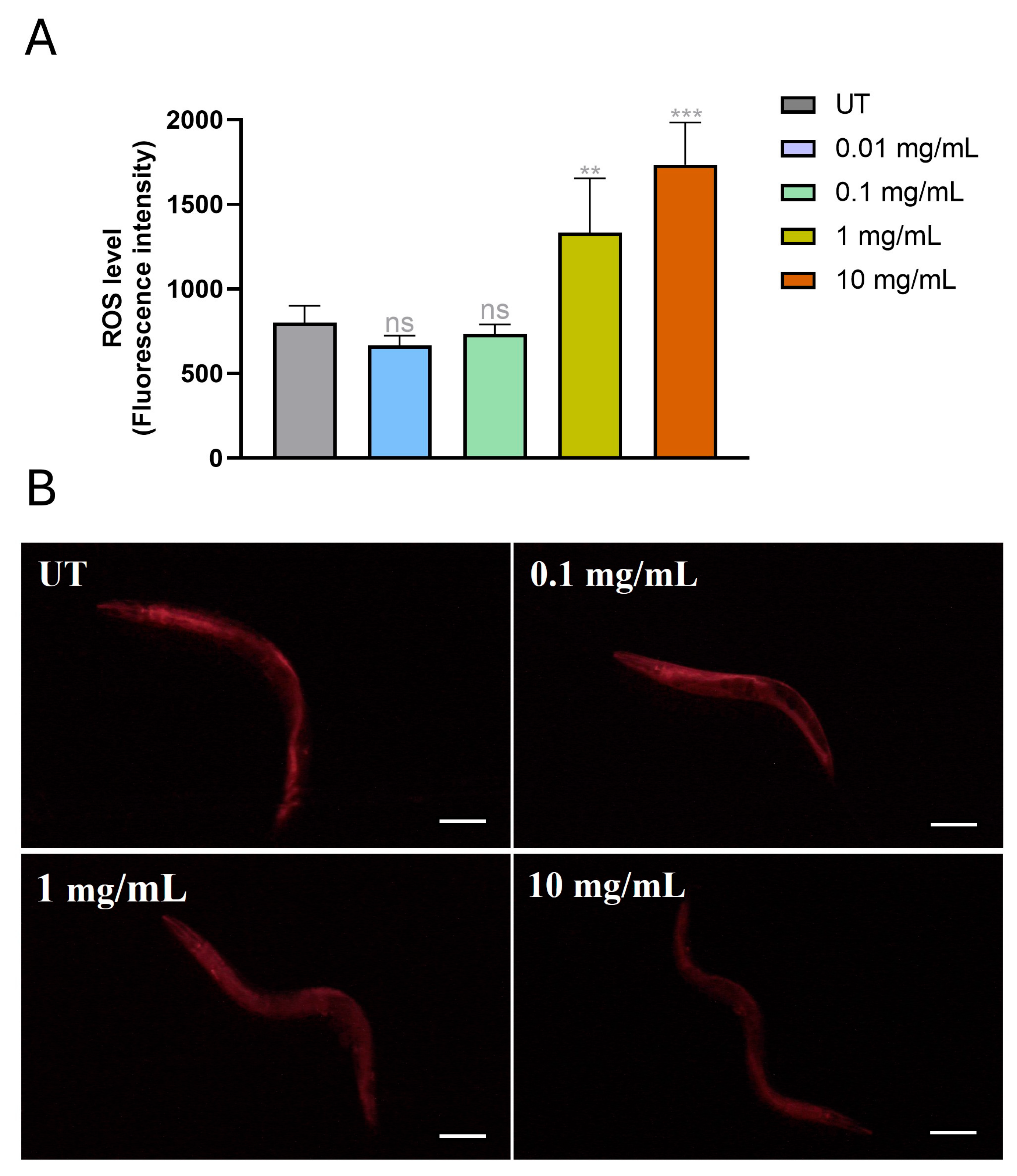
Oxidative stress represents a biological condition that arises when pro-oxidant factors exceed the organism’s antioxidant defense capacity. Under normal physiological conditions, a balance exists between the production of reactive oxygen species (ROS) and the efficacy of antioxidant systems [22]. However, this balance can be disrupted by factors that enhance ROS generation or by compounds that impair antioxidant defense mechanisms. Many xenobiotics, including microplastics (MPs), are known to promote oxidative stress [23]. For this reason, a significant portion of this study was devoted to evaluating the effects of MPs on oxidative damage. To assess ROS accumulation in 4-day-old adult nematodes following exposure to different concentrations of MPs, the fluorescent probe H2DCF-DA was employed. As shown in Figure 5A, no significant differences in ROS levels were observed between nematodes exposed to 0.01 or 0.1 mg/mL MPs the control group at either age. However, exposure to 1 mg/mL and 10 mg/mL MPs resulted in a 2-fold increase in ROS production compared to controls. A similar trend was observed in 4-day-old adults, with ROS levels elevated by 70% and 120% in response to the same concentrations. To evaluate the effect of mitochondrial functionality of treated worms, MitoTracker® Red staining was performed (Figure 5B). The fluorescent dye that accumulates in mitochondria in a membrane potential-dependent manner, provides an indicator of mitochondrial activity and oxidative stress. In worms treated with MPs, a noticeable reduction in fluorescence intensity was observed, suggesting impaired mitochondrial function, in accord with the increased oxidative stress (Figure 5B). Treatments with 0.01 mg/mL MPs did not induce significant changes in mitochondrial functionality compared to controls.

3.6. Activation of Oxidative Stress Machinery After MP Supplementation

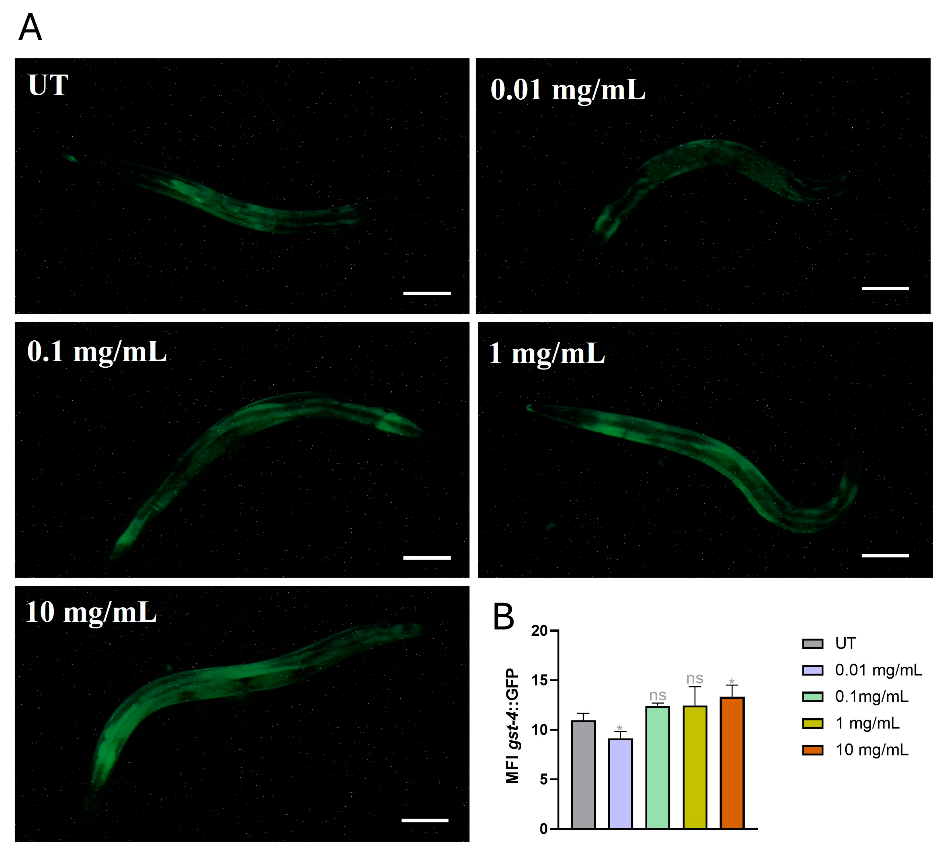
To provide additional insights into the involvement of detoxification mechanisms after MP treatments, the transgenic C. elegans strain gst-4::GFP was employed, in which the expression of the green fluorescent protein (GFP) is regulated by the gst-4 gene promoter. Glutathione S-transferase 4 (GST-4) is another key enzyme involved in the antioxidant defense system, playing a crucial role in cellular detoxification [13]. Adult nematodes at 4 days of age were grown on NGM plates and exposed to MPs at concentrations of 0.01 mg/mL, 0.1 mg/mL, 1 mg/mL, and 10 mg/mL. Analysis of images acquired through fluorescence microscopy (Figure 6A,B) revealed a significant increase in mean fluorescence intensity (MFI) in nematodes treated with 10 mg/mL MPs, with increases of 35% on day 1 and 22% on day 4 compared to the control group not exposed to MPs. Conversely, a reduction in MFI of 17% was observed in 4-day-old adults exposed to 0.01 mg/mL MPs. No significant changes were detected in the other experimental conditions compared to the untreated controls.
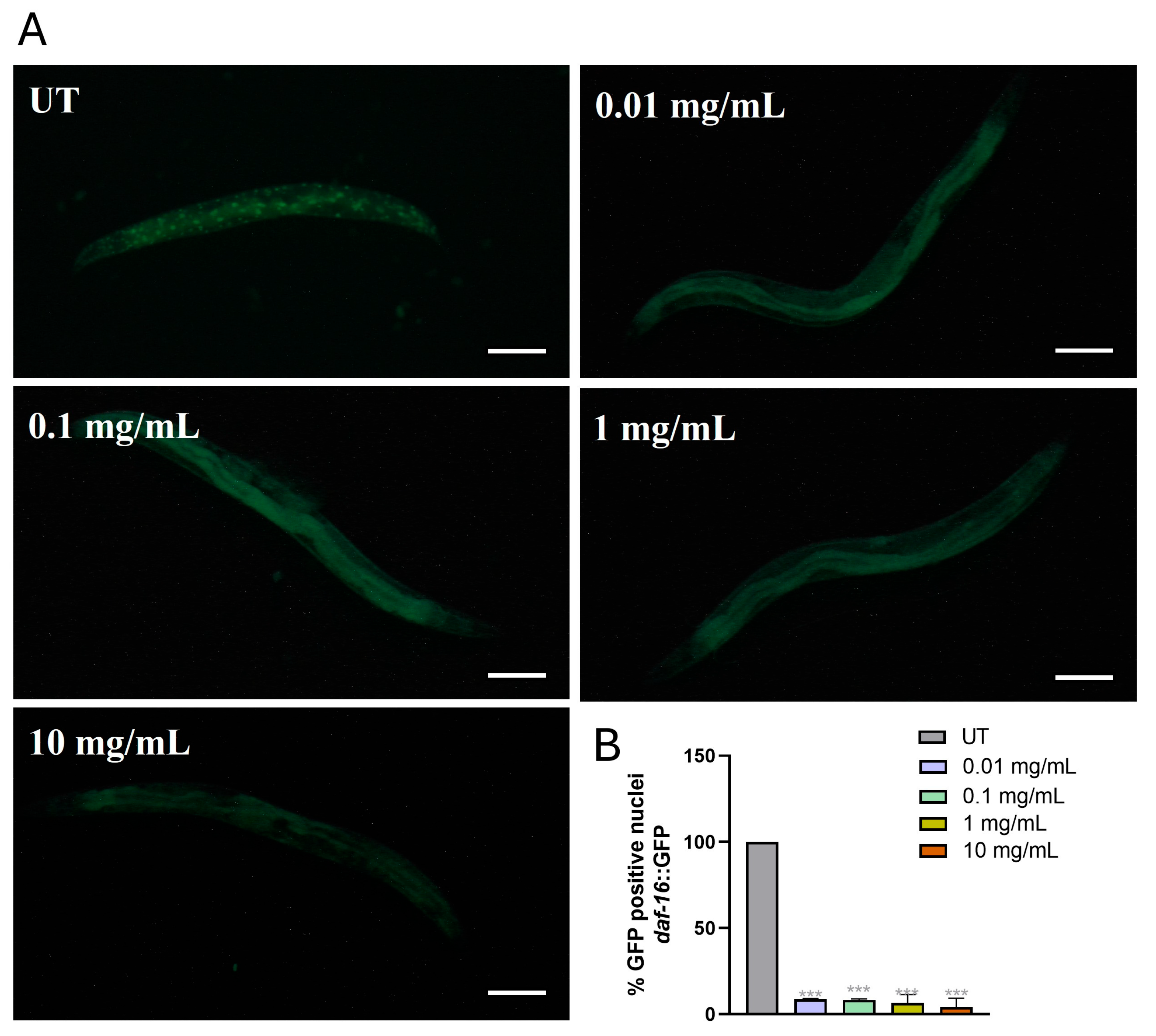
The transcription factor DAF-16, part of the FoxO family, is involved in regulating cellular responses to various stress conditions, including oxidative stress [24]. In response to stress, DAF-16 undergoes dephosphorylation, which promotes its nuclear translocation. Once in the nucleus, it binds to conserved DNA motifs known as DAF-16/FoxO binding elements (DBEs) that regulate the expression of many stress-responsive genes, including gst-4. To visualize this activation process, we employed the transgenic C. elegans strain daf-16::GFP. (Figure 7). After heat stress induction by exposure to 37 °C for 40 min, fluorescence microscopy revealed that, in nematodes exposed to MPs, the nuclear translocation of DAF-16 was significantly reduced (Figure 7A). Subsequently, the number of GFP-positive nuclei (Figure 7B), highlighting a reduction in nuclear localization of DAF-16 in 4-day-old adults by about 90% after MPs treatment, compared to the control group.

3.7. Gene Expression Levels of Oxidative Stress-Related Genes

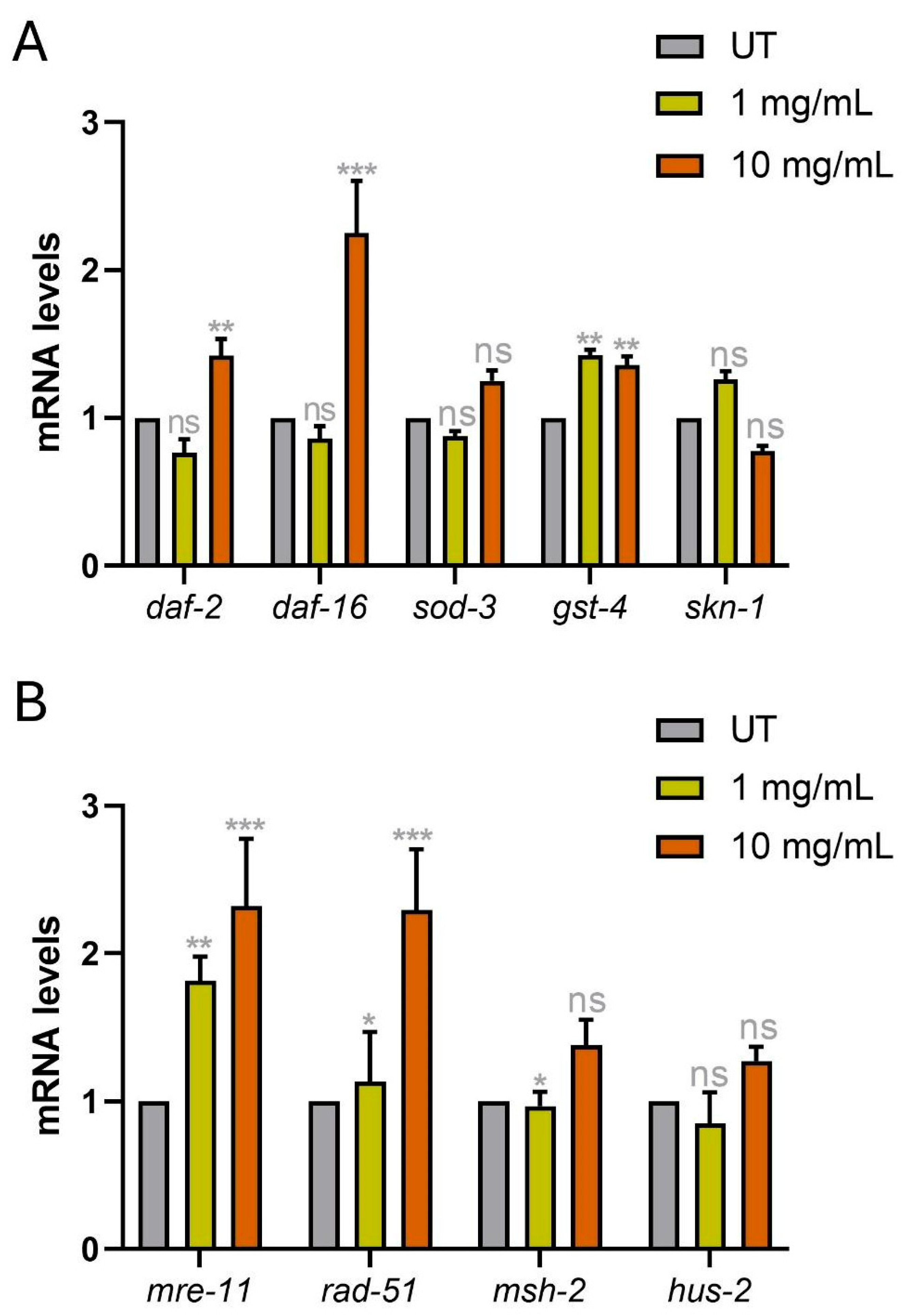
In 4-day-old adult nematodes treated with 1 mg/mL and 10 mg/mL MPs, a molecular analysis was conducted using real-time qPCR to quantify the expression of daf-2, sod-3, gst-4, skn-1, and daf-16, genes involved in oxidative stress responses in C. elegans.
Figure 8A showed that the expression of daf-2 was 30% higher in nematodes treated with 1 mg/mL MPs, while a 20% reduction was observed in those exposed to 10 mg/mL MPs. In nematodes exposed to 10 mg/mL MPs, a 2.5-fold increase in daf-16 mRNA levels was recorded. A significant increase in the transcript levels of gst-4, with 40% increase in the groups exposed to 1 mg/mL and 10 mg/mL MPs, respectively, compared to the control group. Conversely, in samples treated with 1 mg/mL MPs, a 15% decrease in skn-1 transcript levels was observed. Interestingly, all genes involved in DNA repair mechanisms were upregulated after MPs treatment (Figure 8B).
To demonstrate the intake of PMMA and its distribution inside the animals, a μFT-IR analysis was carried out on 4-day adult worms fed from embryos hatching with the 10 mg/mL microparticle suspension. The analysis was also performed on the untreated nematode as control (Figure S1). The peak centered at 1724 cm−1, related to the characteristic ester carbonyl C = O stretching vibration of PMMA [25], was monitored. In the untreated animals, no absorption signal is observed in this range, whereas the C = O stretching vibration of triglycerides is detected at higher wavenumbers (1745 cm−1) [26]. Figure 9 presents the 2D (panel A) and 3D (panel B) chemical mappings, revealing the spatial distribution of the PMMA absorption signal throughout the nematode body.

4. Discussion

Microplastics (MPs) and other environmental contaminants, such as heavy metals, represent an escalating threat to ecosystems, impacting organisms across trophic levels [27]. Understanding their biological effects is essential for developing mitigation strategies. In this study, we evaluated the impact of poly(methyl methacrylate) (PMMA) MPs on the nematode C. elegans, a widely used model in environmental toxicology, by assessing phenotypic, physiological, and molecular endpoints [17]. C. elegans is particularly suited for toxicological screening due to its sensitivity to a broad spectrum of pollutants, including organic compounds, nanoparticles, and MPs. Compared to other invertebrate and vertebrate models, C. elegans often exhibits toxicity responses, especially reproductive impairments, at lower contaminant concentrations [28]. These responses are well correlated with outcomes in vertebrate models [29], and endpoints such as growth, reproduction, and oxidative stress in C. elegans closely mirror those observed in higher organisms exposed to MPs and nanoplastics [30]. Our data clearly show that in C. elegans, PMMA MPs exert concentration-dependent toxicity, with the strongest effects consistently observed at the highest exposure level. Although intermediate doses did not always follow a strictly linear trend, measurable impacts were detected on development, survival, reproduction, and oxidative homeostasis. FT-IR microspectroscopy has been demonstrated to be a powerful technique for obtaining chemical distribution maps in complex biological systems [31] and has been effectively employed to evaluate the distribution of carbon nanomaterials in C. elegans. The PMMA detected by chemical mapping constitute a fine fraction, which may originate either from ultrasonic disintegration of the original MPs, or from a pre-existing fine fraction inherently present in the batch. The localization of PMMA within both the intestinal tract and the gonads suggests a potential for direct interaction with tissues essential for nutrient absorption and reproduction, which could have implications for organismal health and fitness, as suggested from previous results of this study. Similar biodistribution patterns have been reported for other polymeric particles and nanomaterials in C. elegans [32,33]. The significant reduction in body size at higher concentrations of MPs suggests disrupted larval development, consistent with prior reports of developmental delays in MP-exposed invertebrates [17]. Such outcomes may stem from compromised nutrient uptake or stress-induced metabolic reallocation that reduces growth. We indeed observed a significant decline in lifespan, particularly in worms exposed to 1 and 10 mg/mL MPs. This reduction in longevity was accompanied by impaired physiological functions, such as pharyngeal pumping and motility, which are recognized markers of aging in C. elegans (See Figure 2). In addition, intestinal permeability assays demonstrated damage to gut integrity in worms treated with the highest MP concentration. The dye Brilliant Blue FCF, commonly used to assess gut barrier dysfunction [34], diffused beyond the intestinal lumen only in MP-exposed animals, suggesting epithelial compromise, as reported in [35]. As the intestine plays key roles in immunity, metabolism, and stress signaling, its disruption may be central to the observed reductions in fertility and lifespan. PMMA exposure also resulted in substantial reproductive toxicity. Brood size declined significantly across all treatment groups, and morphological abnormalities in embryos were evident at the highest concentration (see Figure 4). Previous studies have shown that the toxicity of MPs and NPs increases with decreasing particle size, likely due to enhanced bioavailability and tissue penetration [36]. These findings support the notion that PMMA-MPs may accelerate the aging process, potentially through cumulative mitochondrial dysfunction and stress pathway activation. Indeed, oxidative stress emerged as a key toxicological mechanism. We recorded a notable increase in cytosolic and mitochondrial ROS levels in MP-treated worms. This finding is consistent with previous work demonstrating that MPs can disrupt the redox balance by overwhelming ROS scavenging systems and inducing expression of detoxification enzymes such as SOD, catalase, and GST [17]. In our experiments, the upregulation of gst-4 and daf-16, alongside activation of gst-4::GFP, points to an adaptive stress response. However, the reduced nuclear localization of DAF-16 and downregulation of skn-1 at higher doses suggest that the protective capacity of these pathways is compromised under severe MP exposure. Sustained ROS accumulation is known to damage nucleic acids, lipids, and proteins, potentially leading to apoptosis or necrosis [37]. Oxidative byproducts can activate MAPK signaling pathways, such as JNK and p38, implicated in cell responses and death [24]. Mitochondrial dysfunction intensifies this cascade, as impaired mitochondria are major ROS producers and are linked to aging, metabolic disorders, and chronic diseases [38]. In our study, increased expression of DNA repair genes, including mre-11, rad-51, hus-1, and msh-2, supports the notion that MPs also cause genotoxic stress. These genes are involved in mismatch repair, DNA damage sensing, and apoptosis signaling, and their activation suggests an attempt to counteract DNA lesions caused by oxidative damage, according to [38].

5. Conclusions

Overall, our findings support the conclusion that MPs act as multifactorial stressors in C. elegans, impairing essential physiological processes and triggering both oxidative and genotoxic stress pathways. Given the evolutionary conservation of many of these mechanisms, these results raise concerns about the broader ecological and health impacts of MPs in higher organisms, including humans. Importantly, our study underscores the value of C. elegans as a cost-effective, sensitive, and genetically tractable model for evaluating the complex toxicological effects of PMMA microplastics. This model can be readily applied to investigate the toxicity of other MPs while adhering to the 3Rs principles (Replace, Reduce, Refinement), making it ethically advantageous compared to higher-order animal models. While commercially available PMMA MPs are homogeneous in size and shape, their toxicity may be further modulated by environmental factors such as degradation, adsorption of pollutants, and formation of an eco-corona, which is a layer of biomolecules and environmental compounds that absorb onto particle surfaces, altering their bioavailability and toxic effects. Controlled experiments allow the assessment of intrinsic toxicity independently from these additional influences.

Supplementary Materials

The following supporting information can be downloaded at https://www.mdpi.com/article/10.3390/environments12100353/s1, Table S1: Primers for real time q-PCR. Figure S1. FT-IR microspectroscopy of 4 day-adult untreated nematodes.

Author Contributions

Conceptualization, D.U., E.S. (Emily Schifano) and L.D.; methodology, S.F., E.S. (Erica Sonaglia) and M.S.; validation, E.S. (Emily Schifano), S.T. and M.L.S.; formal analysis, E.S. (Emily Schifano), L.D. and D.U.; investigation, E.S. (Erica Sonaglia), M.S. and S.T.; data curation, E.S. (Emily Schifano) and M.L.S.; writing—original draft preparation, E.S. (Erica Sonaglia) and E.S. (Emily Schifano), writing—review and editing, D.U., L.D. and M.L.S.; funding acquisition, M.L.S. All authors have read and agreed to the published version of the manuscript.

Funding

This research was partially supported by PRIORITY—COST ACTION CA20101 “Plastics Monitoring Detection Remediation Recovery”.

Data Availability Statement

The original contributions presented in this study are included in the article/Supplementary Material. Further inquiries can be directed to the corresponding author(s).

Conflicts of Interest

The authors declare no conflicts of interest.

References

  1. Thompson, R.C.; Courtene-Jones, W.; Boucher, J.; Pahl, S.; Raubenheimer, K.; Koelmans, A.A. Twenty Years of Microplastic Pollution Research—What Have We Learned? Science 2024, 386, eadl2746. [Google Scholar] [CrossRef]
  2. Li, Y.; Chen, L.; Zhou, N.; Chen, Y.; Ling, Z.; Xiang, P. Microplastics in the Human Body: A Comprehensive Review of Exposure, Distribution, Migration Mechanisms, and Toxicity. Sci. Total Environ. 2024, 946, 174215. [Google Scholar] [CrossRef]
  3. Pannetier, P.; Morin, B.; Le Bihanic, F.; Dubreil, L.; Clérandeau, C.; Chouvellon, F.; Van Arkel, K.; Danion, M.; Cachot, J. Environmental Samples of Microplastics Induce Significant Toxic Effects in Fish Larvae. Environ. Int. 2020, 134, 105047. [Google Scholar] [CrossRef] [PubMed]
  4. De Souza Machado, A.A.; Kloas, W.; Zarfl, C.; Hempel, S.; Rillig, M.C. Microplastics as an Emerging Threat to Terrestrial Ecosystems. Glob. Change Biol. 2018, 24, 1405–1416. [Google Scholar] [CrossRef] [PubMed]
  5. Scheurer, M.; Bigalke, M. Microplastics in Swiss Floodplain Soils. Environ. Sci. Technol. 2018, 52, 3591–3598. [Google Scholar] [CrossRef]
  6. Fuller, S.; Gautam, A. A Procedure for Measuring Microplastics Using Pressurized Fluid Extraction. Environ. Sci. Technol. 2016, 50, 5774–5780. [Google Scholar] [CrossRef]
  7. Hoang, V.-H.; Nguyen, M.-K.; Hoang, T.-D.; Ha, M.C.; Huyen, N.T.T.; Bui, V.K.H.; Pham, M.-T.; Nguyen, C.-M.; Chang, S.W.; Nguyen, D.D. Sources, Environmental Fate, and Impacts of Microplastic Contamination in Agricultural Soils: A Comprehensive Review. Sci. Total Environ. 2024, 950, 175276. [Google Scholar] [CrossRef]
  8. Adomako, M.O.; Wu, J.; Yu, F.-H. Ecological and Evolutionary Responses of Earthworm Holobionts to Environmental Changes. ISME J. 2025, 19, wraf044. [Google Scholar] [CrossRef]
  9. Burcea, A.; Bănățeanu, A.-M.; Poalelungi, C.-V.; Burcea, A.; Forna, N.; Cumpătă, C.N. Enhanced Properties and Multifaceted Applications of Polymethyl Methacrylate (PMMA) in Modern Medicine and Dentistry. RJOR 2024, 16, 108–123. [Google Scholar] [CrossRef]
  10. Edo, G.I.; Ndudi, W.; Ali, A.B.M.; Yousif, E.; Zainulabdeen, K.; Onyibe, P.N.; Akpoghelie, P.O.; Ekokotu, H.A.; Isoje, E.F.; Igbuku, U.A.; et al. An Updated Review on the Modifications, Recycling, Polymerization, and Applications of Polymethyl Methacrylate (PMMA). J. Mater. Sci. 2024, 59, 20496–20539. [Google Scholar] [CrossRef]
  11. Neves, B.; Oliveira, M.; Frazão, C.; Almeida, M.; Pinto, R.J.B.; Figueira, E.; Pires, A. The Role of Life Stages in the Sensitivity of Hediste Diversicolor to Nanoplastics: A Case Study with Poly(Methyl)Methacrylate (PMMA). Toxics 2024, 12, 352. [Google Scholar] [CrossRef]
  12. Yip, Y.J.; Sivananthan, G.D.; Lee, S.S.C.; Neo, M.L.; Teo, S.L.-M.; Valiyaveettil, S. Transfer of Poly(Methyl Methacrylate) Nanoparticles from Parents to Offspring and the Protection Mechanism in Two Marine Invertebrates. ACS Sustain. Chem. Eng. 2022, 10, 37–49. [Google Scholar] [CrossRef]
  13. Maupas, E. La mue et l’enkystement chez les Nématodes. Arch. Zool. Exper. Gén. Ser. 1899, 3, 7. [Google Scholar]
  14. Steinberg, C.E.W.; Stürzenbaum, S.R.; Menzel, R. Genes and Environment—Striking the Fine Balance between Sophisticated Biomonitoring and True Functional Environmental Genomics. Sci. Total Environ. 2008, 400, 142–161. [Google Scholar] [CrossRef] [PubMed]
  15. Polak, N.; Read, D.S.; Jurkschat, K.; Matzke, M.; Kelly, F.J.; Spurgeon, D.J.; Stürzenbaum, S.R. Metalloproteins and Phytochelatin Synthase May Confer Protection against Zinc Oxide Nanoparticle Induced Toxicity in Caenorhabditis Elegans. Comp. Biochem. Physiol. Part C Toxicol. Pharmacol. 2014, 160, 75–85. [Google Scholar] [CrossRef] [PubMed]
  16. Ficociello, G.; Gerardi, V.; Uccelletti, D.; Setini, A. Molecular and Cellular Responses to Short Exposure to Bisphenols A, F, and S and Eluates of Microplastics in C. Elegans. Environ. Sci. Pollut. Res. 2021, 28, 805–818. [Google Scholar] [CrossRef]
  17. Jewett, E.; Arnott, G.; Connolly, L.; Vasudevan, N.; Kevei, E. Microplastics and Their Impact on Reproduction—Can We Learn From the C. Elegans Model? Front. Toxicol. 2022, 4, 748912. [Google Scholar] [CrossRef] [PubMed]
  18. Chen, H.; Chen, M.; Gu, Y.; Jiang, Y.; Ding, P.; Wang, C.; Pan, R.; Shi, C.; Li, H. Microbial Colonization of Microplastics in Wastewater Accelerates the Aging Process Associated with Oxidative Stress and the Insulin/IGF1 Signaling Pathway. Environ. Pollut. 2023, 332, 121954. [Google Scholar] [CrossRef] [PubMed]
  19. Schifano, E.; Cicalini, I.; Pieragostino, D.; Heipieper, H.J.; Del Boccio, P.; Uccelletti, D. In Vitro and in Vivo Lipidomics as a Tool for Probiotics Evaluation. Appl. Microbiol. Biotechnol. 2020, 104, 8937–8948. [Google Scholar] [CrossRef] [PubMed]
  20. Jeayeng, S.; Thongsroy, J.; Chuaijit, S. Caenorhabditis Elegans as a Model to Study Aging and Photoaging. Biomolecules 2024, 14, 1235. [Google Scholar] [CrossRef] [PubMed]
  21. Zhang, H.; Chen, W. Automated Recognition and Analysis of Body Bending Behavior in C. Elegans. BMC Bioinform. 2023, 24, 175. [Google Scholar] [CrossRef] [PubMed]
  22. Armeli, F.; Mengoni, B.; Schifano, E.; Lenz, T.; Archer, T.; Uccelletti, D.; Businaro, R. The Probiotic Yeast, Milmed, Promotes Autophagy and Antioxidant Pathways in BV-2 Microglia Cells and C. Elegans. Antioxidants 2025, 14, 393. [Google Scholar] [CrossRef]
  23. Kadac-Czapska, K.; Ośko, J.; Knez, E.; Grembecka, M. Microplastics and Oxidative Stress—Current Problems and Prospects. Antioxidants 2024, 13, 579. [Google Scholar] [CrossRef] [PubMed]
  24. Singhal, A.; Dubey, K.A.; Bhardwaj, Y.K.; Jain, D.; Choudhury, S.; Tyagi, A.K. UV-Shielding Transparent PMMA/In2O3 Nanocomposite Films Based on In2O3 Nanoparticles. RSC Adv. 2013, 3, 20913. [Google Scholar] [CrossRef]
  25. Bouyanfif, A.; Liyanage, S.; Hequet, E.; Moustaid-Moussa, N.; Abidi, N. Review of FTIR Microspectroscopy Applications to Investigate Biochemical Changes in C. Elegans. Vib. Spectrosc. 2018, 96, 74–82. [Google Scholar] [CrossRef]
  26. Yarahmadi, A.; Heidari, S.; Sepahvand, P.; Afkhami, H.; Kheradjoo, H. Microplastics and Environmental Effects: Investigating the Effects of Microplastics on Aquatic Habitats and Their Impact on Human Health. Front. Public Health 2024, 12, 1411389. [Google Scholar] [CrossRef]
  27. Liu, P.; Zhan, X.; Wu, X.; Li, J.; Wang, H.; Gao, S. Effect of Weathering on Environmental Behavior of Microplastics: Properties, Sorption and Potential Risks. Chemosphere 2020, 242, 125193. [Google Scholar] [CrossRef]
  28. Hunt, P.R. The C. elegans Model in Toxicity Testing. J. Appl. Toxicol. 2017, 37, 50–59. [Google Scholar] [CrossRef]
  29. Zhao, Y.; Wu, Q.; Li, Y.; Wang, D. Translocation, Transfer, and in Vivo Safety Evaluation of Engineered Nanomaterials in the Non-Mammalian Alternative Toxicity Assay Model of Nematode Caenorhabditis Elegans. RSC Adv. 2013, 3, 5741. [Google Scholar] [CrossRef]
  30. Diomede, L.; Cassata, G.; Fiordaliso, F.; Salio, M.; Ami, D.; Natalello, A.; Doglia, S.M.; De Luigi, A.; Salmona, M. Tetracycline and Its Analogues Protect Caenorhabditis Elegans from β Amyloid-Induced Toxicity by Targeting Oligomers. Neurobiol. Dis. 2010, 40, 424–431. [Google Scholar] [CrossRef] [PubMed]
  31. Kim, H.M.; Lee, D.-K.; Long, N.P.; Kwon, S.W.; Park, J.H. Uptake of Nanopolystyrene Particles Induces Distinct Metabolic Profiles and Toxic Effects in Caenorhabditis Elegans. Environ. Pollut. 2019, 246, 578–586. [Google Scholar] [CrossRef]
  32. Wu, T.; He, J.; Ye, Z.; Xia, J.; Chen, M.; Chen, S.; Liu, K.; Xing, P.; Yang, J.; Qian, Y.; et al. Aged Biodegradable Nanoplastics Enhance Body Accumulation Associated with Worse Neuronal Damage in Caenorhabditis elegans. Environ. Sci. Technol. 2025, 59, 4352–4363. [Google Scholar] [CrossRef] [PubMed]
  33. Martins, R.; McCracken, A.; Simons, M.; Henriques, C.; Rera, M. How to Catch a Smurf?—Ageing and Beyond…In Vivo Assessment of Intestinal Permeability in Multiple Model Organisms. BIO-Protoc. 2018, 8, e2722. [Google Scholar] [CrossRef]
  34. Yu, Y.; Chen, H.; Hua, X.; Dang, Y.; Han, Y.; Yu, Z.; Chen, X.; Ding, P.; Li, H. Polystyrene Microplastics (PS-MPs) Toxicity Induced Oxidative Stress and Intestinal Injury in Nematode Caenorhabditis Elegans. Sci. Total Environ. 2020, 726, 138679. [Google Scholar] [CrossRef] [PubMed]
  35. Liu, H.; Tian, L.; Wang, S.; Wang, D. Size-Dependent Transgenerational Toxicity Induced by Nanoplastics in Nematode Caenorhabditis Elegans. Sci. Total Environ. 2021, 790, 148217. [Google Scholar] [CrossRef] [PubMed]
  36. Martins, A.C.; Virgolini, M.B.; Ávila, D.S.; Scharf, P.; Li, J.; Tinkov, A.A.; Skalny, A.V.; Bowman, A.B.; Rocha, J.B.T.; Aschner, M. Mitochondria in the Spotlight: C. Elegans as a Model Organism to Evaluate Xenobiotic-Induced Dysfunction. Cells 2023, 12, 2124. [Google Scholar] [CrossRef] [PubMed]
  37. Dilberger, B.; Baumanns, S.; Schmitt, F.; Schmiedl, T.; Hardt, M.; Wenzel, U.; Eckert, G.P. Mitochondrial Oxidative Stress Impairs Energy Metabolism and Reduces Stress Resistance and Longevity of C. elegans. Oxidative Med. Cell. Longev. 2019, 2019, 6840540. [Google Scholar] [CrossRef] [PubMed]
  38. Ali, I.; Tan, X.; Peng, C.; Naz, I.; Zhang, Y.; Hernández, A.; Marcos, R.; Pervez, R.; Duan, Z.; Ruan, Y. Eco- and Bio-Corona-Based Microplastics and Nanoplastics Complexes in the Environment: Modulations in the Toxicological Behavior of Plastic Particles and Factors Affecting. Process Saf. Environ. Prot. 2024, 187, 356–375. [Google Scholar] [CrossRef]
Figure 1. Impact of MPs on body length and survival in C. elegans. (A) Assessment of worm body length following exposure to increasing concentrations of MPs (0.01, 0.1, 1, and 10 mg/mL) at defined time points. Untreated worms (UT) were taken as reference. Measurements were performed from head to tail, and data represent the average of three independent replicates. (B) Kaplan–Meier survival curves comparing wild-type N2 worms exposed to MPs with untreated controls (UT). N: 80 animals per condition per experiment. Statistical significance is indicated as follows: * p < 0.05, ** p < 0.05; *** p < 0.001; ns: not significant.
Environments 12 00353 g001
Figure 2. Evaluation of aging-related markers in C. elegans exposed to MPs. (A) Pharyngeal pumping rate of 2- or 10-day-old worms following MP exposure, assessed over 30 s. Ten worms were analyzed for each condition. Untreated worms (UT) were used as controls. (B) Body bend frequency measured over a 30 s interval in worms treated with different concentrations of MPs, compared to untreated controls. Statistical analysis was performed using one-way ANOVA followed by Bonferroni post hoc test. Asterisks denote significance (* p < 0.05, ** p < 0.01, *** p < 0.001, ns: not significant). Bars indicate the average of three biological replicates.
Environments 12 00353 g002
Figure 3. Effects of MPs on intestinal permeability. (A) FCF Brilliant Blue staining in 10-day adult worms treated with MPs at the concentrations of 1 or 10 mg/mL, compared to untreated worms (control). Scale bar: 100 μm. (B) Lumen diameter in worms treated with MPs at 10 days of adulthood. Untreated worms were taken as reference. Bars represent the mean of three independent experiments; asterisks indicate the p-values (log-rank test) normalized to the control (*** p < 0.001).
Environments 12 00353 g003
Figure 4. Effect of MPs on C. elegans reproduction. (A) Average embryos production per worm of animals supplemented with different concentrations of MPs. Untreated worms were taken as reference. Bars represent the mean of three independent experiments; asterisks indicate the p-values (log-rank test) normalized to the control (* p < 0.01, *** p < 0.001, ns: not significant). (B) Morphological analysis of embryos in 1-, 2-, and 3-day-old adult nematodes exposed to a concentration of MPs at 10 mg/mL and in untreated controls. Structural alterations in embryos are indicated by arrows. Images were acquired using a Zeiss Axiovert25 microscope at 32× magnification. Scale bar: 20 μm.
Environments 12 00353 g004
Figure 5. Assessment of oxidative stress in MP-treated worms. (A) Reactive Oxygen Species (ROS) levels in 4-day-old adult individuals treated with MPs at concentrations of 0.01 mg/mL, 0.1 mg/mL, 1 mg/mL, and 10 mg/mL, compared to the untreated control group (UT). Asterisks indicate statistically significant differences (** p < 0.01, *** p < 0.001, ns: not significant) relative to the control group. (B) 4-day adults, treated with MPs from embryos hatching, were stained with MitoTracker® Red for mitochondrial ROS. Scale bar is 100 μM.
Environments 12 00353 g005
Figure 6. Visualization of GST-4 in C. elegans nuclei. (A) Analysis of the localization and fluorescence intensity of GST-4 enzyme in the transgenic gst-4::GFP strain. Fluorescence microscopy of worms at the stage of 4-day adult treated with MPs. Scale bar  =  100 μm. (B) Percentage of GFP-positive nuclei. Statistical analysis was evaluated by one-way ANOVA with the Bonferroni post-test; asterisks indicate significant differences (* p  <  0.05; ns: not significant). Bars represent the mean of three independent experiments with n  =  20.
Environments 12 00353 g006
Figure 7. Visualization of DAF-16 translocation in C. elegans nuclei. (A) Analysis of the translocation of DAF-16 in the nucleus in the transgenic daf-16::GFP strain. Fluorescence microscopy of worms at the stage of 4-day adult treated with MPs. Scale bar  =  100 μm. (B) Percentage of GFP-positive nuclei. Statistical analysis was evaluated by one-way ANOVA with the Bonferroni post-test; asterisks indicate significant differences (*** p  <  0.001). Bars represent the mean of three independent experiments with n  =  20.
Environments 12 00353 g007
Figure 8. Impact of MPs on oxidative stress responses and DNA repair mechanisms. Expression of (A) daf-2, daf-16, sek-1, sod-3, gst-4 and skn-1 genes, involved in oxidative stress responses and (B) mre-11, rad-51, msh-2, and hus-2, involved in DNA repair, in N2 worms treated with 1 or 10 mg/mL MPs, compared to untreated nematodes at day 4 of adulthood. Histograms show the expression of genes detected by real-time PCR. Experiments were performed in triplicate. Data are presented as mean ± SD (* p < 0.05, ** p < 0.01 and *** p < 0.001; ns: not significant).
Environments 12 00353 g008
Figure 9. The spatial distribution of the PMMA within the 4 day-adult nematodes treated with 10 mg/mL MPs. Spectroscopic 2D (A) and 3D (B) images. All spectra were collected in reflection mode with a 3 cm–1 spectral resolution, and the images were obtained by monitoring the 1724 cm−1 peak; the absorption intensity was measured and expressed as arbitrary units.
Environments 12 00353 g009
Disclaimer/Publisher’s Note: The statements, opinions and data contained in all publications are solely those of the individual author(s) and contributor(s) and not of MDPI and/or the editor(s). MDPI and/or the editor(s) disclaim responsibility for any injury to people or property resulting from any ideas, methods, instructions or products referred to in the content.