Review

Electric Field Effects on Microbial Cell Properties: Implications for Detection and Control in Wastewater Systems

1
Department of General Chemistry, Faculty of Chemical Engineering and Biotechnologies, The National University of Science and Technology POLITEHNICA Bucharest, Gheorghe Polizu 1-7 Street, 011061 Bucharest, Romania
2
Department of Automation and Industrial Informatics, Faculty of Automatic Control and Computers, The National University of Science and Technology POLITEHNICA Bucharest, Splaiul Independenţei 313 Street, 060042 Bucharest, Romania
3
Department of Analytical Chemistry and Environmental Engineering, Faculty of Chemical Engineering and Biotechnologies, The National University of Science and Technology POLITEHNICA Bucharest, Gheorghe Polizu 1-7 Street, 011061 Bucharest, Romania
4
I.C.P.E. BISTRITA S.A., Parcului 7 Street, 420035 Bistrita, Romania
5
Pharmacy Faculty, University Titu Maiorescu, No. 22 Dâmbovnicului Street, District 4, 040441 Bucharest, Romania
*
Authors to whom correspondence should be addressed.
Environments 2025, 12(10), 343; https://doi.org/10.3390/environments12100343
Submission received: 14 August 2025 / Revised: 17 September 2025 / Accepted: 23 September 2025 / Published: 25 September 2025
(This article belongs to the Special Issue Advanced Technologies for Contaminant Removal from Water)

Abstract

Electric fields (EFs) have emerged as effective, non-chemical tools for modulating microbial populations in complex matrices such as wastewater. This review consolidates current advances on EF-induced alterations in microbial structures and functions, focusing on both vegetative cells and spores. Key parameters affected include membrane thickness, transmembrane potential, electrical conductivity, and dielectric permittivity, with downstream impacts on ion homeostasis, metabolic activity, and viability. Such bioelectrical modifications underpin EF-based detection methods—particularly impedance spectroscopy and dielectrophoresis—which enable rapid, label-free, in situ microbial monitoring. Beyond detection, EFs can induce sublethal or lethal effects, enabling selective inactivation without chemical input. This review addresses the influence of field type (DC, AC, pulsed), intensity, and exposure duration, alongside limitations such as species-specific variability, heterogeneous environmental conditions, and challenges in achieving uniform field distribution. Emerging research highlights the integration of EF-based platforms with biosensors, machine learning, and real-time analytics for enhanced environmental surveillance. By linking microbiological mechanisms with engineering solutions, EF technologies present significant potential for sustainable water quality management. Their multidisciplinary applicability positions them as promising components of next-generation wastewater monitoring and treatment systems, supporting global efforts toward efficient, adaptive, and environmentally benign microbial control strategies.

1. Introduction

Microbial contamination in wastewater poses a major challenge for both environmental quality and public health. Untreated effluents contain diverse microorganisms—including bacteria, viruses, protozoa, archaea, and fungi—that can disseminate pathogens, trigger disease outbreaks, and contribute to antimicrobial resistance.
Monitoring microbial content in wastewater is essential for protecting public health, evaluating treatment performance, and safeguarding natural ecosystems [1]. Wastewater contains a wide range of microorganisms—including pathogenic bacteria, viruses, protozoa, and fungal spores—that signal fecal contamination or potential disease outbreaks. Common bacterial species detected in untreated municipal wastewater include Escherichia coli (up to 108 CFU/mL), Pseudomonas aeruginosa, Enterococcus faecalis, Salmonella spp., and spore-forming Clostridium perfringens [2]. In addition to bacteria, viruses, and protozoa, wastewater also harbors significant populations of archaea, yeasts, and fungi. Archaea, particularly methanogens, are involved in anaerobic digestion and biogas production, while yeasts and filamentous fungi contribute to organic matter degradation, biofilm formation, and in some cases opportunistic pathogenicity. Their presence highlights the need for detection and control strategies that address the full microbial diversity of wastewater ecosystems [2,3,4]. Viral agents such as norovirus and adenovirus, as well as protozoan parasites like Giardia lamblia and Cryptosporidium parvum, are also commonly reported in wastewater samples [5]. The persistence of these microorganisms poses risks to downstream environments, especially when effluents reach surface or groundwater bodies. Monitoring typically relies on indicators such as E. coli and Enterococcus [6], but insufficient control or ineffective inactivation can promote the spread of antibiotic-resistant bacteria—such as multidrug-resistant P. aeruginosa—and other pathogens, threatening human health and ecological balance. Consequently, agencies including the WHO and EPA have established microbial quality standards for wastewater discharges and reclaimed water reuse (e.g., ≤1000 CFU/100 mL fecal coliforms in treated effluents for unrestricted irrigation, per WHO guidelines) [7,8]. Against this background, Figure 1 contrasts conventional microbiological approaches with emerging electric field-based strategies, highlighting the shift toward faster, label-free, and in situ detection. Traditional methods such as culture-based assays, staining, and microscopy remain widely used for cultivable microorganisms [9], while novel techniques including electric field exposure, impedance or capacitance spectroscopy, and biosensor platforms offer real-time, label-free, and more targeted microbial monitoring [10]. Conventional approaches for microbial detection and inactivation in wastewater, although widely used, face several important limitations. Culture-based methods are slow and restricted to cultivable organisms, while molecular techniques may overestimate viable counts. Disinfection strategies such as chlorination, UV irradiation, or ozonation reduce overall microbial loads but are less effective in turbid or complex matrices and may generate undesirable byproducts. These gaps highlight the need for alternative solutions that can provide faster, more selective, and reliable results in real-world wastewater environments. Figure 1 summarizes this transition, focusing on representative technologies rather than an exhaustive survey.
Traditional microbiological methods such as culture-based assays and microscopy remain valuable, yet they are time-consuming, labor-intensive, and restricted to cultivable organisms. Notably, only 1–10% of environmental microorganisms are estimated to grow under standard laboratory conditions. To overcome these limitations, electric field (EF)-based methods have emerged as promising alternatives for microbial monitoring and control. Unlike chemical or thermal approaches, EF techniques interact directly with the structural and functional properties of microbial cells, enabling rapid, label-free, and potentially in situ analyses. Methods such as electrical impedance spectroscopy (EIS), dielectrophoresis (DEP), and pulsed electric fields (PEFs) exploit intrinsic microbial electrical properties for detection, discrimination, or inactivation. These approaches provide faster response times, higher selectivity, and greater adaptability to fluctuating wastewater conditions, making them attractive candidates for next-generation environmental monitoring systems. In contrast, emerging electric field (EF)-based technologies provide faster, more selective, and potentially real-time alternatives for microbial detection, classification, and even inactivation in situ [11,12]. Enhancing microbial assessment strategies that exploit electrical and dielectric properties (e.g., membrane potential, conductivity) is therefore critical for adaptive and resilient wastewater management [13].
Conventional detection approaches—including culture-based assays, staining, and molecular methods such as qPCR—each present important limitation. Culture-based techniques detect only viable and cultivable organisms, failing to capture viable but non-culturable (VBNC) cells and thus underestimating microbial diversity [14,15]. For example, Clostridium perfringens spores, frequently found in sewage, can persist undetected in cultures but later resurface under favorable conditions. Indeed, plate cultures recover only ~10% of microbial taxa in wastewater, while metagenomic analyses reveal hundreds of uncultivable species per milliliter [3,4]. These methods are also time-intensive, often requiring 24–72 h for results. Molecular approaches such as qPCR offer faster and more sensitive detection (2–4 h), but detect total DNA, including from dead cells, potentially overestimating viable load by up to 30–50% in effluents, especially after chlorination [16].
In contrast, inactivation strategies—including chlorination, UV disinfection, and ozonation—are designed to reduce microbial loads but also exhibit drawbacks [17]. Chlorination may generate carcinogenic byproducts (e.g., trihalomethanes), and its efficacy declines in turbid or organic-rich water. UV treatment is ineffective against highly resistant spores (Bacillus subtilis, Cryptosporidium) unless applied at high doses, with turbidity > 10 NTU reducing penetration by more than 50%. Ozonation is highly effective but energy-intensive, requiring advanced infrastructure that is less feasible in decentralized contexts [18,19]. Many of these disinfection approaches depend on centralized facilities, multiple treatment steps, and skilled personnel, limiting their applicability in low-resource or small-scale water reuse systems [20].
These limitations underscore the need for alternative approaches that combine rapid detection with effective microbial control. EF-based methods are attractive because they act selectively on microbial structures such as the membrane, influencing potential, conductivity, and permeability, while enabling real-time, in situ analysis without extensive sample preparation or chemical reagents [21]. Unlike traditional chemical or thermal strategies, EFs provide a non-invasive physical mechanism to probe and manipulate microbial behavior [22,23,24]. Exposure to electric fields has been shown to induce diverse changes—including alterations of membrane potential, thickness, conductivity, and dielectric properties—whose outcomes depend on the intensity, duration, and frequency of the applied field, and may be reversible or permanent [25].
In the case of Escherichia coli, for instance, irreversible electroporation generally occurs at field strengths around 10 kV/cm, while Saccharomyces cerevisiae tends to require slightly lower intensities—approximately 7.5 kV/cm—when subjected to pulses lasting from microseconds to milliseconds [26]. These differences are directly linked to structural and compositional features of the cell envelope. Gram-negative bacteria such as E. coli possess relatively thin membranes (4–6 nm) and a porin-rich outer membrane, which facilitates charge redistribution and lowers the electroporation threshold [27,28,29]. In contrast, yeast cells like S. cerevisiae have thicker and more rigid walls composed mainly of glucans and mannans, providing mechanical resistance but still allowing reversible permeabilization under strong pulsed fields [26,30]. Such structural variability explains the species-dependent sensitivity to electric fields. A notable study by Kunlasubpreedee et al. explored the influence of high-frequency, low-voltage alternating electric fields (AEFs) on membrane permeability and biofilm formation in E. coli and P. aeruginosa. Their results showed that applying an AEF of 3.05 V/cm at 20 MHz significantly limited bacterial adhesion during early biofilm stages. For E. coli, cell length increased from 2.04 µm in control conditions to 3.42 µm upon AEF treatment, while P. aeruginosa exhibited a shift from 1.51 µm to 2.22 µm, pointing to a comparable stress response. These findings suggest that even low-intensity AEFs can provoke notable alterations in cell structure and function, affecting membrane integrity, morphology, and adhesion, without relying on heating or electrochemical pathways [31].
Pulsed Electric Fields (PEFs) have also been extensively explored, especially for their role in electroporation, enhancing membrane permeability, facilitating DNA uptake, or leading to microbial inactivation [32]. Research has further shown that applying high specific energy through PEF can result in microbial reductions of up to 5 log units, particularly in liquid food systems with high electrical conductivity [33].
Beyond inactivation, electric fields open new avenues for non-destructive microbial detection. Techniques like electrical impedance spectroscopy (EIS) and dielectrophoresis (DEP) take advantage of the cells’ intrinsic electrical properties to achieve label-free, real-time analysis [34,35]. For instance, impedance-based sensors have successfully detected E. coli in aqueous media at concentrations as low as 103 CFU/mL, highlighting the sensitivity and applicability of these approaches for rapid, reagent-free microbial monitoring [36].
Moreover, electric field-based strategies can be fine-tuned to selectively affect specific microbial populations—for example, targeting spores differently from vegetative cells—or to improve the performance of biosensors. A notable advantage of these methods is their reduced sensitivity to turbidity or interfering compounds, which makes them highly suitable for use in decentralized or real-time monitoring systems [10]. Given these benefits, electric fields are being increasingly incorporated into bioanalytical tools and environmental surveillance platforms. Their versatility offers valuable opportunities not only for advancing fundamental research but also for practical microbiological applications, particularly in areas such as water reuse, biosecurity, and the surveillance of antimicrobial resistance. In this review, particular attention is given to dielectrophoresis as a promising technique for microbial capture and discrimination, which will be discussed in detail in the dedicated section. Beyond DEP, this review provides a broader integration of electric field-based strategies for microbial detection and control in wastewater. The unique contribution of this work lies in combining both detection and inactivation approaches within a single framework, linking fundamental biophysical mechanisms with engineering applications such as biosensors and treatment processes. By highlighting strengths, limitations, and research opportunities, the review aims to support both scientific progress and practical implementation in environmental biotechnology.

Research Trends and Bibliometric Insights

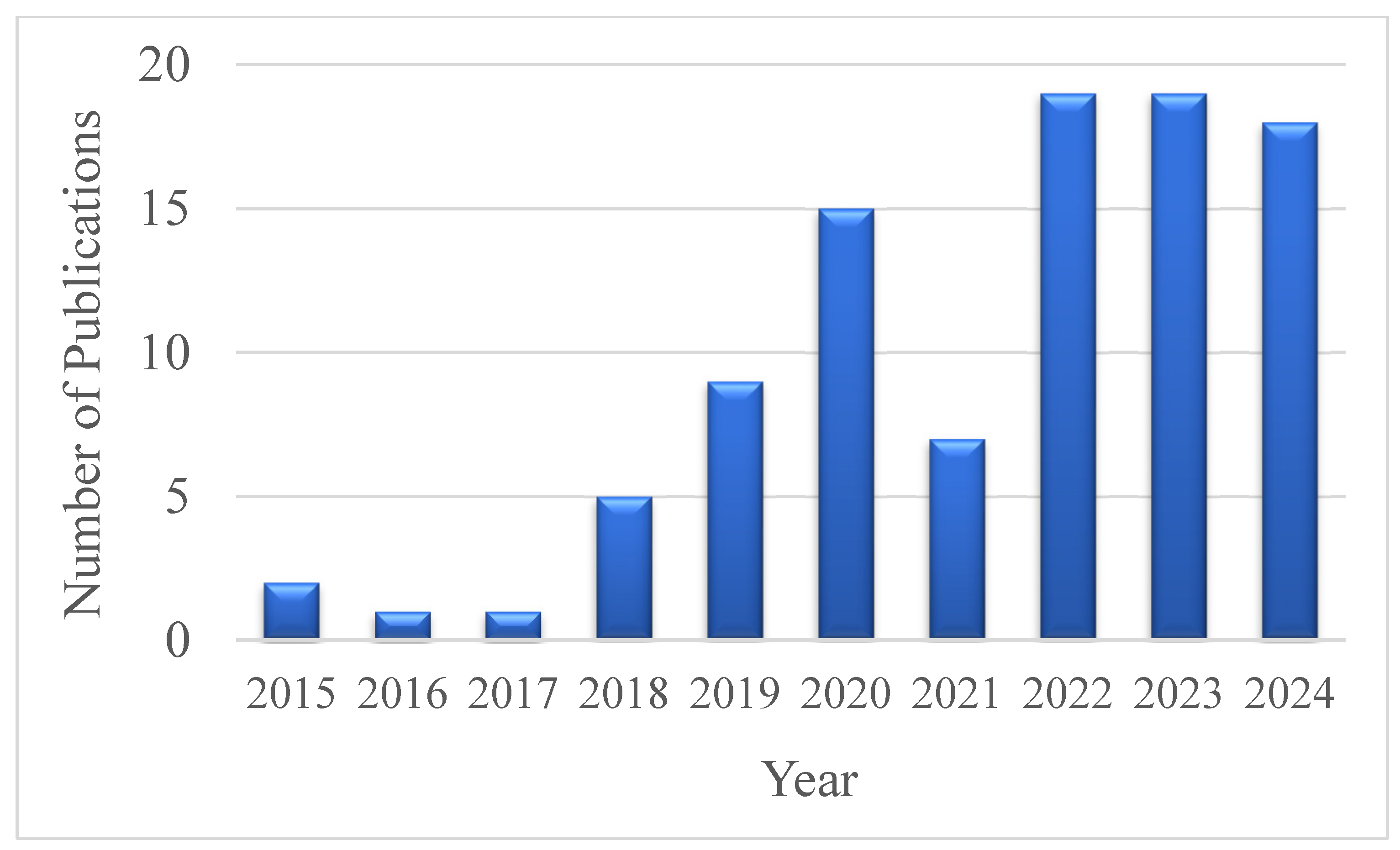
To gain deeper insight into how scientific interest has developed regarding the use of electric fields for microbial control in wastewater, a bibliometric analysis was carried out using data from the Web of Science Core Collection covering the period 2015–2024. The findings reveal a consistent upward trend in the number of publications addressing this topic, with significant peaks observed in 2022 and 2023—each accounting for 19 articles—and 18 publications already indexed in 2024, reflecting continued momentum in the field. This trend (Figure 2) suggests a growing awareness of the potential of electric fields as non-chemical tools for microbial manipulation and detection in environmental systems.
The bibliometric component of this review was based on data retrieved from the Web of Science Core Collection (2015–2024) using the query: (“electric field” OR “pulsed electric field”) AND (“microorganism” OR “bacteria” OR “spores”) AND (“wastewater” OR “waste water”). A total of 126 articles were selected. The records were exported as plain text and processed using VOSviewer v1.6.19 to generate keyword co-occurrence maps and thematic clusters.
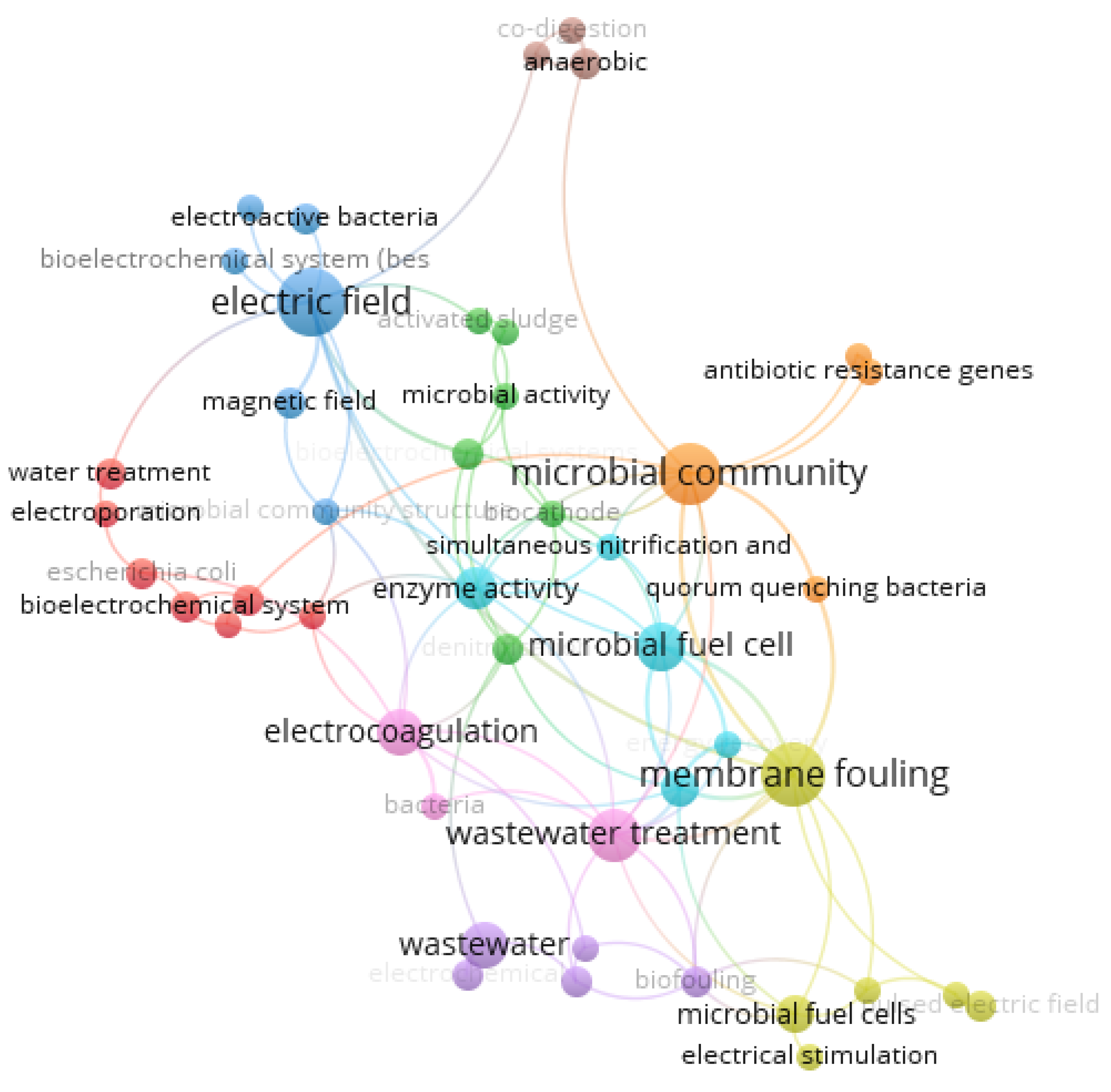
To complement this quantitative analysis, a co-occurrence network of keywords was generated using VOSviewer [37]. The map revealed distinct thematic clusters (Figure 3).
These clusters highlight the interdisciplinary character of the field, combining microbiology, bioelectrochemistry, and environmental engineering. The bibliometric findings confirm that electric field-based strategies are emerging as a robust and innovative direction for future research and practical implementation in wastewater monitoring and treatment. The central positioning of keywords such as “electric field”, “microbial community”, and “membrane fouling” reflects their foundational role across multiple research directions. The green cluster emphasizes biological processes and microbial activity, while the blue cluster is more focused on electrochemical systems and electron transfer mechanisms. The occurrence of distinct yet interconnected terms such as disinfection, photocatalysis, and ultrasound points toward emerging or specialized applications that are likely to gain increased significance soon. At the same time, the intersection of environmental-related keywords with electrochemical terminology reflects a growing integration between conventional wastewater engineering and more refined, bioelectrical approaches.
In parallel with the bibliometric analysis, a narrative synthesis was conducted. Articles were selected based on their relevance to the topic (electric field interactions with microbial systems in wastewater), experimental depth, and thematic coverage. Priority was given to recent publications (2015–2024), papers published in Q1/Q2 journals in relevant fields (Environmental Sciences, Microbiology, Biotechnology), and/or with significant citation impact. Seminal or mechanistic studies were included even when citation counts were low.
This review sets out to consolidate existing knowledge on how electric fields influence microbial cells, focusing on responses at the membrane and cytoplasmic levels. Additionally, it explores how these effects can be harnessed for practical purposes, especially in microbial detection and control strategies within wastewater treatment systems.
To build a solid conceptual basis, the next section introduces the main types of electric fields employed in microbiological and wastewater-related studies, detailing the associated parameters and mechanisms through which they act on microbial structures.

2. Basics of Electric Fields and Microbial Systems

2.1. Types of Electric Fields

In microbiological applications, electric fields are typically categorized according to specific attributes such as waveform, intensity, frequency, and pulse duration [38]. Among these, three main types have garnered the most attention in scientific literature:
(a)
Direct Current (DC) Electric Fields
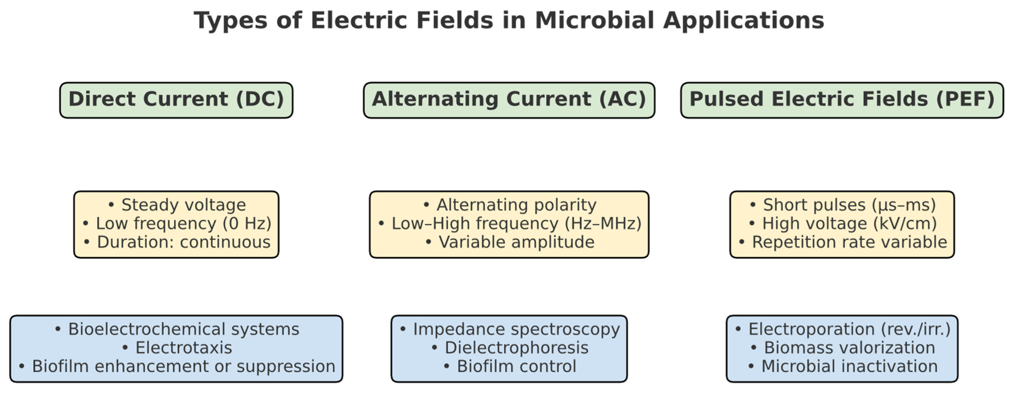
Direct current fields maintain a constant voltage over time and can directly influence microbial physiology by affecting cell orientation, membrane polarization, and electrochemical gradients. These effects make DC fields particularly relevant in bioelectrochemical systems such as microbial fuel cells and microbial electrolysis cells, where they support electrogenic activity and cell migration (electrotaxis) [39].
Applications of DC stimulation have demonstrated enhanced biofilm growth and improved electron transfer efficiency. For example, the use of an encapsulated alginate bioanode in a microbial electrolysis cell significantly increased biofilm formation, boosting current generation up to 4.14 A·m−2 while enriching Geobacter spp. to nearly 80% of the community. At the same time, chemical oxygen demand (COD) removal reached 62–88%, showing the dual benefit of energy recovery and wastewater treatment [40].
Overall, these results show that DC fields can steer microbial organization and enhance bioelectrochemical performance.
(b)
Alternating Current (AC) Electric Fields
Alternating current fields periodically switch polarity across a wide frequency spectrum, from Hz to MHz, and their biological effects are strongly frequency dependent. They are particularly important for analytical applications such as impedance spectroscopy and dielectrophoresis, where microbial dielectric responses provide the basis for label-free discrimination [41].
Studies consistently show that microorganisms exhibit species-specific dielectric signatures under AC exposure. Asami et al. measured E. coli suspensions across 10 kHz–100 MHz, identifying distinct dispersions near 1 MHz and quantifying parameters such as membrane capacitance, cytoplasmic conductivity, and wall permittivity [42]. These features enable real-time classification of mixed microbial populations. At higher frequencies, alternating fields around 20 MHz (≈3 V/cm) significantly inhibited the early adhesion of E. coli and P. aeruginosa, reducing cell attachment during biofilm initiation [31]. Conversely, very low-strength fields (<1 V/cm) primarily caused reversible membrane polarization without damaging cellular structures, supporting their use in continuous monitoring. Taken together, AC fields offer dual advantages: they can generate detailed dielectric fingerprints for microbial detection and, under specific conditions, serve as non-chemical strategies for controlling biofilm formation in water and wastewater systems.
(c)
Pulsed Electric Fields (PEFs)
Pulsed electric fields consist of short, high-voltage pulses in the microsecond to millisecond range and are widely applied to induce either reversible or irreversible electroporation depending on field strength and pulse duration [43]. Beyond their established role in food and medical applications, PEFs are emerging in wastewater and sludge valorization, where pretreatment can release intracellular compounds from microbial or algal cells, enhancing substrate accessibility for bioconversion and improving methane generation or nutrient recovery [44]. Recent studies highlight both inactivation and stimulation effects. For example, Rosenzweig et al. combined nanosecond PEF (600 ns, 21 kV/cm) with low doses of hydrogen peroxide, achieving > 5 log reductions in E. coli, Listeria, and Salmonella—a result not reached by either treatment alone [29]. Similarly, Tongdonyod et al. reported > 6 log reductions in E. coli in coconut water at 20 kV/cm without altering substrate properties, confirming the effectiveness of PEFs in low-conductivity matrices [45]. At the same time, low-intensity PEF can reversibly enhance microbial function: in Pseudomonas putida, stimulation accelerated growth and denitrification, coupled with upregulation of transport and respiration genes [46]. Electroporation has also been used as a delivery tool—for instance, to load gold nanoparticles into vesicles derived from P. aeruginosa—demonstrating PEF’s potential in nano-enabled biotechnologies [47].
Collectively, these studies underline the versatility of PEFs for both microbial inactivation and stimulation, depending on operating conditions. As shown in Figure 4, electric fields relevant to wastewater microbiology can be categorized into Direct Current, Alternating Current, and Pulsed Electric Fields, each associated with characteristic parameters (yellow) and representative applications (blue).

2.2. Microbial Structures Relevant to Electrical Interaction

The interaction between electric fields and microorganisms is largely governed by the structural organization of microbial cells. Several cellular components act as key interfaces for field-induced responses, each contributing differently to the cell’s overall electrical behavior [48].

2.2.1. Cell Membrane and Cell Wall

The structural organization of the microbial envelope plays a decisive role in how cells respond to electric fields. The lipid bilayer, reinforced in many organisms by a rigid cell wall (peptidoglycan in bacteria, chitin in fungi), introduces both mechanical resistance and dielectric heterogeneity [49,50,51,52]. Gram-negative bacteria such as E. coli and P. aeruginosa possess relatively thin envelopes (4–6 nm), which makes them more easily polarized and prone to reversible electroporation at moderate field strengths (~10 kV/cm) [27,28,29]. By contrast, Gram-positive species like B. subtilis and E. faecalis develop thick, multilayered peptidoglycan walls (20–30 nm), which increase the threshold for electroporation and provide greater resilience to electrical stress [53,54]. Experimental evidence confirms these principles: in microfluidic systems, millisecond pulses of 10 kV/cm were sufficient to permanently disrupt E. coli, while S. cerevisiae, with its thicker wall, required slightly higher intensities (~7.5 kV/cm) to achieve lysis and metabolite release [26,30]. Such results demonstrate how envelope composition not only dictates susceptibility to inactivation but also enables practical applications like high-resolution metabolomics and real-time monitoring. Overall, dielectric properties such as membrane capacitance (10−6–10−4 S/m) directly govern whether electrical exposure leads to reversible poration, irreversible breakdown, or controlled extraction of intracellular content [55,56].

2.2.2. Cytoplasm

The cytoplasm functions as a conductive medium where ions, metabolites, and macromolecules interact to define the cell’s dielectric behavior. In E. coli, for example, the total inorganic ion concentration is about 300 mM, with potassium (K+) as the dominant species, although levels fluctuate with osmotic conditions [57,58]. Such ionic content explains why cytoplasmic conductivity typically falls between 0.1 and 1 S/m, depending on metabolic activity [59]. In wastewater isolates like Pseudomonas and Aeromonas, higher conductivity has been linked to stronger electrochemical responsiveness, directly affecting how intracellular gradients shift when exposed to electric fields [60,61]. Structural components also modulate these properties. The outer membrane of P. aeruginosa, enriched in proteins such as β-barrel porins, creates selective diffusion pathways that regulate nutrient flow while contributing to its dielectric signature [62]. On a molecular scale, ions align water molecules and reduce the dielectric constant of cytosolic water compared to pure water, while metabolites such as amino acids can increase modestly [59]. Together, these factors show that the dielectric behavior of the cytoplasm is not static but shaped by ionic balance, metabolic state, and membrane architecture. This interplay is critical for interpreting impedance measurements, where subtle intracellular changes can manifest as detectable shifts in bulk dielectric signals.

2.2.3. Spores and Dormant Forms

Bacterial spores, particularly those produced by Bacillus and Clostridium species, represent some of the most resilient microbial structures. Their multilayered envelopes and dehydrated cores (<30% water) confer both mechanical resistance and markedly reduced dielectric permittivity, setting them apart from vegetative cells [63]. These traits explain why electroporation thresholds for spores often exceed 30 kV/cm, a value consistently reported across experimental and applied studies [64,65]. The physical basis of this resistance lies in their dense architecture and limited water content, which not only hinders inactivation but also provides distinctive dielectric signatures detectable by techniques such as dielectrophoresis or time-domain dielectric spectroscopy [66].
Evidence across multiple models supports this principle. For instance, Clostridium sporogenes spores typically require 30–50 kV/cm to achieve effective inactivation [67], while C. botulinum spores withstand even advanced processes such as high-pressure thermal treatments (300–1200 MPa at 30–75 °C) [68]. These findings emphasize that spore inactivation demands either extreme field intensities or combined approaches, such as coupling PEF with heat or pressure. At the same time, their unique dielectric properties make spores more distinguishable than vegetative cells, enabling label-free detection and selective separation in complex microbial systems.
Overall, the remarkable durability of spores arises from the same characteristics that allow their electrical discrimination. This duality—resistance to inactivation yet amenability to detection—highlights both the challenges and opportunities of targeting dormant forms in wastewater environments.

2.2.4. Surface Structures (e.g., Pili, Flagella, Biofilms)

Surface appendages such as pili and flagella are not only structural features but also key determinants of how bacteria interact with electric fields. By altering local field distribution, these structures influence adhesion, motility, and aggregation. In Pseudomonas aeruginosa, for instance, the retraction of electrically stimulated type IV pili drives electrotaxis—directed movement along electric field gradients—which in turn promotes biofilm development on electrode surfaces [69]. Beyond locomotion, these pili act as mechanosensors: when they contact a surface, their retraction triggers intracellular signaling cascades that elevate cyclic di-GMP, a messenger central to biofilm formation [70,71].
Electric fields themselves can modulate the conformation and function of pili and flagella. Studies have shown that exposure may alter their structure and dynamics, thereby change adhesion capacity or promote aggregation under certain conditions [72]. Once biofilms are established, they evolve into intricate three-dimensional networks that complicate field penetration. In mixed-species wastewater biofilms, the EPS matrix and layered architecture generate strong dielectric heterogeneity and capacitive behavior that can screen the applied field, cause non-uniform penetration and limit selective action on inner layers. As a result, EF responses often reflect interfacial effects and detachment rather than uniform inactivation throughout the biofilm [73,74].
Beyond these shielding effects, biofilms can also display significant electrochemical activity. For example, Geobacter sulfurreducens biofilms can reach current densities of ~2 A/m2 under controlled conditions, highlighting their role in electron transfer. Biofilm thickness, which may range from a few micrometers in early stages to over 50 µm in mature consortia, further influences diffusion processes and the efficacy of antimicrobial treatments. Advanced techniques such as cyclic voltammetry and confocal microscopy have confirmed that electrode-associated biofilms modify hydrogen adsorption and oxygen reduction, underscoring the complexity of their electrical behavior. Low-frequency AC fields (0.1–2 Hz) and direct currents (50–250 µA) have been used to disrupt biofilm integrity and increase susceptibility to antimicrobials like gentamicin, illustrating the potential of electric stimulation for control applications [74].
Together, these findings emphasize that microbial appendages and biofilm architectures are critical modulators of field–microbe interactions. Incorporating such structural and bioelectrical insights into predictive models can greatly improve the specificity and efficiency of electric field-based detection and inactivation strategies, particularly under the complex conditions typical of real wastewater environments. The structural characteristics of selected microorganisms relevant to their interaction with electric fields are summarized in Table 1.
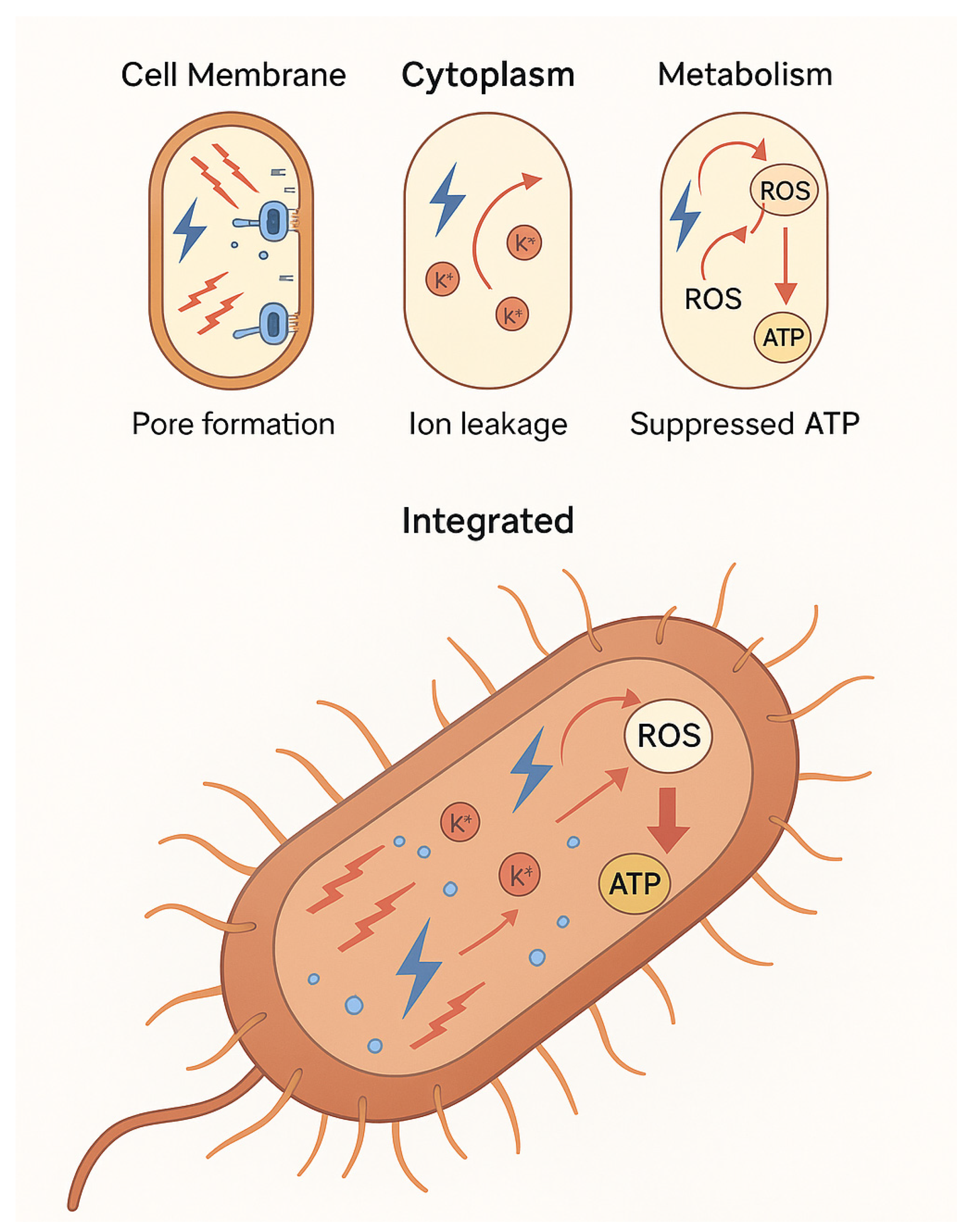
To illustrate these multi-level interactions in a unified view, Figure 5 presents a schematic representation of the main electric field effects on a model bacterium.

3. Membrane-Level Bioelectrical and Dielectric Properties

3.1. Membrane Thickness

Membrane thickness is one of the most decisive structural traits governing microbial interactions with electric fields. Thin envelopes, such as those of Gram-negative bacteria, typically measure ~15 nm in total, with a 4 nm inner bilayer and a minimal peptidoglycan layer, as in E. coli [75]. By contrast, Gram-positive species like B. subtilis or E. faecalis develop peptidoglycan layers of 25–40 nm, interlaced with teichoic acids, producing robust walls that alter their dielectric response [76,77]. These differences mean that thinner membranes polarize more quickly under applied fields, while thicker ones provide greater resistance.
This principle is well captured by the Schwan equation, which shows the inverse relationship between membrane thickness and induced transmembrane potential [78]. In practice, this explains why Gram-negative bacteria reach electroporation thresholds at lower field strengths, whereas Gram-positive bacteria require higher intensities. Spores represent an extreme case: their multilayered envelopes confer strong mechanical resistance, but also create localized hotspots where electric stress can concentrate and induce dielectric breakdown [79].
Recognizing these structural distinctions is essential for predictive modeling. By linking membrane thickness to polarization dynamics, electroporation thresholds, and stress distribution, researchers can better tailor electric field parameters to achieve controlled inactivation or analytical precision, particularly in complex systems such as wastewater.

3.2. Membrane Potential (ΔΨ)

The membrane potential is a fundamental electrochemical property that arises from the uneven distribution of ions across the microbial membrane. In most bacteria, ΔΨ typically ranges from −100 to −200 mV, depending on species, metabolic state, and external conditions [80]. This gradient is sustained by proton pumps and ATP-driven ion transporters, which also maintain pH homeostasis [81,82]. Beyond its role in energy transduction, ΔΨ contributes to active transport [83], motility regulation via flagellar rotation [84], and even antibiotic resistance, as shown by classic studies linking membrane polarization to aminoglycoside susceptibility [85]. These foundational works, although dated, remain essential for understanding bacterial physiology.
In actively respiring cells, ΔΨ is dynamic. For example, E. coli exhibits values around −220 mV during early exponential growth and about −140 mV in late exponential phase, reflecting shifts in metabolic activity and ion balance [86]. This potential is directly affected by external electric fields, which induce asymmetric polarization: depolarization at the anode-facing side and hyperpolarization at the cathode-facing side. Such field-driven effects have been demonstrated in both E. coli and Bacillus subtilis and highlight how proliferative capacity influences polarization responses [87].
Theoretical models such as the Schwan equation [78] predict that the induced transmembrane potential is proportional to both applied field strength and cell size. For a bacterium ~1 µm in diameter, exposure to 10 kV/cm can generate ΔΨ values sufficient to trigger electroporation, a phenomenon confirmed by uptake of propidium iodide in experimental studies [87,88]. When ΔΨ exceeds a critical threshold of ~1 V, nanoscale pores form in the bilayer [89]. These can reseal after short exposures (reversible electroporation) or remain open after strong pulses, leading to irreversible disruption and cell death.
Beyond viability, field-induced polarization also alters intracellular ion fluxes, promotes reactive oxygen species generation, and disrupts metabolism—mechanisms now being explored for antimicrobial and biosensing applications [90]. As emphasized by Pucihar et al., controlling ΔΨ dynamics under EF exposure is central for microbial inactivation, permeability tuning, and real-time monitoring in wastewater treatment [91]. Overall, understanding the relationship between ΔΨ and electric field strength provides the foundation for designing effective EF-based technologies across diverse microbial species.

3.3. Membrane Electrical Conductivity

Membrane electrical conductivity is a critical parameter that reflects the ability of microbial cells to support ionic current flow under an applied electric field. This property depends on membrane composition, lipid phase state, the presence of embedded proteins, and overall ionic permeability. Biological membranes are composed of a dynamic lipid bilayer interspersed with proteins that carry out most of the membrane’s specialized functions—including ion transport, which plays a key role in determining membrane conductivity. As described by Alberts et al. [49], these membrane proteins regulate the selective movement of ions across the bilayer, thereby sustaining ionic gradients essential for cellular function.
In addition to protein activity, the phase state of the lipid bilayer significantly influences membrane permeability and conductivity. Transitions between liquid-disordered and more ordered or gel-like states can alter the membrane’s permeability to small molecules, as shown by Frallicciardi et al. [92]. Such changes directly impact the flow of ions and, thus, the membrane’s electrical properties. Ion channels and transporters embedded in the bilayer contribute further to this conductivity by enabling controlled ionic movement across the membrane [93].
Taken together, membrane electrical conductivity arises from the combined influence of lipid composition, bilayer phase behavior, protein function, and ionic permeability. Understanding this complex interplay is essential for refining electric-field-based techniques aimed at microbial inactivation or bioelectrical diagnostics.
Membrane conductivity reflects the ability of a microbial cell to allow ionic current to pass under the influence of an external electric field. This property is highly dependent on structural and compositional factors, including the nature of the lipids, the presence of membrane proteins, and environmental conditions such as temperature and ionic strength [94].
In Escherichia coli, reported membrane conductivity values typically range between 1 × 10⁻⁶ and 3 × 10⁻⁶ S/m, with variations attributed to environmental influences. This relatively high conductivity is largely due to the abundance of porin proteins in the outer membrane, which facilitate passive ion transport and differentiate E. coli from Gram-positive bacteria [95,96]. In Pseudomonas aeruginosa, conductivity values are slightly higher—around 5 × 10⁻⁶ S/m—reflecting its metabolically active membrane and the presence of anion-selective channels such as protein P, which enhance ionic permeability [97].
By contrast, Bacillus subtilis, a Gram-positive bacterium with a thicker peptidoglycan wall and fewer porin-like channels, exhibits significantly lower membrane conductivity. Values typically range from 0.5 × 10⁻⁶ to 1 × 10⁻⁶ S/m, a difference primarily linked to its dense cell wall structure and limited ion-permeable protein content [28]. Understanding these differences in membrane conductivity among microbial species is fundamental for optimizing electric-field-based applications such as electroporation, antimicrobial treatments, and biosensing technologies. These differences affect how microbial cells respond to applied electric fields. Higher membrane conductivity enables more efficient charge redistribution and faster polarization but also leads to a lower threshold for field-induced membrane destabilization.
Exposure to electric fields can transiently or permanently alter membrane conductivity. Reversible electroporation typically increases membrane conductance by 10–100×, facilitating molecule transport (e.g., dyes, antibiotics, DNA). In E. coli, conductance can increase from 2 × 10⁻⁶ S/m to ~10−4 S/m immediately after exposure to 10–15 kV/cm pulses. In wastewater disinfection contexts, this enhanced conductivity is both a marker of electroporation and a tool for impedance-based microbial detection. Electrical impedance spectroscopy (EIS) systems use these shifts to differentiate between viable, damaged, and lysed cells [98,99]. Monitoring conductivity shifts in microbial membranes enables real-time feedback in disinfection systems. Moreover, integrating conductivity data with field strength allows predictive modeling of cell viability [100]. In one configuration, a continuous electric field of just 2 kV/cm was sufficient to achieve a >4.5-log reduction in E. faecalis, highlighting the strong susceptibility of this Gram-positive bacterium even at moderate field strengths [101]. In mixed microbial communities from wastewater, average membrane conductivity measurements can inform system-wide susceptibility to electric treatments, guiding pulse design and frequency selection in PEF reactors. Understanding membrane conductivity profiles across diverse species is thus essential for optimizing electroporation protocols, improving sensor design, and enhancing microbial inactivation strategies in complex aqueous environments [102].

3.4. Dielectric Permeability of the Membrane

Dielectric permittivity (ε) is a key parameter describing how a material polarizes in response to an external electric field. In microbial membranes, it reflects the ability of lipid and protein components to store electrical energy. This property affects how the cell responds to high-frequency alternating fields and determines the cell’s behavior in techniques like electrical impedance spectroscopy and dielectrophoresis. Microbial membranes typically exhibit relative dielectric permittivity values (εr) in the range of 5–12, depending on composition and hydration state [103,104]. E. coli membranes show dielectric permittivity around 5–9, consistent with fluid-phase lipid bilayers [105]. No direct measurements of the full-cell membrane dielectric permittivity of P. aeruginosa were found. However, typical values for lipid bilayer interiors lie between 2 and 4 εr, and dielectric behavior of bacterial suspensions may exhibit much higher apparent values due to volumetric polarization effects [105,106]. Direct measurements of the full-cell dielectric permittivity for Bacillus subtilis are not available. However, dielectric spectroscopy at 15–16 GHz reveals that B. subtilis exhibits lower dielectric constant responses compared to E. coli, suggesting a lower εr overall [107]. Dormant spores exhibit even lower permittivity values (<4), owing to their dehydrated state and multilamellar protective structures. This is supported by dielectric measurements showing extremely low conductivity and minimal mobile ion content in dormant Bacillus cereus spores at 50 MHz, indicative of very low dielectric response [108]. Dielectric permittivity is frequency dependent. At low frequencies (kHz range), the whole cell polarizes and behaves like a particle in a medium, dominated by membrane properties. At higher frequencies (MHz–GHz), the field penetrates the membrane and interacts with the cytoplasm, where ionic content drives internal polarization [109,110]. In practice, this means at 100 kHz, viable E. coli cells exhibit strong interfacial (β-) polarization, while spores—or cells rendered non-viable—lose this response, evidencing minimal dielectric activity at that frequency range [111]. At frequency above approximately 10 MHz, the cell membrane behaves almost like a short circuit, allowing the electrical current to penetrate the cytoplasm. As a result, intracellular conductivity becomes the dominant contributor to the overall impedance, although membrane permittivity still subtly influences the impedance spectrum at these high frequencies [112]. Dielectric permittivity differences are exploited in DEP-based separation, where cells are sorted according to their dielectric signature—such as spores versus vegetative bacteria—through frequency-selective polarization in non-uniform electric fields [113]. EIS leverages changes in membrane permittivity and conductivity following electroporation or cellular stress as a sensitive, label-free method to assess cell viability in real time [114]. In electroporated E. coli populations, membrane integrity is compromised, and impedance-based measurements—such as those carried out using a capacitive Wheatstone bridge—can detect real-time changes in dielectric properties. While precise quantitative reductions in effective permittivity are not consistently reported, such approaches clearly reveal diminishing dielectric signatures correlating with viability loss confirmed via plating assays [115]. A clear understanding of microbial dielectric permittivity is critical for advancing electric field-based detection systems, fine-tuning operational frequency ranges, and enhancing the sensitivity of in situ biosensors in complex wastewater environments [116]. Following the discussion on structural features, Table 2 focuses on membrane-level parameters—such as electrical conductivity and dielectric behavior—that govern microbial sensitivity to electric stimulation.

4. Cytoplasmic Bioelectrical and Dielectric Properties

4.1. Cytoplasmic Conductivity

Cytoplasmic conductivity plays a central role in shaping the electrical response of microbial cells to external fields. It governs internal charge redistribution and contributes significantly to the complex impedance of cells, especially at intermediate to high frequencies (10 kHz–10 MHz) [117]. This parameter depends on intracellular ion concentrations, metabolic activity, pH, and the macromolecular composition of the cytosol. When an electric field is applied, the cytoplasm behaves as a conductive medium, allowing charge transfer between internal and external environments through electroporated or naturally permeable membranes. In dielectric models, cytoplasmic conductivity affects the reactive (imaginary) component of the cell’s impedance, modulating phase shift and relaxation frequencies observed in impedance spectroscopy [117]. For example, viable E. coli cells with intact membranes typically exhibit cytoplasmic conductivity around 0.22 S/m and a relative permittivity near 100, producing a pronounced β-dispersion in the ~1–10 MHz range [117,118]. Following pulsed electric field exposure, conductivity can decrease markedly due to ion leakage and metabolic arrest, changes that are detectable by electrical impedance spectroscopy or time-domain reflectometry and correlate with viability loss confirmed by plate counts [32]. Cytoplasmic conductivity is highly sensitive to the internal ionic environment and pH. Elevated concentrations of K+, Na+, Cl, and organic acids—such as in metabolically active P. aeruginosa—are associated with higher conductivities compared to nutrient-deprived cells [119,120]. Experimental work has shown that lowering intracellular pH from ~7.2 to ~6.5 can reduce conductivity by up to 20% through altered protein protonation and reduced buffering capacity [121]. In wastewater environments, microbes are frequently exposed to fluctuations in ionic strength and pH during aerobic–anaerobic cycling or after chemical treatment, which alters their impedance signatures. Real-time monitoring systems can exploit these variations to assess microbial viability and stress state [98]. Spore-forming organisms such as Clostridium spp. have very low internal conductivity (<0.05 S/m) due to dehydration and ionic shielding within their core, making them less responsive in impedance-based assays but still distinguishable through dielectric contrast methods [122]. Understanding the dynamic nature of cytoplasmic conductivity is therefore essential for evaluating microbial viability, electrosensitivity, and stress responses—parameters that are increasingly used in wastewater treatment for both detection and selective inactivation.

4.2. Dielectric Permeability of the Cytoplasm

Dielectric permittivity defines how well the cytoplasmic medium becomes polarized under an external electric field. It is vital for understanding microbial responses to electric stimuli, especially at high frequencies where the field penetrates beyond the cellular envelope. Cytoplasm is a richly structured environment, full of water, ions, proteins, nucleic acids, and various inclusions like polyphosphate granules. Its dielectric permittivity determines how effectively the cell’s interior can polarize when subjected to oscillating fields—particularly prominent above ~1 MHz [123]. Studies using a refined multi-shell modeling approach reported that E. coli cells possess a cytoplasm relative permittivity of about 100, paired with a cytoplasmic conductivity near 0.22 S/m. This high permittivity supports substantial polarizability, manifesting as notable β-dispersion around 1–10 MHz [115,118]. Cellular stress—such as electroporation or thermal damage—impairs membrane integrity and ionic balance, lowering polarization capacity. While exact figures are scarce, single-cell impedance studies have shown discernible shifts in the dielectric signature corresponding to compromised viability (e.g., plasma-activated water exposure) [124]. Though bacteria lack membrane-bound organelles, they do accumulate inclusions like glycogen, PHB, or polyphosphate granules. These dense bodies shift cytoplasmic dielectric properties. Moreover, when modeling “dry” bacterial cores (proteins and nucleic acids), cytoplasmic permittivity estimates range from 5 to 6.5. Under ambient (hydrated) conditions, values rise significantly—between 25 and 30 for E. coli—reflecting strong hydration effects and biological complexity [125]. Spore cores, such as those in Clostridium spp., feature dehydrated, densely packed dipicolinic acid–calcium complexes. This structure dramatically limits polarizability—their effective dielectric permittivity remains very low, often under 30, distinguishing them clearly from vegetative cells via dielectric techniques [126]. The cytoplasm inside microbial cells—busy with water, ions, big molecules, and storage inclusions—behaves differently depending on how hydrated or stressed it is. For E. coli, that permittivity hits around 100 when healthy, which helps it polarize sharply under high-frequency fields. Throw in stress or damage, and that ability drops noticeably. Even more interesting: “dry” spores barely polarize, while hydration and internal granules push permittivity way up. That dielectric fingerprint is exactly what tools like high-frequency impedance or dielectrorotation sensors use to tell live cells from stressed or dormant ones. Given that electric fields not only affect microbial membranes but also penetrate the cytoplasm, Table 3 highlights cytoplasmic bioelectrical and dielectric properties that contribute to cellular polarization, electromechanical stress distribution, and inactivation thresholds.
Building upon the cellular-level insights discussed in Section 3 and Section 4, the following section explores how these electrical and dielectric properties are exploited in practice—specifically in the development of detection, monitoring, and disinfection technologies for wastewater systems.

5. Electric Field Effects on Microbial Physiology

Electric fields exert powerful, often species-dependent effects on microorganisms, ranging from reversible physiological changes to complete inactivation. The outcome depends on field intensity, pulse duration and waveform, as well as structural resilience and metabolic state of the target cells.

5.1. Vegetative Cells

In vegetative bacteria such as Escherichia coli and Pseudomonas aeruginosa, EF exposure first causes membrane depolarization followed by pore formation, a process known as electroporation. At moderate field strengths (~10 kV/cm) and short pulses, electroporation is reversible, enabling transient permeability increases useful for biosensing and molecular uptake [127,128,129]. When field intensity exceeds ~15 kV/cm or pulses are prolonged, irreversible poration occurs, leading to membrane rupture, cytoplasmic leakage, and cell death, as confirmed by CFU counts and luminescence assays [130,131]. Sublethal exposure can also alter ATP synthesis, enzyme activity, surface charge, and motility (e.g., reduced swarming in P. aeruginosa) [132], serving as biomarkers of stress [133,134,135].
Overall, vegetative cells are highly responsive to EF exposure, with outcomes ranging from mild physiological stress to complete inactivation, depending on applied parameters.

5.2. Spores

Bacterial spores such as those of Bacillus subtilis and Clostridium perfringens are more resilient due to multilayered, dehydrated structures. PEF in the range of 25–35 kV/cm may trigger germination by releasing dipicolinic acid–calcium complexes or, under suboptimal conditions, inhibit germination and induce membrane reorganization detectable via dielectric relaxation [136,137]. When combined with mild heat (40–50 °C) or high-pressure pretreatments, PEF can synergistically activate spores, offering potential for advanced disinfection strategies, though studies remain limited [138].
Thus, spores require higher EF thresholds than vegetative cells, but their responses open opportunities for combined treatment approaches.

5.3. Broader Metabolic Effects

Beyond direct membrane permeabilization, EFs can disrupt key physiological processes. They collapse the proton motive force (PMF) and membrane potential, induce reactive oxygen species (ROS), impair glucose uptake, and alter NADH activity [139,140]. EF exposure also modulates stress-response gene expression (e.g., soxS, katG), influencing microbial adaptation. In wastewater, baseline stresses from nutrient limitation and redox fluctuations may amplify these effects, sometimes promoting biofilm formation in Pseudomonas spp. or inducing sporulation in B. subtilis under repeated sublethal pulses [141,142]. These metabolic impacts highlight the dual potential of EFs to either damage microbes or trigger adaptive survival responses, depending on context.

5.4. Electrical Impedance Spectroscopy (EIS)

EIS is a label-free method that monitors microbial viability through frequency-dependent impedance patterns. Using low- to mid-frequency AC (1 kHz–10 MHz), it detects changes in membrane integrity, cytoplasmic conductivity, and cell concentration. Viable E. coli typically shows β-dispersion around 100 kHz–1 MHz, whereas inactivated cells display reduced polarization capacity, with capacitance drops of up to 60%. Portable EIS systems achieve sensitivities down to 103 CFU/mL in real effluent without labeling or filtration [143,144,145]. EIS therefore provides a rapid, non-invasive diagnostic tool for real-time monitoring of microbial dynamics in wastewater.

5.5. Dielectrophoresis

DEP exploits the motion of polarizable cells in non-uniform AC fields, enabling separation by size, morphology, or dielectric properties [143,146,147]. It distinguishes viable from non-viable cells and spores from vegetative forms, outperforming filtration or culture-based enrichment. DEP is increasingly used in microelectrode and microfluidic platforms for microbial capture, improving the detection limits of impedance-based biosensors and supporting real-time, label-free analysis [148,149]. However, high ionic strength in wastewater can dampen efficiency, biofilms may mask signatures, and scaling to treatment plants requires optimization of electrode design and flow conditions [150,151,152].
Despite these challenges, DEP offers powerful capabilities for selective microbial detection and discrimination.

5.6. Field-Assisted Biosensors

Biosensors integrated with EF techniques show enhanced sensitivity through improved analyte transport, surface capture, and modulation of microbial activity. Electrochemical impedance sensors with EF-assistance have reached detection limits as low as 2 CFU/mL [153]. In wastewater contexts, DEP-assisted immunosensors combine microbial concentration with amperometric detection, significantly accelerating binding rates and reducing detection times. Field-assisted biosensors thus represent a promising frontier for online pathogen monitoring in complex effluent streams.
Compared to conventional sterilization approaches, such as chlorination, UV or ozonation, which present drawbacks like carcinogenic by-product formation, reduced efficiency in turbid waters, or high energy demand [17,18,19], electric field-based treatments provide a non-thermal alternative. Pulsed electric fields, for instance, can achieve significant microbial reductions while maintaining the physicochemical integrity of the treated substrate, thus avoiding the quality losses typically associated with thermal sterilization [33].

6. Challenges and Research Opportunities

Exploring electric field–based technologies in wastewater treatment has revealed both promise and complexity. These approaches hold great potential for real-time microbial monitoring, selective control, and sustainable disinfection, but their transition from laboratory proof-of-concept to large-scale implementation requires careful consideration of technical and environmental challenges.
Complex mixed-species biofilms introduce additional limitations: EPS-mediated shielding and steep micro-gradients (O2, pH, ionic strength) distort local field distribution, create hot-spots, and confound impedance signatures; detachment may release viable aggregates; deeper layers can remain unaffected unless energy input increases—raising Joule-heating and corrosion risks. These factors complicate reproducibility and scale-up, and call for anti-fouling electrodes, controlled shear/flow, and combined strategies (e.g., EF with mild chemical or enzymatic cleaning) [73,74,148,149,150,151,152]. At the same time, pilot-scale demonstrations have shown that EF-based methods can be successfully applied for real-time microbial monitoring and selective inactivation in wastewater, supporting their transition from proof-of-concept studies to practical implementation. Although impedance biosensors are promising—miniaturized and label-free—they still face reliability constraints in harsh wastewater environments (e.g., biofouling, pH shifts, chemical interferences) [150].
A key practical limitation is the difficulty of achieving uniform electric field distribution in complex matrices such as wastewater. High ionic conductivity, turbidity, and suspended solids can generate shielding effects, local hotspots, and heterogeneous microbial exposure, ultimately reducing reproducibility and scalability [33,44,146,147]. Beyond physical complexity, sensor reliability in harsh wastewater environments remains a critical issue. Impedance biosensors, although promising as miniaturized and label-free tools, often suffer from biofouling, chemical interferences, and pH-driven drift [145]. This highlights the need for standardized protocols, durable electrode materials, and hybrid detection platforms that integrate electrical readouts with complementary molecular or omics-based analyses [149,154].
Promising opportunities are emerging to address these limitations. Advanced signal deconvolution methods, such as Distribution of Relaxation Times (DRT), and machine-learning-based pattern recognition can help extract microbial signals from complex backgrounds [148,149]. Anti-fouling electrode coatings, self-powered sensor designs based on microbial fuel cells (MFCs), and multi-modal detection platforms that couple EIS with optoelectronic or omics modules are under development to enhance both robustness and specificity [149,154]. These innovations, together with pilot-scale demonstrations in real facilities, could provide the basis for reliable, decentralized, and energy-efficient wastewater monitoring and control systems.

7. Outlook: Electric Field-Based Wastewater Monitoring and Control

As global demand for resilient, adaptive, and sustainable wastewater treatment continues to grow, electric field (EF)–based technologies are emerging as transformative tools for microbial monitoring and targeted disinfection. Their compatibility with real-time data acquisition, selective physical interaction with microorganisms, and non-chemical mode of operation position them as strong candidates for integration into next-generation smart water systems.

7.1. Smart Biosensing: Integrating AI with EF-Based Readouts

The convergence of artificial intelligence (AI) and EF-based biosensing is set to redefine microbial diagnostics. AI-driven algorithms can enhance pattern recognition in impedance spectra, predict microbial behavior from dielectric signatures, and optimize EF parameters under dynamic conditions [155,156]. Machine learning models trained on EIS or DEP datasets have been shown to classify microbial species, detect anomalies, and estimate microbial loads with minimal human intervention. Such systems could be embedded in autonomous water quality stations, enabling continuous, intelligent surveillance of microbial risks in wastewater [157,158].

7.2. Targeted Microbial Control in Decentralized Wastewater Systems

EF-based solutions offer unique advantages in decentralized or low-infrastructure settings, including portable PEF devices, field-assisted biosensors, and microfluidic DEP platforms. These can be adapted for on-site treatment facilities, agricultural runoff management, or emergency sanitation deployments [159].
Such systems hold potential for targeted inactivation of pathogens, including antimicrobial-resistant strains, while minimizing disruption of beneficial microbial communities in reuse or bioreactor applications. The low chemical footprint and modular design of EF-based platforms align well with circular economy principles and decentralized water reuse strategies [160].

7.3. Hybrid Biotechnological–Electrical Approaches

Merging EF methods with biotechnological tools opens new opportunities for microbial manipulation and process optimization. Potential applications include electro-fermentation, steering microbial metabolism with low-voltage fields [161]; electro-biostimulation, enhancing biofilm growth for pollutant removal or energy recovery [162]; electroporation-assisted gene transfer, enabling rapid adaptation or functionalization of engineered strains in situ [163]. These hybrid approaches pave the way for programmable microbial system engineered communities designed to respond predictably to electric cues, blending synthetic biology with physical field control. Recent electrochemical studies further expand the applicability of EF-based systems by demonstrating their potential not only for microbial detection but also for the monitoring of heavy metal contaminants in wastewater [164,165]. This broader scope strengthens the case for EF methods as versatile environmental tools. Such practical applications underline that EF-based technologies are not limited to experimental proof-of-concept studies but hold real potential for integration into operational wastewater monitoring, decentralized reuse, and smart treatment systems.

8. Conclusions

Electric field technologies represent a promising and versatile approach for investigating and manipulating microorganisms, with potential in the monitoring and treatment of wastewater. While current results highlight their ability to interact directly with structural and functional cellular components—such as membranes, cytoplasmic content, and dielectric properties—their broader applicability still requires validation at pilot and full scale.
Evidence from the literature shows that exposure to electric fields can modulate membrane potential and integrity, cytoplasmic permittivity and ion fluxes, as well as overall cell viability and metabolic activity. These effects are not inherently destructive; under controlled conditions, they can be tuned or reversed, enabling the selective detection, stimulation, or inactivation of microbial populations.
A major advantage of these technologies lies in their capacity to characterize microbial communities in real time, without labels or reagents, which is particularly valuable in complex matrices such as wastewater. Techniques including impedance spectroscopy, dielectrophoresis, and pulsed electric field inactivation are already being adapted into modular and scalable platforms for environmental biotechnology.
Nevertheless, challenges remain in translating laboratory-scale findings into robust, field-ready solutions. Further investigations are necessary to better understand the relationships between specific electric field parameters and microbial structural or functional responses across diverse species and environmental contexts. Advancing model precision, enhancing sensor integration, and improving the scalability of these systems will be key steps toward enabling their broader application in real-world settings. Overall, electric field-based technologies represent a promising frontier in microbial analysis and control, with the potential to complement and enhance conventional water quality strategies. Their multidisciplinary nature encourages continued exploration at the intersection of microbiology, physics, engineering, and environmental science, paving the way for intelligent and adaptive water management systems.

Author Contributions

Conceptualization, C.U. and D.S.Ș.; methodology, C.U. and D.S.Ș.; software, S.R.; validation, C.U., D.S.Ș., I.L., A.T. and M.Ș.; data curation, M.Ș. and C.U.; writing—original draft preparation, C.U.; writing—review and editing, C.U., D.S.Ș., I.L., A.T. visualization, C.U., M.Ș., D.S.Ș., I.L., A.T. and M.Ș.; supervision, C.U.; project administration, D.S.Ș.; funding acquisition, D.S.Ș. All authors have read and agreed to the published version of the manuscript.

Funding

This work was financially supported by a grant of the Romanian Ministry of Education and Research, CCCDI–UEFISCDI, under the scientific Programme PN-IV-P7-7.1-PTE-2024-0106 „EPDEcoA.

Data Availability Statement

The original contributions presented in this study are included in the article. Further inquiries can be directed to the corresponding authors.

Conflicts of Interest

Authors Iosif Lingvay and Attila Tokos were employed by the company I.C.P.E. BISTRITA S.A. The remaining authors declare that the re-search was conducted in the absence of any commercial or financial relationships that could be construed as a potential conflict of interest.

Abbreviations

The following abbreviations are used in this manuscript:
ACAlternating Current
ATPAdenosine Triphosphate
B. subtilisBacillus subtilis
C. botulinumClostridium botulinum
C. sporogenesClostridium sporogenes
DCDirect Current
DEPDielectrophoresis
DFEDifferential Field Excitation
DNADeoxyribonucleic Acid
E. coliEscherichia coli
E. faecalisEnterococcus faecalis
EISElectrical Impedance Spectroscopy
EPSExtracellular Polymeric Substances
GHzGigahertz
HPTHigh-Pressure Thermal
HzHertz
kHzKilohertz
LPSLipopolysaccharide
MHzMegahertz
mMMillimolar
nmNanometer
pHPotential of Hydrogen
PEFPulsed Electric Field
PHBPolyhydroxybutyrate
ROSReactive Oxygen Species
SFESpecific Field Energy
SHEStandard Hydrogen Electrode
TDDSTime-Domain Dielectric Spectroscopy
UV-VIS-NIRUltraviolet–Visible–Near-Infrared
WHOWorld Health Organization
µmMicrometer
µAMicroampere

References

  1. Chahal, C.; van den Akker, B.; Young, F.; Franco, C.; Blackbeard, J.; Monis, P. Pathogen and Particle Associations in Wastewater: Significance and Implications for Treatment and Disinfection Processes. Adv. Appl. Microbiol. 2016, 97, 63–119. [Google Scholar] [CrossRef]
  2. López, A.; Rodríguez-Chueca, J.; Mosteo, R.; Gómez, J.; Rubio, E.; Goñi, P.; Ormad, M.P. How Does Urban Wastewater Treatment Affect the Microbial Quality of Treated Wastewater? Process Saf. Environ. Prot. 2019, 130, 22–30. [Google Scholar] [CrossRef]
  3. Zhang, S.; Li, X.; Wu, J.; Coin, L.; O’Brien, J.; Hai, F.; Jiang, G. Molecular Methods for Pathogenic Bacteria Detection and Recent Advances in Wastewater Analysis. Water 2021, 13, 3551. [Google Scholar] [CrossRef]
  4. Li, L.; Mendis, N.; Trigui, H.; Oliver, J.D.; Faucher, S.P. The Importance of the Viable but Non-Culturable State in Human Bacterial Pathogens. Front. Microbiol. 2014, 5, 258. [Google Scholar] [CrossRef] [PubMed]
  5. Saaed, F.M.A.; Ongerth, J.E. Giardia and Cryptosporidium in Children with Diarrhea, Kufra, Libya, a North African Migration Route City. Int. J. Hyg. Environ. Health 2019, 222, 840–846. [Google Scholar] [CrossRef]
  6. Sanseverino, I.; Gómez, L.; Navarro, A.; Cappelli, F.; Niegowska, M.; Lahm, A.; Barbiere, M.; Porcel-Rodríguez, E.; Valsecchi, S.; Pedraccini, R.; et al. Holistic Approach to Chemical and Microbiological Quality of Aquatic Ecosystems Impacted by Wastewater Effluent Discharges. Sci. Total Environ. 2022, 835, 155388. [Google Scholar] [CrossRef] [PubMed]
  7. Elfadadny, A.; Ragab, R.F.; AlHarbi, M.; Badshah, F.; Ibáñez-Arancibia, E.; Farag, A.; Hendawy, A.O.; De Los Ríos-Escalante, P.R.; Aboubakr, M.; Zakai, S.A.; et al. Antimicrobial Resistance of Pseudomonas aeruginosa: Navigating Clinical Impacts, Current Resistance Trends, and Innovations in Breaking Therapies. Front. Microbiol. 2024, 15, 1374466. [Google Scholar] [CrossRef]
  8. Shoushtarian, F.; Negahban-Azar, M. Worldwide Regulations and Guidelines for Agricultural Water Reuse: A Critical Review. Water 2020, 12, 971. [Google Scholar] [CrossRef]
  9. Zamorska, J.; Karwowska, E.; Przystaś, W. Assessment of Microbiological Quality of Water Using Culture Methods, Flow Cytometry and Luminometry. Water 2023, 15, 4077. [Google Scholar] [CrossRef]
  10. Fdez-Sanromán, A.; Bernárdez-Rodas, N.; Rosales, E.; Pazos, M.; González-Romero, E.; Sanromán, M.Á. Biosensor Technologies for Water Quality: Detection of Emerging Contaminants and Pathogens. Biosensors 2025, 15, 189. [Google Scholar] [CrossRef]
  11. Ahmad, S.; Lohiya, S.; Taksande, A.; Meshram, R.J.; Varma, A.; Vagha, K. A Comprehensive Review of Innovative Paradigms in Microbial Detection and Antimicrobial Resistance: Beyond Traditional Cultural Methods. Cureus 2024, 16, e61476. [Google Scholar] [CrossRef]
  12. Franco-Duarte, R.; Černáková, L.; Kadam, S.; Kaushik, K.S.; Salehi, B.; Bevilacqua, A.; Corbo, M.R.; Antolak, H.; Dybka-Stępień, K.; Leszczewicz, M.; et al. Advances in Chemical and Biological Methods to Identify Microorganisms—From Past to Present. Microorganisms 2019, 7, 130. [Google Scholar] [CrossRef]
  13. Gonçalves, J.; Pequeno, J.; Diaz, I.; Kržišnik, D.; Žigon, J.; Koritnik, T. Killing Two Crises with One Spark: Cold Plasma for Antimicrobial Resistance Mitigation and Wastewater Reuse. Water 2025, 17, 1218. [Google Scholar] [CrossRef]
  14. González, Y.; Gómez, G.; Moeller-Chávez, G.E.; Vidal, G. UV Disinfection Systems for Wastewater Treatment: Emphasis on Reactivation of Microorganisms. Sustainability 2023, 15, 11262. [Google Scholar] [CrossRef]
  15. Badarne-Abbasi, S.; Armon, R.; Nasser, A. Reduction of Infectious Cryptosporidium and Microbial Indicators in Wastewater Effluents by Disinfection with UV Irradiation or Chlorine. J. Water Resour. Prot. 2022, 14, 407–418. [Google Scholar] [CrossRef]
  16. Delgado-Viscogliosi, P.; Solignac, L.; Delattre, J.M. Viability PCR, a Culture-Independent Method for Rapid and Selective Quantification of Viable Legionella pneumophila Cells in Environmental Water Samples. Appl. Environ. Microbiol. 2009, 75, 3502–3512. [Google Scholar] [CrossRef] [PubMed]
  17. Jütte, M.; Abdighahroudi, M.S.; Waldminghaus, T.; Lackner, S.; Lutze, H.V. Bacterial Inactivation Processes in Water Disinfection—Mechanistic Aspects of Primary and Secondary Oxidants—A Critical Review. Water Res. 2023, 231, 119626. [Google Scholar] [CrossRef]
  18. Madadelahi, M.; Agarwal, R.; Martinez-Chapa, S.O.; Madou, M.J. A Roadmap to High-Speed Polymerase Chain Reaction (PCR): COVID-19 as a Technology Accelerator. Biosens. Bioelectron. 2024, 246, 115830. [Google Scholar] [CrossRef] [PubMed]
  19. Adeyemo, F.E.; Singh, G.; Reddy, P.; Bux, F.; Stenström, T.A. Efficiency of Chlorine and UV in the Inactivation of Cryptosporidium and Giardia in Wastewater. PLoS ONE 2019, 14, e0216040. [Google Scholar] [CrossRef]
  20. Pasciucco, F.; Pecorini, I.; Iannelli, R. Planning the Centralization Level in Wastewater Collection and Treatment: A Review of Assessment Methods. J. Clean. Prod. 2022, 375, 134092. [Google Scholar] [CrossRef]
  21. Olvera, D.; Monaghan, M.G. Electroactive Material-Based Biosensors for Detection and Drug Delivery. Adv. Drug Deliv. Rev. 2021, 170, 396–424. [Google Scholar] [CrossRef]
  22. Bartha, C.; Jipa, M.; Caramitu, A.-R.; Voina, A.; Tókos, A.; Circiumaru, G.; Micu, D.-D.; Lingvay, I. Behavior of Microorganisms from Wastewater Treatments in Extremely Low-Frequency Electric Field. Biointerface Res. Appl. Chem. 2022, 12, 5071–5080. [Google Scholar] [CrossRef]
  23. Emanuel, E.; Dubrovin, I.; Pogreb, R.; Pinhasi, G.A.; Cahan, R. Resuscitation of Pulsed Electric Field-Treated Staphylococcus aureus and Pseudomonas putida in a Rich Nutrient Medium. Foods 2021, 10, 660. [Google Scholar] [CrossRef]
  24. Soltanzadeh, M.; Peighambardoust, S.H.; Gullon, P.; Hesari, J.; Gullón, B.; Alirezalu, K.; Lorenzo, J. Quality Aspects and Safety of Pulsed Electric Field (PEF) Processing on Dairy Products: A Comprehensive Review. Food Rev. Int. 2020, 38 (Suppl. S1), 96–117. [Google Scholar] [CrossRef]
  25. Kotnik, T. Transmembrane Voltage Induced by Applied Electric Fields. In Handbook of Electroporation; Miklavcic, D., Ed.; Springer: Cham, Switzerland, 2016; pp. 1–20. [Google Scholar] [CrossRef]
  26. Rockenbach, A.; Sudarsan, S.; Berens, J.; Kosubek, M.; Lazar, J.; Demling, P.; Hanke, R.; Mennicken, P.; Ebert, B.E.; Blank, L.M.; et al. Microfluidic Irreversible Electroporation—A Versatile Tool to Extract Intracellular Contents of Bacteria and Yeast. Metabolites 2019, 9, 211. [Google Scholar] [CrossRef] [PubMed]
  27. Pluhackova, K.; Horner, A. Native-Like Membrane Models of E. coli Polar Lipid Extract Shed Light on the Importance of Lipid Composition Complexity. BMC Biol. 2021, 19, 4. [Google Scholar] [CrossRef] [PubMed]
  28. Silhavy, T.J.; Kahne, D.; Walker, S. The Bacterial Cell Envelope. Cold Spring Harb. Perspect. Biol. 2010, 2, a000414. [Google Scholar] [CrossRef]
  29. Lyon, R.; Jones, R.A.; Shropshire, H.; Aberdeen, I.; Scanlan, D.J.; Millard, A. Membrane Lipid Renovation in Pseudomonas aeruginosa—Implications for Phage Therapy? Environ. Microbiol. 2022, 24, 4533–4546. [Google Scholar] [CrossRef] [PubMed]
  30. Haberl Meglic, S.; Marolt, T.; Miklavcic, D. Protein Extraction by Means of Electroporation from E. coli with Preserved Viability. J. Membr. Biol. 2015, 248, 893–901. [Google Scholar] [CrossRef]
  31. Kunlasubpreedee, P.; Tobino, T.; Nakajima, F. Influence of High-Frequency, Low-Voltage Alternating Electric Fields on Biofilm Development Processes of Escherichia coli and Pseudomonas aeruginosa. Water 2023, 15, 3055. [Google Scholar] [CrossRef]
  32. Kotnik, T.; Frey, W.; Sack, M.; Haberl Meglič, S.; Peterka, M.; Miklavčič, D. Electroporation-Based Applications in Biotechnology. Trends Biotechnol. 2015, 33, 480–488. [Google Scholar] [CrossRef]
  33. Rosenzweig, Z.; Garcia, J.; Thompson, G.L.; Perez, L.J. Inactivation of Bacteria Using Synergistic Hydrogen Peroxide with Split-Dose Nanosecond Pulsed Electric Field Exposures. PLoS ONE 2024, 19, e0311232. [Google Scholar] [CrossRef]
  34. Turcan, I.; Caras, I.; Schreiner, T.G.; Tucureanu, C.; Salageanu, A.; Vasile, V.; Avram, M.; Tincu, B.; Olariu, M.A. Dielectrophoretic and Electrical Impedance Differentiation of Cancerous Cells Based on Biophysical Phenotype. Biosensors 2021, 11, 401. [Google Scholar] [CrossRef] [PubMed]
  35. Grossi, M.; Riccò, B. Electrical Impedance Spectroscopy (EIS) for Biological Analysis and Food Characterization: A Review. J. Sens. Sens. Syst. 2017, 6, 303–325. [Google Scholar] [CrossRef]
  36. Gutsul, O.; Rutherford, D.; Barinkova, M.; Slobodyan, V.; Rezek, B. The Detection of E. coli and S. aureus on Sensors without Immobilization by Using Impedance Spectroscopy. Eng. Proc. 2023, 58, 79. [Google Scholar] [CrossRef]
  37. Perianes-Rodriguez, A.; Waltman, L.; van Eck, N.J. Constructing Bibliometric Networks: A Comparison between Full and Fractional Counting. J. Informetr. 2016, 10, 1178–1195. [Google Scholar] [CrossRef]
  38. Beretta, G.; Mastorgio, A.F.; Pedrali, L.; Saponaro, S.; Sezenna, E. The Effects of Electric, Magnetic and Electromagnetic Fields on Microorganisms in the Perspective of Bioremediation. Rev. Environ. Sci. Biotechnol. 2019, 18, 29–75. [Google Scholar] [CrossRef]
  39. Chong, P.; Erable, B.; Bergel, A. How Bacteria Use Electric Fields to Reach Surfaces. Biofilm 2021, 3, 100048. [Google Scholar] [CrossRef]
  40. Hirsch, L.O.; Gandu, B.; Chiliveru, A.; Dubrovin, I.A.; Jukanti, A.; Schechter, A.; Cahan, R. Hydrogen Production in Microbial Electrolysis Cells Using an Alginate Hydrogel Bioanode Encapsulated with a Filter Bag. Polymers 2024, 16, 1996. [Google Scholar] [CrossRef]
  41. Pethig, R. Review Article—Dielectrophoresis: Status of the Theory, Technology, and Applications. Biomicrofluidics 2010, 4, 022811. [Google Scholar] [CrossRef] [PubMed]
  42. Asami, K.; Hanai, T.; Koizumi, N. Dielectric Analysis of Escherichia coli Suspensions in the Light of the Theory of Interfacial Polarization. Biophys. J. 1980, 31, 215–228. [Google Scholar] [CrossRef]
  43. Ghoshal, G. Comprehensive Review on Pulsed Electric Field in Food Preservation: Gaps in Current Studies for Potential Future Research. Heliyon 2023, 9, e17532. [Google Scholar] [CrossRef]
  44. Capodaglio, A.G. Pulse Electric Field Technology for Wastewater and Biomass Residues’ Improved Valorization. Processes 2021, 9, 736. [Google Scholar] [CrossRef]
  45. Tongdonyod, S.; Yossanun, Y.; Kunma, N.; Klanreung, A.; Kammeekum, P.; Klangpetch, W. Inactivation of Escherichia coli in Coconut Water Using Pulsed Electric Field. In Proceedings of the 25th Food Innovation Asia Conference, Bangkok, Thailand, 15–17 June 2023. [Google Scholar]
  46. Wang, F.; Li, L.; Li, X.; Hu, X.; Zhang, B. Pulsed Electric Field Promotes the Growth Metabolism of Aerobic Denitrifying Bacteria Pseudomonas putida W207-14 by Improving Cell Membrane Permeability. Environ. Technol. 2023, 44, 2327–2340. [Google Scholar] [CrossRef] [PubMed]
  47. Ayed, Z.; Cuvillier, L.; Dobhal, G.; Goreham, R.V. Electroporation of Outer Membrane Vesicles Derived from Pseudomonas aeruginosa with Gold Nanoparticles. SN Appl. Sci. 2019, 1, 1600. [Google Scholar] [CrossRef]
  48. Cifra, M.; Fields, J.Z.; Farhadi, A. Electromagnetic Cellular Interactions. Prog. Biophys. Mol. Biol. 2011, 105, 223–246. [Google Scholar] [CrossRef]
  49. Alberts, B.; Johnson, A.; Lewis, J.; Raff, M.; Roberts, K.; Walter, P. Molecular Biology of the Cell, 4th ed.; Garland Science: New York, NY, USA, 2002; The Lipid Bilayer. Available online: https://www.ncbi.nlm.nih.gov/books/NBK26871 (accessed on 5 April 2025).
  50. El-Enshasy, H.A. Filamentous Fungal Cultures—Process Characteristics, Products, and Applications. In Bioprocessing for Value-Added Products from Renewable Resources; Yang, S.-T., Ed.; Elsevier: Amsterdam, The Netherlands, 2007; Chapter 9; pp. 225–261. [Google Scholar] [CrossRef]
  51. Garcia-Rubio, R.; de Oliveira, H.C.; Rivera, J.; Trevijano-Contador, N. The Fungal Cell Wall: Candida, Cryptococcus, and Aspergillus Species. Front. Microbiol. 2020, 10, 2993. [Google Scholar] [CrossRef] [PubMed]
  52. Dworkin, J. Detection of Fungal and Bacterial Carbohydrates: Do the Similar Structures of Chitin and Peptidoglycan Play a Role in Immune Dysfunction? PLoS Pathog. 2018, 14, e1007271. [Google Scholar] [CrossRef]
  53. Rajagopal, M.; Walker, S. Envelope Structures of Gram-Positive Bacteria. Curr. Top. Microbiol. Immunol. 2017, 404, 1–44. [Google Scholar] [CrossRef]
  54. Beeby, M.; Gumbart, J.C.; Roux, B.; Jensen, G.J. Gram-Positive Peptidoglycan Structure. Mol. Microbiol. 2013, 88, 664–672. [Google Scholar] [CrossRef]
  55. Schwan, H.P.; Takashima, S. Dielectric Behavior of Biological Cells and Membranes (Commemoration Issue Dedicated to Professor Tetsuya Hanai on the Occasion of His Retirement). Bull. Inst. Chem. Res. Kyoto Univ. 1991, 69, 459–475. [Google Scholar]
  56. Mosgaard, L.D.; Zecchi, K.A.; Heimburg, T. Mechano-capacitive Properties of Polarized Membranes. Soft Matter 2015, 11, 7899–7910. [Google Scholar] [CrossRef]
  57. BioNumbers—The Database of Useful Biological Numbers. Available online: http://www.bionumbers.hms.harvard.edu/ (accessed on 15 July 2025).
  58. Sundararaj, S.; Guo, A.; Habibi-Nazhad, B.; Rouani, M.; Stothard, P.; Ellison, M.; Wishart, D.S. The CyberCell Database (CCDB): A Comprehensive, Self-Updating, Relational Database to Coordinate and Facilitate In Silico Modeling of Escherichia coli. Nucleic Acids Res. 2004, 32, D293–D295. [Google Scholar] [CrossRef]
  59. Theillet, F.X.; Binolfi, A.; Frembgen-Kesner, T.; Hingorani, K.; Sarkar, M.; Kyne, C.; Li, C.; Crowley, P.B.; Gierasch, L.; Pielak, G.J.; et al. Physicochemical Properties of Cells and Their Effects on Intrinsically Disordered Proteins (IDPs). Chem. Rev. 2014, 114, 6661–6714. [Google Scholar] [CrossRef]
  60. Romero-González, L.E.; Montelongo-Martínez, L.F.; González-Valdez, A.; Quiroz-Morales, S.E.; Cocotl-Yañez, M.; Franco-Cendejas, R.; Soberón-Chávez, G.; Pardo-López, L.; Bustamante, V.H.; Drlica, K. Pseudomonas aeruginosa Isolates from Water Samples of the Gulf of Mexico Show Similar Virulence Properties but Different Antibiotic Susceptibility Profiles than Clinical Isolates. Int. J. Microbiol. 2024, 2024, 6959403. [Google Scholar] [CrossRef] [PubMed]
  61. Tolouei Gilani, J.; Goudarztalejerdi, A.; Yavari, M.; Nouri Kalourazi, M. Isolation and Identification of Aeromonas hydrophila from Cyprinidae Suspected with Hemorrhagic Septicemia in Pools of Warm Water Fishes in Gilan Province. Int. J. Nutr. Sci. 2021, 6, 52–58. [Google Scholar] [CrossRef]
  62. Chevalier, S.; Bouffartigues, E.; Bodilis, J.; Maillot, O.; Lesouhaitier, O.; Feuilloley, M.G.J.; Orange, N.; Dufour, A.; Cornelis, P. Structure, Function and Regulation of Pseudomonas aeruginosa Porins. FEMS Microbiol. Rev. 2017, 41, 698–722. [Google Scholar] [CrossRef]
  63. Maeda, Y.; Fujita, T.; Sugiura, Y.; Koga, S. Physical Properties of Water in Spores of Bacillus megaterium. J. Gen. Appl. Microbiol. 1968, 14, 217–226. Available online: https://www.jstage.jst.go.jp/article/jgam1955/14/3/14_3_217/_pdf (accessed on 15 July 2025). [CrossRef]
  64. Wu, W.J.; Chang, J. Inactivation of Vegetative Cells, Germinated Spores, and Dormant Spores of Bacillus atrophaeus by Pulsed Electric Field with Fixed Energy Input. J. Food Process Eng. 2022, 45, e13959. [Google Scholar] [CrossRef]
  65. Yin, Y.; Zhang, Q.H.; Sastry, S.K. High Voltage Pulsed Electric Field Treatment Chambers for the Preservation of Liquid Food Products. Patent EP1028635B1, 25 November 1997. [Google Scholar]
  66. Sunde, E.P.; Setlow, P.; Hederstedt, L.; Halle, B. The Physical State of Water in Bacterial Spores. Proc. Natl. Acad. Sci. USA 2009, 106, 19334–19339. [Google Scholar] [CrossRef] [PubMed]
  67. Singh, S.H.; Ali, M.I.; Korza, G.; Setlow, P.; Sastry, S. Accelerated Inactivation of Bacterial Spores by Interaction of Electric Fields with Key Spore Components. In Proceedings of the 14th International Conference on Engineering and Food (ICEF14), Nantes, France, 20–23 June 2023; Available online: https://www.icef14.com/abstracts/export/export-abstract-pdf-A5897SS.pdf (accessed on 15 July 2025).
  68. Lenz, C.A.; Reineke, K.; Knorr, D.; Vogel, R.F. High-Pressure Thermal Inactivation of Clostridium botulinum Type E Endospores—Kinetic Modeling and Mechanistic Insights. Front. Microbiol. 2015, 6, 652. [Google Scholar] [CrossRef]
  69. Talà, L.; Fineberg, A.; Kukura, P.; Persat, A. Pseudomonas aeruginosa Orchestrates Twitching Motility by Sequential Control of Type IV Pili Movements. Nat. Microbiol. 2019, 4, 774–780. [Google Scholar] [CrossRef]
  70. David, A.; Tahrioui, A.; Tareau, A.-S.; Forge, A.; Gonzalez, M.; Bouffartigues, E.; Lesouhaitier, O.; Chevalier, S. Pseudomonas aeruginosa Biofilm Lifecycle: Involvement of Mechanical Constraints and Timeline of Matrix Production. Antibiotics 2024, 13, 688. [Google Scholar] [CrossRef]
  71. Charles-Orszag, A.; van Wolferen, M.; Lord, S.J.; Albers, S.-V.; Mullins, R.D. Adhesion Pilus Retraction Powers Twitching Motility in the Thermoacidophilic Crenarchaeon Sulfolobus acidocaldarius. Nat. Commun. 2024, 15, 5051. [Google Scholar] [CrossRef]
  72. Gall, I.; Herzberg, M.; Oren, Y. The effect of electric fields on bacterial attachment to conductive surfaces. Soft Matter 2013, 9, 2443–2452. [Google Scholar] [CrossRef]
  73. Stoodley, P.; Sauer, K.; Davies, D.G.; Costerton, J.W. Biofilms as Complex Differentiated Communities. Annu. Rev. Microbiol. 2002, 56, 187–209. [Google Scholar] [CrossRef]
  74. Czerwińska-Główka, D.; Krukiewicz, K. A journey in the complex interactions between electrochemistry and bacteriology: From electroactivity to electromodulation of bacterial biofilms. Bioelectrochemistry 2020, 131, 107401. [Google Scholar] [CrossRef] [PubMed]
  75. Matuła, K.; Richter, Ł.; Janczuk-Richter, M.; Nogala, W.; Grzeszkowiak, M.; Peplińska, B.; Jurga, S.; Wyroba, E.; Suski, S.; Bilski, H.; et al. Phenotypic Plasticity of Escherichia coli upon Exposure to Physical Stress Induced by ZnO Nanorods. Sci. Rep. 2019, 9, 8575. [Google Scholar] [CrossRef] [PubMed]
  76. Graham, L.L.; Beveridge, T.J. Structural Differentiation of the Bacillus subtilis 168 Cell Wall. J. Bacteriol. 1994, 176, 1413–1421. [Google Scholar] [CrossRef] [PubMed]
  77. Krause, A.L.; Stinear, T.P.; Monk, I.R. Barriers to Genetic Manipulation of Enterococci: Current Approaches and Future Directions. FEMS Microbiol. Rev. 2022, 46, fuac036. [Google Scholar] [CrossRef]
  78. Marszalek, P.; Liu, D.S.; Tsong, T.Y. Schwan Equation and Transmembrane Potential Induced by Alternating Electric Field. Biophys. J. 1990, 58, 1053–1058. [Google Scholar] [CrossRef] [PubMed]
  79. Zimmermann, U. Electrical Breakdown, Electropermeabilization and Electrofusion. Rev. Physiol. Biochem. Pharmacol. 1986, 105, 176–256. [Google Scholar]
  80. Benarroch, J.M.; Asally, M. The Microbiologist’s Guide to Membrane Potential Dynamics. Trends Microbiol. 2020, 28, 304–314. [Google Scholar] [CrossRef] [PubMed]
  81. Padan, E.; Zilberstein, D.; Schuldiner, S. pH Homeostasis in Bacteria. Biochim. Biophys. Acta Rev. Biomembr. 1981, 650, 151–166. [Google Scholar] [CrossRef]
  82. Krulwich, T.; Sachs, G.; Padan, E. Molecular aspects of bacterial pH sensing and homeostasis. Nat. Rev. Microbiol. 2011, 9, 330–343. [Google Scholar] [CrossRef]
  83. Poole, R.J. Energy Coupling for Membrane Transport. Annu. Rev. Plant Physiol. 1978, 29, 437–460. [Google Scholar] [CrossRef]
  84. Miller, J.B.; Koshland, D.E., Jr. Sensory Electrophysiology of Bacteria: Relationship of the Membrane Potential to Motility and Chemotaxis in Bacillus subtilis. Proc. Natl. Acad. Sci. USA 1977, 74, 4752–4756. [Google Scholar] [CrossRef] [PubMed]
  85. Damper, P.D.; Epstein, W. Role of the Membrane Potential in Bacterial Resistance to Aminoglycoside Antibiotics. Antimicrob. Agents Chemother. 1981, 20, 803–808. [Google Scholar] [CrossRef]
  86. Bot, C.T.; Prodan, C. Quantifying the Membrane Potential during E. coli Growth Stages. Biophys. Chem. 2010, 146, 133–137. [Google Scholar] [CrossRef]
  87. Stratford, J.P.; Edwards, C.L.A.; Ghanshyam, M.J.; Malyshev, D.; Delise, M.A.; Hayashi, Y.; Asally, M. Electrically Induced Bacterial Membrane-Potential Dynamics Correspond to Cellular Proliferation Capacity. Proc. Natl. Acad. Sci. USA 2019, 116, 9552–9557. [Google Scholar] [CrossRef]
  88. Barauskaitė, N.; Visockis, M.; Bubnytė, D.; Meldaikytė, I.; Gelažunaitė, S.; Šarkinas, A.; Rafanavičius, A.; Ruzgys, P. Using Pulse Electric Fields (PEF) for Selective Inactivation of Coliform Bacteria. Biologija 2022, 68, 240–247. [Google Scholar] [CrossRef]
  89. Chen, C.; Smye, S.W.; Robinson, M.P.; Evans, J.A. Membrane Electroporation Theories: A Review. Med. Biol. Eng. Comput. 2006, 44, 5–14. [Google Scholar] [CrossRef]
  90. Stewart, M.P.; Langer, R.; Jensen, K.F. Intracellular Delivery by Membrane Disruption: Mechanisms, Strategies, and Concepts. Chem. Rev. 2018, 118, 7409–7531. [Google Scholar] [CrossRef]
  91. Pucihar, G.; Mir, L.M.; Miklavčič, D. The Effect of Pulse Repetition Frequency on the Uptake into Electropermeabilized Cells in vitro with Possible Applications in Electrochemotherapy. Bioelectrochemistry 2002, 57, 167–172. [Google Scholar] [CrossRef] [PubMed]
  92. Frallicciardi, J.; Melcr, J.; Siginou, P.; Marrink, S.J.; Poolman, B. Membrane Thickness, Lipid Phase and Sterol Type Are Determining Factors in the Permeability of Membranes to Small Solutes. Nat. Commun. 2022, 13, 1605. [Google Scholar] [CrossRef] [PubMed]
  93. Hille, B. Ion Channels of Excitable Membranes, 3rd ed.; Sinauer Associates: Sunderland, MA, USA, 2001. [Google Scholar]
  94. Pethig, R. Dielectrophoresis: Theory, Methodology and Biological Applications; John Wiley & Sons: Hoboken, NJ, USA, 2017. [Google Scholar]
  95. Carstensen, E.L.; Cox, H.A., Jr.; Mercer, W.B.; Natale, L.A. Passive electrical properties of microorganisms. I. Conductivity of Escherichia coli and Micrococcus lysodeikticus. Biophys. J. 1965, 5, 289–300. [Google Scholar] [CrossRef]
  96. González-Cuevas, J.A.; Argüello, R.; Florentin, M.; André, F.M.; Mir, L.M. Experimental and Theoretical Brownian Dynamics Analysis of Ion Transport during Cellular Electroporation of E. coli Bacteria. Ann. Biomed. Eng. 2024, 52, 103–123. [Google Scholar] [CrossRef]
  97. Benz, R.; Hancock, R.E. Properties of the Large Ion-Permeable Pores Formed from Protein F of Pseudomonas aeruginosa in Lipid Bilayer Membranes. Biochim. Biophys. Acta Biomembr. 1981, 646, 298–308. [Google Scholar] [CrossRef]
  98. Ameer, S.; Ibrahim, H.; Yaseen, M.U.; Kulsoom, F.; Cinti, S.; Sher, M. Electrochemical Impedance Spectroscopy-Based Sensing of Biofilms: A Comprehensive Review. Biosensors 2023, 13, 777. [Google Scholar] [CrossRef]
  99. Napotnik, T.B.; Polajžer, T.; Miklavčič, D. Cell death due to electroporation—A review. Bioelectrochemistry 2021, 141, 107871. [Google Scholar] [CrossRef]
  100. Burnet, J.B.; Demeter, K.; Dorner, S.; Farnleitner, A.H.; Hammes, F.; Pinto, A.J.; Prest, E.I.; Prévost, M.; Stott, R.; van Bel, N. Automation of on-site microbial water quality monitoring from source to tap: Challenges and perspectives. Water Res. 2025, 274, 123121. [Google Scholar] [CrossRef]
  101. Pudasaini, S.; Perera, A.T.K.; Ahmed, S.S.U.; Chong, Y.B.; Ng, S.H.; Yang, C. An Electroporation Device with Microbead-Enhanced Electric Field for Bacterial Inactivation. Inventions 2020, 5, 2. [Google Scholar] [CrossRef]
  102. El-Hag, A.H.; Jayaram, S.H.; Rodriguez Gonzalez, O.; Griffiths, M.W. The Influence of Size and Shape of Microorganism on Pulsed Electric Field Inactivation. IEEE Trans. Nanobiosci. 2011, 10, 133–138. [Google Scholar] [CrossRef] [PubMed]
  103. Suhaimi, A.; Zulkarnain, A.; Zin, N.M.; Abdulhameed, A.; Kayani, A.A.; Buyong, R. Experimentally Profiling Dielectric Properties of Escherichia coli and Staphylococcus aureus by Movement Velocity and Force. Sci. Rep. 2025, 15, 22079. [Google Scholar] [CrossRef] [PubMed]
  104. Checa, M.; Millan-Solsona, R.; Blanco, N.; Torrents, E.; Fabregas, R.; Gomila, G. Mapping the Dielectric Constant of a Single Bacterial Cell at the Nanoscale with Scanning Dielectric Force Volume Microscopy. Nanoscale 2019, 11, 20809–20819. [Google Scholar] [CrossRef] [PubMed]
  105. Yoshimura, F.; Nikaido, H. Permeability of Pseudomonas aeruginosa Outer Membrane to Hydrophilic Solutes. J. Bacteriol. 1982, 152, 636–642. [Google Scholar] [CrossRef]
  106. Manrique, P.D.; Leus, I.V.; López, C.A.; Mehla, J.; Malloci, G.; Gervasoni, S.; Vargiu, A.V.; Kinthada, R.K.; Herndon, L.; Hengartner, N.W.; et al. Predicting Permeation of Compounds across the Outer Membrane of Pseudomonas aeruginosa Using Molecular Descriptors. Commun. Chem. 2024, 7, 84. [Google Scholar] [CrossRef]
  107. Russel, M.; Sophocleous, M.; JiaJia, S.; Xu, W.; Xiao, L.; Maskow, T.; Alam, M.; Georgiou, J. High-Frequency, Dielectric Spectroscopy for the Detection of Electrophysiological/Biophysical Differences in Different Bacteria Types and Concentrations. Anal. Chim. Acta 2018, 1028, 86–95. [Google Scholar] [CrossRef]
  108. Carstensen, E.L.; Marquis, R.E.; Gerhardt, P. Dielectric Study of the Physical State of Electrolytes and Water within Bacillus cereus Spores. J. Bacteriol. 1971, 107, 106–113. [Google Scholar] [CrossRef]
  109. Irimajiri, A.; Asami, K.; Ichinowatari, T.; Kinoshita, Y. Passive Electrical Properties of the Membrane and Cytoplasm of Cultured Rat Basophil Leukemia Cells. I. Dielectric Behavior of Cell Suspensions in 0.01–500 MHz and Its Simulation with a Single-Shell Model. Biochim. Biophys. Acta 1987, 896, 203–213. [Google Scholar] [CrossRef]
  110. Giannoukos, G.; Min, M.; Rang, T. Relative Complex Permittivity and Its Dependence on Frequency. World J. Eng. 2017, 14, 532–537. [Google Scholar] [CrossRef]
  111. Asami, K. Low-Frequency Dielectric Dispersion of Bacterial Cell Suspensions. Colloids Surf. B Biointerfaces 2014, 119, 1–5. [Google Scholar] [CrossRef]
  112. Sun, T.; Green, N.G.; Morgan, H. Analytical and Numerical Modeling Methods for Impedance Analysis of Single Cells On-Chip. Nano 2008, 3, 55–63. [Google Scholar] [CrossRef]
  113. Houssin, T.; Bridle, H.; Senez, V. Electrochemical Detection. In Waterborne Pathogens, 2nd ed.; Bridle, H., Ed.; Academic Press: Cambridge, MA, USA, 2021; Chapter 6; pp. 147–187. [Google Scholar] [CrossRef]
  114. Castellví, Q.; Mercadal, B.; Ivorra, A. Assessment of Electroporation by Electrical Impedance Methods. In Handbook of Electroporation; Miklavcic, D., Ed.; Springer: Cham, Switzerland, 2016. [Google Scholar] [CrossRef]
  115. Zarrinkhat, F.; Jofre-Roca, L.; Jofre, M.; Rius, J.M.; Romeu, J. Experimental Verification of Dielectric Models with a Capacitive Wheatstone Bridge Biosensor for Living Cells: E. coli. Sensors 2022, 22, 2441. [Google Scholar] [CrossRef]
  116. Sohrabi, H.; Hemmati, A.; Majidi, M.R.; Eyvazi, S.; Jahanban-Esfahlan, A.; Baradaran, B.; Adlpour-Azar, R.; Mokhtarzadeh, A.; de la Guardia, M. Recent advances on portable sensing and biosensing assays applied for detection of main chemical and biological pollutant agents in water samples: A critical review. TrAC Trends Anal. Chem. 2021, 143, 116344. [Google Scholar] [CrossRef]
  117. Zhang, Z.; Huang, X.; Liu, K.; Lan, T.; Wang, Z.; Zhu, Z. Recent Advances in Electrical Impedance Sensing Technology for Single-Cell Analysis. Biosensors 2021, 11, 470. [Google Scholar] [CrossRef] [PubMed]
  118. Bai, W.; Zhao, K.S.; Asami, K. Dielectric properties of E. coli cell as simulated by the three-shell spheroidal model. Biophys. Chem. 2006, 122, 136–142. [Google Scholar] [CrossRef]
  119. Tomaś, N.; Myszka, K.; Wolko, Ł.; Juzwa, W. Global transcriptome analysis of Pseudomonas aeruginosa NT06 response to potassium chloride, sodium lactate, sodium citrate, and microaerophilic conditions in a fish ecosystem. FEMS Microbiol. Lett. 2024, 371, fnae043. [Google Scholar] [CrossRef]
  120. Tokonami, A.; Kawanaka, M.; Ikeda, H.; Nishii, S.; Kamegawa, T.; Yamamoto, Y.; Sadanaga, Y.; Shiigi, H. Monitoring the Metabolic Activity of a Single Bacterial Cell Based on Scattering Intensity. Anal. Chem. 2025, 97, 8293–8300. [Google Scholar] [CrossRef]
  121. Goldsmith, D.J.A.; Hilton, P.J. Relationship between intracellular proton buffering capacity and intracellular pH. Kidney Int. 1992, 41, 43–49. [Google Scholar] [CrossRef]
  122. Shen, A.; Edwards, A.N.; Sarker, M.R.; Paredes-Sabja, D. Sporulation and germination in Clostridial pathogens. Microbiol. Spectr. 2019, 7, 903–926. [Google Scholar] [CrossRef]
  123. Molines, A.T.; Lemière, J.; Gazzola, M.; Steinmark, I.E.; Edrington, C.H.; Hsu, C.T.; Real-Calderon, P.; Suhling, K.; Goshima, G.; Holt, L.J.; et al. Physical properties of the cytoplasm modulate the rates of microtubule polymerization and depolymerization. Dev. Cell 2022, 57, 466–479.e6. [Google Scholar] [CrossRef]
  124. Myers, B.; Agus, R.; Bouharrak, B.; Avino, F.; Furno, I. Investigating the effects of plasma-activated water on E. coli with the novel application of single-cell impedance flow cytometry. Sci. Rep. 2025, 15, 24064. [Google Scholar] [CrossRef] [PubMed]
  125. Li, H.; Multari, C.; Palego, C.; Ma, X.; Du, X.; Ning, Y.; Buceta, J.; Hwang, J.C.M.; Cheng, X. Differentiation of live and heat-killed E. coli by microwave impedance spectroscopy. Sens. Actuators B Chem. 2018, 255 Pt 2, 1614–1622. [Google Scholar] [CrossRef]
  126. Jamroskovic, J.; Chromikova, Z.; List, C.; Bartova, B.; Barak, I.; Bernier-Latmani, R. Variability in DPA and Calcium Content in the Spores of Clostridium Species. Front. Microbiol. 2016, 7, 1791. [Google Scholar] [CrossRef]
  127. Fu, P.; Ramchandran, R.; Sudhadevi, T.; Kumar, P.P.K.; Krishnan, Y.; Liu, Y.; Zhao, Y.; Parinandi, N.L.; Harijith, A.; Sadoshima, J.; et al. NOX4 Mediates Pseudomonas aeruginosa-Induced Nuclear Reactive Oxygen Species Generation and Chromatin Remodeling in Lung Epithelium. Antioxidants 2021, 10, 477. [Google Scholar] [CrossRef]
  128. Garcia, P.A.; Ge, Z.; Moran, J.L.; Buie, C.R. Microfluidic Screening of Electric Fields for Electroporation. Sci. Rep. 2016, 6, 21238. [Google Scholar] [CrossRef]
  129. Saraeva, I.; Zayarny, D.; Tolordava, E.; Nastulyavichus, A.; Khmelnitsky, R.; Khmelenin, D.; Shelygina, S.; Kudryashov, S. Locally Enhanced Electric Field Treatment of E. coli: TEM, FT-IR and Raman Spectrometry Study. Chemosensors 2023, 11, 361. [Google Scholar] [CrossRef]
  130. Novickij, V.; Zinkevičienė, A.; Stanevičienė, R.; Gruškienė, R.; Servienė, E.; Vepštaitė-Monstavičė, I.; Krivorotova, T.; Lastauskienė, E.; Sereikaitė, J.; Girkontaitė, I.; et al. Inactivation of Escherichia coli Using Nanosecond Electric Fields and Nisin Nanoparticles: A Kinetics Study. Front. Microbiol. 2018, 9, 3006. [Google Scholar] [CrossRef]
  131. Zand, E.; Schottroff, F.; Steinacker, E.; Mae-Gano, J.; Schoenher, C.; Wimberger, T.; Wassermann, K.J.; Jaeger, H. Advantages and Limitations of Various Treatment Chamber Designs for Reversible and Irreversible Electroporation in Life Sciences. Bioelectrochemistry 2021, 141, 107841. [Google Scholar] [CrossRef] [PubMed]
  132. García, D.; Mañas, P.; Gómez, N.; Raso, J.; Pagán, R. Biosynthetic Requirements for the Repair of Sublethal Membrane Damage in Escherichia coli Cells after Pulsed Electric Fields. J. Appl. Microbiol. 2006, 100, 428–435. [Google Scholar] [CrossRef]
  133. Berthelot, R.; Doxsee, K.; Neethirajan, S. Electroceutical Approach for Impairing the Motility of Pathogenic Bacterium Using a Microfluidic Platform. Micromachines 2017, 8, 207. [Google Scholar] [CrossRef]
  134. Jakstys, B.; Jakutaviciute, M.; Uzdavinyte, D.; Satkauskiene, I.; Satkauskas, S. Correlation between the Loss of Intracellular Molecules and Cell Viability after Cell Electroporation. Bioelectrochemistry 2020, 135, 107550. [Google Scholar] [CrossRef]
  135. Pillet, F.; Formosa-Dague, C.; Baaziz, H.; Dague, E.; Rols, M.-P. Cell Wall as a Target for Bacteria Inactivation by Pulsed Electric Fields. Sci. Rep. 2016, 6, 19778. [Google Scholar] [CrossRef] [PubMed]
  136. Qiu, X.; Chang, J.; Jin, Y.; Wu, W.J. Pulsed Electric Field Treatments with Nonlethal Field Strength Alter the Properties of Bacterial Spores. J. Food Prot. 2022, 85, 1053–1060. [Google Scholar] [CrossRef] [PubMed]
  137. Siemer, C.; Toepfl, S.; Heinz, V. Inactivation of Bacillus subtilis Spores by Pulsed Electric Fields (PEF) in Combination with Thermal Energy—I. Influence of Process- and Product Parameters. Food Control 2014, 39, 163–171. [Google Scholar] [CrossRef]
  138. Al-Sharify, Z.T.; Al-Najjar, S.Z.; Anumudu, C.K.; Hart, A.; Miri, T.; Onyeaka, H. Non-Thermal Technologies in Food Processing: Implications for Food Quality and Rheology. Appl. Sci. 2025, 15, 3049. [Google Scholar] [CrossRef]
  139. Fila, G.; Kawiak, A.; Grinholc, M.S. Blue Light Treatment of Pseudomonas aeruginosa: Strong Bactericidal Activity, Synergism with Antibiotics and Inactivation of Virulence Factors. Virulence 2017, 8, 938–958. [Google Scholar] [CrossRef]
  140. da Cruz Nizer, W.S.; Adams, M.E.; Allison, K.N.; Montgomery, M.C.; Mosher, H.; Cassol, E.; Overhage, J. Oxidative Stress Responses in Biofilms. Biofilm 2024, 7, 100203. [Google Scholar] [CrossRef]
  141. Mehmood, N.; Saeed, M.; Zafarullah, S.; Hyder, S.; Rizvi, Z.F.; Gondal, A.S.; Jamil, N.; Iqbal, R.; Ali, B.; Ercisli, S.; et al. Multifaceted Impacts of Plant-Beneficial Pseudomonas spp. in Managing Various Plant Diseases and Crop Yield Improvement. ACS Omega 2023, 8, 22296–22315. [Google Scholar] [CrossRef]
  142. Wang, R.; Wang, S.; Liu, L.; Qiu, C.; Xiao, S.; Ouyang, Q.; Ji, M. Research Progress on the Influence Factors of the Quorum Sensing System Regulating the Growth of Wastewater Treatment Biofilm. Water 2025, 17, 1944. [Google Scholar] [CrossRef]
  143. Zhang, H.; Sun, Z.; Sun, K.; Liu, Q.; Chu, W.; Fu, L.; Dai, D.; Liang, Z.; Lin, C.-T. Electrochemical Impedance Spectroscopy-Based Biosensors for Label-Free Detection of Pathogens. Biosensors 2025, 15, 443. [Google Scholar] [CrossRef]
  144. Hargol Zadeh, S.; Kashanian, S.; Nazari, M. A Label-Free Carbohydrate-Based Electrochemical Sensor to Detect Escherichia coli Pathogenic Bacteria Using D-mannose on a Glassy Carbon Electrode. Biosensors 2023, 13, 619. [Google Scholar] [CrossRef]
  145. Razmi, N.; Lazouskaya, M.; Pajcin, I.; Petrovic, B.; Grahovac, J.; Simic, M.; Willander, M.; Nur, O.; Stojanovic, G.M. Monitoring the Effect of pH on the Growth of Pathogenic Bacteria Using Electrical Impedance Spectroscopy. Results Eng. 2023, 20, 101425. [Google Scholar] [CrossRef]
  146. Fernandez, R.E.; Rohani, A.; Farmehini, V.; Swami, N.S. Review: Microbial Analysis in Dielectrophoretic Microfluidic Systems. Anal. Chim. Acta 2017, 966, 11–33. [Google Scholar] [CrossRef] [PubMed]
  147. Cai, D.; Xiao, M.; Xu, P.; Xu, Y.-C.; Du, W. An Integrated Microfluidic Device Utilizing Dielectrophoresis and Multiplex Array PCR for Point-of-Care Detection of Pathogens. Lab Chip 2014, 14, 3917–3924. [Google Scholar] [CrossRef]
  148. Wang, H.; Long, X.; Sun, Y.; Wang, D.; Wang, Z.; Meng, H.; Jiang, C.; Dong, W.; Lu, N. Electrochemical Impedance Spectroscopy Applied to Microbial Fuel Cells: A Review. Front. Microbiol. 2022, 13, 973501. [Google Scholar] [CrossRef]
  149. You, E.; Sarmadi, M.; Sangree, C. Cutting-Edge Advancements in EIS Technologies for Rapid Detection of Pathogenic Bacteria in Water. J. Stud. Res. 2023, 12, 1–10. [Google Scholar] [CrossRef]
  150. Song, X.; Fredj, Z.; Zheng, Y.; Zhang, H.; Rong, G.; Bian, S.; Sawan, M. Biosensors for Waterborne Virus Detection: Challenges and Strategies. J. Pharm. Anal. 2023, 13, 1252–1268. [Google Scholar] [CrossRef] [PubMed] [PubMed Central]
  151. Kivirand, K.; Min, M.; Rinken, T. Challenges and Applications of Impedance-Based Biosensors in Water Analysis. In Biosensing Technologies for the Detection of Pathogens—A Prospective Way for Rapid Analysis; IntechOpen: London, UK, 2019. [Google Scholar] [CrossRef]
  152. Ojha, R.; Dash, J.; Satpathy, S.S.; Singh, V.; Kumar, A.; Mohanty, P.; Sethi, J.K.; Kim, H. A Brief Review on Factors Affecting the Performance of Microbial Fuel Cell and Integration of Artificial Intelligence. Discov. Sustain. 2025, 6, 702. [Google Scholar] [CrossRef]
  153. Sreelakshmi, C.S.; Kini, V.; Singh, M.; Mukhopadhyay, C.; Nag, P.; Sadani, K. Disposable Electrochemical Biosensors for the Detection of Bacteria in the Light of Antimicrobial Resistance. Biotechnol. Bioeng. 2024, 121, 1351–1366. [Google Scholar] [CrossRef]
  154. McDaniel, E.A.; Wahl, S.A.; Ishii, S.; Pinto, A.; Ziels, R.; Nielsen, P.H.; McMahon, K.D.; Williams, R.B.H. Prospects for Multi-omics in the Microbial Ecology of Water Engineering. arXiv 2021, arXiv:2105.08856. [Google Scholar] [CrossRef]
  155. Shajari, S.; Kuruvinashetti, K.; Komeili, A.; Sundararaj, U. The Emergence of AI-Based Wearable Sensors for Digital Health Technology: A Review. Sensors 2023, 23, 9498. [Google Scholar] [CrossRef] [PubMed]
  156. Mpofu, T.; Mthunzi-Kufa, P. Recent Advances in Artificial Intelligence and Machine Learning Based Biosensing Technologies. In IntechOpen; IntechOpen: London, UK, 2025. [Google Scholar] [CrossRef]
  157. Hanifa, R.; Cha, M.; Kang, W.; Yu, J.; Kim, K.-J.; Yun, Y.-M.; Kim, S. Development of Water Quality Analysis for Anomaly Detection and Correlation with Case Studies in Water Supply Systems. Electronics 2025, 14, 1933. [Google Scholar] [CrossRef]
  158. The Potential of Artificial Intelligence in Water Quality Monitoring. Available online: https://www.waterandwastewater.com/the-potential-of-artificial-intelligence-in-water-quality-monitoring/ (accessed on 15 July 2025).
  159. Kumar, S.; Kaushal, J.B.; Lee, H.P. Sustainable Sensing with Paper Microfluidics: Applications in Health, Environment, and Food Safety. Biosensors 2024, 14, 300. [Google Scholar] [CrossRef]
  160. Osman, A.I.; Chen, Z.; Elgarahy, A.M.; Farghali, M.; Mohamed, I.M.A.; Priya, A.K.; Hawash, H.B.; Yap, P.-S. Membrane Technology for Energy Saving: Principles, Techniques, Applications, Challenges, and Prospects. Adv. Energy Sustain. Res. 2024, 5, 202400011. [Google Scholar] [CrossRef]
  161. Yousfi, N.; Merbahi, N.; Bouajila, J.; Taillandier, P.; Debouba, M. Microbial Fermentation Assisted by Pulsed Electric Fields, Magnetic Fields and Cold Atmospheric Plasma: State of the Art. Fermentation 2025, 11, 417. [Google Scholar] [CrossRef]
  162. Ahmadi, S.; Rezaee, A. Environmental Pollution Removal Using Electrostimulation of Microorganisms by Alternative Current. Enzyme Microb. Technol. 2024, 174, 110369. [Google Scholar] [CrossRef]
  163. Cavazza, A.; Molina-Estévez, F.J.; Plaza Reyes, Á.; Ronco, V.; Naseem, A.; Malenšek, Š.; Pečar, P.; Santini, A.; Heredia, P.; Aguilar-González, A.; et al. Advanced Delivery Systems for Gene Editing: A Comprehensive Review from the GenE-HumDi COST Action Working Group. Mol. Ther. Nucleic Acids 2025, 36, 102457. [Google Scholar] [CrossRef] [PubMed]
  164. Buica, G.-O.; Birzan, L.; Tecuceanu, V.; Razus, A.C.; Arnold, G.-L.; Ungureanu, E.-M. Modified Electrodes Based on Poly[(2E)-2-(Azulen-1-ylmethylidene)hydrazinecarbothioamide] for Heavy Metal Ions Complexation. Electroanalysis 2017, 29, 244–252. [Google Scholar] [CrossRef]
  165. Tenea, A.-G.; Dinu, C.; Buica, G.-O.; Vasile, G.-G. Electrochemical System for Field Control of Hg2+ Concentration in Wastewater Samples. Sensors 2023, 23, 1084. [Google Scholar] [CrossRef] [PubMed]
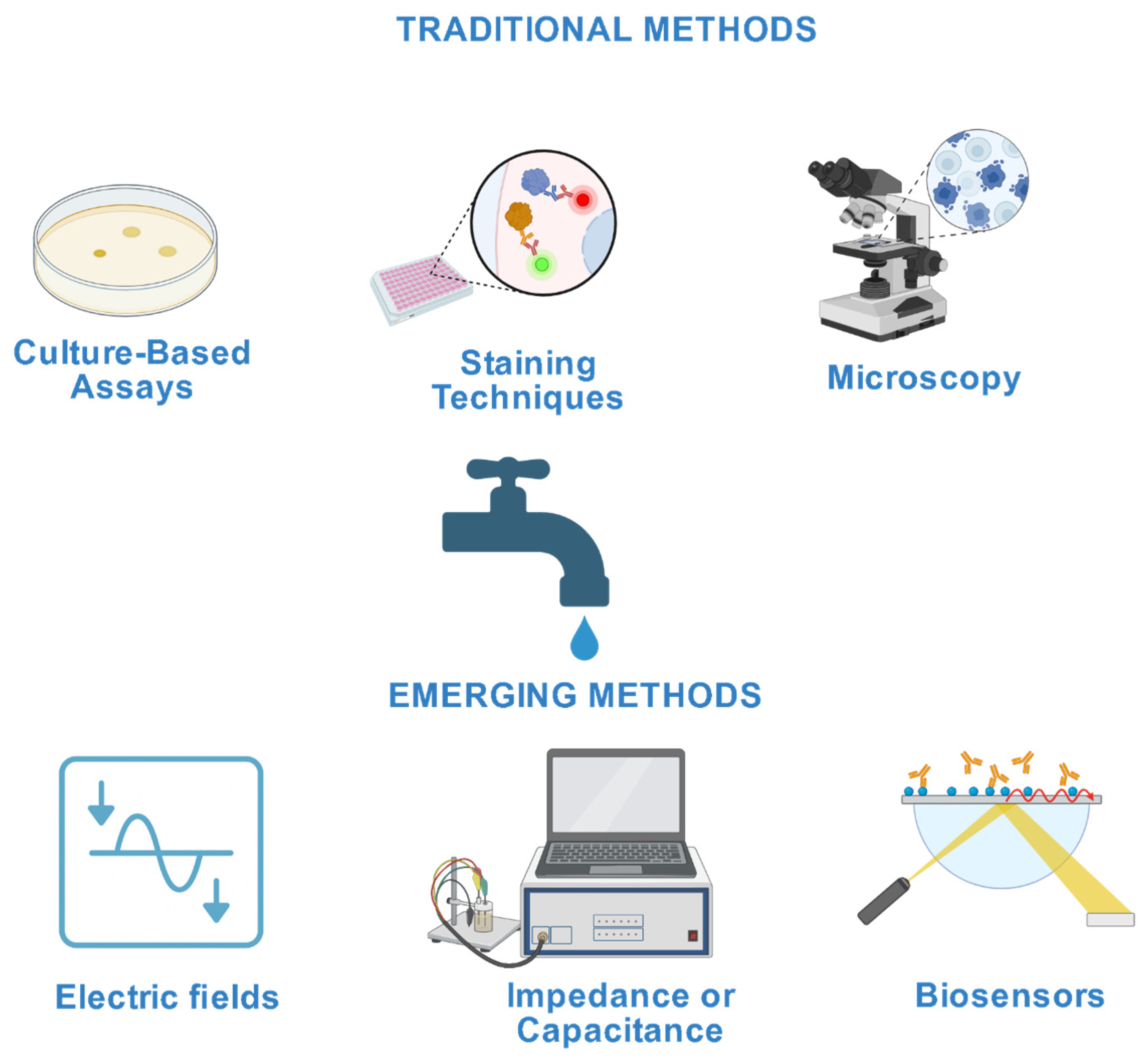
Figure 1. Illustrative comparison between representatives traditional and emerging methods used for microbial assessment in wastewater; In this context, “staining techniques” refers to microscopy combined with Gram staining and viability assays (e.g., Live/Dead). Created in BioRender, (2025) https://BioRender.com/srqe3zd (accessed on 15 March 2025).
Environments 12 00343 g001
Figure 2. Number of publications per year (2015–2024) related to the application of electric fields to microbial systems in wastewater. Data extracted from the Web of Science Core Collection (search terms: “electric field” OR “pulsed electric field” AND “microorganism” OR “bacteria” OR “spores” AND “wastewater” OR “waste water”). Source of raw data: Document Search—SciELO Citation Index (webofscience.com) (accessed on 15 May 2025).
Environments 12 00343 g002
Figure 3. Clustered keyword co-occurrence map generated using VOSviewer based on author keywords from 126 articles indexed in Web of Science (2015–2024) on the application of electric fields to microbial systems in wastewater. Node size reflects keyword frequency, link thickness represents co-occurrence strength, and colors denote distinct thematic clusters.
Environments 12 00343 g003
Figure 4. Classification of electric fields used in microbial applications, organized by field type (Direct Current, Alternating Current, Pulsed Electric Fields), with characteristic parameters (yellow) and representative applications (blue) relevant to wastewater microbiology and environmental biotechnology.
Environments 12 00343 g004
Figure 5. Representative schematic of electric field (EF) effects a model Gram-negative bacterium (E. coli). The figure highlights key mechanisms at different structural levels: membrane (pore formation, polarization), cytoplasm (ion flux, conductivity changes), and macromolecular components (DNA/RNA, proteins). The integrated image shows how these processes occur simultaneously, influencing microbial physiology and viability. Created in BioRender, (2025) https://BioRender.com/srqe3zd (accessed on 15 April 2025).
Environments 12 00343 g005
Table 1. Structural characteristics of selected microorganisms relevant to their interaction with electric fields.
Microorganism/StructureRelevant Structural CharacteristicsElectrical Parameters/Response to EFRefs.
Gram-negative bacteria (E. coli, P. aeruginosa)Membrane thickness 4–6 nm; thin peptidoglycan layer; P. aeruginosa has asymmetric outer membrane with LPS; porins for ionic exchangeReversible electroporation at ~10 kV/cm; membrane capacitance 10−6–10−4 S/m; cytoplasmic conductivity 0.1–1 S/m; detectable via EIS and DEP[26,27,28,29,30,53,59]
Gram-positive bacteria (B. subtilis, E. faecalis)Thick peptidoglycan wall (20–30 nm); multilayered envelopeHigher electroporation threshold than Gram-negatives; lower membrane conductivity (~0.5–1 × 10⁻⁶ S/m)[53,54]
Yeast (S. cerevisiae)Thick wall with glucans and mannansElectroporation at ~7.5 kV/cm; used for intracellular extraction[26,30]
Spores (Bacillus, Clostridium spp.)Multilayered structure; low water content (<30%); highly resistant wallActivation/inactivation threshold >30 kV/cm; low dielectric permittivity; high PEF resistance[63,64,65,66,67,68]
Surface structures (pili, flagella, biofilm)Type IV pili in P. aeruginosa responsive to EF; biofilm thickness 1–50 μm, high mechanical resistanceEF can induce electrotaxis, modify adhesion, detach biofilm (AC 0.1–2 Hz, DC 50–250 μA); affects local field distribution[69,70,71,72,73,74]
Cytoplasmic propertiesMain ions: K+, Na+; cytoplasmic conductivity ~0.1–1 S/m; dielectric constant ~25–30 (hydrated)EF influences ion distribution and polarization at mid–high frequencies; measurable via impedance[57,58,59]
Table 2. Membrane-level bioelectrical and dielectric properties of selected microorganisms.
PropertyMicroorganism/ExampleReported Values/CharacteristicsNotes on Electric Field InteractionRefs.
Membrane ThicknessE. coli (Gram-negative)Inner membrane ~4 nm; peptidoglycan ~3 nm; total ~15 nmThin membranes polarize faster; lower electroporation threshold (~10 kV/cm)[75,78]
B. subtilis (Gram-positive)Peptidoglycan layer 25–40 nmThicker wall increases mechanical resistance; higher electroporation threshold[76,79]
E. faecalis (Gram-positive)Cell wall ~40 nm with teichoic acidsHigh structural robustness under EF[77]
Membrane PotentialGeneral bacteriaResting potential −100 to −200 mV; E. coli: −220 mV (early exponential) to −140 mV (late exponential)Drives ion transport, motility; EF causes depolarization/hyperpolarization depending on orientation[80,81,82,83,84,85,86,87]
Membrane ConductivityE. coli1 × 10−6 to 3 × 10⁻⁶ S/m; increases to ~10−4 S/m after electroporationHigher conductivity → faster polarization, lower EF threshold for breakdown[95,98]
P. aeruginosa~5 × 10⁻⁶ S/mLinked to fluid membrane and protein P channels[97]
B. subtilis0.5 × 10−6 to 1 × 10⁻⁶ S/mThick wall, fewer ion channels → lower conductivity[28]
Dielectric PermittivityE. coliεr ≈ 5–9Fluid-phase lipid bilayer; strong β-polarization at kHz[103,111]
B. subtilisLower εr than E. coli (exact value NA)Reduced dielectric response at GHz[107]
Spores (Bacillus, Clostridium)<4 (dehydrated core)Low water content → high EF resistance (>30 kV/cm); distinct DEP separation[108,113]
Table 3. Cytoplasmic Bioelectrical and Dielectric Properties.
PropertyMicroorganism/StateValue/RangeNotesRefs.
Cytoplasmic ConductivityE. coli (viable)0.22 S/mTypical intact membrane; pronounced β-dispersion at 1–10 MHz[117,118]
E. coli (post-PEF)↓ significantlyReduction due to ion leakage and metabolic arrest[32]
P. aeruginosa (active)Higher than E. coliLinked to high intracellular K+, Na+, Cl, and organic acids[119,120]
Low pH (~6.5)↓ ~20%Reduced protein protonation and buffering capacity[121]
Clostridium spores<0.05 S/mDehydration and ionic shielding; low impedance response[122]
Dielectric PermittivityE. coli (hydrated)εr ~100High polarizability; β-dispersion around 1–10 MHz[115,118]
E. coli (dry core model)5–6.5Represents proteins and nucleic acids without hydration[125]
E. coli (ambient hydrated)25–30Strong hydration effects[125]
Clostridium spores<30Dense dipicolinic acid–calcium complexes; low polarizability[126]
Notes: “↓” indicates a decrease compared to the control or initial value.
Disclaimer/Publisher’s Note: The statements, opinions and data contained in all publications are solely those of the individual author(s) and contributor(s) and not of MDPI and/or the editor(s). MDPI and/or the editor(s) disclaim responsibility for any injury to people or property resulting from any ideas, methods, instructions or products referred to in the content.