


Abstract
Differentiated thyroid carcinoma (DTC) is the most common malignant endocrine tumor, with a generally favorable prognosis. Imaging, including iodine radioactive isotope scintigraphy (IRIS), is crucial for diagnosis and follow-up. While 131I has long been used for both therapeutic and diagnostic purposes, 123I is reserved for diagnostic imaging due to its shorter half-life and γ emissions. This review highlights the utility of 123I scintigraphy, especially in pre-treatment assessment and dosimetry for DTC. It is particularly valuable before radioiodine (RAI) ablation, providing accurate imaging in patients with iodine-refractory (IR) or biochemically incomplete response (BIR) DTC. When compared to post-therapeutic 131I scans, 123I scintigraphy appears to have a lower sensitivity for detecting metastatic lesions, particularly in lymph nodes and lungs. However, its diagnostic performance compared to low-dose diagnostic 131I is more variable, with some studies suggesting superiority due to the absence of stunning. Further research is needed to standardize its use and optimize its role in guiding DTC management.
1. Introduction
Differentiated thyroid carcinoma (DTC) is the most common malignant tumor of the endocrine system [1,2], and it presents a higher incidence in females than in males [3]. DTC is also generally characterized by a low incidence of distant metastases, which are the only features that greatly influence mortality and consequently a favorable prognosis [4,5,6,7]. Imaging examinations are fundamental in the staging and follow-up of the DTC, and they may include ultrasound (US), iodine radioactive isotope scintigraphy (IRIS) [8,9,10], and, in specific cases, 18F-fluorodesoxyglucose positron emission tomography-computed tomography (18F-FDG PET-CT) [11,12,13,14].
Since the 1940s, the IRIS represents the most specific and used diagnostic imaging examination in the management of DTC, with 131I and 123I as the most common isotopes. 131I has a half-life (T1/2) of about 8 days and emits low-penetrating β particles (maximum energy 606 kiloelectronvolts (keV), mean energy 190 keV). It is usually administered for DTC treatment [15,16]. However, due to its γ emissions, it can also be used to obtain whole-body scan (WBS) and single-photon emission tomography/computed tomography (SPECT/CT) images after both diagnostic and therapeutic doses [16,17,18]. 123I presents a prevalence of γ emission (primary emission of 159 keV) and a T1/2 of about 13 h, and it is used only for diagnostic imaging [17,18,19]. Despite being included in the most recent and relevant guidelines for the management of DTC [20], the exact role of 123I scintigraphy—and the clinical contexts in which it may be particularly useful—has yet to be clearly delineated.
A significant concern with using 131I for diagnostic scanning is the potential ‘stunning’ effect, where the diagnostic dose of 131I can impair the subsequent uptake of the therapeutic dose, potentially reducing treatment efficacy. Due to its shorter half-life and the absence of high-energy β emissions, 123I is theoretically free from this effect, making it an attractive agent for pre-therapeutic planning. However, its integration into clinical practice is heterogeneous, and its diagnostic performance relative to 131I, both in diagnostic and therapeutic settings, requires clarification.
The aim of this comprehensive narrative review is to summarize the current evidence regarding the role of 123I scintigraphy in dosimetry and pre-treatment RAI planning and to evaluate its diagnostic performance, particularly in comparison with 131I scintigraphy using diagnostic and therapeutic doses.
2. Radioiodine and Dosimetry
To quantify the red marrow absorbed dose after 131I administration, the 2008 European Association of Nuclear Medicine (EANM) procedure guidelines [21] recommend collecting serial blood samples at 2, 6, 24, 96, and 144 h following the administration of a low diagnostic activity (≤15 MBq), using blood activity as a surrogate for red marrow dosimetry and applying a safety threshold of 2 Gy. In addition, whole-body measurements are advised at 2, 6, and 24 h, with at least one further acquisition at approximately 96 h.
Hänscheid et al. [22] subsequently introduced a simplified method for estimating the blood absorbed dose per unit of administered 131I, relying on a single total-body retention measurement. This approach achieves its greatest accuracy when the measurement is acquired at 24–48 h and appears adequate for routine assessment of individual radiation exposure during radioiodine therapy, although it may carry a risk of underestimating the Maximum Tolerated Activity (MTA).
More recently, Atkins et al. [23] described a bi-exponential model corrected for body surface area (BSA), showing that the 48 h fractional whole-body retention can predict the measured MTA without requiring blood sampling, with an average deviation of only ±1.2%. The authors recommend administering no more than 70% of the calculated MTA, due to the risk of overestimation—particularly in patients prepared with rhTSH: this can occur due to faster RAI biological washout in patients prepared with rhTSH than in patients treated with hormone withdrawal; particularly, it be due to constipation and mild reduction of kidney function, the two main ways of RAI excretion through feces and urine, which are frequent during hypothyroidism.
Despite the increasing use of 123I scintigraphy, often combined with multiple SPECT/CT acquisitions, to determine the optimal therapeutic activity of 131I, the timing and methodology of 123I-based dosimetry remain non-standardized [24,25,26].
In preclinical work, Kim et al. [24] investigated the optimal timing for determining tissue-absorbed dose in DTC xenograft mouse models. They identified two key imaging time-points, at the estimated Tmax of 2.91 h and at 26 h following 123I administration.
Following the recommendations of Atkins et al., Durski et al. [25] tested a protocol involving two scintigraphic acquisitions at 24 and 48 h after 123I administration to estimate the 131I MTA in a patient with metastatic DTC; subsequently, 70% of the calculated MTA was administered.
Brown SR et al. [26] conducted a clinical trial investigating the potential role of selumetinib in promoting redifferentiation in DTC. The protocol incorporated 123I scintigraphy both for patient selection and for pre-treatment dosimetry in 131I therapy. Although the precise imaging schedule was not reported, the authors indicated that three to four acquisitions were performed, consisting of a standard scan followed by two or three additional time-point measurements.
Finally, the 2025 American Thyroid Association (ATA) guidelines for DTC management [20] also do not provide a standardized protocol for 123I-based dosimetry.
In summary, while 131I-based dosimetry is relatively well established, 123I dosimetry represents a promising and more patient-friendly alternative that eliminates the need for multiple blood samples. Emerging protocols—often based on two imaging time-points (e.g., 24 and 48 h)—appear feasible for estimating the MTA for 131I therapy, particularly in metastatic disease. However, the lack of a standardized 123I dosimetry procedure in the 2025 ATA guidelines highlights the need for larger, prospective studies to validate and harmonize these methods before they can be broadly implemented in clinical practice.
3. 123I WBS in Pre- RAI Treatment: Comparison with 131I Diagnostic Dose Scintigraphies
As reported in the recent 2025 ATA guidelines [20], the use of WBS prior to RAI therapy appeared to be a possible procedure to guide the treatment, preferably the use of WBS with 123I rather than WBS with 131I. Possible changes determined by WBS include the following:
- (1)
- Detection of very large thyroid remnants, which require additional surgery prior to ablation;
- (2)
- Absence of a remnant with negative thyroglobulin (Tg), particularly in patients at low risk of recurrence who do not require RAI therapy or who may require a low dose of RAI in other cases;
- (3)
- Detection of RAI-avid metastases, offering the opportunity for high-dose treatment. Instead, the strongest arguments against this practice are provided by the 2008 EANM guidelines, which suggest not performing diagnostic WBS in the presence of a clear indication for RAI therapy, and the 2022 ETA guidelines, which state that diagnostic WBS should not be used routinely [27,28]. In addition, multiple studies analyzed its usefulness and concluded that, in several cases, 123I WBS findings modified the therapeutic approach [29,30,31]. In some cases, this involved making a conservative decision in the absence of 123I uptake; in others, it implies increasing the 131I dose administered in the presence of suspected metastases. 123I appeared to be favored over 131I due to its pure gamma emission, shorter half-life, and lower probability of stunning effect. In this context, it is interesting to evaluate the experience of using 123I WBS compared to 131I diagnostic WBS, considering the studies published in the literature.
Sarkar SD et al. [32] retrospectively reviewed 12 cases of patients who underwent thyroidectomy for DTC and compared the results of 123I whole-body scintigraphy (WBS), performed 24–96 h after administering 2–5 mCi (74–185 MBq) of 123I, with those of 131I WBS, performed at the same time points using a diagnostic dose of 111–185 MBq. They found that both 123I and 131I WBS correctly identified residual thyroid tissue in nine patients, but only 131I WBS detected metastatic lesions in five scans from four patients. No lesion was more clearly visualized with 123I than with 131I, leading the authors to conclude that 123I WBS is less sensitive than 131I WBS for imaging DTC metastases. The study’s limitations included the small sample size and the lack of single-photon emission computed tomography (SPECT/CT) imaging.
Mandel SJ et al. [33] prospectively evaluated 14 patients with DTC who underwent total thyroidectomy and were subsequently staged using 123I and 131I diagnostic scintigraphy. For the 123I WBS, images were acquired five hours after the administration of 48–56 MBq of 123I, and for the 131I scintigraphy, images were acquired 48 h after the administration of 111 MBq of 131I. They found that 123I scintigraphy detected three more foci of increased uptake than 131I WBS (35 versus 32 foci). The study’s limitations include the small sample size and the absence of SPECT/CT image acquisition.
Siddiqi A et al. [34] prospectively evaluated 12 patients with DTC previously treated with thyroidectomy and RAI and with negative 131I diagnostic scan and raised Tg levels. They performed a 123I scan on all the patients before RAI therapy, revealing positive findings in 10 out of 12 patients. A fundamental limitation was the selection bias, considering only patients with a negative 131I scan.
The comparison between 123I and diagnostic-dose 131I WBS yields conflicting results, as summarized in Table 1. The study by Mandel et al. [31], which reported superior performance for 123I, utilized an early acquisition time (5 h), potentially capitalizing on 123I’s favorable early pharmacokinetics. In contrast, Sarkar et al. [30] used later acquisitions (24–96 h), which may disadvantage 123I due to its physical short half-life. This highlights that the timing of image acquisition is a critical and often overlooked variable that significantly influences the perceived sensitivity of 123I scintigraphy. In addition, there are specific limitations that affect both RAI WBS with diagnostic dose. These include the potential for restricted RAI uptake in small thyroid remnants, the high cost of 123I, the possibility of misinterpretation of thyroid remnants as nodal metastases on planar images, and, finally, the limited sensitivity in the presence of unfavorable mutations (e.g., BRAF, TERT) [20].
Table 1.
Summary of articles comparing 123I with diagnostic-dose 131I WBS.
4. 123I Diagnostic Dose vs. 131I Therapeutic Dose Scintigraphies
Several studies have attempted to compare the diagnostic performance of 123I WBS and 131I WBS after RAI therapy, reaching conflicting conclusions [35,36,37,38,39,40,41,42,43,44,45,46]. Most of the studies describe lower sensitivity of 123I WBS, with the possibility of misrecognizing some RAI-avid lesions that are detectable with 131I WBS [35,37,38,39,42,44]. However, other studies do not confirm these findings [40,41,43,45,46] and report comparable sensitivities, albeit often with lower values for 123I WBS than for 131I WBS. This would seem to confirm the latter’s slight superiority. Only one study described superiority of the 123I scan [36].
Considering single study, Alzahrani AS et al. [35] performed a retrospective study which compared 238 diagnostic WBS performed 24 h after the oral administration of 185–555 MBq of 123I with their corresponding 131I post-therapy WBS obtained 4–5 days after 131I therapy. They studied scans in three clinical situations: with the first 131I therapy, with the second 131I therapy, and in cases of elevated Tg levels and negative diagnostic scan. They found that 177 pairs were obtained with the first 131I therapy and showed concordance between pre-treatment and post-treatment scans in 166 pairs with a concordance rate (CR) of 93.8%: particularly, six post-treatment scans showed foci in thyroid bed and five in metastatic locations. Considering the second 131I therapy, 34 pairs were obtained and showed concordance in 28 pairs with a CR of 82.4%: particularly, in six cases, metastases were not detected with 123I WBS. Finally, of twenty-seven pairs of scans in patients with elevated Tg levels and negative pre-treatment WBS, fifteen post-treatment scans remained negative, six showed an uptake in the thyroid bed, three showed benign lung uptake, and three showed definite uptake (in thyroid bed, in thyroid bed and lung, and in lymph nodes) which was also weakly detectable retrospectively in the 123I WBS: CR in this setting was 55.6%.
Thomas DL et al. [36] retrospectively evaluated and compared pretherapy diagnostic 123I scans with 7-day post-therapy 131I scans in detecting remnant thyroid disease as well as locoregional metastases in 53 patients. They found that 123I scans performed 24 h after an oral administration of 1–1.6 mCi (37–59.2 MBq) were more sensitive and provided a better lesion-to-background ratio than 131I scans performed seven days after 131I oral administration. The study was probably affected by the late acquisition of the 131I WBS, and the authors acknowledge this in their conclusion.
Cohen JB et al. [37] retrospectively compared 30 pre-therapy diagnostic 123I scans performed 24 h after the administration of 74 MBq of 123I with 30 post-therapy 131I scans performed 2–10 (mean 5.8) days after the administration of RAI in 29 patients. They found that WBS showed concordance in nineteen cases (63.3%), while in four cases (13.3%), 123I WBS detected more lesions in the neck than 131I WBS. However, in seven cases (23.3%), 131I WBS were more sensitive than 123I WBS, particularly in cases involving distant metastases.
Bravo PE et al. [38] retrospectively evaluated 342 individuals diagnosed with DTC, underwent RAI therapy, and performed a 123I WBS one day before RAI administration and 131I WBS one week after RAI therapy. SPECT/CT acquisitions were also performed, and the administered 123I activities were standardized: 1.5 mCi for patients treated with thyroid hormone withdrawal and 2.0 mCi for patients prepared with recombinant human TSH (rhTSH). The patients were divided into three groups: 311 patients RAI-naive without known distant metastatic disease, 23 patients with history of prior RAI and persistent disease, and 8 patients with known distant metastases. Their analysis showed that 22 patients (7%) in the first group had discordant scan findings but in only four cases did these discrepancies reflect actual disease. In the second and third groups, discordant scans were present in seven (30%) and five (62.5%) patients, respectively, with misinterpretation of metastatic disease at the 123I WBS scan. Bravo PE et al. concluded that the sensitivity of 123I WBS depends on the clinical setting.
Iwano S et al. [39] published a study evaluating 69 patients with DTC treated with RAI. They compared WBS images taken 24 h after the administration of 37 MBq of 123I with images obtained 34 days after administration of 2.22–7.4 GBq (median 5.55 GBq) of 131I. They identified 108 sites of RAI uptake in the 131I WBSs, but only 77 of these were also detected with the 123I, giving a CR of 71%. CR was higher for uptake in the thyroid bed (89%) and bone metastases (86%) but lower for lymph nodes (61%) and lung metastases (39%). The authors concluded that WBSs performed 24 h after the administration of 37 MBq of 123I were not always predictive of the 131I WBS results.
Urhan M et al. [40] evaluated 292 123I WBS (dose administered: 50–111 MBq) with their corresponding post-treatment 131I images. All patients treated with RAI were in a hypothyroid state. They found that in 228 out of 263 patients with a positive diagnostic scan, 123I and 131I WBS findings were concordant (CR 87%). However, 44 additional foci of abnormal uptake in 22 discordant cases were found in 131I WBS but with no impact on therapeutic management of the patients. In the other 13 patients, there was at least one new site on post-treatment images that had been missed on pre-treatment 123I images. Finally, 29 patients with a negative diagnostic WBS were treated with 131I due to high Tg levels (range 11.3–480 ng/mL) with RAI uptake sites not detected with 123I WBS seen in eight post-treatment 131I scans (CR 72%). The authors concluded that in DTC management pre-treatment, 123I WBS is comparable to high-dose 131I post-treatment imaging, but data apparently do not support this conclusion: in fact, 43 of 292 WBS (14.7%) had at least an RAI-avid lesion not detected with 123I WBS.
Gulzar Z et al. [41] prospectively evaluated 27 patients who underwent near total thyroidectomy and performed subsequent RAI therapy. They compared WBS obtained 4 and 24 h after the administration of 185 MBq 123I, with WBS performed 5–7 days after administration of therapeutic dose of 131I. They found CR of 92.6% and 83.2%, respectively, for WBS performed after 4 and 24 h following 123I administration compared with 131I WBS. Particularly, 7.8% of patients had at least one RAI-avid lesion that was not detected with the 123I WBS.
De Geus-Oei LF et al. [42] studied 55 patients who had been treated with 131I (1.85–5.55 GBq) and had previously undergone diagnostic 123I WBS (111–370 MBq). They compared WBS obtained 24 h after 123I administration with WBS obtained 3–15 days (median 8.6 days) after 131I administration in the 36 patients for whom these data were available. They found that more lesions were visible on the post-therapeutic 131I WBS than on the corresponding diagnostic 123I scan in thirteen patients; in two patients, 123I WBS appeared more sensitive; and in twenty-one, the findings were equal (CR 58.3%).
Ali N et al. [43] retrospectively evaluated 58 patients who underwent at least two 131I therapy and made a comparison between a 20 min scintigraphy obtained 2 and 24 h after the administration of 123I 185–270 MBq and a 10–12 min scintigraphy obtained 4–7 days after the administration of 5.55 GBq of 131I. The study, which initially included 135 patients, showed that despite a high complete response rate (94.8%), more lesions were visible on the post-therapeutic 131I WBS than on the corresponding diagnostic 123I scan. Notably, in three patients, the 123I WBS was negative while the 131I scan was positive. In one patient, 123I scintigraphy detected an uptake that was not seen on the 131I scan; however, a subsequent scan performed after the third 131I therapy eventually revealed it.
Siddiqi A et al. [34], as previously reported, prospectively evaluated 12 patients with DTC previously treated with thyroidectomy and RAI and with negative 131I diagnostic scan and raised Tg levels (>2.5 pg/L): in these patients, a diagnostic 123I WBS was performed 2 h and 24 h after administration of 123I tracer dose 185 MBq and 4–7 days after 131I therapy dose 5.55 GBq. They performed a total of 18 123I scans and found 16 positive 123I WBS with corresponding 14 positive 131I WBS: in the two cases of discordance, one was a false negative uptake; in the second one, the Tg levels decreased after therapy; and no imaging or histopathological confirmation of the bone uptake at 123I scan was performed. In two cases, 131I WBS detected thyroid bed uptakes not revealed by 123I. The primary limitation of the study—aside from the small sample size and the absence of SPECT/CT images—was the patient selection criterion, which included only patients with a negative 131I diagnostic WBS.
Schoelwer MJ et al. [44] retrospectively evaluated 33 pediatric patients who performed total thyroidectomy, diagnostic scintigraphy, and RAI therapy for DTC. Thirty-seven pairs of scans with different iodine isotopes were performed. For diagnostic scanning, five received 74 MBq of 131I, twenty-one received 74 MBq of 123I, and eleven received 111 mCi of 123I; images were acquired 24 h after RAI administration. Therapeutic dose of 131I was variable (27–190 mCi) and images acquired after 7 days. Of the 31 131I scans considered, 24/31 were concordant with 131I therapeutic scans (CR 77%), particularly in two cases with missing mediastinal uptake and four cases with missing lung uptake (false negative 123I rate: 19.4%). One case was represented by a false positive 131I finding.
Yaakob W et al. [45] retrospectively evaluated 13 patients with DTC who underwent thyroidectomy and were given 123I whole-body scans 24 h after receiving 0.8–1.0 mCi of 123I. All patients then received a therapeutic dose of 131I (29.9–250 mCi), after which a WBS was performed 7–10 days later. Excluding one patient with a false positive 123I WBS scan, the authors found excellent correlation between scans in 11 out of 12 patients, particularly in the case where 123I underestimated the lung burden (CR 91.7%).
Berbano R et al. [46] conducted a study involving 16 patients with DTC treated with RAI. They compared whole-body scans performed 24 h after administration of 10 mCi (370 MBq) of 123I with images acquired 5–7 days after 131I administration (dose range: 75–200 mCi). They observed concordant findings in 15 of the 16 scans (CR 93.8%), with a single discordant case in which the 123I WBS failed to detect a lung lesion.
Most of the studies were affected by the limitation of the absence of SPECT/CT images, resulting in low diagnostic performance of the scintigraphic images. Furthermore, some studies [34,38,41,44,45,46] had a small sample size (<30 patients). Another possible source of bias is the heterogeneity in the 123I scintigraphy protocol, including the administered dose and timing of image acquisition. A similar problem is also present for 131I scintigraphy after a therapeutic dose. Finally, excluding the paper by Gulzar Z et al. [41], most studies had a retrospective design.
Table 2 summarizes the findings of the studies reported in the comparison of 123I diagnostic scintigraphy and scintigraphy performed after a therapeutic dose of 131I.
Table 2.
Summary of articles that comparing 123I scintigraphy with therapeutic-dose 131I WBS.
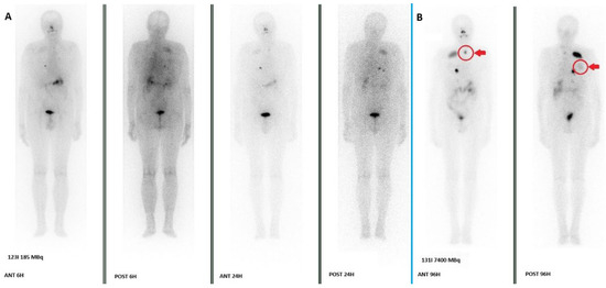
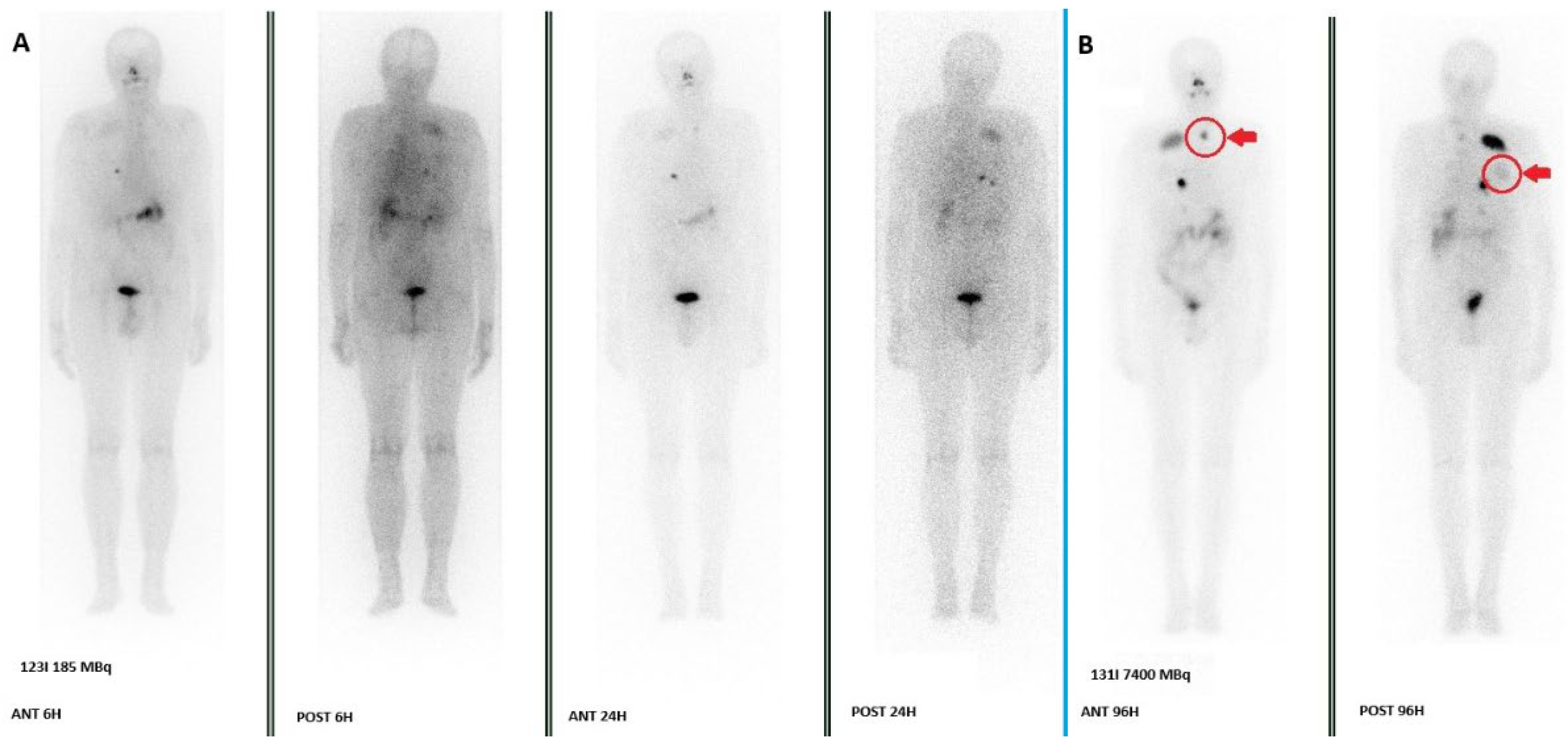
The bulk of evidence from Table 2 indicates that post-therapeutic 131I WBS detects a higher number of RAI-avid lesions than pre-therapeutic 123I WBS. The concordance rates are generally high in treatment-naïve patients with low tumor burden but drop significantly in the setting of persistent or metastatic disease. The most common sites for 123I false negatives are lymph nodes and pulmonary metastases, likely due to lower lesion uptake and poorer count statistics compared to the high activity used in therapy. Therefore, a negative 123I scan in a high-risk patient, particularly one with elevated Tg levels, should be interpreted with caution, as it does not definitively exclude the presence of RAI-avid disease. Figure 1 shows an example of WBSs confrontation in a patient who was treated for metastatic DTC.
Figure 1.
A comparison of a WBS performed 6 and 24 h after the injection of 185 MBq of 123I (A) and a WBS performed 96 h after RAI therapy (7400 MBq) (B) was conducted in a patient with known bone and pulmonary metastases who was treated in our department in 2024. The images show the detection of a new nodal localization in the neck (arrow and circle in ANT 96H) and weak uptake in known rib metastases (arrow and circle in POST 96H) in the 131I WBS, whereas only faint and unclear uptakes were present in the 123I WBS.
5. 123I Scan and Thyroglobulin Level
Tg is a protein produced only by follicular thyroid cells. After thyroidectomy and, in particular, RAI ablation, it represents a fundamental tumor marker for the management and follow-up of DTC [47]. Its level and/or rising trend are well known to be associated with disease recurrence, as reported in the ATA 2025 guidelines. They defined dynamic risk stratification classes according to the response to therapy and the Tg levels. This includes, other than an excellent response (ER) and a structural incomplete response (SIR), an indeterminate response (IR) in the presence of a basal Tg (bTg) range of 0.2–1.0 ng/mL and a biochemically incomplete response (BIR) in the presence of a bTg range of 1.0–10.0 ng/mL, which is associated with an increased risk of structural recurrence of the disease. In this context, some studies have analyzed the usefulness of 123I scintigraphy in relation to Tg levels [48,49,50,51].
Campennì et al. [48] conducted a retrospective evaluation of 124 patients with DTC who underwent thyroidectomy and RAI therapy during follow-up. The study focused on the utility of the 123I scan. They found that the diagnostic performance of 123I WBS SPECT/CT significantly increased in patients with basal Tg values of at least 0.39 ng/mL.
Sol B et al. [49] retrospectively evaluated 40 patients with DTC who had undergone total thyroidectomy, focusing on 24 patients with undetectable basal Tg level determined with a highly sensitive Tg assay (below 0.1 ng/mL) six months after thyroidectomy. They found that none of these patients had stimulated Tg level above 1 ng/mL or a remnant on the 123I WBS after one year of follow-up. They concluded that 123I scintigraphy is unnecessary in this patient group.
Again, Campennì et al. [50] retrospectively evaluated 241 low- to intermediate-risk patients with histologically confirmed DTC and negative Tg antibodies (AbTg) all treated with total thyroidectomy and RAI therapy. During the follow-up 8–12 months after RAI, a 123I WBS was performed in 51 patients, particularly in 16 patients without excellent response. They found that seven out of sixteen (43.7%) DTC patients with IR (mean stimulated Tg = 4.5 ng/mL) and/or BIR (mean stimulated Tg = 13.5 ng/mL) 123I WBS and SPECT/CT were able to identify five local and two locoregional lymph-node metastases.
Villani MF et al. [51] evaluated 55 pediatric patients with DTC, and, particularly, they considered 41 scans performed after 123I administration with rhTSH and before RAI therapy. They found that thyroglobulin alone apparently was not a good predictor for staging modification (AUC = 0.6855 in ROC analysis), while 123I WBS modified staging in 12/41 (29%): in 3/12 (25%) for the presence of lung metastases and in 9/12 (75%) for lymph node involvement. In all these patients, the therapeutic management were modified.
Table 3 summarizes the findings of the studies reported.
Table 3.
Summary of articles evaluating the relationship between 123I scintigraphy and Tg levels.
6. Discussion
123I scintigraphy remains a key tool in the management of DTC, in particular in the diagnostic setting during the follow-up, especially in patients with IR and/or BIR as reported by Campennì A et al. [48,50], although its sensitivity is limited [35,38]. Its possible utility in performing it after total thyroidectomy and prior to RAI therapy has also been reported [29,30,31,51]. Execution of 123I diagnostic images allows the assessment of metastatic DTC localization or can underline the presence of unexpected alterations of radioiodine distribution in several cases [28]. These findings can help to define the appropriate RAI dose, if indicated, or can modify the therapeutic approach to these subjects: this applies to both initial therapy after total thyroidectomy and follow-up therapy.
In contrast with its utility in cases of dosable bTg, the absence of incremental diagnostic value in the presence of negative bTg was also described in one study. Particularly, the study of Sol B et al. [49] reported that the presence of bTg < 0.1 ng/mL during the early follow-up after initial therapy apparently made unuseful the 123I scintigraphy. These data appear to agree with the literature and particularly with the high negative predictive value of the bTg data [52,53]. Therefore, there is no benefit in routinely performing 123I WBS during the follow-up of patients with ER.
It is important to highlight that studies comparing 123I and 131I scintigraphy reported in the literature describe an apparent superiority of the 123I diagnostic scan over the 131I scan [33,34], while 131I scans performed after RAI therapy appear to be more sensitive than 123I diagnostic WBS scans [35,37,38,39,42,44]. Studies reporting comparable diagnostic values for 131I WBS after RAI therapy and 123I diagnostic WBS mostly describe a slight superiority of the former. In particular, few patients in these studies had a DTC lesion missing on the 123I WBS that was subsequently detected with the 131I. Urhan et al. [40] reported this to be 14.7%, Gulzar et al. [41] 7.8%, Ali et al. [43] 5.2%, Yaakob et al. [45] 8.3%, and Berbano et al. [46] 6.2%. Given that the presence of metastases is one of the few factors that can significantly affect outcomes in patients with DTC [54], the possibility of missing them in 5.2–14.7% of cases warrants caution when using 123I diagnostic scintigraphy during follow-up. In summary, 123I WBS scintigraphy appears to provide useful information when positive results are reported. However, a negative result could not exclude the presence of a RAI-avid lesion in a few cases, despite its good sensitivity. In conclusion, the 131I WBS performed after the administration of therapeutic activity remains the gold standard for evaluating RAI avidity in the localization of DTC disease.
It is important to emphasize that no clear standardization protocol on 123I dose or acquisition time were described for patient dosimetry, even though diagnostic practice studies reported the administration of different 123I doses. Almost all these studies agreed on acquiring images 24 h after administration, whereas the utility of an earlier acquisition is still being debated. In the same context, no studies focused on comparing preparation with hormone withdrawal in relation to rhTSH were found in the literature. This lack of standardized protocols requires an expert consensus to establish a well-known and widely utilized practice, especially considering the large number of studies reported in the literature. Standardization of 123I WBS dosimetry and diagnostic protocols, including the dose, timing, and patient preparation, could provide a basis for further studies evaluating its usefulness and enabling better comparison.
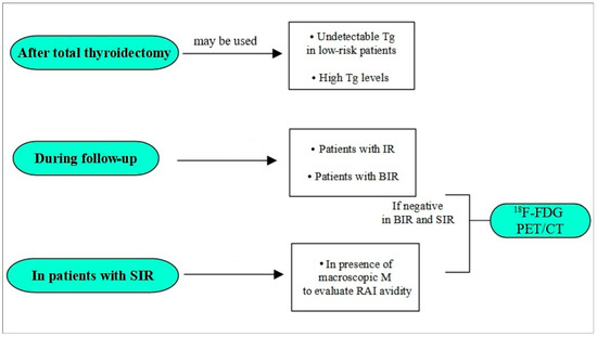
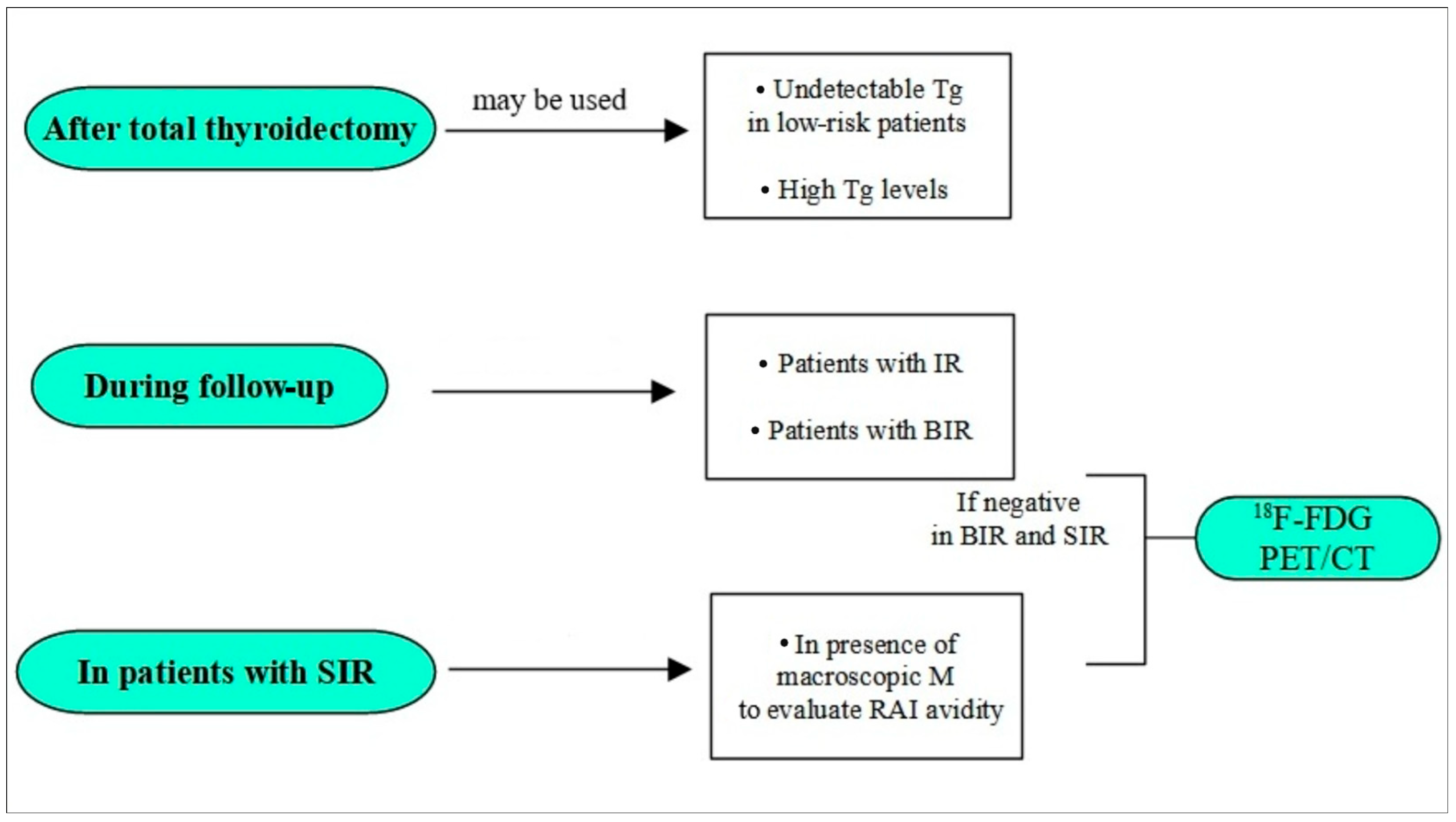
Furthermore, it is fundamental to highlight that the choice of imaging in DTC is not merely between 123I and 131I, and performing an RAI WBS does not necessarily conclude the diagnostic process. In this setting, 18F-FDG PET/CT has a well-established role in RAI refractory DTC where lesions lose the ability to concentrate iodine [55,56]. A key clinical challenge is identifying which patients could benefit from 18F-FDG PET/CT and where 123I can serve as a triage tool. Considering patients with BIR, a positive scan confirms the presence of RAI-avid lesions and guides therapy, albeit with some limitations, while a negative scan should prompt investigation for iodine-refractory disease with 18F-FDG PET/CT. Similarly, in patients with known SIR, the possibility to evaluate both the RAI and FDG uptake in every single metastases could guide the therapeutic choice. Possible roles of the 123I scintigraphy in the management of DTC are highlighted in Figure 2.
Figure 2.
A flow chart about possible roles of 123I scintigraphy inside the DTC management.
Finally, beyond conventional planar or SPECT-based approaches, it is important to acknowledge the additional contribution that PET imaging can offer in the evaluation of iodine-avid disease. In this context, 124I PET/CT provides superior sensitivity and true quantitative capabilities, enabling improved detection of small-volume lesions—particularly cervical lymph node involvement and micronodular pulmonary metastases—and offering a more accurate basis for patient-specific dosimetry. Several studies [57,58,59] have demonstrated that 124I PET outperforms 123I scintigraphy in lesion conspicuity and may better predict post-therapeutic 131I uptake, supporting its potential role in treatment planning. Nevertheless, its routine clinical use remains limited by restricted availability and higher costs.
We can actually suggest the use of 123I scintigraphy for pre-ablation assessment in cases of unclear RAI indication, IR, BIR, and SIR, bearing in mind its limited sensitivity. This can be followed by an 18F-FDG PET/CT scan if the WBS is negative. Finally, 123I should be used for dosimetry when required.
The review has several limitations. Firstly, most of the studies appeared quite old and lacked SPECT/CT image acquisition, which is actually the gold standard for managing DTC as recommended by several international guidelines, including the recent SNMMI/EANM 2022 and ATA 2025 guidelines [20,60]. This could lead to an underestimation of the sensitivity of the two tracers considered. Secondly, the absence of standardization in administration and acquisition protocols resulted in heterogeneous use of dosage and acquisition time in 123I diagnostic scintigraphy. Similarly, significant heterogeneity in the 131I therapeutic dose administered to patients, as well as in the timing and acquisition protocol of the scintigraphic images, was reported. This heterogeneity makes it difficult to compare individual studies and surely affects the sensitivity of scintigraphic studies. Thirdly, most of the studies considered were designed with retrospective analyses. Finally, another important possible limitation is represented by the small sample size of some studies considered.
Further studies with a large sample size and a prospective design should be desirable to define the real discrepancy between 123I and 131I scintigraphy after RAI therapy. Other variable such as 18F-FDG PET/CT, Tg levels, the staging, the risk factor, etc., should be also considered in the analysis. This approach could confirm whether the latter is superior and identify patients in whom the possibility of a false negative result with 123I is present.
7. Conclusions
In conclusion, 123I scintigraphy represents a valuable diagnostic tool in the management of DTC in specific settings, particularly in cases of raised Tg levels (IR and/or BIR), patients with SIR, and for dosimetry in metastatic disease. Instead, its role in pre-ablation assessment is still debated. Moreover, it is fundamental to highlight its possible lower sensitivity compared to post-therapeutic 131I scans. The development of standardized protocols for administered activity and acquisition timing is needed. Future prospective, multi-center studies with large sample sizes, standardized 123I protocols, and mandatory SPECT/CT are essential to definitively establish its sensitivity and integrate it into a cost-effective, patient-tailored diagnostic algorithm.
Author Contributions
Conceptualization, P.B., V.Z., F.D. and F.B.; methodology, P.B.; software, C.I., C.R., F.S. and G.V.; validation, P.B., F.D. and V.Z.; formal analysis, P.B., F.D., C.I., C.R. and G.V.; investigation, P.B.; data curation, P.B., F.D., C.I. and C.R.; writing—original draft preparation, P.B.; writing—review and editing, F.D., E.G., R.M., C.C. (Claudio Casella), D.L., F.B., C.C. (Carlo Cappelli) and M.C.; visualization; supervision, F.B. and L.S. All authors have read and agreed to the published version of the manuscript.
Funding
This research received no external funding.
Institutional Review Board Statement
Not applicable.
Informed Consent Statement
Not applicable.
Data Availability Statement
No new data were created or analyzed in this study.
Conflicts of Interest
The authors declare no conflicts of interest.
References
- Albano, D.; Dondi, F.; Bellini, P.; Bertagna, F. Biomarkers and Molecular Imaging in Postoperative DTC Management. In Integrated Diagnostics and Theranostics of Thyroid Diseases; Giovanella, L., Ed.; Springer: Cham, Switzerland, 2023. [Google Scholar] [CrossRef] [Scilit]
- Cabanillas, M.E.; McFadden, D.G.; Durante, C. Thyroid cancer. Lancet 2016, 388, 2783–2795. [Google Scholar] [CrossRef] [Scilit] [PubMed]
- Filetti, S.; Durante, C.; Hartl, D.; Leboulleux, S.; Locati, L.; Newbold, K.; Papotti, M.; Berruti, A. Thyroid cancer: ESMO Clinical Practice Guidelines for diagnosis, treatment and follow-up. Ann. Oncol. 2019, 30, 1856–1883. [Google Scholar] [CrossRef] [Scilit] [PubMed]
- van Velsen, E.F.S.; Peeters, R.P.; Stegenga, M.T.; van Kemenade, F.J.; van Ginhoven, T.M.; van Balkum, M.; A Verburg, F.; Visser, W.E. Evaluating Disease-specific Survival Prediction of Risk Stratification and TNM Systems in Differentiated Thyroid Cancer. J. Clin. Endocrinol. Metab. 2023, 108, e267–e274. [Google Scholar] [CrossRef] [Scilit] [PubMed Central]
- Roman, B.R.; Morris, L.G.; Davies, L. The thyroid cancer epidemic, 2017 perspective. Curr. Opin. Endocrinol. Diabetes 2017, 24, 332–336. [Google Scholar] [CrossRef] [Scilit] [PubMed] [PubMed Central]
- Bellini, P.; Dondi, F.; Gatta, E.; Zilioli, V.; Albano, D.; Cappelli, C.; Bertagna, F. Prognostic role and characteristics of the indeterminate response in differentiated thyroid cancer: A systematic review. Endocrine 2024, 84, 812–821. [Google Scholar] [CrossRef] [Scilit] [PubMed]
- Albano, D.; Bellini, P.; Dondi, F.; Calabrò, A.; Casella, C.; Taboni, S.; Lombardi, D.; Treglia, G.; Bertagna, F. Temporal Evolution and Prognostic Role of Indeterminate Response Sub-Groups in Patients with Differentiated Thyroid Cancer after Initial Therapy with Radioiodine. Cancers 2023, 15, 1270. [Google Scholar] [CrossRef] [Scilit] [PubMed] [PubMed Central]
- Avram, A.M. Radioiodine scintigraphy with SPECT/CT: An important diagnostic tool for thyroid cancer staging and risk stratification. J. Nucl. Med. Technol. 2014, 42, 170–180. [Google Scholar] [CrossRef] [Scilit] [PubMed]
- Zilioli, V.; Peli, A.; Panarotto, M.B.; Magri, G.; Alkraisheh, A.; Wiefels, C.; Rodella, C.; Giubbini, R. Differentiated thyroid carcinoma: Incremental diagnostic value of 131I SPECT/CT over planar whole body scan after radioiodine therapy. Endocrine 2017, 56, 551–559. [Google Scholar] [CrossRef] [Scilit] [PubMed]
- Sun, Y.-Q.; Sun, D.; Zhang, X.; Zhang, Y.-Q.; Lin, Y.-S. Radioiodine adjuvant therapy in differentiated thyroid cancer: An update and reconsideration. Front. Endocrinol. 2022, 13, 994288. [Google Scholar] [CrossRef] [Scilit] [PubMed] [PubMed Central]
- Albano, D.; Dondi, F.; Mazzoletti, A.; Bellini, P.; Rodella, C.; Bertagna, F. Prognostic Role of 2-[18F]FDG PET/CT Metabolic Volume Parameters in Patients Affected by Differentiated Thyroid Carcinoma with High Thyroglobulin Level, Negative 131I WBS and Positive 2-[18F]-FDG PET/CT. Diagnostics 2021, 11, 2189. [Google Scholar] [CrossRef] [Scilit] [PubMed] [PubMed Central]
- Nanni, C.; Rubello, D.; Fanti, S.; Farsad, M.; Ambrosini, V.; Rampin, L.; Banti, E.; Carpi, A.; Muzzio, P.; Franchi, R. Role of 18F-FDG-PET and PET/CT imaging in thyroid cancer. Biomed. Pharmacother. 2006, 60, 409–413. [Google Scholar] [CrossRef] [Scilit] [PubMed]
- Zampella, E.; Klain, M.; Pace, L.; Cuocolo, A. PET/CT in the management of differentiated thyroid cancer. Diagn. Interv. Imaging 2021, 102, 515–523. [Google Scholar] [CrossRef] [Scilit] [PubMed]
- Albano, D.; Piccardo, A.; Rizzo, A.; Cuzzocrea, M.; Bottoni, G.; Bellini, P.; Bertagna, F.; Treglia, G. Diagnostic performance of 2-[18F]FDG PET/CT in recurrent differentiated thyroid cancer and elevated antithyroglobulin antibodies: An updated systematic review and bivariate meta-analysis. Endocrine 2025, 87, 351–361. [Google Scholar] [CrossRef] [Scilit] [PubMed] [PubMed Central]
- Bellini, P.; Dondi, F.; Cappelli, C.; Gatta, E.; Lombardi, D.; Casella, C.; Morandi, R.; Viganò, G.; Camoni, L.; Cossandi, M.; et al. The Impact of Radioiodine (131I) Therapy of Thyroid Disease on Salivary Glands Function and Inflammation: A Comprehensive Review. Biomedicines 2025, 13, 1404. [Google Scholar] [CrossRef] [Scilit] [PubMed] [PubMed Central]
- Campennì, A.; Siracusa, M.; Ruggeri, R.M. Oldie but Goldie: The Fundamental Role of Radioiodine in the Management of Thyroid Cancer. J. Clin. Med. 2024, 13, 6400. [Google Scholar] [CrossRef] [Scilit] [PubMed] [PubMed Central]
- Singh, N.; Lewington, V. Molecular radiotheragnostics in thyroid disease. Clin. Med. 2017, 17, 453–457. [Google Scholar] [CrossRef] [Scilit] [PubMed] [PubMed Central]
- Giovanella, L.; Deandreis, D.; Vrachimis, A.; Campenni, A.; Ovcaricek, P.P. Molecular Imaging and Theragnostics of Thyroid Cancers. Cancers 2022, 14, 1272. [Google Scholar] [CrossRef] [Scilit] [PubMed] [PubMed Central]
- Könik, A.; Auer, B.; De Beenhouwer, J.; Kalluri, K.; Zeraatkar, N.; Furenlid, L.R.; A King, M. Primary, scatter, and penetration characterizations of parallel-hole and pinhole collimators for I-123 SPECT. Phys. Med Biol. 2019, 64, 245001. [Google Scholar] [CrossRef] [Scilit] [PubMed] [PubMed Central]
- Ringel, M.D.; Sosa, J.A.; Baloch, Z.; Bischoff, L.; Bloom, G.; Brent, G.A.; Brock, P.L.; Chou, R.; Flavell, R.R.; Goldner, W.; et al. 2025 American Thyroid Association Management Guidelines for Adult Patients with Differentiated Thyroid Cancer. Thyroid 2025, 35, 841–985. [Google Scholar] [CrossRef] [Scilit] [PubMed]
- Lassmann, M.; Hänscheid, H.; Chiesa, C.; Hindorf, C.; Flux, G.; Luster, M. EANM Dosimetry Committee. EANM Dosimetry Committee series on standard operational procedures for pre-therapeutic dosimetry I: Blood and bone marrow dosimetry in differentiated thyroid cancer therapy. Eur. J. Nucl. Med. Mol. Imaging 2008, 35, 1405–1412. [Google Scholar] [CrossRef] [Scilit] [PubMed]
- Hänscheid, H.; Lassmann, M.; Luster, M.; Kloos, R.T.; Reiners, C. Blood dosimetry from a single measurement of the whole body radioiodine retention in patients with differentiated thyroid carcinoma. Endocr. Relat. Cancer. 2009, 16, 1283–1289. [Google Scholar] [CrossRef] [Scilit] [PubMed]
- Atkins, F.; Van Nostrand, D.; Moreau, S.; Burman, K.; Wartofsky, L. Validation of a Simple Thyroid Cancer Dosimetry Model Based on the Fractional Whole-Body Retention at 48 Hours Post-Administration of 131I. Thyroid 2015, 25, 1347–1350. [Google Scholar] [CrossRef] [Scilit] [PubMed]
- Bin Kim, S.; Lee, M.S.; Song, I.H.; Park, H.S.; Kim, S.E. Theranostic Surrogacy of [123I]NaI for Differentiated Thyroid Cancer Radionuclide Therapy. Mol Pharm. 2023, 20, 3460–3470. [Google Scholar] [CrossRef] [Scilit] [PubMed]
- Durski, J.M.; Hruska, C.B.; Bogsrud, T.V.; Ryder, M.; Johnson, G.B. 123I Scan With Whole-Body Retention Measurement at 48 Hours for Simplified Dosimetry Before 131I Treatment of Metastatic Thyroid Cancer. Clin. Nucl. Med. 2021, 46, e151–e153. [Google Scholar] [CrossRef] [Scilit] [PubMed]
- Brown, S.R.; Hall, A.; Buckley, H.L.; Flanagan, L.; de Castro, D.G.; Farnell, K.; Moss, L.; Gregory, R.; Newbold, K.; Du, Y.; et al. Investigating the potential clinical benefit of Selumetinib in resensitising advanced iodine refractory differentiated thyroid cancer to radioiodine therapy (SEL-I-METRY): Protocol for a multicentre UK single arm phase II trial. BMC Cancer 2019, 19, 582. [Google Scholar] [CrossRef] [Scilit] [PubMed] [PubMed Central]
- Luster, M.; Clarke, S.E.; Dietlein, M.; Lassmann, M.; Lind, P.; Oyen, W.J.G.; Tennvall, J.; Bombardieri, E. European Association of Nuclear Medicine (EANM). Guidelines for radioiodine therapy of differentiated thyroid cancer. Eur. J. Nucl. Med. Mol. Imaging 2008, 35, 1941–1959. [Google Scholar] [CrossRef] [Scilit] [PubMed]
- Pacini, F.; Fuhrer, D.; Elisei, R.; Handkiewicz-Junak, D.; Leboulleux, S.; Luster, M.; Schlumberger, M.; Smit, J.W. 2022 ETA Consensus Statement: What are the indications for post-surgical radioiodine therapy in differentiated thyroid cancer? Eur. Thyroid. J. 2022, 11, e210046. [Google Scholar] [CrossRef] [Scilit] [PubMed] [PubMed Central]
- Chen, M.-K.; Yasrebi, M.; Samii, J.; Staib, L.H.; Doddamane, I.; Cheng, D.W. The utility of I-123 pretherapy scan in I-131 radioiodine therapy for thyroid cancer. Thyroid 2012, 22, 304–309. [Google Scholar] [CrossRef] [Scilit] [PubMed]
- Van Nostrand, D.; Aiken, M.; Atkins, F.; Moreau, S.; Garcia, C.; Acio, E.; Burman, K.; Wartofsky, L. The utility of radioiodine scans prior to iodine 131 ablation in patients with well-differentiated thyroid cancer. Thyroid 2009, 19, 849–855. [Google Scholar] [CrossRef] [Scilit] [PubMed]
- Song, H.; Mosci, C.; Akatsu, H.; Basina, M.; Dosiou, C.; Iagaru, A. Diagnostic 123I Whole Body Scan Prior to Ablation of Thyroid Remnant in Patients With Papillary Thyroid Cancer: Implications for Clinical Management. Clin. Nucl. Med. 2018, 43, 705–709. [Google Scholar] [CrossRef] [Scilit] [PubMed]
- Sarkar, S.D.; Kalapparambath, T.P.; Palestro, C.J. Comparison of 123I and 131I for whole-body imaging in thyroid cancer. J. Nucl. Med. 2002, 43, 632–634. [Google Scholar] [PubMed]
- Mandel, S.J.; Shankar, L.K.; Benard, F.; Yamamoto, A.; Alavi, A. Superiority of iodine-123 compared with iodine-131 scanning for thyroid remnants in patients with differentiated thyroid cancer. Clin. Nucl. Med. 2001, 26, 6–9. [Google Scholar] [CrossRef] [Scilit] [PubMed]
- Siddiqi, A.; Foley, R.R.; Britton, K.E.; Sibtain, A.; Plowman, P.N.; Grossman, A.B.; Monson, J.P.; Besser, G.M. The role of 123I-diagnostic imaging in the follow-up of patients with differentiated thyroid carcinoma as compared to 131I-scanning: Avoidance of negative therapeutic uptake due to stunning. Clin. Endocrinol. 2001, 55, 515–521. [Google Scholar] [CrossRef] [Scilit] [PubMed]
- Alzahrani, A.S.; Bakheet, S.; Al Mandil, M.; Al-Hajjaj, A.; Almahfouz, A.; Al Haj, A. 123I isotope as a diagnostic agent in the follow-up of patients with differentiated thyroid cancer: Comparison with post 131I therapy whole body scanning. J. Clin. Endocrinol. Metab. 2001, 86, 5294–5300. [Google Scholar] [CrossRef] [PubMed]
- Thomas, D.L.; Menda, Y.; Bushnell, D. A comparison between diagnostic I-123 and posttherapy I-131 scans in the detection of remnant and locoregional thyroid disease. Clin. Nucl. Med. 2009, 34, 745–748. [Google Scholar] [CrossRef] [Scilit] [PubMed]
- Cohen, J.B.; Kalinyak, J.E.; McDougall, I.R. Clinical implications of the differences between diagnostic 123I and post-therapy 131I scans. Nucl. Med. Commun. 2004, 25, 129–134. [Google Scholar] [CrossRef] [Scilit] [PubMed]
- E Bravo, P.; Goudarzi, B.; Rana, U.; Filho, P.T.; Castillo, R.; Rababy, C.; Ewertz, M.; A Ziessman, H.; Cooper, D.S.; Ladenson, P.W.; et al. Clinical significance of discordant findings between pre-therapy 123I and post-therapy 131I whole body scan in patients with thyroid cancer. Int. J. Clin. Exp. Med. 2013, 6, 320–333. [Google Scholar] [PubMed] [PubMed Central]
- Iwano, S.; Kato, K.; Nihashi, T.; Ito, S.; Tachi, Y.; Naganawa, S. Comparisons of I-123 diagnostic and I-131 post-treatment scans for detecting residual thyroid tissue and metastases of differentiated thyroid cancer. Ann. Nucl. Med. 2009, 23, 777–782. [Google Scholar] [CrossRef] [Scilit] [PubMed]
- Urhan, M.; Dadparvar, S.; Mavi, A.; Houseni, M.; Chamroonrat, W.; Alavi, A.; Mandel, S.J. Iodine-123 as a diagnostic imaging agent in differentiated thyroid carcinoma: A comparison with iodine-131 post-treatment scanning and serum thyroglobulin measurement. Eur. J. Nucl. Med. Mol Imaging 2007, 34, 1012–1017. [Google Scholar] [CrossRef] [Scilit] [PubMed]
- Gulzar, Z.; Jana, S.; Young, I.; Bukberg, P.; Yen, V.; Naddaf, S.; Abdel-Dayem, H.M. Neck and whole-body scanning with 5-mCi dose of 123I as diagnostic tracer in patients with well-differentiated thyroid cancer. Endocr. Pract. 2001, 7, 244–249. [Google Scholar] [PubMed]
- de Geus-Oei, L.-F.; Oei, H.-Y.; Hennemann, G.; Krenning, E.P. Sensitivity of 123I whole-body scan and thyroglobulin in the detection of metastases or recurrent differentiated thyroid cancer. Eur. J. Nucl. Med. Mol. Imaging 2002, 29, 768–774. [Google Scholar] [CrossRef] [Scilit] [PubMed]
- Ali, N.; Sebastian, C.; Foley, R.R.; Murray, I.; Canizales, A.L.; Jenkins, P.J.; Drake, W.M.; Plowman, P.N.; Besser, G.M.; Chew, S.L.; et al. The management of differentiated thyroid cancer using 123I for imaging to assess the need for 131I therapy. Nucl. Med. Commun. 2006, 27, 165–169. [Google Scholar] [CrossRef] [Scilit] [PubMed]
- Schoelwer, M.J.; Zimmerman, D.; Shore, R.M.; Josefson, J.L. The Use of 123I in Diagnostic Radioactive Iodine Scans in Children with Differentiated Thyroid Carcinoma. Thyroid 2015, 25, 935–941. [Google Scholar] [CrossRef] [Scilit] [PubMed]
- Yaakob, W.; Gordon, L.; Spicer, K.M.; Nitke, S.J. The usefulness of iodine-123 whole-body scans in evaluating thyroid carcinoma and metastases. J. Nucl. Med. Technol. 1999, 27, 279–281. [Google Scholar] [PubMed]
- Berbano, R.; Naddaf, S.; Echemendia, E.; Barsa, J.; Turoglu, H.; Akisik, M.; Abdel-Dayem, H.M.; Young, I. Use of iodine-123 as a diagnostic tracer for neck and whole-body scanning in patients with well-differentiated thyroid cancer. Endocr Pract. 1998, 4, 11–16. [Google Scholar] [CrossRef] [Scilit] [PubMed]
- Malandrino, P.; Tumino, D.; Russo, M.; Marescalco, S.; Fulco, R.A.; Frasca, F. Surveillance of patients with differentiated thyroid cancer and indeterminate response: A longitudinal study on basal thyroglobulin trend. J. Endocrinol. Investig. 2019, 42, 1223–1230. [Google Scholar] [CrossRef] [Scilit] [PubMed]
- Campennì, A.; Ruggeri, R.M.; Siracusa, M.; Romano, D.; Giacoppo, G.; Crocè, L.; Rosarno, H.; Russo, S.; Cardile, D.; Capoccetti, F.; et al. Thyroglobulin Value Predict Iodine-123 Imaging Result in Differentiated Thyroid Cancer Patients. Cancers 2023, 15, 2242. [Google Scholar] [CrossRef] [Scilit] [PubMed] [PubMed Central]
- Sol, B.; Bravenboer, B.; Velkeniers, B.; Raeymaeckers, S.; Keyaerts, M.; Andreescu, C.E. Undetectable thyroglobulin makes 123I whole-body scan and stimulated thyroglobulin obsolete in follow-up care of differentiated thyroid cancer: A retrospective study. Thyroid. Res. 2021, 14, 23. [Google Scholar] [CrossRef] [Scilit] [PubMed] [PubMed Central]
- Campennì, A.; Vrachimis, A.; Siracusa, M.; Baldari, S.; Giovanella, L. Usefulness of 123I-spect/ct to assess the response to initial therapy in differentiated thyroid cancer patients. Endocrine 2021, 74, 193–196. [Google Scholar] [CrossRef] [Scilit] [PubMed]
- Villani, M.F.; Grossi, A.; Cassano, B.; Pizzoferro, M.; Ubertini, G.; Longo, M.; Garganese, M.C. Usefulness of iodine-123 whole-body scan in planning iodine-131 treatment of the differentiated thyroid carcinoma in children and adolescence. Nucl. Med. Commun. 2018, 39, 1121–1128. [Google Scholar] [CrossRef] [Scilit] [PubMed]
- Giovanella, L.; Treglia, G.; Sadeghi, R.; Trimboli, P.; Ceriani, L.; Verburg, F.A. Unstimulated highly sensitive thyroglobulin in follow-up of differentiated thyroid cancer patients: A meta-analysis. J. Clin. Endocrinol. Metab. 2014, 99, 440–447. [Google Scholar] [CrossRef] [Scilit] [PubMed]
- Pabst, K.M.; Seifert, R.; Hirmas, N.; Broecker-Preuss, M.; Weber, M.; Fendler, W.P.; Bartel, T.; Theurer, S.; Herrmann, K.; Görges, R. Predictive value of highly sensitive basal versus stimulated thyroglobulin measurement in long-term follow-up of thyroid cancer. Endocr. Connect. 2023, 12, e220312. [Google Scholar] [CrossRef] [Scilit] [PubMed] [PubMed Central]
- Verburg, F.A.; Hänscheid, H.; Luster, M. Radioactive iodine (RAI) therapy for metastatic differentiated thyroid cancer. Best Pract. Res. Clin. Endocrinol. Metab. 2017, 31, 279–290. [Google Scholar] [CrossRef] [Scilit] [PubMed]
- Volpe, F.; Nappi, C.; Zampella, E.; Di Donna, E.; Maurea, S.; Cuocolo, A.; Klain, M. Current Advances in Radioactive Iodine-Refractory Differentiated Thyroid Cancer. Curr. Oncol. 2024, 31, 3870–3884. [Google Scholar] [CrossRef] [Scilit] [PubMed] [PubMed Central]
- Tang, X.M.; Shi, L.; Zhao, Z.M.; Wang, J.; Yang, R.B.; Huang, Y.B.; Tang, J.; Chen, Z.; Wang, F. Clinical role of 18F-FDG PET/CT for detection of radioactive iodine refractory differentiated thyroid cancer. Medicine 2023, 102, e33878. [Google Scholar] [CrossRef] [Scilit] [PubMed] [PubMed Central]
- Phan, H.T.T.; Jager, P.L.; Paans, A.M.J.; Plukker, J.T.M.; Sturkenboom, M.G.G.; Sluiter, W.J.; Wolffenbuttel, B.H.R.; Dierckx, R.A.J.O.; Links, T.P. Thediagnostic value of 124I-PET in patients with differentiated thyroid cancer. Eur. J. Nucl. Med. Mol. Imaging 2008, 35, 958–965. [Google Scholar] [CrossRef] [Scilit]
- Capoccetti, F.; Criscuoli, B.; Rossi, G.; Ferretti, F.; Manni, C.; Brianzoni, E. The effectiveness of 124I-PET/CT in patients with differentiated thyroid cancer. Q. J. Nucl. Med. Mol. Imaging 2009, 53, 536–545. [Google Scholar] [PubMed]
- Rosenbaum-Krumme, S.; Nagarajah, J.; Ruhlmann, M.; Bockisch, A.; Jentzen, W. 124-I PET/CT images of differentiated thyroid cancer patients. Nuklearmedizin 2012, 51, 213–216. [Google Scholar] [CrossRef] [Scilit]
- Avram, A.M.; Giovanella, L.; Greenspan, B.; Lawson, S.A.; Luster, M.; Van Nostrand, D.; Peacock, J.G.; Ovcaricek, P.P.; Silberstein, E.; Tulchinsky, M.; et al. SNMMI Procedure Standard/EANM Practice Guideline for Nuclear Medicine Evaluation and Therapy of Differentiated Thyroid Cancer: Abbreviated Version. J. Nucl. Med. 2022, 63, 15N–35N. [Google Scholar] [PubMed]

Disclaimer/Publisher’s Note: The statements, opinions and data contained in all publications are solely those of the individual author(s) and contributor(s) and not of MDPI and/or the editor(s). MDPI and/or the editor(s) disclaim responsibility for any injury to people or property resulting from any ideas, methods, instructions or products referred to in the content. |
© 2026 by the authors. Licensee MDPI, Basel, Switzerland. This article is an open access article distributed under the terms and conditions of the Creative Commons Attribution (CC BY) license.