Hypoxia-Inducible Factor 1-α in Autoimmune Diseases—Insights from the Paradigm of Hashimoto’s Thyroiditis: A Narrative Review
 , , , , ,
  ,
,  and
and
Abstract
1. Introduction
2. Methods
3. Hypoxia-Inducible Factor (HIF)
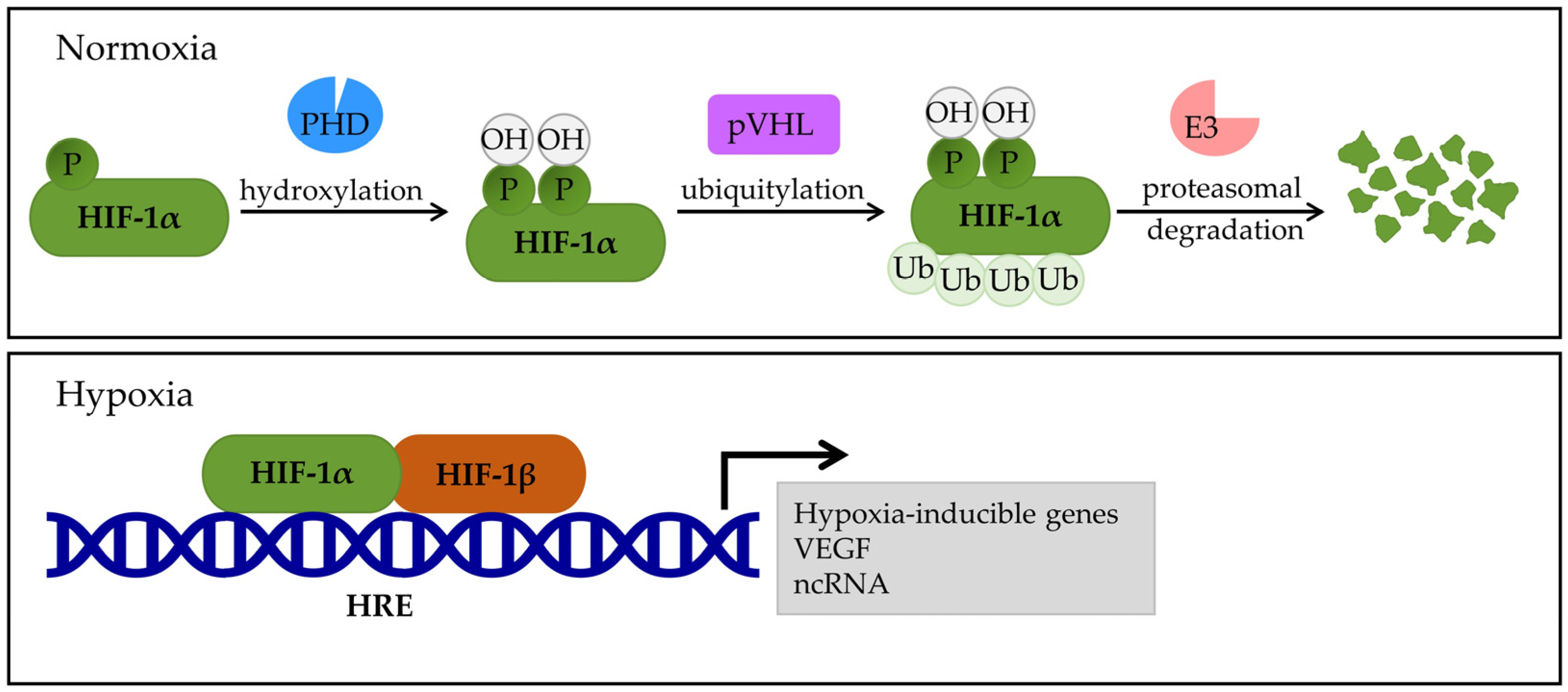
3.1. HIF-1 in Hypoxic vs. Normoxic Environments
3.2. HIF and Cell Signaling Pathways
4. HIF-1α and Autoimmune Diseases
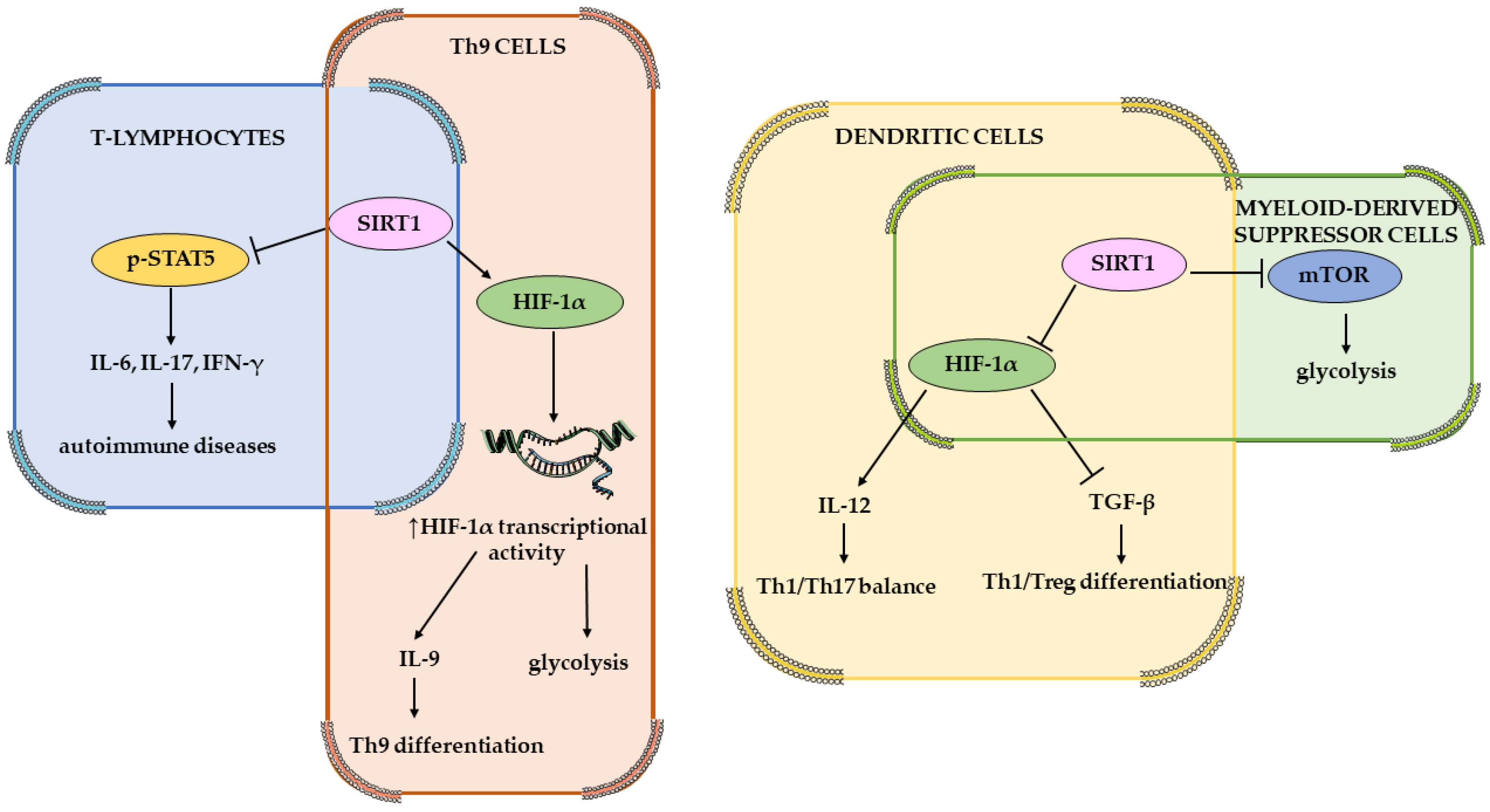
4.1. HIF-1α Effect on Immune Cells
4.2. Single-Nucleotide Polymorphisms and Autoimmune Diseases
5. Hashimoto’s Thyroiditis
5.1. Pathophysiology of HT
5.2. Hashimoto’s Encephalopathy
5.3. Role of HIF-1α in Hashimoto Thyroiditis
5.4. HIF-1α as a Therapeutic Target
6. Limitation of Current Evidence and Future Research Directions
7. Conclusions
Author Contributions
Funding
Institutional Review Board Statement
Informed Consent Statement
Data Availability Statement
Conflicts of Interest
References
- Danieli, M.G.; Casciaro, M.; Paladini, A.; Bartolucci, M.; Sordoni, M.; Shoenfeld, Y.; Gangemi, S. Exposome: Epigenetics and Autoimmune Diseases. Autoimmun. Rev. 2024, 23, 103584. [Google Scholar] [CrossRef]
- Dumas, G.; Arabi, Y.M.; Bartz, R.; Ranzani, O.; Scheibe, F.; Darmon, M.; Helms, J. Diagnosis and Management of Autoimmune Diseases in the ICU. Intensive Care Med. 2024, 50, 17–35. [Google Scholar] [CrossRef] [PubMed]
- Classification of Autoimmune Diseases-Autoimmune Disease|Johns Hopkins Pathology. Available online: https://pathology.jhu.edu/autoimmune/classification (accessed on 18 December 2024).
- A-Z Autoimmune Disease List|Global Autoimmune Institute. Available online: https://www.autoimmuneinstitute.org/resources/autoimmune-disease-list/ (accessed on 18 December 2024).
- Miller, F.W. The Increasing Prevalence of Autoimmunity and Autoimmune Diseases: An Urgent Call to Action for Improved Understanding, Diagnosis, Treatment and Prevention. Curr. Opin. Immunol. 2023, 80, 102266. [Google Scholar] [CrossRef] [PubMed]
- Ralli, M.; Angeletti, D.; Fiore, M.; D’Aguanno, V.; Lambiase, A.; Artico, M.; de Vincentiis, M.; Greco, A. Hashimoto’s Thyroiditis: An Update on Pathogenic Mechanisms, Diagnostic Protocols, Therapeutic Strategies, and Potential Malignant Transformation. Autoimmun. Rev. 2020, 19, 102649. [Google Scholar] [CrossRef]
- Jonklaas, J. Optimal Thyroid Hormone Replacement. Endocr. Rev. 2021, 43, 366–404. [Google Scholar] [CrossRef]
- Hu, X.; Chen, Y.; Shen, Y.; Tian, R.; Sheng, Y.; Que, H. Global Prevalence and Epidemiological Trends of Hashimoto’s Thyroiditis in Adults: A Systematic Review and Meta-Analysis. Front. Public Health 2022, 10, 1020709. [Google Scholar] [CrossRef]
- Tang, Y.-Y.; Wang, D.-C.; Wang, Y.-Q.; Huang, A.-F.; Xu, W.-D. Emerging Role of Hypoxia-Inducible Factor-1α in Inflammatory Autoimmune Diseases: A Comprehensive Review. Front. Immunol. 2023, 13, 1073971. [Google Scholar] [CrossRef] [PubMed]
- Zhang, F.; Zhang, Y.; Zhou, J.; Cai, Y.; Li, Z.; Sun, J.; Xie, Z.; Hao, G. Metabolic Effects of Quercetin on Inflammatory and Autoimmune Responses in Rheumatoid Arthritis Are Mediated through the Inhibition of JAK1/STAT3/HIF-1α Signaling. Mol. Med. 2024, 30, 170. [Google Scholar] [CrossRef]
- Guo, X.; Chen, G. Hypoxia-Inducible Factor Is Critical for Pathogenesis and Regulation of Immune Cell Functions in Rheumatoid Arthritis. Front. Immunol. 2020, 11, 1668. [Google Scholar] [CrossRef]
- Chen, S.; Duan, Y.; Zhang, Y.; Cheng, L.; Cai, L.; Hou, X.; Li, W. Associations Between Single Nucleotide Polymorphisms of Hypoxia-Related Genes and Capsule Formation in Hepatocellular Carcinoma. J. Hepatocell. Carcinoma 2023, 10, 1785–1797. [Google Scholar] [CrossRef]
- Chervona, Y.; Costa, M. The Control of Histone Methylation and Gene Expression by Oxidative Stress, Hypoxia, and Metals. Free Radic. Biol. Med. 2012, 53, 1041–1047. [Google Scholar] [CrossRef]
- Škrlec, I.; Kolomeichuk, S.N. Hypoxia-Inducible Factor-1α in Myocardial Infarction. World J. Cardiol. 2024, 16, 181–185. [Google Scholar] [CrossRef] [PubMed]
- Gladek, I.; Ferdin, J.; Horvat, S.; Calin, G.; Kunej, T. HIF1A Gene Polymorphisms and Human Diseases: Graphical Review of 97 Association Studies. Genes. Chromosomes Cancer 2017, 56, 439–452. [Google Scholar] [CrossRef]
- Zimna, A.; Kurpisz, M. Hypoxia-Inducible Factor-1 in Physiological and Pathophysiological Angiogenesis: Applications and Therapies. Biomed. Res. Int. 2015, 2015, 549412. [Google Scholar] [CrossRef] [PubMed]
- Li, H.; Wu, Q.-Y.; Teng, X.-H.; Li, Z.-P.; Zhu, M.-T.; Gu, C.-J.; Chen, B.-J.; Xie, Q.-Q.; LuO, X.-J. The Pathogenesis and Regulatory Role of HIF-1 in Rheumatoid Arthritis. Cent. Eur. J. Immunol. 2023, 48, 338–345. [Google Scholar] [CrossRef]
- Pezzuto, A.; Carico, E. Role of HIF-1 in Cancer Progression: Novel Insights. A Review. Curr. Mol. Med. 2018, 18, 343–351. [Google Scholar] [CrossRef]
- Jaskiewicz, M.; Moszynska, A.; Serocki, M.; Króliczewski, J.; Bartoszewska, S.; Collawn, J.F.; Bartoszewski, R. Hypoxia-Inducible Factor (HIF)-3a2 Serves as an Endothelial Cell Fate Executor during Chronic Hypoxia. EXCLI J. 2022, 21, 454–469. [Google Scholar] [CrossRef]
- McGettrick, A.F.; O’Neill, L.A.J. The Role of HIF in Immunity and Inflammation. Cell Metab. 2020, 32, 524–536. [Google Scholar] [CrossRef]
- Kolonko, M.; Greb-Markiewicz, B. bHLH–PAS Proteins: Their Structure and Intrinsic Disorder. Int. J. Mol. Sci. 2019, 20, 3653. [Google Scholar] [CrossRef]
- Daffern, N.; Radhakrishnan, I. Per-ARNT-Sim (PAS) Domains in Basic Helix-Loop-Helix (bHLH)-PAS Transcription Factors and Coactivators: Structures and Mechanisms. J. Mol. Biol. 2024, 436, 168370. [Google Scholar] [CrossRef] [PubMed]
- He, W.; Batty-Stuart, S.; Lee, J.E.; Ohh, M. HIF-1α Hydroxyprolines Modulate Oxygen-Dependent Protein Stability Via Single VHL Interface with Comparable Effect on Ubiquitination Rate. J. Mol. Biol. 2021, 433, 167244. [Google Scholar] [CrossRef]
- Albanese, A.; Daly, L.A.; Mennerich, D.; Kietzmann, T.; Sée, V. The Role of Hypoxia-Inducible Factor Post-Translational Modifications in Regulating Its Localisation, Stability, and Activity. Int. J. Mol. Sci. 2021, 22, 268. [Google Scholar] [CrossRef]
- Zhang, J.; Yao, M.; Xia, S.; Zeng, F.; Liu, Q. Systematic and Comprehensive Insights into HIF-1 Stabilization under Normoxic Conditions: Implications for Cellular Adaptation and Therapeutic Strategies in Cancer. Cell. Mol. Biol. Lett. 2025, 30, 2. [Google Scholar] [CrossRef]
- Islam, S.M.T.; Won, J.; Khan, M.; Mannie, M.D.; Singh, I. Hypoxia-inducible Factor-1 Drives Divergent Immunomodulatory Functions in the Pathogenesis of Autoimmune Diseases. Immunology 2021, 164, 31–42. [Google Scholar] [CrossRef]
- Gong, X.; Yang, S.-Y.; Wang, Z.-Y.; Tang, M. The Role of Hypoxic Microenvironment in Autoimmune Diseases. Front. Immunol. 2024, 15, 1435306. [Google Scholar] [CrossRef] [PubMed]
- Tian, Y.; Zhao, L.; Gui, Z.; Liu, S.; Liu, C.; Yu, T.; Zhang, L. PI3K/AKT Signaling Activates HIF1α to Modulate the Biological Effects of Invasive Breast Cancer with Microcalcification. npj Breast Cancer 2023, 9, 93. [Google Scholar] [CrossRef]
- Pan, T.; Sun, S.; Chen, Y.; Tian, R.; Chen, E.; Tan, R.; Wang, X.; Liu, Z.; Liu, J.; Qu, H. Immune Effects of PI3K/Akt/HIF-1α-Regulated Glycolysis in Polymorphonuclear Neutrophils during Sepsis. Crit. Care 2022, 26, 29. [Google Scholar] [CrossRef]
- Oliver, K.M.; Garvey, J.F.; Ng, C.T.; Veale, D.J.; Fearon, U.; Cummins, E.P.; Taylor, C.T. Hypoxia Activates NF-kappaB-Dependent Gene Expression through the Canonical Signaling Pathway. Antioxid. Redox Signal 2009, 11, 2057–2064. [Google Scholar] [CrossRef] [PubMed]
- Peyssonnaux, C.; Cejudo-Martin, P.; Doedens, A.; Zinkernagel, A.S.; Johnson, R.S.; Nizet, V. Cutting Edge: Essential Role of Hypoxia Inducible Factor-1alpha in Development of Lipopolysaccharide-Induced Sepsis. J. Immunol. 2007, 178, 7516–7519. [Google Scholar] [CrossRef]
- Pawlus, M.; Wang, L.; Hu, C.-J. STAT3 and HIF1α Cooperatively Activate HIF1 Target Genes in MDA-MB-231 and RCC4 Cells. Oncogene 2014, 33, 1670–1679. [Google Scholar] [CrossRef] [PubMed]
- Dinarello, A.; Betto, R.M.; Diamante, L.; Tesoriere, A.; Ghirardo, R.; Cioccarelli, C.; Meneghetti, G.; Peron, M.; Laquatra, C.; Tiso, N.; et al. STAT3 and HIF1α Cooperatively Mediate the Transcriptional and Physiological Responses to Hypoxia. Cell Death Discov. 2023, 9, 226. [Google Scholar] [CrossRef]
- Cheng, Y.-L.; Park, J.-S.; Manzanero, S.; Choi, Y.; Baik, S.-H.; Okun, E.; Gelderblom, M.; Fann, D.Y.-W.; Magnus, T.; Launikonis, B.S.; et al. Evidence That Collaboration between HIF-1α and Notch-1 Promotes Neuronal Cell Death in Ischemic Stroke. Neurobiol. Dis. 2014, 62, 286–295. [Google Scholar] [CrossRef] [PubMed]
- Vitale, C.; Griggio, V.; Riganti, C.; Todaro, M.; Kopecka, J.; Jones, R.; Salvetti, C.; Boccellato, E.; Perutelli, F.; Voena, C.; et al. Targeting HIF-1α Regulatory Pathways as a Strategy to Hamper Tumor-Microenvironment Interactions in CLL. Cancers 2021, 13, 2883. [Google Scholar] [CrossRef]
- Strickland, J.; Garrison, D.; Copple, B.L. Hypoxia Upregulates Cxcl12 in Hepatocytes by a Complex Mechanism Involving Hypoxia-Inducible Factors and Transforming Growth Factor-β. Cytokine 2020, 127, 154986. [Google Scholar] [CrossRef]
- Zhao, L.; Wu, Q.; Wang, X.; Wang, S.; Shi, X.; Shan, Z.; Teng, W. Reversal of Abnormal CD4+ T Cell Metabolism Alleviates Thyroiditis by Deactivating the mTOR/HIF1a/Glycolysis Pathway. Front. Endocrinol. 2021, 12, 659738. [Google Scholar] [CrossRef]
- Kiliç, Y.; Guzel Erdogan, D.; Baykul, M.; Nas, K. Examining the Functions of the Vascular Endothelial Growth Factor/Hypoxia-Inducible Factor Signaling Pathway in Psoriatic Arthritis. Arch. Rheumatol. 2023, 38, 579–589. [Google Scholar] [CrossRef]
- Li, Y.; Chen, S.; Yang, Q.; Liu, X.; Zhou, W.; Kang, T.; Wu, W.; Ou, S. The ANGPTL4-HIF-1α Loop: A Critical Reg-ulator of Renal Interstitial Fibrosis. J. Transl. Med. 2024, 22, 649. [Google Scholar] [CrossRef]
- Saravani, M.; Sandoughi, M.; Heidary, Z.; Ebrahimi, G.; Mirzamohammadi, S.; Haddadi, M.; Nematollahi, M.H. HIF-1α and VEGF Polymorphisms and Systemic Lupus Erythematosus Susceptibility. Meta Gene 2021, 30, 100982. [Google Scholar] [CrossRef]
- Qi, X.; Bie, M.; Jiang, R.; Kang, F. HIF-1α Regulates Osteoclastogenesis and Alveolar Bone Resorption in Periodontitis via ANGPTL4. Arch. Oral. Biol. 2023, 153, 105736. [Google Scholar] [CrossRef] [PubMed]
- He, Y.; Yang, W.; Gan, L.; Liu, S.; Ni, Q.; Bi, Y.; Han, T.; Liu, Q.; Chen, H.; Hu, Y.; et al. Silencing HIF-1α Aggravates Non-Alcoholic Fatty Liver Disease in Vitro through Inhibiting PPAR-α/ANGPTL4 Singling Pathway. Gastroenterol. Hepatol. 2021, 44, 355–365. [Google Scholar] [CrossRef]
- Zhu, J.; Tang, Y.; Wu, Q.; Ji, Y.-C.; Feng, Z.-F.; Kang, F.-W. HIF-1α Facilitates Osteocyte-Mediated Osteoclastogenesis by Activating JAK2/STAT3 Pathway in Vitro. J. Cell Physiol. 2019, 234, 21182–21192. [Google Scholar] [CrossRef]
- Wang, D.; Liu, L.; Qu, Z.; Zhang, B.; Gao, X.; Huang, W.; Feng, M.; Gong, Y.; Kong, L.; Wang, Y.; et al. Hypoxia-Inducible Factor 1α Enhances RANKL-Induced Osteoclast Differentiation by Upregulating the MAPK Pathway. Ann. Transl. Med. 2022, 10, 1227. [Google Scholar] [CrossRef]
- Hepp, M.; Werion, A.; De Greef, A.; de Ville de Goyet, C.; de Bournonville, M.; Behets, C.; Lengelé, B.; Daumerie, C.; Mourad, M.; Ludgate, M.; et al. Oxidative Stress-Induced Sirtuin1 Downregulation Correlates to HIF-1α, GLUT-1, and VEGF-A Upregulation in Th1 Autoimmune Hashimoto’s Thyroiditis. Int. J. Mol. Sci. 2021, 22, 3806. [Google Scholar] [CrossRef] [PubMed]
- Ryu, D.R.; Yu, M.R.; Kong, K.H.; Kim, H.; Kwon, S.H.; Jeon, J.S.; Han, D.C.; Noh, H. Sirt1-Hypoxia-Inducible Factor-1α Interaction Is a Key Mediator of Tubulointerstitial Damage in the Aged Kidney. Aging Cell 2019, 18, e12904. [Google Scholar] [CrossRef] [PubMed]
- Li, M.; Ying, M.; Gu, S.; Zhou, Z.; Zhao, R. SIRT6 Inhibits Hypoxia-Induced Pulmonary Arterial Smooth Muscle Cells Proliferation via HIF-1α/PDK4 Signaling. Life Sci. 2023, 312, 121192. [Google Scholar] [CrossRef] [PubMed]
- You, Q.; Wang, J.; Yu, Y.; Li, F.; Meng, L.; Chen, M.; Yang, Q.; Xu, Z.; Sun, J.; Zhuo, W.; et al. The Histone Deacetylase SIRT6 Promotes Glycolysis through the HIF-1α/HK2 Signaling Axis and Induces Erlotinib Resistance in Non-Small Cell Lung Cancer. Apoptosis 2022, 27, 883–898. [Google Scholar] [CrossRef]
- Chen, X.; Zhao, J.; Herjan, T.; Hong, L.; Liao, Y.; Liu, C.; Vasu, K.; Wang, H.; Thompson, A.; Fox, P.L.; et al. IL-17-Induced HIF1α Drives Resistance to Anti-PD-L1 via Fibroblast-Mediated Immune Exclusion. J. Exp. Med. 2022, 219, e20210693. [Google Scholar] [CrossRef]
- Subudhi, I.; Konieczny, P.; Prystupa, A.; Castillo, R.L.; Sze-Tu, E.; Xing, Y.; Rosenblum, D.; Reznikov, I.; Sidhu, I.; Loomis, C.; et al. Metabolic Coordination between Skin Epithelium and Type 17 Immunity Sustains Chronic Skin Inflammation. Immunity 2024, 57, 1665–1680.e7. [Google Scholar] [CrossRef]
- Gao, Y.; Cai, L.; Wu, Y.; Jiang, M.; Zhang, Y.; Ren, W.; Song, Y.; Li, L.; Lei, Z.; Wu, Y.; et al. Emerging Functions and Therapeutic Targets of IL-38 in Central Nervous System Diseases. CNS Neurosci. Ther. 2024, 30, e14550. [Google Scholar] [CrossRef]
- Gkotinakou, I.-M.; Befani, C.; Simos, G.; Liakos, P. ERK1/2 Phosphorylates HIF-2α and Regulates Its Activity by Controlling Its CRM1-Dependent Nuclear Shuttling. J. Cell Sci. 2019, 132, jcs225698. [Google Scholar] [CrossRef]
- Koukoulas, K.; Giakountis, A.; Karagiota, A.; Samiotaki, M.; Panayotou, G.; Simos, G.; Mylonis, I. ERK Signaling Controls Productive HIF-1 Binding to Chromatin and Cancer Cell Adaptation to Hypoxia through HIF-1α Interaction with NPM1. Mol. Oncol. 2021, 15, 3468–3489. [Google Scholar] [CrossRef]
- Dodd, K.M.; Yang, J.; Shen, M.H.; Sampson, J.R.; Tee, A.R. mTORC1 Drives HIF-1α and VEGF-A Signalling via Multiple Mechanisms Involving 4E-BP1, S6K1 and STAT3. Oncogene 2015, 34, 2239–2250. [Google Scholar] [CrossRef]
- Li, J.; Quan, C.; He, Y.-L.; Cao, Y.; Chen, Y.; Wang, Y.-F.; Wu, L.-Y. Autophagy Regulated by the HIF/REDD1/mTORC1 Signaling Is Progressively Increased during Erythroid Differentiation under Hypoxia. Front. Cell Dev. Biol. 2022, 10, 896893. [Google Scholar] [CrossRef] [PubMed]
- Huang, Y.; Chen, Z.; Lu, T.; Bi, G.; Li, M.; Liang, J.; Hu, Z.; Zheng, Y.; Yin, J.; Xi, J.; et al. HIF-1α Switches the Functionality of TGF-β Signaling via Changing the Partners of Smads to Drive Glucose Metabolic Reprogramming in Non-Small Cell Lung Cancer. J. Exp. Clin. Cancer Res. 2021, 40, 398. [Google Scholar] [CrossRef] [PubMed]
- Selnø, A.T.H.; Schlichtner, S.; Yasinska, I.M.; Sakhnevych, S.S.; Fiedler, W.; Wellbrock, J.; Klenova, E.; Pavlova, L.; Gibbs, B.F.; Degen, M.; et al. Transforming Growth Factor Beta Type 1 (TGF-β) and Hypoxia-Inducible Factor 1 (HIF-1) Transcription Complex as Master Regulators of the Immunosuppressive Protein Galectin-9 Expression in Human Cancer and Embryonic Cells. Aging 2020, 12, 23478–23496. [Google Scholar] [CrossRef]
- Kokeza, J.; Strikic, A.; Ogorevc, M.; Kelam, N.; Vukoja, M.; Dilber, I.; Zekic Tomas, S. The Effect of GLUT1 and HIF-1α Expressions on Glucose Uptake and Patient Survival in Non-Small-Cell Lung Carcinoma. Int. J. Mol. Sci. 2023, 24, 10575. [Google Scholar] [CrossRef]
- Yi, H.; Han, Y.; Li, Q.; Lin, R.; Zhang, J.; Yang, Y.; Wang, X.; Zhang, L. Prognostic Impact of the Combination of HIF-1α and GLUT1 in Patients with Oesophageal Squamous Cell Carcinoma. Oncol. Lett. 2023, 26, 404. [Google Scholar] [CrossRef]
- Hsu, T.-S.; Lai, M.-Z. Hypoxia-Inducible Factor 1α Plays a Predominantly Negative Role in Regulatory T Cell Functions. J. Leukoc. Biol. 2018, 104, 911–918. [Google Scholar] [CrossRef]
- Zhang, M.; Wu, D.; Xu, J.; Liu, L.; Jiao, W.; Yu, J.; Chen, G. Suppression of NLRP3 Inflammasome by Dihydroarteannuin via the HIF-1α and JAK3/STAT3 Signaling Pathway Contributes to Attenuation of Collagen-Induced Arthritis in Mice. Front. Pharmacol. 2022, 13, 884881. [Google Scholar] [CrossRef] [PubMed]
- Phenekos, C.; Vryonidou, A.; Gritzapis, A.D.; Baxevanis, C.N.; Goula, M.; Papamichail, M. Th1 and Th2 Serum Cytokine Profiles Characterize Patients with Hashimoto’s Thyroiditis (Th1) and Graves’ Disease (Th2). Neuroimmunomodulation 2004, 11, 209–213. [Google Scholar] [CrossRef]
- Joo, H.-Y.; Yun, M.; Jeong, J.; Park, E.-R.; Shin, H.-J.; Woo, S.R.; Jung, J.K.; Kim, Y.-M.; Park, J.-J.; Kim, J.; et al. SIRT1 Deacetylates and Stabilizes Hypoxia-Inducible Factor-1α (HIF-1α) via Direct Interactions during Hypoxia. Biochem. Biophys. Res. Commun. 2015, 462, 294–300. [Google Scholar] [CrossRef]
- Bettelli, E.; Carrier, Y.; Gao, W.; Korn, T.; Strom, T.B.; Oukka, M.; Weiner, H.L.; Kuchroo, V.K. Reciprocal Developmental Pathways for the Generation of Pathogenic Effector TH17 and Regulatory T Cells. Nature 2006, 441, 235–238. [Google Scholar] [CrossRef]
- Chen, X.; Lu, Y.; Zhang, Z.; Wang, J.; Yang, H.; Liu, G. Intercellular Interplay between Sirt1 Signalling and Cell Metabolism in Immune Cell Biology. Immunology 2015, 145, 455–467. [Google Scholar] [CrossRef] [PubMed]
- Yu, Q.; Dong, L.; Li, Y.; Liu, G. SIRT1 and HIF1α Signaling in Metabolism and Immune Responses. Cancer Lett. 2018, 418, 20–26. [Google Scholar] [CrossRef] [PubMed]
- Liu, G.; Bi, Y.; Shen, B.; Yang, H.; Zhang, Y.; Wang, X.; Liu, H.; Lu, Y.; Liao, J.; Chen, X.; et al. SIRT1 Limits the Function and Fate of Myeloid-Derived Suppressor Cells in Tumors by Orchestrating HIF-1α-Dependent Glycolysis. Cancer Res. 2014, 74, 727–737. [Google Scholar] [CrossRef] [PubMed]
- Liu, G.; Bi, Y.; Xue, L.; Zhang, Y.; Yang, H.; Chen, X.; Lu, Y.; Zhang, Z.; Liu, H.; Wang, X.; et al. Dendritic Cell SIRT1-HIF1α Axis Programs the Differentiation of CD4+ T Cells through IL-12 and TGF-Β1. Proc. Natl. Acad. Sci. USA 2015, 112, E957–E965. [Google Scholar] [CrossRef]
- Sarumaru, M.; Watanabe, M.; Inoue, N.; Hisamoto, Y.; Morita, E.; Arakawa, Y.; Hidaka, Y.; Iwatani, Y. Association between Functional SIRT1 Polymorphisms and the Clinical Characteristics of Patients with Autoimmune Thyroid Disease. Autoimmunity 2016, 49, 329–337. [Google Scholar] [CrossRef]
- Camagni, G.F.; Minervini, G.; Tosatto, S.C.E. Structural Characterization of Hypoxia Inducible Factor α—Prolyl Hydroxylase Domain 2 Interaction through MD Simulations. Int. J. Mol. Sci. 2023, 24, 4710. [Google Scholar] [CrossRef]
- Ding, M.; Guan, T.-J.; Wei, C.-Y.; Chen, B.-H. Identification of Pathways Significantly Associated with Spondyloarthropathy/Ankylosing Spondylitis Using the Sub-Pathway Method. Mol. Med. Rep. 2018, 18, 3825–3833. [Google Scholar] [CrossRef]
- Jiang, J.; Zhan, X.; Qu, H.; Liang, T.; Li, H.; Chen, L.; Huang, S.; Sun, X.; Jiang, W.; Chen, J.; et al. Upregulated of ANXA3, SORL1, and Neutrophils May Be Key Factors in the Progressionof Ankylosing Spondylitis. Front. Immunol. 2022, 13, 861459. [Google Scholar] [CrossRef]
- Wong, W.J.; Richardson, T.; Seykora, J.T.; Cotsarelis, G.; Simon, M.C. Hypoxia Inducible Factors Regulate Filaggrin Expression and Epidermal Barrier Function. J. Invest. Dermatol. 2015, 135, 454–461. [Google Scholar] [CrossRef]
- Gwon, M.-G.; Leem, J.; An, H.-J.; Gu, H.; Bae, S.; Kim, J.H.; Park, K.-K. The Decoy Oligodeoxynucleotide against HIF-1α and STAT5 Ameliorates Atopic Dermatitis-like Mouse Model. Mol. Ther.-Nucleic Acids 2023, 34, 102036. [Google Scholar] [CrossRef]
- Xie, A.; Robles, R.J.; Mukherjee, S.; Zhang, H.; Feldbrügge, L.; Csizmadia, E.; Wu, Y.; Enjyoji, K.; Moss, A.C.; Otterbein, L.E.; et al. HIF-1α-Induced Xenobiotic Transporters Promote Th17 Responses in Crohn’s Disease. J. Autoimmun. 2018, 94, 122–133. [Google Scholar] [CrossRef]
- De Luna, N.; Suárez-Calvet, X.; Lleixà, C.; Diaz-Manera, J.; Olivé, M.; Illa, I.; Gallardo, E. Hypoxia Triggers IFN-I Production in Muscle: Implications in Dermatomyositis. Sci. Rep. 2017, 7, 8595. [Google Scholar] [CrossRef] [PubMed]
- Preuße, C.; Allenbach, Y.; Hoffmann, O.; Goebel, H.-H.; Pehl, D.; Radke, J.; Doeser, A.; Schneider, U.; Alten, R.H.E.; Kallinich, T.; et al. Differential Roles of Hypoxia and Innate Immunity in Juvenile and Adult Dermatomyositis. Acta Neuropathol. Commun. 2016, 4, 45. [Google Scholar] [CrossRef] [PubMed]
- Görtz, G.-E.; Horstmann, M.; Aniol, B.; Reyes, B.D.; Fandrey, J.; Eckstein, A.; Berchner-Pfannschmidt, U. Hypoxia-Dependent HIF-1 Activation Impacts on Tissue Remodeling in Graves’ Ophthalmopathy—Implications for Smoking. J. Clin. Endocrinol. Metab. 2016, 101, 4834–4842. [Google Scholar] [CrossRef]
- Chng, C.L.; Lai, O.F.; Chew, C.S.-M.; Peh, Y.P.; Fook-Chong, S.M.-C.; Seah, L.L.; Khoo, D.H.-C. Hypoxia Increases Adipogenesis and Affects Adipocytokine Production in Orbital Fibroblasts–a Possible Explanation of the Link between Smoking and Graves’ Ophthalmopathy. Int. J. Ophthalmol. 2014, 7, 403–407. [Google Scholar] [CrossRef]
- Zhang, L.; Sun, X.; Liu, L.; Wang, P.; Qian, L. Excessive Iodine Induces Thyroid Follicular Epithelial Cells Apoptosis by Activating HIF-1α-Mediated Hypoxia Pathway in Hashimoto Thyroiditis. Mol. Biol. Rep. 2023, 50, 3633–3640. [Google Scholar] [CrossRef]
- Robinson, A.; Keely, S.; Karhausen, J.; Gerich, M.E.; Furuta, G.T.; Colgan, S.P. Mucosal Protection by Hypoxia-Inducible Factor Prolyl Hydroxylase Inhibition. Gastroenterology 2008, 134, 145–155. [Google Scholar] [CrossRef]
- Keely, S.; Glover, L.E.; MacManus, C.F.; Campbell, E.L.; Scully, M.M.; Furuta, G.T.; Colgan, S.P. Selective Induction of Integrin Beta1 by Hypoxia-Inducible Factor: Implications for Wound Healing. FASEB J. 2009, 23, 1338–1346. [Google Scholar] [CrossRef] [PubMed]
- Yu, L.; Ran, H.; Lu, Y.; Ma, Q.; Huang, H.; Liu, W. Targeting HIF-1α Alleviates the Inflammatory Responses and Rebuilds the CD4+ T Cell Subsets Balance in the Experimental Autoimmune Myasthenia Gravis Inflammation Model via Regulating Cellular and Humoral Immunity. Life Sci. 2024, 336, 122287. [Google Scholar] [CrossRef]
- Altınönder, İ.; Kaya, M.; Yentür, S.P.; Çakar, A.; Durmuş, H.; Yegen, G.; Özkan, B.; Parman, Y.; Sawalha, A.H.; Saruhan-Direskeneli, G. Thymic Gene Expression Analysis Reveals a Potential Link between HIF-1A and Th17/Treg Imbalance in Thymoma Associated Myasthenia Gravis. J. Neuroinflamm. 2024, 21, 126. [Google Scholar] [CrossRef] [PubMed]
- Davies, A.L.; Desai, R.A.; Bloomfield, P.S.; McIntosh, P.R.; Chapple, K.J.; Linington, C.; Fairless, R.; Diem, R.; Kasti, M.; Murphy, M.P.; et al. Neurological Deficits Caused by Tissue Hypoxia in Neuroinflammatory Disease. Ann. Neurol. 2013, 74, 815–825. [Google Scholar] [CrossRef] [PubMed]
- Aboul-Enein, F.; Rauschka, H.; Kornek, B.; Stadelmann, C.; Stefferl, A.; Brück, W.; Lucchinetti, C.; Schmidbauer, M.; Jellinger, K.; Lassmann, H. Preferential Loss of Myelin-Associated Glycoprotein Reflects Hypoxia-like White Matter Damage in Stroke and Inflammatory Brain Diseases. J. Neuropathol. Exp. Neurol. 2003, 62, 25–33. [Google Scholar] [CrossRef] [PubMed]
- Seo, Y.; Ji, Y.W.; Lee, S.M.; Shim, J.; Noh, H.; Yeo, A.; Park, C.; Park, M.S.; Chang, E.J.; Lee, H.K. Activation of HIF-1α (Hypoxia Inducible Factor-1α) Prevents Dry Eye-Induced Acinar Cell Death in the Lacrimal Gland. Cell Death Dis. 2014, 5, e1309. [Google Scholar] [CrossRef]
- Hu, F.; Shi, L.; Mu, R.; Zhu, J.; Li, Y.; Ma, X.; Li, C.; Jia, R.; Yang, D.; Li, Y.; et al. Hypoxia-Inducible Factor-1α and Interleukin 33 Form a Regulatory Circuit to Perpetuate the Inflammation in Rheumatoid Arthritis. PLoS ONE 2013, 8, e72650. [Google Scholar] [CrossRef]
- Chen, X.; Jiang, S.; Zhou, Z.; Xu, X.; Ying, S.; Du, L.; Qiu, K.; Xu, Y.; Wu, J.; Wang, X. Increased Expression of Interleukin-21-Inducible Genes in Minor Salivary Glands Are Associated with Primary Sjögren’s Syndrome Disease Characteristics. Rheumatology 2021, 60, 2979–2989. [Google Scholar] [CrossRef]
- Hernández-Molina, G.; Rodríguez-Pérez, J.M.; Fernández-Torres, J.; Lima, G.; Pérez-Hernández, N.; López-Reyes, A.; Martínez-Nava, G.A. HIF1A (Rs11549465) and AKNA (Rs10817595) Gene Polymorphisms Are Associated with Primary Sjögren’s Syndrome. Biomed. Res. Int. 2017, 2017, 5845849. [Google Scholar] [CrossRef]
- Min, J.H.; Lee, C.H.; Ji, Y.W.; Yeo, A.; Noh, H.; Song, I.; Kim, E.K.; Lee, H.K. Activation of Dll4/Notch Signaling and Hypoxia-Inducible Factor-1 Alpha Facilitates Lymphangiogenesis in Lacrimal Glands in Dry Eye. PLoS ONE 2016, 11, e0147846. [Google Scholar] [CrossRef]
- Liao, H.-J.; Chu, C.-L.; Wang, S.-C.; Lee, H.-Y.; Wu, C.-S. Increased HIF-1α Expression in T Cells and Associated with Enhanced Th17 Pathway in Systemic Lupus Erythematosus. J. Formos. Med. Assoc. 2022, 121, 2446–2456. [Google Scholar] [CrossRef]
- Little, A.J.; Chen, P.-M.; Vesely, M.D.; Khan, R.N.; Fiedler, J.; Garritano, J.; Maisha, F.I.; McNiff, J.M.; Craft, J. HIF-1 Regulates Pathogenic Cytotoxic T Cells in Lupus Skin Disease. JCI Insight 2023, 8, e166076. [Google Scholar] [CrossRef]
- Cheng, K.; Ho, K.; Stokes, R.; Scott, C.; Lau, S.M.; Hawthorne, W.J.; O’Connell, P.J.; Loudovaris, T.; Kay, T.W.; Kulkarni, R.N.; et al. Hypoxia-Inducible Factor-1alpha Regulates Beta Cell Function in Mouse and Human Islets. J. Clin. Invest. 2010, 120, 2171–2183. [Google Scholar] [CrossRef]
- Catrina, S.-B.; Okamoto, K.; Pereira, T.; Brismar, K.; Poellinger, L. Hyperglycemia Regulates Hypoxia-Inducible Factor-1alpha Protein Stability and Function. Diabetes 2004, 53, 3226–3232. [Google Scholar] [CrossRef]
- Botusan, I.R.; Sunkari, V.G.; Savu, O.; Catrina, A.I.; Grünler, J.; Lindberg, S.; Pereira, T.; Ylä-Herttuala, S.; Poellinger, L.; Brismar, K.; et al. Stabilization of HIF-1alpha Is Critical to Improve Wound Healing in Diabetic Mice. Proc. Natl. Acad. Sci. USA 2008, 105, 19426–19431. [Google Scholar] [CrossRef] [PubMed]
- Capone, A.; Volpe, E. Transcriptional Regulators of T Helper 17 Cell Differentiation in Health and Autoimmune Diseases. Front. Immunol. 2020, 11, 348. [Google Scholar] [CrossRef]
- Charles, A.; Janeway, J.; Travers, P.; Walport, M.; Shlomchik, M.J. Autoimmune Responses Are Directed against Self Antigens. In Immunobiology: The Immune System in Health and Disease, 5th ed.; Garland Science: New York, NY, USA, 2001. [Google Scholar]
- Zambrano-Zaragoza, J.F.; Romo-Martínez, E.J.; Durán-Avelar, M.d.J.; García-Magallanes, N.; Vibanco-Pérez, N. Th17 Cells in Autoimmune and Infectious Diseases. Int. J. Inflam. 2014, 2014, 651503. [Google Scholar] [CrossRef] [PubMed]
- Goswami, T.K.; Singh, M.; Dhawan, M.; Mitra, S.; Emran, T.B.; Rabaan, A.A.; Mutair, A.A.; Alawi, Z.A.; Alhumaid, S.; Dhama, K. Regulatory T Cells (Tregs) and Their Therapeutic Potential against Autoimmune Disorders-Advances and Challenges. Hum. Vaccin. Immunother. 2022, 18, 2035117. [Google Scholar] [CrossRef]
- Sun, L.; Su, Y.; Jiao, A.; Wang, X.; Zhang, B. T Cells in Health and Disease. Sig Transduct. Target. Ther. 2023, 8, 235. [Google Scholar] [CrossRef]
- Arias, C.; Sepúlveda, P.; Castillo, R.L.; Salazar, L.A. Relationship between Hypoxic and Immune Pathways Activation in the Progression of Neuroinflammation: Role of HIF-1α and Th17 Cells. Int. J. Mol. Sci. 2023, 24, 3073. [Google Scholar] [CrossRef] [PubMed]
- Goździk, M.; Żelaźniewicz, A.; Nowak-Kornicka, J.; Pawłowska-Seredyńska, K.; Umławska, W.; Pawłowski, B. Autoimmune Hashimoto’s Disease and Feminization Level—Testing the Immunocompetence Hypothesis. Evol. Psychol. 2024, 22, 14747049241259187. [Google Scholar] [CrossRef]
- Salari, N.; Heidarian, P.; Jalili, F.; Babajani, F.; Shohaimi, S.; Nasirian, M.; Mohammadi, M. The Sexual Dysfunction in Women with Thyroid Disorders: A Meta-Analysis. BMC Endocr. Disord. 2024, 24, 279. [Google Scholar] [CrossRef]
- Padmanabhan, R.A.; Johnson, B.S.; Dhyani, A.K.; Pillai, S.M.; Jayakrishnan, K.; Laloraya, M. Autoimmune Regulator (AIRE): Takes a Hypoxia-Inducing Factor 1A (HIF1A) Route to Regulate FOXP3 Expression in PCOS. Am. J. Reprod. Immunol. 2023, 89, e13637. [Google Scholar] [CrossRef] [PubMed]
- Szeliga, A.; Calik-Ksepka, A.; Maciejewska-Jeske, M.; Grymowicz, M.; Smolarczyk, K.; Kostrzak, A.; Smolarczyk, R.; Rudnicka, E.; Meczekalski, B. Autoimmune Diseases in Patients with Premature Ovarian Insufficiency—Our Current State of Knowledge. Int. J. Mol. Sci. 2021, 22, 2594. [Google Scholar] [CrossRef]
- Szczuko, M.; Syrenicz, A.; Szymkowiak, K.; Przybylska, A.; Szczuko, U.; Pobłocki, J.; Kulpa, D. Doubtful Justification of the Gluten-Free Diet in the Course of Hashimoto’s Disease. Nutrients 2022, 14, 1727. [Google Scholar] [CrossRef] [PubMed]
- Betterle, C.; Furmaniak, J.; Sabbadin, C.; Scaroni, C.; Presotto, F. Type 3 Autoimmune Polyglandular Syndrome (APS-3) or Type 3 Multiple Autoimmune Syndrome (MAS-3): An Expanding Galaxy. J. Endocrinol. Invest. 2023, 46, 643–665. [Google Scholar] [CrossRef]
- Corcoran, S.E.; O’Neill, L.A.J. HIF1α and Metabolic Reprogramming in Inflammation. J. Clin. Invest. 2016, 126, 3699–3707. [Google Scholar] [CrossRef] [PubMed]
- Dvornikova, K.A.; Platonova, O.N.; Bystrova, E.Y. Hypoxia and Intestinal Inflammation: Common Molecular Mechanisms and Signaling Pathways. Int. J. Mol. Sci. 2023, 24, 2425. [Google Scholar] [CrossRef]
- Single Nucleotide Polymorphisms (SNPs). Available online: https://www.genome.gov/genetics-glossary/Single-Nucleotide-Polymorphisms-SNPs (accessed on 20 February 2025).
- Definition of SNP-NCI Dictionary of Genetics Terms-NCI. Available online: https://www.cancer.gov/publications/dictionaries/genetics-dictionary/def/snp (accessed on 20 February 2025).
- Harun-Or-Roshid, M.; Ali, M.B.; Jesmin; Mollah, M.N.H. Association of Hypoxia Inducible Factor 1-Alpha Gene Polymorphisms with Multiple Disease Risks: A Comprehensive Meta-Analysis. PLoS ONE 2022, 17, e0273042. [Google Scholar] [CrossRef]
- Huang, Y.; Jin, L.; Yu, H.; Jiang, G.; Tam, C.H.T.; Jiang, S.; Zheng, C.; Jiang, F.; Zhang, R.; Zhang, H.; et al. SNPs in PRKCA-HIF1A-GLUT1 Are Associated with Diabetic Kidney Disease in a Chinese Han Population with Type 2 Diabetes. Eur. J. Clin. Invest. 2020, 50, e13264. [Google Scholar] [CrossRef]
- Wipff, J.; Dieude, P.; Avouac, J.; Tiev, K.; Hachulla, E.; Granel, B.; Diot, E.; Sibilia, J.; Mouthon, L.; Meyer, O.; et al. Association of Hypoxia-Inducible Factor 1A (HIF1A) Gene Polymorphisms with Systemic Sclerosis in a French European Caucasian Population. Scand. J. Rheumatol. 2009, 38, 291–294. [Google Scholar] [CrossRef]
- Maciejewska, M.; Sikora, M.; Stec, A.; Zaremba, M.; Maciejewski, C.; Pawlik, K.; Rudnicka, L. Hypoxia-Inducible Factor-1α (HIF-1α) as a Biomarker for Changes in Microcirculation in Individuals with Systemic Sclerosis. Dermatol. Ther. 2023, 13, 1549–1560. [Google Scholar] [CrossRef]
- Cai, T.; Li, J.; An, X.; Yan, N.; Li, D.; Jiang, Y.; Wang, W.; Shi, L.; Qin, Q.; Song, R.; et al. Polymorphisms in MIR499A and MIR125A Gene Are Associated with Autoimmune Thyroid Diseases. Mol. Cell. Endocrinol. 2017, 440, 106–115. [Google Scholar] [CrossRef]
- Feng, C.-C.; Ye, Q.-L.; Zhu, Y.; Leng, R.-X.; Chen, G.-M.; Yang, J.; Cen, H.; Yang, X.-K.; Li, R.; Xu, W.-D.; et al. Lack of Association between the Polymorphisms of Hypoxia-Inducible Factor 1A (HIF1A) Gene and SLE Susceptibility in a Chinese Population. Immunogenetics 2014, 66, 9–13. [Google Scholar] [CrossRef]
- Urganci, B.E.; Acikbas, I.; Er, F.R. Investigation of Immunovascular Polymorphisms and Intersections in Psoriasis. Indian. J. Dermatol. 2019, 64, 187–191. [Google Scholar] [CrossRef] [PubMed]
- Nagy, G.; Kovacs-Nagy, R.; Kereszturi, E.; Somogyi, A.; Szekely, A.; Nemeth, N.; Hosszufalusi, N.; Panczel, P.; Ronai, Z.; Sasvari-Szekely, M. Association of Hypoxia Inducible Factor-1 Alpha Gene Polymorphism with Both Type 1 and Type 2 Diabetes in a Caucasian (Hungarian) Sample. BMC Med. Genet. 2009, 10, 79. [Google Scholar] [CrossRef]
- Rs11549465 RefSNP Report-dbSNP-NCBI. Available online: https://www.ncbi.nlm.nih.gov/snp/rs11549465#frequency_tab (accessed on 28 November 2025).
- Rs11549467 RefSNP Report-dbSNP-NCBI. Available online: https://www.ncbi.nlm.nih.gov/snp/rs11549467 (accessed on 28 November 2025).
- Li, Q.; Yang, W.; Li, J.; Shan, Z. Emerging Trends and Hot Spots in Autoimmune Thyroiditis Research from 2000 to 2022: A Bibliometric Analysis. Front. Immunol. 2022, 13, 953465. [Google Scholar] [CrossRef]
- Yan, M.; Wu, H.; Zhang, K.; Gong, P.; Wang, Y.; Wei, H. Analysis of the Correlation between Hashimoto’s Thyroiditis and Food Intolerance. Front. Nutr. 2024, 11, 1452371. [Google Scholar] [CrossRef] [PubMed]
- Petranović Ovčariček, P.; Görges, R.; Giovanella, L. Autoimmune Thyroid Diseases. Semin. Nucl. Med. 2024, 54, 219–236. [Google Scholar] [CrossRef] [PubMed]
- Valea, A.; Costachescu, M.; Stanciu, M.; Nistor, C.; Sima, O.-C.; Carsote, M.; Nistor, T.V.I.; Tanasescu, D.; Popa, F.L.; Ciobica, M.-L. A Real-Life Study in Patients Newly Diagnosed with Autoimmune Hashimoto’s Thyroiditis: Analysis of Asthenia as Admission Complaint. Life 2024, 14, 1380. [Google Scholar] [CrossRef]
- Couto, B.; Neves, C.; Neves, J.S.; Delgado, L. Thyroid Function, Autoimmunity, Thyroid Volume, and Metabolic Profile in People with Hashimoto Thyroiditis. BMC Endocr. Disord. 2024, 24, 281. [Google Scholar] [CrossRef]
- Avramidou, E.; Gkantaras, A.; Dermitzakis, I.; Sapalidis, K.; Manthou, M.E.; Theotokis, P. Histological Alterations in Hashimoto’s Disease: A Case-Series Ultrastructural Study. Medicines 2023, 10, 51. [Google Scholar] [CrossRef]
- Szanto, I.; Pusztaszeri, M.; Mavromati, M. H2O2 Metabolism in Normal Thyroid Cells and in Thyroid Tumorigenesis: Focus on NADPH Oxidases. Antioxidants 2019, 8, 126. [Google Scholar] [CrossRef]
- Tang, P.; Dang, H.; Huang, J.; Xu, T.; Yuan, P.; Hu, J.; Sheng, J.-F. NADPH Oxidase NOX4 Is a Glycolytic Regulator through mROS-HIF1α Axis in Thyroid Carcinomas. Sci. Rep. 2018, 8, 15897. [Google Scholar] [CrossRef]
- Lee, H.-C.; Tsai, S.-J. Endocrine Targets of Hypoxia-Inducible Factors. J. Endocrinol. 2017, 234, R53–R65. [Google Scholar] [CrossRef]
- Peeters, R.P.; Wouters, P.J.; Kaptein, E.; van Toor, H.; Visser, T.J.; Van den Berghe, G. Reduced Activation and Increased Inactivation of Thyroid Hormone in Tissues of Critically Ill Patients. J. Clin. Endocrinol. Metab. 2003, 88, 3202–3211. [Google Scholar] [CrossRef]
- Simonides, W.S.; Mulcahey, M.A.; Redout, E.M.; Muller, A.; Zuidwijk, M.J.; Visser, T.J.; Wassen, F.W.J.S.; Crescenzi, A.; da-Silva, W.S.; Harney, J.; et al. Hypoxia-Inducible Factor Induces Local Thyroid Hormone Inactivation during Hypoxic-Ischemic Disease in Rats. J. Clin. Invest. 2008, 118, 975–983. [Google Scholar] [CrossRef] [PubMed]
- Manocchio, N.; Magro, V.M.; Massaro, L.; Sorbino, A.; Ljoka, C.; Foti, C. Hashimoto’s Encephalopathy: Clinical Features, Therapeutic Strategies, and Rehabilitation Approaches. Biomedicines 2025, 13, 726. [Google Scholar] [CrossRef] [PubMed]
- Sliwinska, A.; Fumuso, P.; Stringer, B.; Ansar, M.; Baldwin, J. Hashimoto Encephalopathy with Status Epilepticus. Cureus 2020, 12, e11857. [Google Scholar] [CrossRef] [PubMed]
- Dumrikarnlert, C.; Thakolwiboon, S.; Senanarong, V. Clinical Presentations and Treatment Outcomes of Hashimoto Encephalopathy at Siriraj Hospital-Thailand’s Largest National Tertiary Referral Center. BMC Neurol. 2023, 23, 334. [Google Scholar] [CrossRef]
- Singh, A.; Verma, L. Hashimoto’s Encephalopathy with Psychiatric Presentation. Ind. Psychiatry J. 2022, 31, 162–164. [Google Scholar] [CrossRef]
- Fiore, A.A.; Pfeiffer, W.B.; Rizvi, S.A.A.; Cortes, A.; Ziembinski, C.; Pham, R.; Graves, S.; Patel, U. Hashimoto Encephalopathy as a Complication of Autoimmune Thyroiditis. Med. Princ. Pract. 2019, 28, 91–95. [Google Scholar] [CrossRef]
- Kitamura, E.; Kondo, Y.; Kanazawa, N.; Akutsu, T.; Nishiyama, K.; Iizuka, T. Autoimmune Encephalitis as an Extra-Articular Manifestation of Rheumatoid Arthritis. Intern. Med. 2019, 58, 1007–1009. [Google Scholar] [CrossRef]
- Lee, S.W.; Donlon, S.; Caplan, J.P. Steroid Responsive Encephalopathy Associated with Autoimmune Thyroiditis (SREAT) or Hashimoto’s Encephalopathy: A Case and Review. Psychosomatics 2011, 52, 99–108. [Google Scholar] [CrossRef]
- Falb, V.; Costanzo, L.; Avalos, C.; Feoktistov, A. Autoimmune Encephalopathy Associated with Anti-Thyroid Antibodies: A Case Report. Cureus 2022, 14, e28183. [Google Scholar] [CrossRef]
- Tani, N.; Ishikawa, M.; Watanabe, M.; Ikeda, T.; Ishikawa, T. Thyroid-Related Hormones as Potential Markers of Hypoxia/Ischemia. Hum. Cell 2020, 33, 545–558. [Google Scholar] [CrossRef]
- Ostrowski, R.P.; Colohan, A.R.T.; Zhang, J.H. Mechanisms of Hyperbaric Oxygen-Induced Neuroprotection in a Rat Model of Subarachnoid Hemorrhage. J. Cereb. Blood Flow. Metab. 2005, 25, 554–571. [Google Scholar] [CrossRef]
- Akhtar, S.; Hartmann, P.; Karshovska, E.; Rinderknecht, F.-A.; Subramanian, P.; Gremse, F.; Grommes, J.; Jacobs, M.; Kiessling, F.; Weber, C.; et al. Endothelial Hypoxia-Inducible Factor-1α Promotes Atherosclerosis and Monocyte Recruitment by Upregulating MicroRNA-19a. Hypertension 2015, 66, 1220–1226. [Google Scholar] [CrossRef] [PubMed]
- Feng, S.; Bowden, N.; Fragiadaki, M.; Souilhol, C.; Hsiao, S.; Mahmoud, M.; Allen, S.; Pirri, D.; Ayllon, B.T.; Akhtar, S.; et al. Mechanical Activation of Hypoxia-Inducible Factor 1α Drives Endothelial Dysfunction at Atheroprone Sites. Arterioscler. Thromb. Vasc. Biol. 2017, 37, 2087–2101. [Google Scholar] [CrossRef]
- Zhang, X.; Zhou, K.; Wang, R.; Cui, J.; Lipton, S.A.; Liao, F.-F.; Xu, H.; Zhang, Y. Hypoxia-Inducible Factor 1alpha (HIF-1alpha)-Mediated Hypoxia Increases BACE1 Expression and Beta-Amyloid Generation. J. Biol. Chem. 2007, 282, 10873–10880. [Google Scholar] [CrossRef] [PubMed]
- Chen, H.; Ma, D.; Yue, F.; Qi, Y.; Dou, M.; Cui, L.; Xing, Y. The Potential Role of Hypoxia-Inducible Factor-1 in the Progression and Therapy of Central Nervous System Diseases. Curr. Neuropharmacol. 2022, 20, 1651–1666. [Google Scholar] [CrossRef] [PubMed]
- Maas, A.; Braun, K.P.J.; Geleijns, K.; Jansen, F.E.; van Royen-Kerkhof, A. Risks and Benefits of Rituximab in the Treatment of Hashimoto Encephalopathy in Children: Two Case Reports and a Mini Review. Pediatr. Neurol. 2017, 66, 28–31. [Google Scholar] [CrossRef]
- Schnedl, W.J.; Mirzaei, S.; Wallner-Liebmann, S.J.; Tafeit, E.; Mangge, H.; Krause, R.; Lipp, R.W. Improvement of Cerebral Hypoperfusion with Levothyroxine Therapy in Hashimoto’s Encephalopathy Demonstrated by (99m)Tc-HMPAO-SPECT. Eur. Thyroid. J. 2013, 2, 116–119. [Google Scholar] [CrossRef][Green Version]
- Chaudhuri, J.; Mukherjee, A.; Chakravarty, A. Hashimoto’s Encephalopathy: Case Series and Literature Review. Curr. Neurol. Neurosci. Rep. 2023, 23, 167–175. [Google Scholar] [CrossRef]
- Şorodoc, V.; Constantin, M.; Asaftei, A.; Lionte, C.; Ceasovschih, A.; Sîrbu, O.; Haliga, R.E.; Şorodoc, L. The Use of Intravenous Immunoglobulin in the Treatment of Hashimoto’s Encephalopathy: Case Based Review. Front. Neurol. 2023, 14, 1243787. [Google Scholar] [CrossRef]
- Charron, C.E.; Chou, P.-C.; Coutts, D.J.C.; Kumar, V.; To, M.; Akashi, K.; Pinhu, L.; Griffiths, M.; Adcock, I.M.; Barnes, P.J.; et al. Hypoxia-Inducible Factor 1α Induces Corticosteroid-Insensitive Inflammation via Reduction of Histone Deacetylase-2 Transcription. J. Biol. Chem. 2009, 284, 36047–36054. [Google Scholar] [CrossRef]
- Tang, H.; Yuan, L.; Xu, Z.; Jiang, G.; Liang, Y.; Li, C.; Ding, P.; Qian, M. Glucocorticoids Induce Femoral Head Necrosis in Rats through the HIF-1α/VEGF Signaling Pathway. Sci. Rep. 2025, 15, 29205. [Google Scholar] [CrossRef] [PubMed]
- Clayton, S.A.; Lockwood, C.; O’Neil, J.D.; Daley, K.K.; Hain, S.; Abdelmottaleb, D.; Bolimowska, O.O.; Tennant, D.A.; Clark, A.R. The Glucocorticoid Dexamethasone Inhibits HIF-1α Stabilization and Metabolic Reprogramming in Lipopolysaccharide-Stimulated Primary Macrophages. Discov. Immunol. 2023, 2, kyad027. [Google Scholar] [CrossRef] [PubMed]
- Scher, J.; Dabao, C.; Rukat, C. Thyroid Autoimmunity: The Treatment of Hashimoto’s Disease, or the Presence of Antithyroid Antibodies Alone, in Pregnancy. Am. J. Reprod. Immunol. 2025, 93, e70042. [Google Scholar] [CrossRef] [PubMed]
- Hashimoto’s Disease-Hashimoto’s Disease-Diagnosis & Treatment. Available online: https://www.mayoclinic.org/diseases-conditions/hashimotos-disease/diagnosis-treatment/drc-20351860 (accessed on 14 January 2025).
- Klubo-Gwiezdzinska, J.; Wartofsky, L. Hashimoto Thyroiditis: An Evidence-Based Guide to Etiology, Diagnosis and Treatment. Pol. Arch. Intern. Med. 2022, 132, 16222. [Google Scholar] [CrossRef]
- Bindra, A.; Braunstein, G.D. Thyroiditis. afp 2006, 73, 1769–1776. [Google Scholar]
- Huwiler, V.V.; Maissen-Abgottspon, S.; Stanga, Z.; Mühlebach, S.; Trepp, R.; Bally, L.; Bano, A. Selenium Supplementation in Patients with Hashimoto Thyroiditis: A Systematic Review and Meta-Analysis of Randomized Clinical Trials. Thyroid. 2024, 34, 295–313. [Google Scholar] [CrossRef] [PubMed]
- Kachouei, A.; Rezvanian, H.; Amini, M.; Aminorroaya, A.; Moradi, E. The Effect of Levothyroxine and Selenium versus Levothyroxine Alone on Reducing the Level of Anti-Thyroid Peroxidase Antibody in Autoimmune Hypothyroid Patients. Adv. Biomed. Res. 2018, 7, 1. [Google Scholar] [CrossRef]
- Luciano, N.; Orlandella, F.M.; Braile, M.; Cavaliere, C.; Aiello, M.; Franzese, M.; Salvatore, G. Association of Radiomic Features with Genomic Signatures in Thyroid Cancer: A Systematic Review. J. Transl. Med. 2024, 22, 1088. [Google Scholar] [CrossRef]
- Gallo, E.; Smaldone, G.; Cimmino, L.; Braile, M.; Orlandella, F.M.; Luciano, N.; Accardo, A.; Salvatore, G. Fmoc-FF Nanogel-Mediated Delivery of Doxorubicin and Curcumin in Thyroid Cancer Cells. Pharmaceutics 2025, 17, 263. [Google Scholar] [CrossRef]
- Le Moli, R.; Vella, V.; Tumino, D.; Piticchio, T.; Naselli, A.; Belfiore, A.; Frasca, F. Inflammasome Activation as a Link between Obesity and Thyroid Disorders: Implications for an Integrated Clinical Management. Front. Endocrinol. 2022, 13, 959276. [Google Scholar] [CrossRef]
- Yang, H.; Xia, Q.; Shen, Y.; Chen, T.-L.; Wang, J.; Lu, Y.-Y. Gender-Specific Impact of Metabolic Obesity Phenotypes on the Risk of Hashimoto’s Thyroiditis: A Retrospective Data Analysis Using a Health Check-Up Database. J. Inflamm. Res. 2022, 15, 827–837. [Google Scholar] [CrossRef]
- He, Q.; Gao, Z.; Yin, J.; Zhang, J.; Yun, Z.; Ye, J. Regulation of HIF-1α Activity in Adipose Tissue by Obesity-Associated Factors: Adipogenesis, Insulin, and Hypoxia. Am. J. Physiol. -Endocrinol. Metab. 2011, 300, E877–E885. [Google Scholar] [CrossRef]
- López-Reyes, A.; Rodríguez-Pérez, J.M.; Fernández-Torres, J.; Martínez-Rodríguez, N.; Pérez-Hernández, N.; Fuentes-Gómez, A.J.; Aguilar-González, C.A.; Álvarez-León, E.; Posadas-Romero, C.; Villarreal-Molina, T.; et al. The HIF1A Rs2057482 Polymorphism Is Associated with Risk of Developing Premature Coronary Artery Disease and with Some Metabolic and Cardiovascular Risk Factors. The Genetics of Atherosclerotic Disease (GEA) Mexican Study. Exp. Mol. Pathol. 2014, 96, 405–410. [Google Scholar] [CrossRef]
- Andersen, C.J.; Murphy, K.E.; Fernandez, M.L. Impact of Obesity and Metabolic Syndrome on Immunity. Adv. Nutr. 2016, 7, 66–75. [Google Scholar] [CrossRef] [PubMed]
- De Vito, P.; Balducci, V.; Leone, S.; Percario, Z.; Mangino, G.; Davis, P.J.; Davis, F.B.; Affabris, E.; Luly, P.; Pedersen, J.Z.; et al. Nongenomic Effects of Thyroid Hormones on the Immune System Cells: New Targets, Old Players. Steroids 2012, 77, 988–995. [Google Scholar] [CrossRef] [PubMed]
- Gabryelska, A.; Szmyd, B.; Szemraj, J.; Stawski, R.; Sochal, M.; Białasiewicz, P. Patients with Obstructive Sleep Apnea Present with Chronic Upregulation of Serum HIF-1α Protein. J. Clin. Sleep. Med. 2020, 16, 1761–1768. [Google Scholar] [CrossRef] [PubMed]
- Xerfan, E.M.S.; Facina, A.S.; Andersen, M.L.; Tufik, S.; Tomimori, J. Hashimoto Thyroiditis as a Cause or Consequence of Obstructive Sleep Apnea. J. Clin. Sleep. Med. 2019, 15, 1703. [Google Scholar] [CrossRef] [PubMed]
- Wang, L.; Fang, X.; Xu, C.; Pan, N.; Wang, Y.; Xue, T.; Zhang, M.; Cao, J.; Zhang, J. Epworth Sleepiness Scale Is Associated with Hypothyroidism in Male Patients with Obstructive Sleep Apnea. Front. Endocrinol. 2022, 13, 1010646. [Google Scholar] [CrossRef] [PubMed]
- Yakovlev, A.V.; Chernyshev, R.S.; Sakhonchik, K.S.; Shilov, S.N.; Grakova, E.V.; Kop’eva, K.V.; Teplyakov, A.T.; Efremov, I.A.; Kessler, M.S.; Yakovleva, N.F.; et al. Prognostic value of hypoxia-inducible factor-1 alpha gene polymorphism in patients with heart failure with preserved ejection fraction and obstructive sleep apnea. Cardiovasc. Ther. Prev. 2022, 21, 3276. [Google Scholar] [CrossRef]
- Балабанoвич, Т.И.; Шишкo, В.И.; Степурo, Т.Л.; Шулика, В.Р. The relationship of HIF-1α gene polymorphism to the risk of obstructive apnea/hypopnea syndrome in patients with atrial fibrillation suffering from coronary heart disease and/or arterial hypertension. Emerg. Cardiol. Cardio-Vasc. Risks 2019, 3, 666–671. [Google Scholar]
- Lee, J.; Lee, J.; Baek, H.; Lim, D.-J.; Lee, S.-B.; Lee, J.-M.; Jang, S.-A.; Kang, M.I.; Yang, S.-W.; Kim, M.-H. In Vitro Investigation of HIF-1α as a Therapeutic Target for Thyroid-Associated Ophthalmopathy. Endocrinol. Metab. 2024, 39, 767–776. [Google Scholar] [CrossRef]
- Liu, Y.-M.; Ying, S.-P.; Huang, Y.-R.; Pan, Y.; Chen, W.-J.; Ni, L.-Q.; Xu, J.-Y.; Shen, Q.-Y.; Liang, Y. Expression of HIF-1α and HIF-2α Correlates to Biological and Clinical Significance in Papillary Thyroid Carcinoma. World J. Surg. Oncol. 2016, 14, 30. [Google Scholar] [CrossRef]
- Condorelli, R.A.; Cannarella, R.; Aversa, A.; Basile, L.; Avola, O.; Calogero, A.E.; La Vignera, S. Effects of Slow-Acting Metformin Treatment on the Hormonal and Morphological Thyroid Profile in Patients with Insulin Resistance. Pharmaceutics 2022, 14, 1987. [Google Scholar] [CrossRef]
- Corcoran, C.; Jacobs, T.F. Metformin. In StatPearls; StatPearls Publishing: Treasure Island, FL, USA, 2025. [Google Scholar]
- Tamer, G.; Arik, S.; Tamer, I.; Coksert, D. Relative Vitamin D Insufficiency in Hashimoto’s Thyroiditis. Thyroid. 2011, 21, 891–896. [Google Scholar] [CrossRef]
- Botelho, I.M.B.; Moura Neto, A.; Silva, C.A.; Tambascia, M.A.; Alegre, S.M.; Zantut-Wittmann, D.E. Vitamin D in Hashimoto’s Thyroiditis and Its Relationship with Thyroid Function and Inflammatory Status. Endocr. J. 2018, 65, 1029–1037. [Google Scholar] [CrossRef]
- Luty, J.; Ruckemann-Dziurdzińska, K.; Witkowski, J.M.; Bryl, E. Immunological Aspects of Autoimmune Thyroid Disease-Complex Interplay between Cells and Cytokines. Cytokine 2019, 116, 128–133. [Google Scholar] [CrossRef] [PubMed]
- Roehlen, N.; Doering, C.; Hansmann, M.-L.; Gruenwald, F.; Vorlaender, C.; Bechstein, W.O.; Holzer, K.; Badenhoop, K.; Penna-Martinez, M. Vitamin D, FOXO3a, and Sirtuin1 in Hashimoto’s Thyroiditis and Differentiated Thyroid Cancer. Front. Endocrinol. 2018, 9, 527. [Google Scholar] [CrossRef] [PubMed]
- Wang, S.; Wu, Y.; Zuo, Z.; Zhao, Y.; Wang, K. The Effect of Vitamin D Supplementation on Thyroid Autoantibody Levels in the Treatment of Autoimmune Thyroiditis: A Systematic Review and a Meta-Analysis. Endocrine 2018, 59, 499–505. [Google Scholar] [CrossRef]
- Tang, J.; Shan, S.; Li, F.; Yun, P. Effects of Vitamin D Supplementation on Autoantibodies and Thyroid Function in Patients with Hashimoto’s Thyroiditis: A Systematic Review and Meta-Analysis. Medicine 2023, 102, e36759. [Google Scholar] [CrossRef]
- Wimalawansa, S.J. Physiology of Vitamin D-Focusing on Disease Prevention. Nutrients 2024, 16, 1666. [Google Scholar] [CrossRef]
- Ruihong, J. Clinical Observation of Calcitriol Combined with Levothyroxine Sodium Tablets in the Treatment of Hashimoto’s Thyroiditis with Hypothyroidism. J. Med. Forum 2020, 41, 133–135. [Google Scholar]



| Pathway | Function and HIF-1α Interplay | References |
|---|---|---|
| Phosphatidylinositol 3-kinase/protein kinase B (PI3K/Akt) | Regulates HIF-1α levels by activating its transcription and translation (human cell lines); Affects the immune function of neutrophils (human patients). | [28,29] |
| Nuclear factor kappa-light-chain-enhancer of activated B cells (NF-κB) | HIF-1α promotes the expression of NF-κb-regulated inflammatory cytokines in macrophages after lipopolysaccharide stimulation (mouse models); HIF-1α mediates NF-κb activation in anoxic neutrophils (mouse models); NF-κb signaling is oxygen-dependent (human cell lines). | [30,31] |
| Signal transducer and activator of transcription 3 (STAT3) | STAT3 activates HIF-1 target genes by binding to specific HIF-1 target gene promoters (human cell lines and animal models—husbandry and zebrafish lines). | [32,33] |
| Neurogenic locus notch homolog protein 1 (Notch-1) | Notch and HIF-1α bind directly to each other in response to ischemia-like conditions (mouse models). | [34] |
| C-X-C motif chemokine ligand 12 (CXCL12) | HIF-1 and the CXCL12/CXCR4 axis regulate the interaction of chronic lymphocytic leukemia cells and the tumor microenvironment (human patients); In hypoxia HIF-1α regulates Cxcl12 in hepatocytes (mouse models). | [35,36] |
| Phosphoinositide 3-kinase/protein kinase B/mammalian target of rapamycin pathway (PI3K/Akt/mTOR) | PI3K/Akt/mtor enhances HIF-1α translation (human patients); Thyroiditis elevates the mtor/HIF-1α/HK2/glycolysis pathway in CD4+ T cells (human patients). | [37] |
| Vascular endothelial growth factor (VEGF) | VEGF is upregulated by HIF-1 (human patients). | [38,39,40] |
| Angiopoietin-like 4 (ANGPTL4) | HIF-1α regulates the expression of ANGPTL4 (rat models and human cell lines). | [41,42] |
| Receptor activator of nuclear factor kappa-Β ligand (RANKL) | HIF-1α promotes the expression of RANKL by activating JAK2/STAT3 pathway (mouse cell lines). | [43,44] |
| Janus Kinase/signal transducers and activators of transcription (JAK2/STAT3s) | HIF-1α promotes the expression of RANKL by activating JAK2/STAT3 pathway (mouse cell lines); HIF-1α expression activates the JAK2/STAT3 pathway (mouse cell lines). | [43] |
| Sirtuin 1 (SIRT1) | SIRT1 induces the deacetylation of HIF-1α, therefore inhibiting cellular accumulation of HIF-1α (human patients and mouse models). | [45,46] |
| Sirtuin 6 (SIRT6) | SIRT6 over-expression inhibits the transcriptional activation of HIF-1α/PDK4 signaling (rat models); SIRT6 increases glycolysis through the HIF-1α/HK2 (hexokinase-2) signaling pathway (human tissue). | [47,48] |
| Interleukin-17 (IL-17) | IL-17 induces expression of HIF-1α (mouse models and human tissue samples). | [49,50] |
| Interleukin-38 (IL-38) | IL-38 down-regulates HIF-1α (mouse models). | [51] |
| Extracellular signal-regulated kinase (ERK) | ERK1/2 regulates the nucleocytoplasmic transport of HIF-1α (human cell lines); ERK signaling controls productive HIF-1 binding to chromatin (human cell lines), | [52,53] |
| The target of rapamycin complex 1 (TORC1) | Mtorc1 mediates VEGF-A expression via HIF-1α (mice and human cell lines). | [54,55] |
| Transforming growth factor-beta (TGF-β) | HIF-1α is a critical factor in the dual role (inhibits glycolysis under normoxia while significantly promoting tumor cells’ glycolysis under hypoxia) of TGF-β in tumor cells (human cell lines). | [56,57] |
| Glucose transporter 1 (GLUT1) | HIF-1α induces GLUT1 expression (human patients, human tissue, and human cell lines). | [45,58,59] |
| Retinoic acid-related orphan receptor gamma t (RORγt) | HIF-1α induces expression of RORγt (mouse models). | [60] |
| Autoimmune Disease | Role of HIF | Reference |
|---|---|---|
| Ankylosing spondylitis (AS) | Disruption of long non-coding RNA (LncRNA)-mediated competitive HIF pathways may be involved in the pathogenesis of AS (human tissue samples); Hypoxia in AS lesions could serve as a trigger for immune cells (neutrophils), to become activated and contribute to the pathogenesis of AS through the MAPK and HIF-1 signaling pathways (human tissue samples). | [71,72] |
| Atopic dermatitis (AD) | Using a decoy strategy, the inhibition of HIF-1α and STAT5 transcription factors effectively attenuates mast cell survival and alleviates AD-like skin disease in vitro and in vivo models (mouse models); Lack of HIF-1α and HIF-2α in the epidermis leads to dry skin, impaired permeability barrier, and elevated sensitivity to cutaneous allergens (mouse models). | [73,74] |
| Crohn’s disease (CD) | Inhibition of HIF-1α/ABC transporters restores Th17 response to unconjugated bilirubin in CD (human cell lines and mouse models). | [75] |
| Dermatomyositis (DM) | Vascular neogenesis in dermatomyositis patients is regulated by the VEGF/HIF pathway (human tissue samples); HIF-1α within the muscle fibers of dermatomyositis patients; hypoxia may contribute to the pathogenesis of DM (human tissue samples). | [76,77] |
| Graves’ ophthalmopathy (GO) | HIF-1-dependent pathways stimulate angiogenesis and adipogenesis, thus impacting tissue remodeling in GO (human tissue samples). | [78,79] |
| Hashimoto’s thyroiditis (HT) | Upregulation of HIF-1α in Th1 cytokines induces an increase in ROS and a decrease in antioxidants; Excessive iodine adsorption activates HIF-1α pathway to promote apoptosis of thyroid follicular cell (risk factor for HT development) (human tissue samples). | [45,80] |
| Inflammatory bowel disease (IBD) | Stabilization of HIF-1α through prolyl hydroxylase inhibition (PHDi) results in earlier and increased epithelial integrin β1 expression, leading to accelerated mucosal healing and restitution of epithelial barrier function (human cell lines in mouse model). | [81,82] |
| Myasthenia gravis (MG) | HIF-1α alleviates the inflammatory responses and rebuilds the CD4+ T cell subsets balance (rat models); Th17/Treg imbalance in myasthenia gravis may relate to increased HIF-1A (human tissue samples). | [83,84] |
| Multiple sclerosis (MS) | Elevated HIF-1α at mRNA and protein levels in the early lesions of MS (human tissue samples); Hypoxia leads to neuroinflammation and tissue expression of HIF-1α correlates with neurological deficits (rat models). | [85,86] |
| Psoriatic arthritis (PsA) | Serum levels of VEGF and HIF-1 were elevated in PsA patient groups (human patients); Stiffness-dependent lysyl oxidase regulation through HIF 1 leads to extracellular matrix modifications in PsA (human tissue samples). | [38,87] |
| Rheumatoid arthritis (RA) | JAK1/STAT3/HIF-1α pathway is essential for the proliferation of fibroblast-like synoviocytes and pannus formation (rat models); IL-33 induces HIF-1α expression in fibroblasts, forming a HIF-1α/IL-33 regulatory unit that perpetuates the inflammation process in RA (human tissue samples). | [10,88] |
| Sjögren’s syndrome (SS) | HIF-1α activation prevents dry eye (mouse models); HIF-1α can directly transcribe and activate RORγt to enhance Th17 development causing imbalance of T cell subsets and enhanced inflammation (human tissue samples); HIF-1α Pro582Ser T allele and C/T genotype polymorphism was identified as a genetic factor associated with SS (human tissue sample). | [89,90,91] |
| Systemic lupus erythematosus (SLE) | HIF-1α (rs11549465)-CT was identified as a protective factor (human tissue sample); HIF-1α contributes to the activation of Th17 cells in SLE (human tissue and mouse models); T cell-rich inflammatory infiltrate exhibited higher levels of HIF-1 and a cytotoxic signature (mouse models). | [40,92,93] |
| Diabetes mellitus (DM) | HIF-1α plays a key role in β cell reserve and regulation of ARNT expression (human tissue and mouse models); Hyperglycemia impairs hypoxia-dependent protection of HIF-1alpha against proteasomal degradation (mouse model); HIF-1 promotes wound healing by elevating angiogenesis and fibroblast proliferation; Protein stabilization and transactivation activity of HIF-1 are inhibited in wounds of patients with diabetes and in animal models. | [94,95,96] |
Disclaimer/Publisher’s Note: The statements, opinions and data contained in all publications are solely those of the individual author(s) and contributor(s) and not of MDPI and/or the editor(s). MDPI and/or the editor(s) disclaim responsibility for any injury to people or property resulting from any ideas, methods, instructions or products referred to in the content. |
© 2026 by the authors. Licensee MDPI, Basel, Switzerland. This article is an open access article distributed under the terms and conditions of the Creative Commons Attribution (CC BY) license.
Share and Cite
Srb, N.; Milostić-Srb, A.; Sarić, L.; Holik, D.; Šapina, M.; Fureš, R.; Talapko, J.; Škrlec, I.; Katalinić, D.; Kovačić, B. Hypoxia-Inducible Factor 1-α in Autoimmune Diseases—Insights from the Paradigm of Hashimoto’s Thyroiditis: A Narrative Review. Med. Sci. 2026, 14, 61. https://doi.org/10.3390/medsci14010061
Srb N, Milostić-Srb A, Sarić L, Holik D, Šapina M, Fureš R, Talapko J, Škrlec I, Katalinić D, Kovačić B. Hypoxia-Inducible Factor 1-α in Autoimmune Diseases—Insights from the Paradigm of Hashimoto’s Thyroiditis: A Narrative Review. Medical Sciences. 2026; 14(1):61. https://doi.org/10.3390/medsci14010061
Chicago/Turabian StyleSrb, Nika, Andrea Milostić-Srb, Lea Sarić, Dubravka Holik, Matej Šapina, Rajko Fureš, Jasminka Talapko, Ivana Škrlec, Darko Katalinić, and Borna Kovačić. 2026. "Hypoxia-Inducible Factor 1-α in Autoimmune Diseases—Insights from the Paradigm of Hashimoto’s Thyroiditis: A Narrative Review" Medical Sciences 14, no. 1: 61. https://doi.org/10.3390/medsci14010061
APA StyleSrb, N., Milostić-Srb, A., Sarić, L., Holik, D., Šapina, M., Fureš, R., Talapko, J., Škrlec, I., Katalinić, D., & Kovačić, B. (2026). Hypoxia-Inducible Factor 1-α in Autoimmune Diseases—Insights from the Paradigm of Hashimoto’s Thyroiditis: A Narrative Review. Medical Sciences, 14(1), 61. https://doi.org/10.3390/medsci14010061