Article

FOXP3+ Cells in Tertiary Lymphoid Structures Have Adverse Impact on Overall Survival in Patients with Gastric Cancer

by
Ana Paparella Karaman
1,
Tomislav Ivanović
2,
Krešimir Mustapić
2,
Katarina Vukojević
3,
Luka Minarik
4,
Merica Glavina Durdov
1 and
Petar Đolonga
1,*
1
Department of Pathology, Forensic Medicine and Cytology, University Hospital of Split, 21000 Split, Croatia
2
Department of Surgery, University Hospital of Split, 21000 Split, Croatia
3
Department of Anatomy, Histology and Embryology, University of Split School of Medicine, 21000 Split, Croatia
4
Department of ENT, Čakovec County Hospital, 40000 Čakovec, Croatia
*
Author to whom correspondence should be addressed.
Med. Sci. 2026, 14(1), 145; https://doi.org/10.3390/medsci14010145
Submission received: 6 February 2026 / Revised: 13 March 2026 / Accepted: 16 March 2026 / Published: 18 March 2026

Abstract

Background/Objectives: Patients with local/locally advanced gastric cancer (GC) undergo gastrectomy/lymphadenectomy, but recurrences are common and the disease usually progresses to death. Tertiary lymphoid structures (TLS) of varying maturity can be observed in the immune microenvironment of the primary tumor. The aim of the study was to analyze the association of TLSs and their immune cellular composition with clinicopathological variables and overall survival (OS). Methods: In a cohort of 92 GC patients who underwent gastrectomy, the characteristics of tumor core TLSs were assessed and the density of cytotoxic CD8+ T cells and regulatory FOXP3+ T cells was analyzed. Results: Patients with TLS had a better OS than patients without TLS, 19.4 months vs. 9.2 months (p = 0.001). Immature TLSs were more frequently associated with lymphovascular invasion and regional lymph node metastasis (p = 0.014 and p = 0.034). Mature TLSs had a higher FOXP3+ T lymphocyte density and lower CD8+/FOXP3+ ratio than immature TLSs (p = 0.029 and p = 0.013), and patients had a longer OS than patients with immature TLSs, 34.55 months vs. 15.2 months (p = 0.033). In patients with TLS-positive GC, cases with FOXP3+ T cells had a shorter OS, 12.7 months vs. 47.5 months (p < 0.001). Conclusions: The presence of FOXP3+ cells in TLS is associated with significantly shorter OS of patients with local/locally advanced GC.

1. Introduction

Gastric cancer (GC) remains among the five leading causes of cancer-related morbidity and mortality worldwide, representing a major public health challenge [1]. Despite significant advances in diagnostics and treatment, patient prognosis continues to be poor, largely due to late diagnosis and biological heterogeneity [2,3]. Although the tumor-node-metastasis (TNM) classification remains the gold standard for GC staging, additional prognostic criteria could be valuable, particularly considering the variability in clinical outcomes even among patients classified within the same TNM category [4]. Traditionally, gastric adenocarcinoma has been classified according to the Lauren classification into intestinal and diffuse types [5], while the World Health Organization (WHO) classification further distinguishes several histological variants [6]. Moreover, WHO even suggests further molecular subtyping into four categories: Epstein–Barr virus (EBV)-positive, microsatellite instability-high, genomically stable, and chromosomal instability types [7]. This molecular framework complements morphology-based systems and offers potential for refined prognostic assessment and personalized therapy [8].
Incorporating additional parameters such as tumor budding, tumor–stroma ratio, HER2 expression, claudin-4 status, and Stroma A-Reactive Invasion Front Areas (SARIFA) could enhance prognostic accuracy and support individualized treatment strategies [7,9,10,11].
Over the past decade, the tumor immune microenvironment (TIM) has been recognized as a crucial determinant of tumor progression and patient outcome [12,13]. Accordingly, there is increasing interest in understanding the mechanisms that enable an effective antitumor immune response, including the role of tertiary lymphoid structures (TLSs) [2]. TLSs are ectopic, organized, non-encapsulated aggregates of immune cells that develop within non-lymphoid tissues in response to persistent pathological stimuli, such as infection, chronic inflammation or malignancy. Identified in various non-lymphoid organs, TLSs structurally and developmentally resemble secondary lymphoid organs, but exhibit different degrees of organization, ranging from immature lymphoid aggregates to mature, well-structured formations [2,4,14]. TLSs are mainly composed of B and T lymphocytes, follicular dendritic cells, and high endothelial venules [14,15,16]. Several studies have reported that the presence of TLSs within the TIM correlates with better prognosis in various malignancies, including gastric cancer [2,9,10,11]. Nevertheless, conflicting evidence also exists, suggesting that their prognostic role may vary depending on tumor type, localization, and immune composition [3,4,17]. The most available data originate from studies conducted in Eastern populations, while information on Western cohorts remains limited [4]. The aim of this study was to elucidate the prognostic significance of TLSs in a cohort of patients from a single institution in Southern Croatia.

2. Materials and Methods

2.1. Patient Selection

This retrospective study included 92 patients with GC who underwent total gastrectomy and lymphadenectomy at the Department of Surgery, University Hospital of Split between 2015 and 2019. Clinicopathological data: age, sex, histological subtype of GC, lymphovascular and perineural invasion, and pathological stage, were collected from pathology reports. Exclusion criteria were lack of clinical follow-up and previous neoadjuvant therapy. Data were obtained from hospital registries and a national mortality database. OS was defined as the interval between the date of pathological diagnosis and death from any cause or the last day of follow-up on 31 May 2025.
Representative paraffin blocks were retrieved from the archives of the Department of Pathology, Forensic Medicine and Cytology, University Hospital Split and stained with standard H&E staining, and immunohistochemically with primary antibodies CD8+ and FOXP3+ (both Ventana, Tucson, AZ, USA) on immunostainer Ultra benchmark (Ventana, Tucson, AZ, USA).

2.2. Evaluation of Size and Maturity of Tertiary Lymphoid Structures

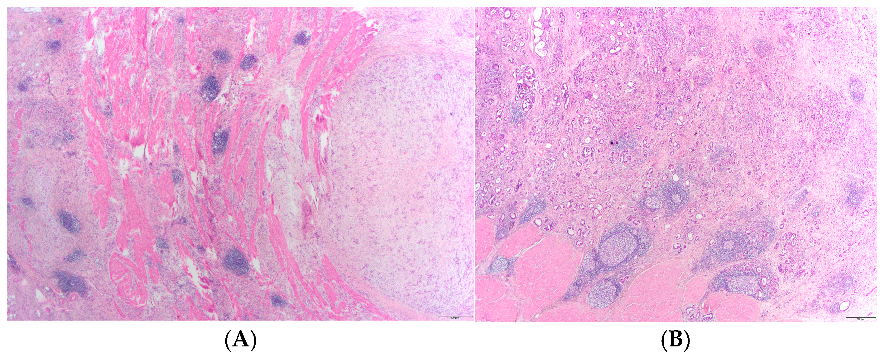
Representative histological slides of tumor tissue were examined under a light microscope (Olympus 63, Okazaki, Japan), photographed with a digital camera (Olympus, Okazaki, Japan) and analyzed using Cell Sens software (Olympus, Okazaki, Japan). TLS is defined as an unencapsulated organized lymphoid aggregate at least 200 µm in diameter, located in the mucosa, tumor itself, or at the tumor margin [4]. TLS density was determined by the number of TLSs in 3 consecutive microscopic fields at 40× magnification (the area of one field was approximately 9.0 mm2). All sections were independently analyzed by two observers (APK, MGD), and any discrepancies were resolved by consensus review. The average TLS area (mm2) was calculated as the mean of 4 consecutive TLSs on the slide. Mature TLSs were defined as well-organized lymphoid structures containing B-cell follicles with a germinal center, while immature TLSs consisted of loosely arranged aggregates of lymphocytes without recognizable follicular organization [18,19]. Lymphoid aggregates are classified as immature TLSs [19,20]. Cases with only immature TLSs were classified as immature, whereas cases containing mature TLSs or a mixture of mature and immature TLSs were classified as mature (Figure 1).

2.3. Immune Cell Density in Tertiary Lymphoid Structures

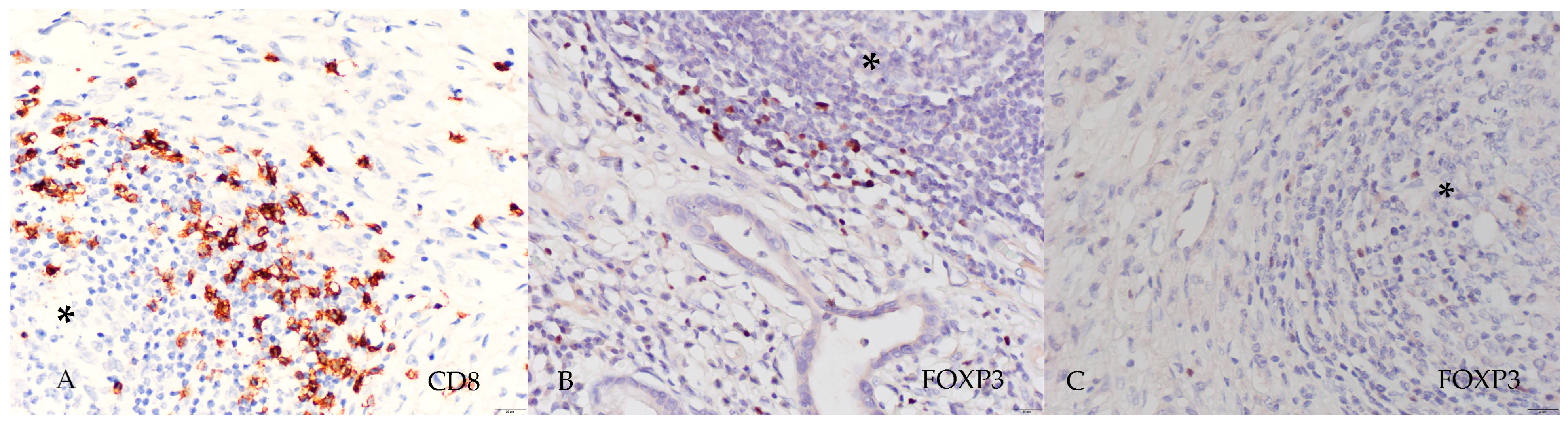
The slides were microscopically analyzed and photographed at 400× magnification (Figure 2). The number of lymphocytes positive for CD8+ (brown membranous staining) and FOXP3+ (brown nuclear staining) was counted using CellSens software v. 1.17 and expressed as the absolute number of positive cells per mean TLS area (number of cells/mm2).

2.4. Transcriptomic Data

To further evaluate the transcriptomic expression of FOXP3, molecular and clinical data were obtained from The Cancer Genome Atlas (TCGA). The dataset was accessed via the cBioPortal platform (https://www.cbioportal.org) on 5 March 2026, using the STAD TCGA PanCancer Atlas dataset. FOXP3 mRNA expression values (z-scores) were extracted and matched with corresponding clinical data using unique patient identifiers. Overall survival (OS) was defined as the time from diagnosis to death or last follow-up and was expressed in months.

2.5. Statistical Analysis

Categorical data are presented as absolute frequencies (n) and percentages (%). Differences between categorical variables were determined using the chi-square test. For numerical data, the normality of distribution was tested using the Shapiro–Wilk test. Due to significant deviation from normality, numerical data were expressed as median and interquartile range (IQR). Significance of differences between groups was established using the Mann–Whitney U test. Survival analysis was performed by the Kaplan–Meier method and multivariable Cox proportional hazards regression. A p-value of <0.05 was considered statistically significant. All statistical analyses were performed using IBM SPSS Statistics for Windows, Version 26 (IBM Corp., Armonk, NY, USA), and RStudio software (v. 2024.12.1+563).

3. Results

3.1. Clinicopathological Characteristics of the Study Cohort

The study included 92 patients with local/locally advanced GC, 63 (68.5%) male and 29 (31.5%) female. The median age was 75 years (IQR: 64–81). Intestinal-type adenocarcinoma predominated (63.3%). According to Bormann, ulcerative-vegetative (27.5%) and ulcerative (33.0%) types were the most frequent, followed by infiltrative (23.1%) and polypoid (16.5%) types. Most tumors were classified as pT3–4 (79.3%) with regional lymph node metastases in 73.9% of cases. Lymphovascular invasion was present in 78.6%, and perineural invasion in 62.3% of cases. TLSs were identified in 66 cases (71.7%), whereas 26 tumors (28.3%) lacked TLS formation.
When patients were stratified according to the presence of TLSs (Table 1), no statistically significant differences were observed between the groups in terms of sex, age, histological or macroscopic type, depth of invasion, lymph node involvement, or the presence of lymphovascular and perineural invasion. However, patients with TLS-positive tumors had a significantly longer overall survival (OS) than patients with TLS-negative tumors (p = 0.006).

3.2. Survival Analysis According to the Presence of TLSs

Kaplan–Meier survival analysis was performed on 85 patients, with follow-up from the time of diagnosis to death or censoring in June 2025. A total of 9 patients (10.6%) were censored, and 7 patients were excluded from the analysis due to missing survival data. The median survival time was 13.5 months (95% CI: 7.979–19.021). Median overall survival times stratified by clinical stage were calculated and are presented in Supplementary Table S1.
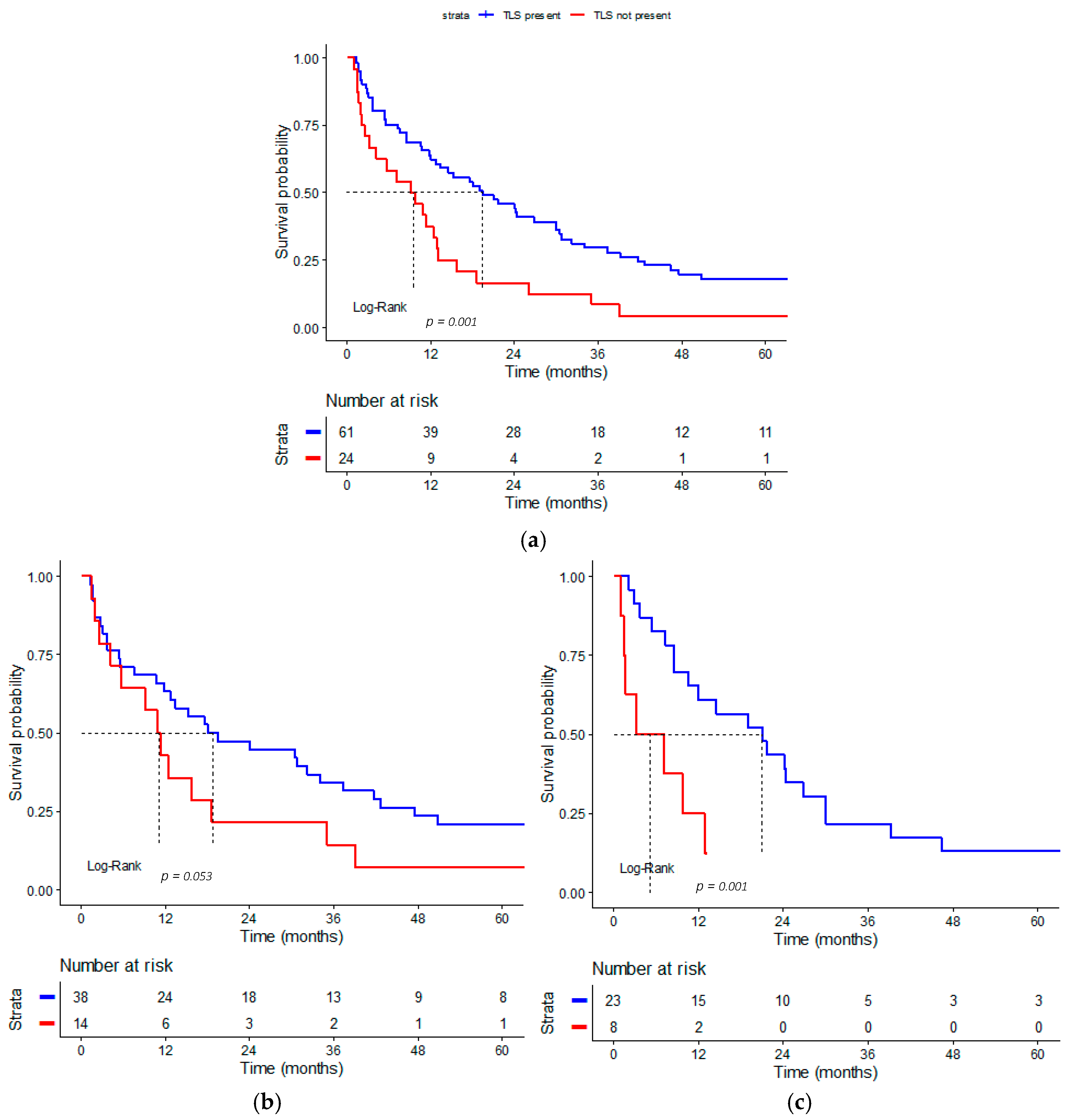
There was a statistically significant difference in OS between patients with and without intratumoral TLS (Figure 3a). A significant difference in 2-year survival was found between patients with TLS-positive and TLS-negative tumors. Median survival of patients who had TLS-positive tumors was 19.4 months (95% CI: 9.779–29.021), and 9.2 months (95% CI: 3.079–15.321) in patients with TLS-negative tumors (Log-Rank test, p = 0.001). The estimated two-year survival rate was 44.3% (95% CI: 33.4–58.7) for TLS-positive patients and 16.7% (95% CI: 6.8–40.8) for TLS-negative patients.
When analyzed by Lauren histological subtype (Figure 3b,c), similar survival trends were observed. In intestinal-type tumors, median survival time was 15.218 months (95% CI: 8.604–21.796) in TLS-positive cases and 10.8 months (95% CI: 6.950–14.650) in TLS-negative cases (Log-Rank test, p = 0.053). In diffuse/mixed tumors, median survival reached 21 months (95% CI: 9.887–32.113) versus 3.3 months (95% CI: 0–10.784) in TLS-negative cases (Log-Rank test, p = 0.001).
Multivariable Cox regression was performed, adjusted for age, pathological stage (pT), nodal status, and Lauren subtype (Table 2). The presence of TLS remained an independent predictor of improved OS (HR 0.497, 95% CI 0.281–0.88, p = 0.017).

3.3. Clinicopathological Variables in Relation to the Number and Maturity of TLSs

Clinicopathological features and immune cell densities in TLSs were analyzed in relation to TLS maturation (Table 3). Most TLSs were classified as lymphoid aggregates or non-mature forms (49, 74.2%), while 17 (25.8%) were classified as mature. Samples with mature TLSs exhibited a higher number of TLSs (p = 0.001) and larger dimensions of individual TLSs (p = 0.016). Immature forms of TLSs were associated with more prevalent lymphovascular invasion (ꭓ2 = 6.081, p = 0.014) and more frequently exhibited lymph node metastases (ꭓ2 = 4.520, p = 0.034). On the other hand, mature TLSs demonstrated higher density of regulatory FOXP3+ T lymphocytes (p = 0.029) and a lower CD8+/FOXP3+ ratio (p = 0.013), while CD8+ lymphocyte density did not differ significantly between groups (p = 0.795). Although there was no significant difference in distribution of pathological and clinical stages between the groups, patients with mature forms reached better OS than those with immature forms of TLSs, 34.55 months (IQR: 18.93–87.03) versus 15.2 months (IQR: 5.4–34) (p = 0.033).

3.4. Impact of FOXP3+ Regulatory T Lymphocytes in TLSs on Patients’ Survival

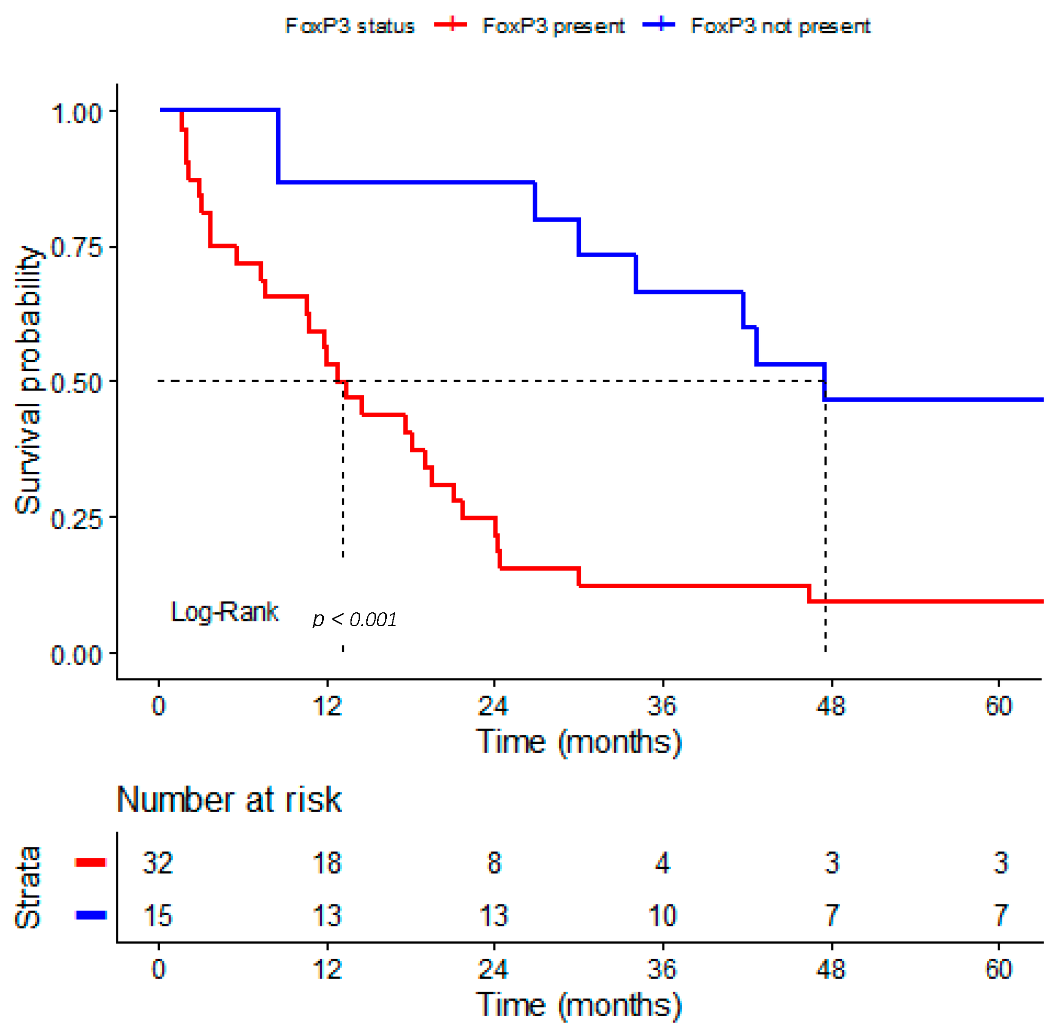
In 47 patients, survival analysis was performed in relation to regulatory FOXP3+ T lymphocytes in intratumoral TLSs (Figure 4). OS was significantly shorter in patients who had regulatory FOXP3+ T lymphocytes in TLSs (median 12.7 months, 95% CI: 8.958–16.442) compared to those without these cells (median 47.5 months, 95% CI: 0–101.655) (Log-Rank test, p < 0.001). The estimated 2-year survival rate was 21.9% (95% CI: 11.4–42.1) for patients with FOXP3+ regulatory T cells in TLSs and 86.7% (95% CI: 71–100) for patients without them.
Multivariable Cox regression was performed, adjusted for age, pathological stage (pT), and nodal status (Table 4). The presence of regulatory T cells in TLSs remained an independent predictor of worse OS (HR 8.165, 95% CI 3.341–19.955, p < 0.001).

3.5. Transcriptomic Data

Analysis of transcriptomic data from the TCGA cohort demonstrated that patients with higher FOXP3 expression did not exhibit longer OS compared with patients with lower FOXP3 expression (log-rank test, p = 0.48) (Supplementary Figure S1).

4. Discussion

The tumor microenvironment interacts with tumor cells, shaping tumor biological behavior, proliferation, angiogenesis, and invasiveness, while modulating the host immune response [21]. In GC, the microenvironment plays a key role in establishing an immunosuppressive milieu [22] that supports tumor progression and resistance to therapy, but can also promote the development of TLSs as a sign of antitumor immunity activation [2]. Chronic antigenic stimulation recruits antigen-presenting cells and lymphocytes, leading to their accumulation within the tumor [23,24,25,26,27]. Activated fibroblasts acquire a phenotype resembling nodal fibroblastic reticular cells [28], and the endothelium differentiates into high endothelial venules, enabling continuous lymphocyte circulation from the bloodstream [29,30,31]. The establishment of distinct T- and B-cell zones results in the formation of TLSs, an organized structure that mirrors the architecture of the lymph node [32,33].
TLSs have been described in a variety of solid tumors, including those of the lung, breast, colon, and melanoma, where their presence generally correlates with favourable clinical outcomes [15,16,33].
TLSs represent a frequent histopathological feature of GC, although their reported prevalence varies considerably among published studies. Xie et al. observed the presence of TLSs in approximately 55% of GC cases [2], while Jiang et al. identified TLSs in as many as 95% of analyzed tumors [13]. Similarly, Zhang et al. detected that only 7.5% of resected GCs lacked both peritumoral and intratumoral TLSs, demonstrating a significant association with favorable survival outcomes [34]. In our cohort, TLSs were present in 66% of cases, placing our findings within the range of previously reported data. This wide range in the reported prevalence may reflect differences in patient population, tumor biology and biological differences within the TIM, and methodological approach used for TLS assessment.
Generally, our cohort exhibited a relatively short overall survival compared with previously reported European cohorts [35,36,37] (median 13.5 months). This likely reflects the high-risk profile of the included patients, who were all of older age (median 75 years) and predominantly presented with locally advanced disease, with stage III tumors accounting for 54.1% of cases.
In our cohort, patients with TLS-positive GCs had significantly better survival outcomes compared to patients with TLS-negative GCs, with the same trend observed across both histological subtypes. TLSs may support local activation and proliferation of lymphocytes within the tumor microenvironment. By enabling antigen presentation and priming of immune cells directly at the tumor site, TLSs reduce the need for lymphocyte recruitment from remote lymph nodes, allowing for an accelerated and effective cytotoxic response against tumor cells [26,27]. This locally organized immune activity may therefore explain the improved survival observed in patients with TLS-positive tumors. According to Zhang et al., the formation of TLSs predicts favorable lymphocytic infiltration and may serve as a biomarker for OS risk stratification [34]. In a spatial mapping analysis, Xie Y et al. demonstrated that TLSs, particularly those located in the tumor core, are associated with better prognosis and improved response to immune checkpoint blockade [2]. Meta-analyses and multicenter studies confirm these findings. Yu A et al. showed that the presence of TLSs is significantly associated with improved outcomes in gastrointestinal tumors [38]. However, the impact of TLSs on outcome depends on tumor type, location, and TLS maturation stage. According to the meta-analysis by Zao et al., intratumoral TLSs are associated with a better prognosis, while peritumoral TLS is favorable in some tumors (esophageal and colorectal cancer) and unfavorable in others (breast and liver cancer) [15]. Kemi N et al. did not confirm the association of the presence/density of TLSs in GC with OS in a multivariate analysis [4].
In our study, lymphovascular invasion and positive regional lymph nodes were more frequently observed in GCs with immature forms of TLSs, suggesting less effective local immune surveillance. In contrast, mature TLSs were larger, more numerous, and characterized by a higher density of FOXP3+ regulatory cells and a lower CD8+/FOXP3+ ratio, suggesting a more structured and functionally coordinated immune response. Comparable findings have been reported in other studies: higher maturity of TLS, usually defined by follicular organization, presence of germinal centers, and developed high endothelial venules, is associated with more favorable clinicopathological features and longer survival [15,39,40,41].
Furthermore, Posh et al. showed that a high proportion of mature TLSs is a stronger prognostic indicator than the total number of TLSs in non-metastatic colon cancer, which highlights the importance of TLS composition/maturation stage [42].
In line with previous reports, our analysis demonstrated significantly shorter overall survival in GCs containing only immature TLSs compared with tumors harboring mixed or fully mature TLSs.
Immature TLSs are often functionally inactive or even immunosuppressive, as they mainly contain FOXP3+ regulatory T cells and M2 macrophages that suppress cytotoxic response. Our finding of a correlation between immature TLSs with lymphovascular invasion and lymph node metastasis is consistent with previous reports [43,44]. While GCs with immature TLSs are typically enriched in immunosuppressive cell populations, mature TLSs contain abundant B cell clusters at various stages of differentiation, actively involved in antigen presentation and antibody production [3]. Moreover, plasma cells derived from TLSs have been shown to migrate into tumor nests, where they secrete antibodies directed against tumor antigens, thus contributing to the antitumor humoral immune response [45]. Meta-analyses across digestive system tumors confirm that only mature TLSs with germinal centers consistently confer survival benefit [38,46].
FOXP3+ regulatory T cells are essential for maintaining immune tolerance and preventing autoimmunity under physiological conditions. Within the tumor microenvironment, their function becomes detrimental. By suppressing cytotoxic activity and dampening antitumor immunity, TLSs enriched in regulatory T cells lose their usual protective role and may even promote tumor progression by creating a locally immunosuppressive milieu. As a result, cytotoxic immune activity is reduced, allowing tumor cells to persist, proliferate, and metastasize. In our series, TLS-positive tumors with FOXP3+ regulatory T cells present displayed higher mortality, supporting the notion that regulatory predominance within TLS may counteract their otherwise favorable prognostic effect. Previous studies have also linked high intratumoral FOXP3+ cells infiltration to poorer prognosis in GC [2,47]. According to Hennequin et al., in localized GC, marked infiltration of FOXP3+ cells within the tumor stroma was associated with shorter/reduced relapse-free survival (RFS) [48]. These results suggest that TLS confer prognostic advantage only when their internal structure supports effective immune activation. Immature or functionally inactive FOXP3+ rich TLSs fail to initiate an effective antitumor response, which contributes to faster disease progression and shorter patient survival.
In an exploratory analysis based on publicly available transcriptomic datasets, no statistically significant association was observed between FOXP3 mRNA expression and overall survival (p = 0.48). However, bulk transcriptomic analyses assess gene expression across the entire tumor tissue and therefore do not allow spatial localization of FOXP3+ immune cells. In contrast, the present study specifically evaluated FOXP3+ lymphocytes within tertiary lymphoid structures (TLS). This distinction is important because the biological and prognostic role of immune cells may depend on their spatial organization and functional context within the tumor microenvironment. TLS represent highly organized immune niches that resemble secondary lymphoid organs and provide a microenvironment for coordinated immune cell interactions [33]. Such spatial organization cannot be captured by bulk transcriptomic analyses [49,50]. Furthermore, mRNA expression levels do not necessarily correspond to protein expression levels due to post-transcriptional regulation [51]. Therefore, spatially resolved protein-level analyses, such as immunohistochemical assessment of FOXP3+ cells within TLS, may provide a more accurate understanding of their prognostic significance in gastric cancer.
The total number of CD8+ cells did not differ significantly, while, as expected, CD8+/FOXP3+ ratio was significantly higher in GCs with immature forms of TLSs, underscoring that the prognostic impact is determined not merely by cytotoxic T cell abundance, but by the interplay of effector and regulatory components within the TLS. These observations are consistent with the results of Hennequin et al., who analyzed survival outcomes of patients with GC, highlighting the importance of a coordinated Th1 and B cell response [48].
In summary, our findings suggest a trend toward improved clinical outcomes in patients with TLS-positive GCs and indicate a potential role of regulatory T cells in disease progression. However, these results should be interpreted with caution due to the relatively small sample size and the homogeneity of the studied population. Further large-scale and more rigorous studies are needed to validate our findings and to explore novel biomarkers in the context of the tumor microenvironment and contemporary therapeutic regimens.

5. Conclusions

The presence and maturation of TLSs in GC are associated with improved prognostic stratification. Assessment of TLS maturation and immune cell composition may provide valuable insights into disease progression. Further studies on larger and more diverse patient cohorts are warranted to elucidate the biological role of effector and regulatory T cells in the tumor immune microenvironment and their impact on clinical outcomes.

Supplementary Materials

The following supporting information can be downloaded at: https://www.mdpi.com/article/10.3390/medsci14010145/s1, Figure S1: Kaplan–Meier overall survival curves according to FOXP3 transcript levels in bulk tumor tissue. Table S1: Median overall survival (OS) times according to clinical stage.

Author Contributions

Conceptualization, M.G.D. and A.P.K.; methodology, P.Đ.; software, P.Đ. and L.M.; validation, A.P.K., P.Đ. and M.G.D.; formal analysis, P.Đ.; investigation, A.P.K.; resources, T.I. and K.M.; data curation, A.P.K.; writing—original draft preparation, A.P.K.; writing—review and editing, M.G.D. and P.Đ.; visualization, A.P.K.; supervision, K.V. All authors have read and agreed to the published version of the manuscript.

Funding

This research received no external funding.

Institutional Review Board Statement

The study was conducted in accordance with the Declaration of Helsinki, and approved by the Institutional Ethics Committee UNIVERSITY HOSPITAL SPLIT, Croatia (protocol code 520-03/19-01/102, date of approval 30 October 2025).

Informed Consent Statement

Patient consent was waived due to research on paraffin blocks of tumor tissue.

Data Availability Statement

The original contributions presented in this study are included in the article/Supplementary Material. Further inquiries can be directed to the corresponding authors.

Acknowledgments

The authors have reviewed and edited the output and take full responsibility for the content of this publication.

Conflicts of Interest

The authors declare no conflicts of interest.

Abbreviations

The following abbreviations are used in this manuscript:
GCGastric cancer
TLSTertiary lymphoid structure
OSOverall survival
TNMTumor-Node-Metastasis
WHOWorld Health Organization
EBVEpstein–Barr virus
HER2Human epidermal growth factor receptor 2
SARIFAStroma AReactive Invasion Front Areas
TIMTumor immune microenvironment
FDCsFollicular dendritic cells
HEVsHigh endothelial venules
H&EHematoxylin and eosin
IHCImmunohistochemistry
IQRInterquartile range
RFSRelapse-free survival

References

  1. Bray, F.; Laversanne, M.; Sung, H.; Ferlay, J.; Siegel, R.L.; Soerjomataram, I.; Jemal, A. Global Cancer Statistics 2022: GLOBOCAN Estimates of Incidence and Mortality Worldwide for 36 Cancers in 185 Countries. CA Cancer J. Clin. 2024, 74, 229–263. [Google Scholar] [CrossRef] [Scilit]
  2. Xie, Y.; Peng, H.; Hu, Y.; Jia, K.; Yuan, J.; Liu, D.; Li, Y.; Feng, X.; Li, J.; Zhang, X.; et al. Immune Microenvironment Spatial Landscapes of Tertiary Lymphoid Structures in Gastric Cancer. BMC Med. 2025, 23, 59. [Google Scholar] [CrossRef] [Scilit]
  3. Groen-van Schooten, T.S.; Franco Fernandez, R.; van Grieken, N.C.T.; Bos, E.N.; Seidel, J.; Saris, J.; Martínez-Ciarpaglini, C.; Fleitas, T.C.; Thommen, D.S.; de Gruijl, T.D.; et al. Mapping the Complexity and Diversity of Tertiary Lymphoid Structures in Primary and Peritoneal Metastatic Gastric Cancer. J. Immunother. Cancer 2024, 12, e009243. [Google Scholar] [CrossRef] [Scilit]
  4. Kemi, N.; Ylitalo, O.; Väyrynen, J.P.; Helminen, O.; Junttila, A.; Mrena, J.; Böhm, J.; Kauppila, J.H. Tertiary Lymphoid Structures and Gastric Cancer Prognosis. Acta Pathol. Microbiol. Immunol. Scand. 2023, 131, 19–25. [Google Scholar] [CrossRef] [Scilit]
  5. WHO Classification of Tumours Editorial Board. Digestive System Tumours, 5th ed.; International Agency for Research on Cancer: Lyon, France, 2019. [Google Scholar]
  6. Rugge, M.; Carneiro, F.; Fukayama, M.; Yasui, W.; Grabsch, H.; Znaor, A. Gastric Adenocarcinoma. In WHO Classification of Tumours: Digestive System Tumours; International Agency for Research on Cancer: Lyon, France, 2019. [Google Scholar]
  7. Nagtegaal, I.D.; Odze, R.D.; Klimstra, D.; Paradis, V.; Rugge, M.; Schirmacher, P.; Washington, K.M.; Carneiro, F.; Cree, I.A. The 2019 WHO Classification of Tumours of the Digestive System. Histopathology 2020, 76, 182–188. [Google Scholar] [CrossRef] [Scilit]
  8. Cancer Genome Atlas Research Network. Comprehensive Molecular Characterization of Gastric Adenocarcinoma. Nature 2014, 513, 202–209. [Google Scholar] [CrossRef] [Scilit] [PubMed]
  9. Cristescu, R.; Lee, J.; Nebozhyn, M.; Kim, K.-M.; Ting, J.C.; Wong, S.S.; Liu, J.; Yue, Y.G.; Wang, J.; Yu, K.; et al. Molecular Analysis of Gastric Cancer Identifies Subtypes Associated with Distinct Clinical Outcomes. Nat. Med. 2015, 21, 449–456. [Google Scholar] [CrossRef] [Scilit] [PubMed]
  10. Grabsch, H.I.; Tan, P. Gastric Cancer Pathology and Underlying Molecular Mechanisms. Dig. Surg. 2013, 30, 150–158. [Google Scholar] [CrossRef] [Scilit] [PubMed]
  11. Ulase, D.; Behrens, H.-M.; Röcken, C. Stroma AReactive Invasion Front Areas (SARIFA) Predict Poor Survival in Adenocarcinomas of the Stomach and Gastrooesophageal Junction: A Validation Study. Virchows Arch. Int. J. Pathol. 2024, 485, 527–534. [Google Scholar] [CrossRef] [Scilit]
  12. Pernot, S.; Terme, M.; Radosevic-Robin, N.; Castan, F.; Badoual, C.; Marcheteau, E.; Penault-Llorca, F.; Bouche, O.; Bennouna, J.; Francois, E.; et al. Infiltrating and Peripheral Immune Cell Analysis in Advanced Gastric Cancer According to the Lauren Classification and Its Prognostic Significance. Gastric Cancer 2020, 23, 73–81. [Google Scholar] [CrossRef] [Scilit]
  13. Jiang, S.; Liao, X.; Ding, X. Maturity and Density of Tertiary Lymphoid Structures Associate with Tumor Metastasis and Chemotherapy Response. Front. Med. 2024, 11, 1435620. [Google Scholar] [CrossRef] [Scilit]
  14. Munoz-Erazo, L.; Rhodes, J.L.; Marion, V.C.; Kemp, R.A. Tertiary Lymphoid Structures in Cancer-Considerations for Patient Prognosis. Cell. Mol. Immunol. 2020, 17, 570–575. [Google Scholar] [CrossRef] [Scilit]
  15. Zhao, L.; Jin, S.; Wang, S.; Zhang, Z.; Wang, X.; Chen, Z.; Wang, X.; Huang, S.; Zhang, D.; Wu, H. Tertiary Lymphoid Structures in Diseases: Immune Mechanisms and Therapeutic Advances. Signal Transduct. Target. Ther. 2024, 9, 225. [Google Scholar] [CrossRef] [Scilit]
  16. Dieu-Nosjean, M.-C.; Goc, J.; Giraldo, N.A.; Sautès-Fridman, C.; Fridman, W.H. Tertiary Lymphoid Structures in Cancer and Beyond. Trends Immunol. 2014, 35, 571–580. [Google Scholar] [CrossRef] [Scilit]
  17. Mao, Y.; Wang, X.; Xi, L.; Dong, M.; Song, P.; Miao, J.; Lu, C.; Sun, S.; Li, Q.; Yu, C.; et al. Prediction Values of Tertiary Lymphoid Structures in the Prognosis of Patients with Left- and Right-Sided Colon Cancer: A Multicenter Propensity Score-Matched Study. Int. J. Surg. Lond. Engl. 2023, 109, 2344–2358. [Google Scholar] [CrossRef] [Scilit]
  18. Li, H.; Zhang, M.-J.; Zhang, B.; Lin, W.-P.; Li, S.-J.; Xiong, D.; Wang, Q.; Wang, W.-D.; Yang, Q.-C.; Huang, C.-F.; et al. Mature Tertiary Lymphoid Structures Evoke Intra-Tumoral T and B Cell Responses via Progenitor Exhausted CD4(+) T Cells in Head and Neck Cancer. Nat. Commun. 2025, 16, 4228. [Google Scholar] [CrossRef] [Scilit] [PubMed]
  19. Chen, Y.; Wu, Y.; Yan, G.; Zhang, G. Tertiary Lymphoid Structures in Cancer: Maturation and Induction. Front. Immunol. 2024, 15, 1369626. [Google Scholar] [CrossRef] [Scilit] [PubMed]
  20. Vanhersecke, L.; Bougouin, A.; Crombé, A.; Brunet, M.; Sofeu, C.; Parrens, M.; Pierron, H.; Bonhomme, B.; Lembege, N.; Rey, C.; et al. Standardized Pathology Screening of Mature Tertiary Lymphoid Structures in Cancers. Lab. Investig. J. Tech. Methods Pathol. 2023, 103, 100063. [Google Scholar] [CrossRef] [Scilit]
  21. de Visser, K.E.; Joyce, J.A. The Evolving Tumor Microenvironment: From Cancer Initiation to Metastatic Outgrowth. Cancer Cell 2023, 41, 374–403. [Google Scholar] [CrossRef] [Scilit]
  22. Cozac-Szőke, A.-R.; Cozac, D.A.; Negovan, A.; Tinca, A.C.; Vilaia, A.; Cocuz, I.-G.; Sabău, A.H.; Niculescu, R.; Chiorean, D.M.; Tomuț, A.N.; et al. Immune Cell Interactions and Immune Checkpoints in the Tumor Microenvironment of Gastric Cancer. Int. J. Mol. Sci. 2025, 26, 1156. [Google Scholar] [CrossRef] [Scilit] [PubMed]
  23. Barone, F.; Gardner, D.H.; Nayar, S.; Steinthal, N.; Buckley, C.D.; Luther, S.A. Stromal Fibroblasts in Tertiary Lymphoid Structures: A Novel Target in Chronic Inflammation. Front. Immunol. 2016, 7, 477. [Google Scholar] [CrossRef] [Scilit] [PubMed]
  24. Aloisi, F.; Pujol-Borrell, R. Lymphoid Neogenesis in Chronic Inflammatory Diseases. Nat. Rev. Immunol. 2006, 6, 205–217. [Google Scholar] [CrossRef] [Scilit]
  25. Drayton, D.L.; Liao, S.; Mounzer, R.H.; Ruddle, N.H. Lymphoid Organ Development: From Ontogeny to Neogenesis. Nat. Immunol. 2006, 7, 344–353. [Google Scholar] [CrossRef] [Scilit]
  26. Sautès-Fridman, C.; Lawand, M.; Giraldo, N.A.; Kaplon, H.; Germain, C.; Fridman, W.H.; Dieu-Nosjean, M.-C. Tertiary Lymphoid Structures in Cancers: Prognostic Value, Regulation, and Manipulation for Therapeutic Intervention. Front. Immunol. 2016, 7, 407. [Google Scholar] [CrossRef] [Scilit]
  27. Colbeck, E.J.; Ager, A.; Gallimore, A.; Jones, G.W. Tertiary Lymphoid Structures in Cancer: Drivers of Antitumor Immunity, Immunosuppression, or Bystander Sentinels in Disease? Front. Immunol. 2017, 8, 1830. [Google Scholar] [CrossRef] [Scilit]
  28. Denton, A.E.; Roberts, E.W.; Linterman, M.A.; Fearon, D.T. Fibroblastic Reticular Cells of the Lymph Node Are Required for Retention of Resting but Not Activated CD8+ T Cells. Proc. Natl. Acad. Sci. USA 2014, 111, 12139–12144. [Google Scholar] [CrossRef] [Scilit] [PubMed]
  29. Grasso, C.; Pierie, C.; Mebius, R.E.; van Baarsen, L.G.M. Lymph Node Stromal Cells: Subsets and Functions in Health and Disease. Trends Immunol. 2021, 42, 920–936. [Google Scholar] [CrossRef] [Scilit] [PubMed]
  30. Fletcher, A.L.; Acton, S.E.; Knoblich, K. Lymph Node Fibroblastic Reticular Cells in Health and Disease. Nat. Rev. Immunol. 2015, 15, 350–361. [Google Scholar] [CrossRef] [Scilit] [PubMed]
  31. Moussion, C.; Girard, J.-P. Dendritic Cells Control Lymphocyte Entry to Lymph Nodes through High Endothelial Venules. Nature 2011, 479, 542–546. [Google Scholar] [CrossRef] [Scilit]
  32. Schumacher, T.N.; Thommen, D.S. Tertiary Lymphoid Structures in Cancer. Science 2022, 375, eabf9419. [Google Scholar] [CrossRef] [Scilit]
  33. Sautès-Fridman, C.; Petitprez, F.; Calderaro, J.; Fridman, W.H. Tertiary Lymphoid Structures in the Era of Cancer Immunotherapy. Nat. Rev. Cancer 2019, 19, 307–325. [Google Scholar] [CrossRef] [Scilit]
  34. Zhang, N.; Zhang, G.; Wang, D.; Liu, H.; Zhang, Y.; Ayarick, V.A.; Han, X.; Lv, Y.; Wang, Y. The Relationship of the Tertiary Lymphoid Structures with the Tumor-Infiltrating Lymphocytes and Its Prognostic Value in Gastric Cancer. Arch. Med. Sci. 2024, 20, 255–266. [Google Scholar] [CrossRef] [Scilit] [PubMed]
  35. Al-Batran, S.-E.; Homann, N.; Pauligk, C.; Goetze, T.O.; Meiler, J.; Kasper, S.; Kopp, H.-G.; Mayer, F.; Haag, G.M.; Luley, K.; et al. Perioperative Chemotherapy with Fluorouracil plus Leucovorin, Oxaliplatin, and Docetaxel versus Fluorouracil or Capecitabine plus Cisplatin and Epirubicin for Locally Advanced, Resectable Gastric or Gastro-Oesophageal Junction Adenocarcinoma (FLOT4): A Randomised, Phase 2/3 Trial. Lancet Lond. Engl. 2019, 393, 1948–1957. [Google Scholar] [CrossRef] [Scilit]
  36. Cunningham, D.; Allum, W.H.; Stenning, S.P.; Thompson, J.N.; Van de Velde, C.J.H.; Nicolson, M.; Scarffe, J.H.; Lofts, F.J.; Falk, S.J.; Iveson, T.J.; et al. Perioperative Chemotherapy versus Surgery Alone for Resectable Gastroesophageal Cancer. N. Engl. J. Med. 2006, 355, 11–20. [Google Scholar] [CrossRef] [Scilit]
  37. Smalley, S.R.; Benedetti, J.K.; Haller, D.G.; Hundahl, S.A.; Estes, N.C.; Ajani, J.A.; Gunderson, L.L.; Goldman, B.; Martenson, J.A.; Jessup, J.M.; et al. Updated Analysis of SWOG-Directed Intergroup Study 0116: A Phase III Trial of Adjuvant Radiochemotherapy versus Observation after Curative Gastric Cancer Resection. J. Clin. Oncol. 2012, 30, 2327–2333. [Google Scholar] [CrossRef] [Scilit]
  38. Yu, A.; Cao, M.; Zhang, K.; Yang, Y.; Ma, L.; Zhang, X.; Zhao, Y.; Ma, X.; Fan, Z.; Han, Z.; et al. The Prognostic Value of the Tertiary Lymphoid Structure in Gastrointestinal Cancers. Front. Immunol. 2023, 14, 1256355. [Google Scholar] [CrossRef] [Scilit]
  39. Sun, H.; Liu, Y.; Cheng, W.; Xiong, R.; Gu, W.; Zhang, X.; Wang, X.; Wang, X.; Tan, C.; Weng, W.; et al. The Distribution and Maturation of Tertiary Lymphoid Structures Can Predict Clinical Outcomes of Patients with Gastric Adenocarcinoma. Front. Immunol. 2024, 15, 1396808. [Google Scholar] [CrossRef] [Scilit]
  40. Liu, X.; Xu, D.; Zhou, C.; Zhong, Y.; Geng, H.; Huang, C.; Shen, Y.; Xia, X.; Wang, C.; Zhu, C.; et al. Association of PD-1 + Treg/PD-1 + CD8 Ratio and Tertiary Lymphoid Structures with Prognosis and Response in Advanced Gastric Cancer Patients Receiving Preoperative Treatment. J. Transl. Med. 2024, 22, 1152. [Google Scholar] [CrossRef] [Scilit] [PubMed]
  41. Yin, Y.-X.; Ling, Y.-H.; Wei, X.-L.; He, C.-Y.; Wang, B.-Z.; Hu, C.-F.; Lin, W.-P.; Nie, R.-C.; Chen, J.-W.; Lin, J.-L.; et al. Impact of Mature Tertiary Lymphoid Structures on Prognosis and Therapeutic Response of Epstein-Barr Virus-Associated Gastric Cancer Patients. Front. Immunol. 2022, 13, 973085. [Google Scholar] [CrossRef] [Scilit]
  42. Posch, F.; Silina, K.; Leibl, S.; Mündlein, A.; Moch, H.; Siebenhüner, A.; Samaras, P.; Riedl, J.; Stotz, M.; Szkandera, J.; et al. Maturation of Tertiary Lymphoid Structures and Recurrence of Stage II and III Colorectal Cancer. Oncoimmunology 2018, 7, e1378844. [Google Scholar] [CrossRef] [Scilit] [PubMed]
  43. Pande, K.; Rauniyar, S.K.; Pudasaini, S. Assessment of Lymphovascular Invasion in Gastric Carcinoma; Do They Always Indicate Lymph Node Metastasis? J. Pathol. Nepal. 2018, 8, 1251–1256. [Google Scholar] [CrossRef] [Scilit]
  44. Sumiyoshi, S.; Ohashi, T.; Kubota, T.; Nishibeppu, K.; Owada, K.; Kiuchi, J.; Shimizu, H.; Arita, T.; Iitaka, D.; Yamamoto, Y.; et al. Lymphovascular Invasion Is Associated with Poor Long-Term Outcomes in Patients with pT1N0-3 or pT2-3N0 Remnant Gastric Cancer: A Retrospective Cohort Study. World J. Surg. Oncol. 2024, 22, 86. [Google Scholar] [CrossRef] [Scilit] [PubMed]
  45. Meylan, M.; Petitprez, F.; Becht, E.; Bougoüin, A.; Pupier, G.; Calvez, A.; Giglioli, I.; Verkarre, V.; Lacroix, G.; Verneau, J.; et al. Tertiary Lymphoid Structures Generate and Propagate Anti-Tumor Antibody-Producing Plasma Cells in Renal Cell Cancer. Immunity 2022, 55, 527–541.e5. [Google Scholar] [CrossRef] [Scilit] [PubMed]
  46. Sun, H.; Shi, Y.; Ran, H.; Peng, J.; Li, Q.; Zheng, G.; He, Y.; Liu, S.; Chang, W.; Xiao, Y. Prognostic Value of Tertiary Lymphoid Structures (TLS) in Digestive System Cancers: A Systematic Review and Meta-Analysis. BMC Cancer 2023, 23, 1248. [Google Scholar] [CrossRef] [Scilit]
  47. Perrone, G.; Ruffini, P.A.; Catalano, V.; Spino, C.; Santini, D.; Muretto, P.; Spoto, C.; Zingaretti, C.; Sisti, V.; Alessandroni, P.; et al. Intratumoural FOXP3-Positive Regulatory T Cells Are Associated with Adverse Prognosis in Radically Resected Gastric Cancer. Eur. J. Cancer 2008, 44, 1875–1882. [Google Scholar] [CrossRef] [Scilit]
  48. Hennequin, A.; Derangère, V.; Boidot, R.; Apetoh, L.; Vincent, J.; Orry, D.; Fraisse, J.; Causeret, S.; Martin, F.; Arnould, L.; et al. Tumor Infiltration by Tbet+ Effector T Cells and CD20+ B Cells Is Associated with Survival in Gastric Cancer Patients. Oncoimmunology 2016, 5, e1054598. [Google Scholar] [CrossRef] [Scilit]
  49. Ji, A.L.; Rubin, A.J.; Thrane, K.; Jiang, S.; Reynolds, D.L.; Meyers, R.M.; Guo, M.G.; George, B.M.; Mollbrink, A.; Bergenstråhle, J.; et al. Multimodal Analysis of Composition and Spatial Architecture in Human Squamous Cell Carcinoma. Cell 2020, 182, 497–514.e22. [Google Scholar] [CrossRef] [Scilit]
  50. Longo, S.K.; Guo, M.G.; Ji, A.L.; Khavari, P.A. Integrating Single-Cell and Spatial Transcriptomics to Elucidate Intercellular Tissue Dynamics. Nat. Rev. Genet. 2021, 22, 627–644. [Google Scholar] [CrossRef] [Scilit] [PubMed]
  51. Vogel, C.; Marcotte, E.M. Insights into the Regulation of Protein Abundance from Proteomic and Transcriptomic Analyses. Nat. Rev. Genet. 2012, 13, 227–232. [Google Scholar] [CrossRef] [Scilit]
Figure 1. Tertiary lymphoid structures in gastric adenocarcinoma: immature (A) and mature (B) with germinal centers (20×).
Medsci 14 00145 g001
Figure 2. Immunohistochemical staining of CD8+ T cells (A) and FOXP3+ regulatory T cells in tertiary lymphoid structures (*) observed in intestinal (B) and diffuse (C) subtypes of gastric adenocarcinoma (400×).
Medsci 14 00145 g002
Figure 3. Kaplan–Meier survival curves according to TLS status: (a) all patients; (b) patients with intestinal-type GC; (c) patients with diffuse/mixed-type GC. The dashed lines represent the median survival time.
Medsci 14 00145 g003
Figure 4. Kaplan–Meier survival analysis according to the presence of FOXP3+ regulatory T cells in TLSs in gastric adenocarcinoma. The dashed lines represent the median survival time.
Medsci 14 00145 g004
Table 1. Clinicopathological characteristics according to the TLS status of GC.
VariableTLS
n (%) or Median (IQR)
p
Present
n = 66 (71.7%)
Not Present
n = 26 (28.3%)
Sexmale46 (69.7)17 (65.4)0.689 *
female20 (30.3)9 (34.6)
Age, years74.45 (62.5–81.25)76 (67–80.25)0.529 #
Lauren subtypeintestinal41 (62.1)16 (66.7)0.692 *
diffuse/mixed25 (37.9)8 (33.3)
Bormann typepolypoid9 (13.6)6 (24)0.564 *
ulcerative24 (36.4)6 (24)
ulcerovegetative18 (27.3)7 (28)
infiltrative15 (22.7)6 (24)
pTpT1, 216 (24.2)3 (11.5)0.175 *
pT3, 450 (75.8)23 (88.5)
pNnegative18 (27.3)6 (23.1)0.680 *
positive48 (72.7)20 (76.9)
Clinical stageI, II32 (49.2)9 (34.6)0.206 *
III, IV33 (50.8)17 (65.4)
Lymphovascular invasionno10 (20.4)5 (23.8)0.751 *
yes39 (79.6)16 (76.2)
Perineural invasionno20 (42.6)6 (27.3)0.222 *
yes27 (57.4)16 (72.7)
Overall survival, months19.4 (6.5–42.1)9.45 (2.23–15.05)0.006 #
*—ꭓ2 test; # Mann–Whitney U test; bold indicates p < 0.05. TLS, tertiary lymphoid structures.
Table 2. Impact of TLS presence and clinicopathologic factors on overall survival in gastric cancer patients: Multivariable Cox regression.
VariableHR (95% CI)p-Value
Age1.027 (1–1.055)0.051
pT1.258 (0.608–2.602)0.536
pN1.588 (0.82–3.077)0.171
Lauren subtype1.367 (0.771–2.421)0.284
TLS status0.497 (0.281–0.88)0.017
Bold indicates p < 0.05.
Table 3. Impact of TLS maturation on clinicopathological features and immune composition in GC.
VariableTLS
n (%) or Median (IQR)
p
Lymphoid Aggregate/Non-Mature
n = 49 (74.2%)
Mature
n = 17 (25.8%)
SexMale35 (71.4)11 (64.7)0.602 *
Female14 (28.6)6 (35.3)
Age, year 71.3 (59.5–80.3)77 (67.5–82.5)0.284 #
Lauren subtypeIntestinal31 (63.3)10 (58.8)0.745 *
Diffuse/mixed18 (36.7)7 (41.2)
Bormann classificationPolypoid7 (14.3)2 (11.8)0.487 *
Ulcerative18 (36.7)6 (35.3)
Ulcerovegetative15 (30.6)3 (17.6)
Infiltrative9 (18.4)6 (35.3)
pTpT1, 210 (20.4)6 (35.3)0.217 *
pT3, 439 (79.6)11 (64.7)
pNnegative10 (20.4)8 (47.1)0.034 *
positive39 (79.6)9 (52.9)
Clinical stageI, II23 (47.9)9 (52.9)0.722 *
III, IV25 (52.1)8 (47.1)
Lymphovascular invasionNo4 (11.4)6 (42.9)0.014 *
Yes31 (88.6)8 (57.1)
Perineural invasionNo14 (42.4)6 (42.9)0.978 *
Yes19 (57.6)8 (57.1)
Overall survival, months15.2 (5.4–34)34.55 (18.93–87.03)0.033 #
TLS mean area, mm2 19 (9–125)59 (19.25–190.75)0.016 #
Number of TLS **4 (2–6)8 (6–12.5)0.001 #
CD8+/mm2 of TLS ** 750 (590–999)790 (578.25–980.5)0.795 #
FOXP3+/mm2 of TLS ** 27.27 (11.25–60)169.05 (24.84–223.21)0.029 #
CD8+/FOXP3+26.49 (19.73–73.3)3.61 (3.3–28.37)0.013 #
*—ꭓ2 test; # Mann–Whitney U test; bold indicates p < 0.05. ** measured in three low-magnification fields. TLS, tertiary lymphoid structures.
Table 4. Impact of FOXP3+ regulatory T lymphocytes in TLSs and clinicopathologic factors on overall survival in gastric cancer patients: Multivariable Cox regression.
VariableHR (95% CI)p-Value
Age1.059 (1.024–1.097)0.001
pT1.328 (0.527–3.345)0.547
pN2.061 (0.813–5.222)0.127
FOXP3+ lymphocyte status8.165 (3.341–19.955)<0.001
Bolded p value < 0.05.
Disclaimer/Publisher’s Note: The statements, opinions and data contained in all publications are solely those of the individual author(s) and contributor(s) and not of MDPI and/or the editor(s). MDPI and/or the editor(s) disclaim responsibility for any injury to people or property resulting from any ideas, methods, instructions or products referred to in the content.