RhoA, Claudin 18, and c-MET in Gastric Cancer: Clinicopathological Characteristics and Prognostic Significance in Curative Resected Patients

 ,
, 

Abstract
:1. Introduction
2. Materials and Methods
2.1. Patients
2.2. Tissue Microarray Construction (TMA)
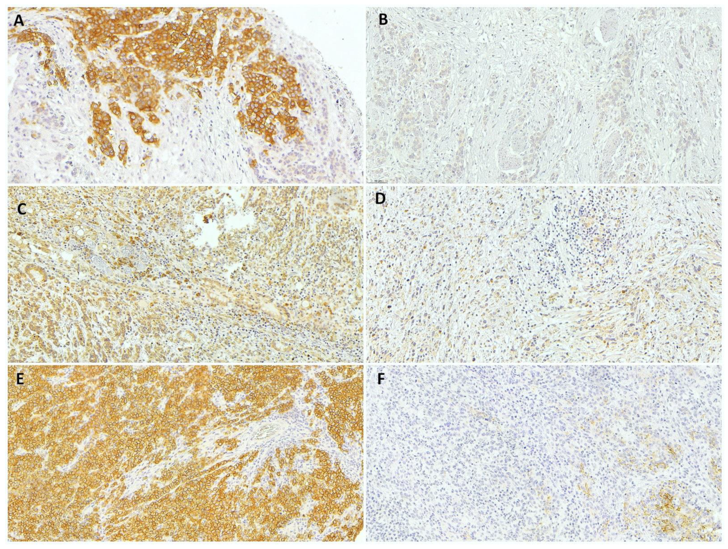
2.3. Immunohistochemistry
2.4. Statistical Analysis
3. Results
3.1. C-MET and Clinicopathological Characteristics
3.2. RhoA and Clinicopathological Characteristics
3.3. CLDN18 and Clinicopathological Characteristics
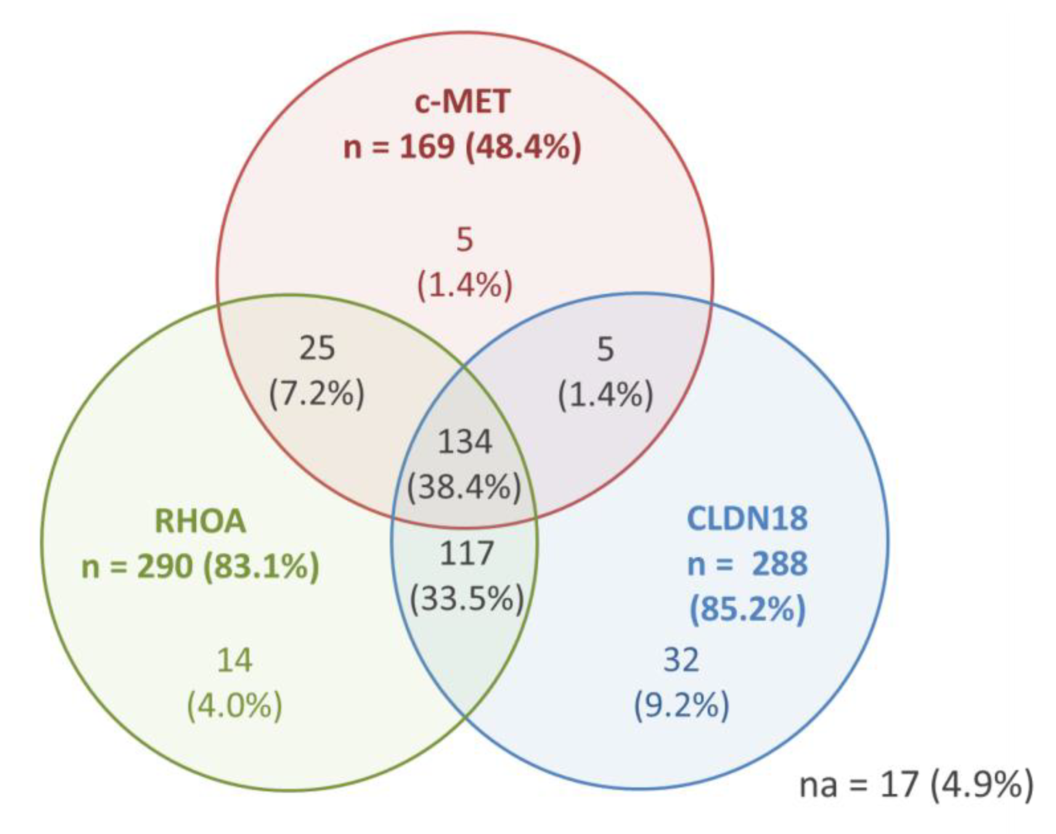
3.4. Immunohistochemical Analysis and Correlation between c-MET, RhoA, and CLDN18
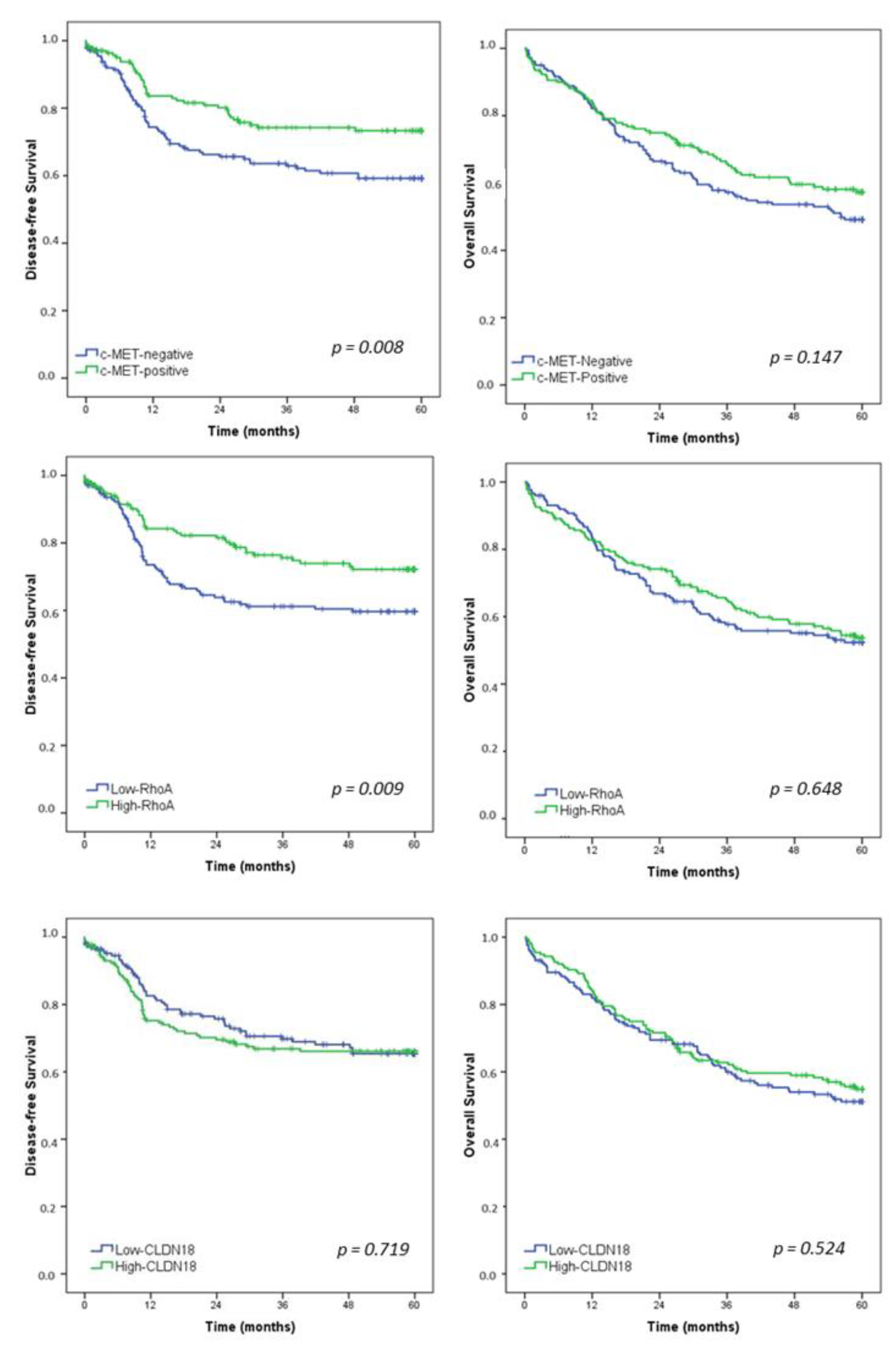
3.5. Survival Analysis
4. Discussion
5. Conclusions
Supplementary Materials
Author Contributions
Funding
Institutional Review Board Statement
Informed Consent Statement
Data Availability Statement
Conflicts of Interest
References
- Bray, F.; Ferlay, J.; Soerjomataram, I.; Siegel, R.L.; Torre, L.A.; Jemal, A. Global cancer statistics 2018: GLOBOCAN estimates of incidence and mortality worldwide for 36 cancers in 185 countries. CA: A Cancer J. Clin. 2018, 68, 394–424. [Google Scholar] [CrossRef]
- Japanese Gastric Cancer Association. Japanese gastric cancer treatment guidelines 2014 (ver. 4). Gastric Cancer Off. J. Int. Gastric Cancer Assoc. Jpn. Gastric Cancer Assoc. 2017, 20, 1–19. [Google Scholar] [CrossRef]
- Barchi, L.C.; Ramos, M.F.K.P.; Dias, A.R.; Andreollo, N.A.; Weston, A.C.; Lourenço, L.G.; Malheiros, C.A.; Kassab, P.; Zilberstein, B.; Ferraz, Á.A.B.; et al. II brazilian consensus on gastric cancer by the brazilian gastric cancer association. ABCD Arq. Bras. De Cir. Dig. 2020, 33, e1535. [Google Scholar] [CrossRef]
- Cancer Genome Atlas Research Network. Comprehensive molecular characterization of gastric adenocarcinoma. Nature 2014, 513, 202–209. [Google Scholar] [CrossRef]
- Pereira, M.A.; Ramos, M.; Dias, A.R.; Faraj, S.F.; Ribeiro, R.R.E.; de Castria, T.B.; Zilberstein, B.; Alves, V.A.F.; Ribeiro, U.; de Mello, E.S., Jr. Expression profile of markers for targeted therapy in gastric cancer patients: HER2, microsatellite instability and PD-L1. Mol. Diagn. Ther. 2019, 23, 761–771. [Google Scholar] [CrossRef]
- Ramos, M.; Pereira, M.A.; de Mello, E.S.; Cirqueira, C.D.S.; Zilberstein, B.; Alves, V.A.F.; Ribeiro-Junior, U.; Cecconello, I. Gastric cancer molecular classification based on immunohistochemistry and in situ hybridization: Analysis in western patients after cura-tive-intent surgery. World J. Clin. Oncol. 2021, 12, 688–701. [Google Scholar] [CrossRef]
- Pereira, M.A.; Ramos, M.; Dias, A.R.; Ribeiro, R.; Cardili, L.; Zilberstein, B.; Cecconello, I.; Ribeiro, U.; de Mello, E.S., Jr.; de Castria, T.B. Scoring systems for PD-L1 expression and their prognostic impact in patients with resectable gastric cancer. Virchows Arch. Int. J. Pathol. 2021, 478, 1039–1048. [Google Scholar] [CrossRef] [PubMed]
- Amin, M.B.; Edge, S.; Greene, F.; Byrd, D.R.; Brookland, R.K.; Washington, M.K.; Gershenwald, J.E.; Compton, C.C.; Hess, K.R. AJCC Cancer Staging Manual, 8th ed.; Springer International Publishing: New York, NY, USA; American Joint Commission on Cancer: Chicago, IL, USA, 2017. [Google Scholar]
- Sunakawa, Y.; Lenz, H.J. Molecular classification of gastric adenocarcinoma: Translating new insights from the cancer genome atlas research network. Curr. Treat. Options Oncol. 2015, 16, 17. [Google Scholar] [CrossRef]
- Zhang, W. TCGA divides gastric cancer into four molecular subtypes: Implications for individualized therapeutics. Chin. J. Cancer 2014, 33, 469–470. [Google Scholar] [CrossRef] [PubMed]
- Hao, T.; Jiang, J.; Wu, W.; Li, M.; Li, L.; Zhang, C.; He, Y. RhoA mutations in diffuse-type gastric cancer. Dig. Med. Res. 2020, 3, 4. [Google Scholar] [CrossRef]
- Baek, J.H.; Park, D.J.; Kim, G.Y.; Cheon, J.; Kang, B.W.; Cha, H.J.; Kim, J.G. Clinical Implications of Claudin18.2 Expression in Patients with Gastric Cancer. Anticancer. Res. 2019, 39, 6973–6979. [Google Scholar] [CrossRef]
- Kawakami, H.; Okamoto, I. MET-targeted therapy for gastric cancer: The importance of a biomarker-based strategy. Gastric Cancer Off. J. Int. Gastric Cancer Assoc. Jpn. Gastric Cancer Assoc. 2016, 19, 687–695. [Google Scholar] [CrossRef]
- Dottermusch, M.; Krüger, S.; Behrens, H.M.; Halske, C.; Röcken, C. Expression of the potential therapeutic target claudin-18.2 is frequently decreased in gastric cancer: Results from a large caucasian cohort study. Virchows Arch. Int. J. Pathol. 2019, 475, 563–571. [Google Scholar] [CrossRef] [PubMed]
- Chang, H.R.; Nam, S.; Lee, J.; Kim, J.H.; Jung, H.R.; Park, H.S.; Park, S.; Ahn, Y.Z.; Huh, I.; Balch, C.; et al. Systematic approach identifies RHOA as a potential biomarker therapeutic target for Asian gastric cancer. Oncotarget 2016, 7, 81435–81451. [Google Scholar] [CrossRef] [PubMed]
- Charlson, M.E.; Pompei, P.; Ales, K.L.; MacKenzie, C.R. A new method of classifying prognostic comorbidity in longitudinal studies: Development and validation. J. Chronic Dis. 1987, 40, 373–383. [Google Scholar] [CrossRef]
- Bosman, T.F.; Carneiro, F.; Hruban, R.H.; Theise, N.D. WHO Classification of Tumours of the Digestive System, 4th ed.; International Agency for Research on Cancer (IARC), IARC Press: Lyon, France, 2010. [Google Scholar]
- Dindo, D.; Demartines, N.; Clavien, P.-A. Classification of surgical complications. Ann. Surg. 2004, 240, 205–213. [Google Scholar] [CrossRef]
- Pereira, M.A.; Ramos, M.; Dias, A.R.; Faraj, S.F.; Cirqueira, C.D.S.; de Mello, E.S.; Zilberstein, B.; Alves, V.A.F.; Ribeiro, U., Jr. Immunohistochemical expression of thymidylate synthase and prognosis in gastric cancer patients submitted to fluoropyrimi-dine-based chemotherapy. Chin. J. Cancer Res. 2018, 30, 526–536. [Google Scholar] [CrossRef]
- Rohde, C.; Yamaguchi, R.; Mukhina, S.; Sahin, U.; Itoh, K.; Türeci, Ö. Comparison of Claudin 18.2 expression in primary tumors and lymph node metastases in Japanese patients with gastric adenocarcinoma. Jpn. J. Clin. Oncol. 2019, 49, 870–876. [Google Scholar] [CrossRef]
- Huang, K.H.; Lan, Y.T.; Chen, M.H.; Chao, Y.; Lo, S.S.; Li, A.F.; Wu, C.W.; Chiou, S.H.; Yang, M.H.; Shyr, Y.M.; et al. The correlation between RhoA expression and clinicopathological characteristics in gastric cancer patients after curative surgery. World J. Surg. 2015, 39, 2289–2299. [Google Scholar] [CrossRef] [PubMed]
- Hofmann, M.; Stoss, O.; Shi, D.; Buttner, R.; van de Vijver, M.; Kim, W.; Ochiai, A.; Ruschoff, J.; Henkel, T. Assessment of a HER2 scoring system for gastric cancer: Results from a validation study. Histopathology 2008, 52, 797–805. [Google Scholar] [CrossRef] [PubMed]
- Kim, K.C.; Koh, Y.W.; Chang, H.M.; Kim, T.H.; Yook, J.H.; Kim, B.S.; Jang, S.J.; Park, Y.S. Evaluation of HER2 protein expression in gastric carcinomas: Comparative analysis of 1414 cases of whole-tissue sections and 595 cases of tissue microarrays. Ann. Surg. Oncol. 2011, 18, 2833–2840. [Google Scholar] [CrossRef]
- Carpenter, P.M.; Al-Kuran, R.A.; Theuer, C.P. Paranuclear E-Cadherin in gastric adenocarcinoma. Am. J. Clin. Pathol. 2002, 118, 887–894. [Google Scholar] [CrossRef]
- Ahn, S.; Lee, S.J.; Kim, Y.; Kim, A.; Shin, N.; Choi, K.U.; Lee, C.H.; Huh, G.Y.; Kim, K.M.; Setia, N.; et al. High-throughput protein and mRNA expression-based classification of gastric cancers can identify clinically distinct subtypes, concordant with recent molecular classifications. Am. J. Surg. Pathol. 2017, 41, 106–115. [Google Scholar] [CrossRef]
- Setia, N.; Agoston, A.T.; Han, H.S.; Mullen, J.T.; Duda, D.G.; Clark, J.W.; Deshpande, V.; Mino-Kenudson, M.; Srivastava, A.; Lennerz, J.K.; et al. A protein and mRNA expression-based classification of gastric cancer. Mod. Pathol. Off. J. United States Can. Acad. Pathol. Inc. 2016, 29, 772–784. [Google Scholar] [CrossRef] [PubMed]
- Cristescu, R.; Lee, J.; Nebozhyn, M.; Kim, K.M.; Ting, J.C.; Wong, S.S.; Liu, J.; Yue, Y.G.; Wang, J. Molecular analysis of gastric cancer identifies subtypes associated with distinct clinical outcomes. Nat. Med. 2015, 21, 449–456. [Google Scholar] [CrossRef] [PubMed]
- Gonzalez, R.S.; Messing, S.; Tu, X.; McMahon, L.A.; Whitney-Miller, C.L. Immunohistochemistry as a surrogate for molecular subtyping of gastric adenocarcinoma. Hum. Pathol. 2016, 56, 16–21. [Google Scholar] [CrossRef] [PubMed]
- Garattini, S.K.; Basile, D.; Cattaneo, M.; Fanotto, V.; Ongaro, E.; Bonotto, M.; Negri, F.V.; Berenato, R.; Ermacora, P.; Cardellino, G.G.; et al. Molecular classifications of gastric cancers: Novel insights and possible future applications. World J. Gastrointest. Oncol. 2017, 9, 194–208. [Google Scholar] [CrossRef]
- Kuboki, Y.; Yamashita, S.; Niwa, T.; Ushijima, T.; Nagatsuma, A.; Kuwata, T.; Yoshino, T.; Doi, T.; Ochiai, A.; Ohtsu, A. Comprehensive analyses using next-generation sequencing and immunohistochemistry enable precise treatment in advanced gastric cancer. Ann. Oncol. Off. J. Eur. Soc. Med. Oncol. 2016, 27, 127–133. [Google Scholar] [CrossRef]
- Kim, H.S.; Shin, S.J.; Beom, S.H.; Jung, M.; Choi, Y.Y.; Son, T.; Kim, H.I.; Cheong, J.H.; Hyung, W.J.; Noh, S.H.; et al. Comprehensive expression profiles of gastric cancer molecular subtypes by immunohistochemistry: Implications for individualized therapy. Oncotarget 2016, 7, 44608–44620. [Google Scholar] [CrossRef]
- Lee, H.E.; Kim, M.A.; Lee, H.S.; Jung, E.J.; Yang, H.K.; Lee, B.L.; Bang, Y.J.; Kim, W.H. MET in gastric carcinomas: Comparison between protein expression and gene copy number and impact on clinical outcome. Br. J. Cancer 2012, 107, 325–333. [Google Scholar] [CrossRef]
- Drebber, U.; Baldus, S.E.; Nolden, B.; Grass, G.; Bollschweiler, E.; Dienes, H.P.; Hölscher, A.H.; Mönig, S.P. The overexpression of c-met as a prognostic indicator for gastric carcinoma compared to p53 and p21 nuclear accumulation. Oncol. Rep. 2008, 19, 1477–1483. [Google Scholar]
- Nakajima, M.; Sawada, H.; Yamada, Y.; Watanabe, A.; Tatsumi, M.; Yamashita, J.; Matsuda, M.; Sakaguchi, T.; Hirao, T.; Nakano, H. The prognostic significance of amplification and overexpression of c-met and c-erb B-2 in human gastric carcinomas. Cancer 1999, 85, 1894–1902. [Google Scholar] [CrossRef]
- Retterspitz, M.F.; Mönig, S.P.; Schreckenberg, S.; Schneider, P.M.; Hölscher, A.H.; Dienes, H.P.; Baldus, S.E. Expression of {be-ta}-catenin, MUC1 and c-met in diffuse-type gastric carcinomas: Correlations with tumour progression and prognosis. Anticancer. Res. 2010, 30, 4635–4641. [Google Scholar]
- Kim, Y.N.; Koo, K.H.; Sung, J.Y.; Yun, U.J.; Kim, H. Anoikis resistance: An essential prerequisite for tumor metastasis. Int. J. Cell Biol. 2012, 2012, 306879. [Google Scholar] [CrossRef] [PubMed]
- Korourian, A.; Roudi, R.; Shariftabrizi, A.; Madjd, Z. MicroRNA-31 inhibits RhoA-mediated tumor invasion and chemotherapy resistance in MKN-45 gastric adenocarcinoma cells. Exp. Biol. Med. 2017, 242, 1842–1847. [Google Scholar] [CrossRef] [PubMed]
- Yoon, J.H.; Choi, W.S.; Kim, O.; Choi, B.J.; Nam, S.W.; Lee, J.Y.; Park, W.S. Gastrokine 1 inhibits gastric cancer cell migration and invasion by downregulating RhoA expression. Gastric Cancer Off. J. Int. Gastric Cancer Assoc. Jpn. Gastric Cancer Assoc. 2017, 20, 274–285. [Google Scholar] [CrossRef] [PubMed]
- Matsuda, Y.; Semba, S.; Ueda, J.; Fuku, T.; Hasuo, T.; Chiba, H.; Sawada, N.; Kuroda, Y.; Yokozaki, H. Gastric and intestinal claudin expression at the invasive front of gastric carcinoma. Cancer Sci. 2007, 98, 1014–1019. [Google Scholar] [CrossRef]
- Fuse, N.; Kuboki, Y.; Kuwata, T.; Nishina, T.; Kadowaki, S.; Shinozaki, E.; Machida, N.; Yuki, S.; Ooki, A.; Kajiura, S.; et al. Prognostic impact of HER2, EGFR, and c-MET status on overall survival of advanced gastric cancer patients. Gastric Cancer Off. J. Int. Gastric Cancer Assoc. Jpn. Gastric Cancer Assoc. 2016, 19, 183–191. [Google Scholar] [CrossRef] [PubMed]
- Ushiku, T.; Ishikawa, S.; Kakiuchi, M.; Tanaka, A.; Katoh, H.; Aburatani, H.; Lauwers, G.Y.; Fukayama, M. RHOA mutation in diffuse-type gastric cancer: A comparative clinicopathology analysis of 87 cases. Gastric Cancer Off. J. Int. Gastric Cancer Assoc. Jpn. Gastric Cancer Assoc. 2016, 19, 403–411. [Google Scholar] [CrossRef]
- Nam, S.; Kim, J.H.; Lee, D.H. RHOA in gastric cancer: Functional roles and therapeutic potential. Front. Genet. 2019, 10, 438. [Google Scholar] [CrossRef]
- Wang, K.; Yuen, S.T.; Xu, J.; Lee, S.P.; Yan, H.H.; Shi, S.T.; Siu, H.C.; Deng, S.; Chu, K.M.; Law, S.; et al. Whole-genome sequencing and comprehensive molecular profiling identify new driver mutations in gastric cancer. Nat. Genet. 2014, 46, 573–582. [Google Scholar] [CrossRef]
- O’Hayre, M.; Inoue, A.; Kufareva, I.; Wang, Z.; Mikelis, C.M.; Drummond, R.A.; Avino, S.; Finkel, K.; Kalim, K.W.; DiPasquale, G.; et al. Inactivating mutations in GNA13 and RHOA in Burkitt’s lymphoma and diffuse large B-cell lymphoma: A tumor suppressor function for the Gα13/RhoA axis in B cells. Oncogene 2016, 35, 3771–3780. [Google Scholar] [CrossRef]
- Kalpana, G.; Figy, C.; Yeung, M.; Yeung, K.C. Reduced RhoA expression enhances breast cancer metastasis with a concomitant increase in CCR5 and CXCR4 chemokines signaling. Sci. Rep. 2019, 9, 16351. [Google Scholar] [CrossRef] [PubMed]
- Rodrigues, P.; Macaya, I.; Bazzocco, S.; Mazzolini, R.; Andretta, E.; Dopeso, H.; Mateo-Lozano, S.; Bilić, J.; Cartón-García, F.; Nieto, R.; et al. RHOA inactivation enhances Wnt signalling and promotes colorectal cancer. Nat. Commun. 2014, 5, 5458. [Google Scholar] [CrossRef]
- Sanada, Y.; Oue, N.; Mitani, Y.; Yoshida, K.; Nakayama, H.; Yasui, W. Down-regulation of the claudin-18 gene, identified through serial analysis of gene expression data analysis, in gastric cancer with an intestinal phenotype. J. Pathol. 2006, 208, 633–642. [Google Scholar] [CrossRef] [PubMed]
- Bang, Y.J.; Van Cutsem, E.; Feyereislova, A.; Chung, H.C.; Shen, L.; Sawaki, A.; Lordick, F.; Ohtsu, A.; Omuro, Y.; Satoh, T.; et al. Trastuzumab in combination with chemotherapy versus chemotherapy alone for treatment of HER2-positive advanced gastric or gastro-oesophageal junction cancer (ToGA): A phase 3, open-label, randomised controlled trial. Lancet 2010, 376, 687–697. [Google Scholar] [CrossRef]
- Schuler, M.; Al-Batran, S.E.; Zvirbule, Z.; Manikhas, G.; Lordick, F.; Rusyn, A.; Vinnyk, Y.; Vynnychenko, I.; Fadeeva, N.; Nechaeva, M.; et al. Final results of the FAST study, an international, multicenter, randomized, phase II trial of epirubicin, oxaliplatin, and capecitabine (EOX) with or without the an-ti-CLDN18.2 antibody IMAB362 as first-line therapy in patients with advanced CLDN18.2+ gastric and gastroesophageal junction (GEJ) adenocarcinoma. Ann. Oncol. 2016, 27, vi208. [Google Scholar] [CrossRef]
- Al-Batran, S.-E.; Schuler, M.H.; Zvirbule, Z.; Manikhas, G.; Lordick, F.; Rusyn, A.; Vynnyk, Y.; Vynnychenko, I.; Fadeeva, N.; Nechaeva, M.; et al. FAST: An international, multicenter, randomized, phase II trial of epirubicin, oxaliplatin, and capecitabine (EOX) with or without IMAB362, a first-in-class anti-CLDN18.2 antibody, as first-line therapy in patients with advanced CLDN18.2+ gastric and gastroesophageal junction (GEJ) adenocarcinoma. J. Clin. Oncol. 2016, 34 (Suppl. 18), LBA4001. [Google Scholar] [CrossRef]


| Variables | c-MET-Negative n = 180 (%) | c-MET-Positive n = 169 (%) | p | |
|---|---|---|---|---|
| Sex | 0.358 | |||
| Female | 68 (37.8) | 72 (42.6) | ||
| Male | 112 (62.2) | 97 (57.4) | ||
| Age (years) | 0.173 | |||
| Mean (SD) | 63.1 (11.9) | 61.4 (11.6) | ||
| ASA classification | 0.686 | |||
| I/II | 155 (86.1) | 148 (87.6) | ||
| III/IV | 25 (13.9) | 21 (12.4) | ||
| Type of resection | 0.026 | |||
| Subtotal | 83 (46.1) | 98 (58) | ||
| Total | 97 (53.9) | 71 (42) | ||
| Tumor size (cm) | 0.143 | |||
| Mean (SD) | 5.3 (3.2) | 4.8 (3.3) | ||
| Lauren type | 0.619 | |||
| Intestinal | 90 (50) | 89 (52.7) | ||
| Diffuse/mixed | 90 (50) | 80 (47.3) | ||
| Grade of histological differentiation | 0.004 | |||
| Well/moderately differentiated | 68 (37.8) | 90(53.3) | ||
| Poorly differentiated | 112 (62.2) | 79 (46.7) | ||
| Peritumoral inflammatory infiltrate | 0.001 | |||
| Absent/mild | 104 (58.4) | 128 (75.7) | ||
| Moderate/intense | 74 (41.6) | 41 (24.3) | ||
| Lymphatic invasion | 0.097 | |||
| No | 82 (45.6) | 92 (54.4) | ||
| Yes | 98 (54.4) | 77 (45.6) | ||
| Venous invasion | 0.680 | |||
| No | 122 (67.8) | 118 (69.8) | ||
| Yes | 58 (32.2) | 51 (30.2) | ||
| Perineural Invasion | 0.879 | |||
| No | 88 (48.9) | 84 (49.7) | ||
| Yes | 91 (51.1) | 85 (50.3) | ||
| pT status | 0.058 | |||
| pT1/T2 | 59 (32.8) | 72 (42.6) | ||
| pT3/T4 | 121 (67.2) | 97 (57.4) | ||
| No of lymph nodes | 0.600 | |||
| Mean (SD) | 38.6 (18.4) | 39.6 (18.6) | ||
| pN status | 0.088 | |||
| pN0 | 70 (38.9) | 81 (47.9) | ||
| pN+ | 110 (61.1) | 88 (52.1) | ||
| pTNM status | 0.164 | |||
| I/II | 91 (50.6) | 98 (58) | ||
| III/IV | 89 (49.4) | 71 (42) | ||
| RhoA | <0.001 | |||
| Low-RhoA | 116 (64.4) | 58 (34.3) | ||
| High-RhoA | 64 (35.6) | 111 (65.7) | ||
| Claudin 18 | 0.122 | |||
| Low-CLDN18 | 82 (45.6) | 91 (53.8) | ||
| High-CLDN18 | 90 (54.4) | 78 (46.2) | ||
| HER2 * | 0.014 | |||
| HER2 (0/+1) | 151 (86.8) | 119 (76.3) | ||
| HER2 (+2/+3) | 23 (13.2) | 37 (23.7) | ||
| E-cadherin * | 0.013 | |||
| Normal | 150 (86.7) | 146 (94.8) | ||
| Loss of expression | 23 (13.3) | 8 (5.2) | ||
| Variables | Low-RhoA n = 174 (%) | High-RhoA n = 169 (%) | p | |
|---|---|---|---|---|
| Sex | 0.793 | |||
| Female | 71 (40.8) | 69 (39.4) | ||
| Male | 103 (59.2) | 106 (60.6) | ||
| Age (years) | 0.260 | |||
| Mean (SD) | 61.5 (12.8) | 63.0 (10.7) | ||
| ASA classification | 0.198 | |||
| I/II | 147 (84.5) | 156 (89.1) | ||
| III/IV | 27 (15.5) | 19 (10.9) | ||
| Type of resection | 0.871 | |||
| Subtotal | 91 (52.3) | 90 (51.4) | ||
| Total | 83 (47.7) | 85 (48.6) | ||
| Tumor size (cm) | 0.170 | |||
| Mean (SD) | 5.3 (3.3) | 4.8 (3.3) | ||
| Lauren type | <0.001 | |||
| Intestinal | 72 (41.4) | 107 (61.1) | ||
| Diffuse/mixed | 102 (58.6) | 68 (38.9) | ||
| Grade of histological differentiation | ||||
| Well/moderately differentiated | 62 (35.6) | 96 (54.9) | <0.001 | |
| Poorly differentiated | 112 (64.4) | 79 (45.1) | ||
| Peritumoral inflammatory infiltrate | 0.012 | |||
| Absent/mild | 104 (60.5) | 128 (73.1) | ||
| Moderate/intense | 68 (39.5) | 47 (26.9) | ||
| Lymphatic invasion | 0.097 | |||
| No | 79 (45.4) | 95 (54.3) | ||
| Yes | 95 (54.6) | 80 (45.7) | ||
| Venous invasion | 0.191 | |||
| No | 114 (65.5) | 126 (72) | ||
| Yes | 60 (34.5) | 49 (28) | ||
| Perineural Invasion | 0.872 | |||
| No | 85 (48.9) | 87 (49.7) | ||
| Yes | 89 (51.1) | 88 (50.3) | ||
| pT status | 0.023 | |||
| pT1/T2 | 55 (31.6) | 76 (43.4) | ||
| pT3/T4 | 119 (68.4) | 99 (56.6) | ||
| No of lymph nodes | 0.269 | |||
| Mean (SD) | 40.2 (18.1) | 38.0 (18.8) | ||
| pN status | 0.355 | |||
| pN0 | 71 (40.8) | 80 (45.7) | ||
| pN+ | 103 (59.2) | 95 (54.3) | ||
| pTNM status | 0.047 | |||
| I/II | 85 (48.9) | 104 (59.4) | ||
| III/IV | 89 (51.1) | 71 (40.6) | ||
| c-MET | <0.001 | |||
| c-MET-negative | 116 (66.7) | 64 (36.6) | ||
| c-MET-positive | 58 (33.3) | 111 (63.4) | ||
| Claudin 18 | 0.422 | |||
| Low-CLDN18 | 90 (51.7) | 83 (47.4) | ||
| High-CLDN18 | 84 (48.3) | 91 (52.6) | ||
| HER2 * | 0.533 | |||
| HER2 (0/+1) | 138 (83.1) | 132 (80.5) | ||
| HER2 (+2/+3) | 28 (16.9) | 32 (19.5) | ||
| E-cadherin * | 0.040 | |||
| Normal | 143 (87.2) | 153 (93.9) | ||
| Loss of expression | 21 (12.8) | 10 (6.1) | ||
| Variables | Low-CLDN18 n = 173 (%) | High-CLDN18 n = 176 (%) | p | |
|---|---|---|---|---|
| Sex | 0.931 | |||
| Female | 69 (39.3) | 71 (40.3) | ||
| Male | 104 (60.1) | 105 (59.7) | ||
| Age (years) | 0.407 | |||
| Mean (SD) | 62.8 (12.0) | 61.7 (11.6) | ||
| ASA classification | 0.705 | |||
| I/II | 149 (86.1) | 154 (87.5) | ||
| III/IV | 24 (13.9) | 22 (12.5) | ||
| Type of resection | 0.258 | |||
| Subtotal | 95 (54.9) | 86 (48.9) | ||
| Total | 78 (45.1) | 90 (51.1) | ||
| Tumor size (cm) | 0.145 | |||
| Mean (SD) | 5.3 (3.4) | 4.8 (3.1) | ||
| Lauren type | 0.627 | |||
| Intestinal | 91 (52.6) | 88 (50) | ||
| Diffuse/mixed | 82 (47.4) | 88 (50) | ||
| Grade of histological differentiation | 0.151 | |||
| Well/moderately differentiated | 85 (49.1) | 73 (41.5) | ||
| Poorly differentiated | 88 (50.9) | 103 (58.5) | ||
| Peritumoral inflammatory infiltrate | 0.080 | |||
| Absent/mild | 122 (71,3) | 110 (62.5) | ||
| Moderate/intense | 49 (28.7) | 66 (37.5) | ||
| Lymphatic invasion | 0.120 | |||
| No | 79 (45.7) | 95 (54) | ||
| Yes | 94 (54.3) | 81 (46) | ||
| Venous invasion | 0.038 | |||
| No | 110 (63.6) | 130 (73.9) | ||
| Yes | 63 (36.4) | 46 (26.1) | ||
| Perineural Invasion | 0.874 | |||
| No | 86 (49.7) | 86 (48.9) | ||
| Yes | 87 (50.3) | 90 (51.1) | ||
| pT status | 0.079 | |||
| pT1/T2 | 57 (32.9) | 74 (42) | ||
| pT3/T4 | 116 (67.1) | 102 (58) | ||
| No of lymph nodes | 0.562 | |||
| Mean (SD) | 39.7 (17.8) | 38.5 (19.1) | ||
| pN status | 0.854 | |||
| pN0 | 74 (42.8) | 77 (43.8) | ||
| pN+ | 99 (57.2) | 99 (56.2) | ||
| pTNM status | 0.883 | |||
| I/II | 93 (53.8) | 96 (54.5) | ||
| III/IV | 80 (46.2) | 80 (45.5) | ||
| c-MET | 0.122 | |||
| c-MET-negative | 82 (47.4) | 98 (55.7) | ||
| c-MET-positive | 91 (52.6) | 78 (44.3) | ||
| RhoA | 0.422 | |||
| Low-RhoA | 90 (52) | 84 (47.7) | ||
| High-RhoA | 83 (48) | 92 (52.3) | ||
| HER2 * | 0.046 | |||
| HER2 (0/+1) | 128 (77.6) | 142 (86.1) | ||
| HER2 (+2/+3) | 37 (22.4) | 23 (13.9) | ||
| E-cadherin * | 0.608 | |||
| Normal | 148 (91.4) | 148 (89.7) | ||
| Loss of expression | 14 (8.6) | 17 (10.3) | ||
| Disease-Free Survival | Univariate Analysis | Multivariate Analysis | ||||
| Variables | HR | 95% CI | p | HR | 95% CI | p |
| Male (vs female) | 1.12 | 0.75–1.66 | 0.579 | ― | ― | ― |
| Age > 65 (vs <65 years) | 0.84 | 0.57–1.24 | 0.384 | ― | ― | ― |
| Total Gastrectomy (vs. subtotal) | 2.52 | 1.69–3.76 | <0.001 | 2.05 | 1.37–3.09 | 0.001 |
| Diffuse/mixed Lauren type (vs. others) | 1.65 | 0.12–2.44 | 0.011 | 1.11 | 0.74–1.65 | 0.620 |
| pT3/T4 status (vs. pT1/T2) | 7.68 | 4.00–14.75 | <0.001 | 3.83 | 1.92–7.64 | <0.001 |
| pN+ (vs. pN0) | 5.92 | 3.42–10.24 | <0.001 | 3.32 | 1.86–5.92 | <0.001 |
| c-MET-negative (vs. c-MET-positive) | 1.70 | 1.14–2.52 | 0.009 | 1.26 | 0.84–1.90 | 0.258 |
| Low-RhoA (vs. High-RhoA) | 1.67 | 1.13–2.47 | 0.010 | 1.35 | 0.90–2.02 | 0.152 |
| Low-CLDN18 (vs. High-CLDN18) | 0.93 | 0.64–1.37 | 0.719 | ― | ― | ― |
| non-CMT (vs. CMT) | 0.72 | 0.48–1.07 | 0.099 | ― | ― | ― |
| Overall Survival | Univariate Analysis | Multivariate Analysis | ||||
| Variables | HR | 95% CI | p | HR | 95% CI | p |
| Male (vs. female) | 1.21 | 0.87–1.67 | 0.253 | ― | ― | ― |
| Age > 65 (vs <65 years) | 1.22 | 0.89–1.67 | 0.218 | ― | ― | ― |
| Total Gastrectomy (vs. subtotal) | 2.08 | 1.51–2.86 | <0.001 | 1.87 | 1.35–2.58 | <0.001 |
| Diffuse/mixed Lauren type (vs. others) | 1.25 | 0.91–1.71 | 0.162 | ― | ― | ― |
| pT3/T4 status (vs. pT1/T2) | 3.07 | 2.08–4.54 | <0.001 | 2.21 | 1.44–3.39 | <0.001 |
| pN+ (vs. pN0) | 2.55 | 1.79–3.62 | <0.001 | 1.74 | 1.18–2.56 | 0.005 |
| c-MET-negative (vs. c-MET-positive) | 1.26 | 0.92–1.73 | 0.149 | ― | ― | ― |
| Low-RhoA (vs. High-RhoA) | 1.08 | 0.79–1.47 | 0.648 | ― | ― | ― |
| Low-CLDN18 (vs. High-CLDN18) | 1.11 | 0.81–1.51 | 0.525 | ― | ― | ― |
| non-CMT (vs. CMT) | 1.01 | 0.74–1.39 | 0.932 | ― | ― | ― |
Publisher’s Note: MDPI stays neutral with regard to jurisdictional claims in published maps and institutional affiliations. |
© 2021 by the authors. Licensee MDPI, Basel, Switzerland. This article is an open access article distributed under the terms and conditions of the Creative Commons Attribution (CC BY) license (https://creativecommons.org/licenses/by/4.0/).
Share and Cite
Pereira, M.A.; Ramos, M.F.K.P.; Dias, A.R.; Cardili, L.; Ribeiro, R.R.e.; de Castria, T.B.; Zilberstein, B.; Nahas, S.C.; Ribeiro, U., Jr.; de Mello, E.S. RhoA, Claudin 18, and c-MET in Gastric Cancer: Clinicopathological Characteristics and Prognostic Significance in Curative Resected Patients. Med. Sci. 2022, 10, 4. https://doi.org/10.3390/medsci10010004
Pereira MA, Ramos MFKP, Dias AR, Cardili L, Ribeiro RRe, de Castria TB, Zilberstein B, Nahas SC, Ribeiro U Jr., de Mello ES. RhoA, Claudin 18, and c-MET in Gastric Cancer: Clinicopathological Characteristics and Prognostic Significance in Curative Resected Patients. Medical Sciences. 2022; 10(1):4. https://doi.org/10.3390/medsci10010004
Chicago/Turabian StylePereira, Marina Alessandra, Marcus Fernando Kodama Pertille Ramos, Andre Roncon Dias, Leonardo Cardili, Renan Ribeiro e Ribeiro, Tiago Biachi de Castria, Bruno Zilberstein, Sergio Carlos Nahas, Ulysses Ribeiro, Jr., and Evandro Sobroza de Mello. 2022. "RhoA, Claudin 18, and c-MET in Gastric Cancer: Clinicopathological Characteristics and Prognostic Significance in Curative Resected Patients" Medical Sciences 10, no. 1: 4. https://doi.org/10.3390/medsci10010004
APA StylePereira, M. A., Ramos, M. F. K. P., Dias, A. R., Cardili, L., Ribeiro, R. R. e., de Castria, T. B., Zilberstein, B., Nahas, S. C., Ribeiro, U., Jr., & de Mello, E. S. (2022). RhoA, Claudin 18, and c-MET in Gastric Cancer: Clinicopathological Characteristics and Prognostic Significance in Curative Resected Patients. Medical Sciences, 10(1), 4. https://doi.org/10.3390/medsci10010004