Anaesthetic and Perioperative Management of 14 Male New Zealand White Rabbits for Calvarial Bone Surgery

 and
and
Simple Summary
Abstract
1. Introduction
2. Materials and Methods
2.1. Animals and Husbandry
2.2. Anaesthetic Management
2.3. Surgical Procedure
2.4. Postoperative Management
2.5. Euthanasia
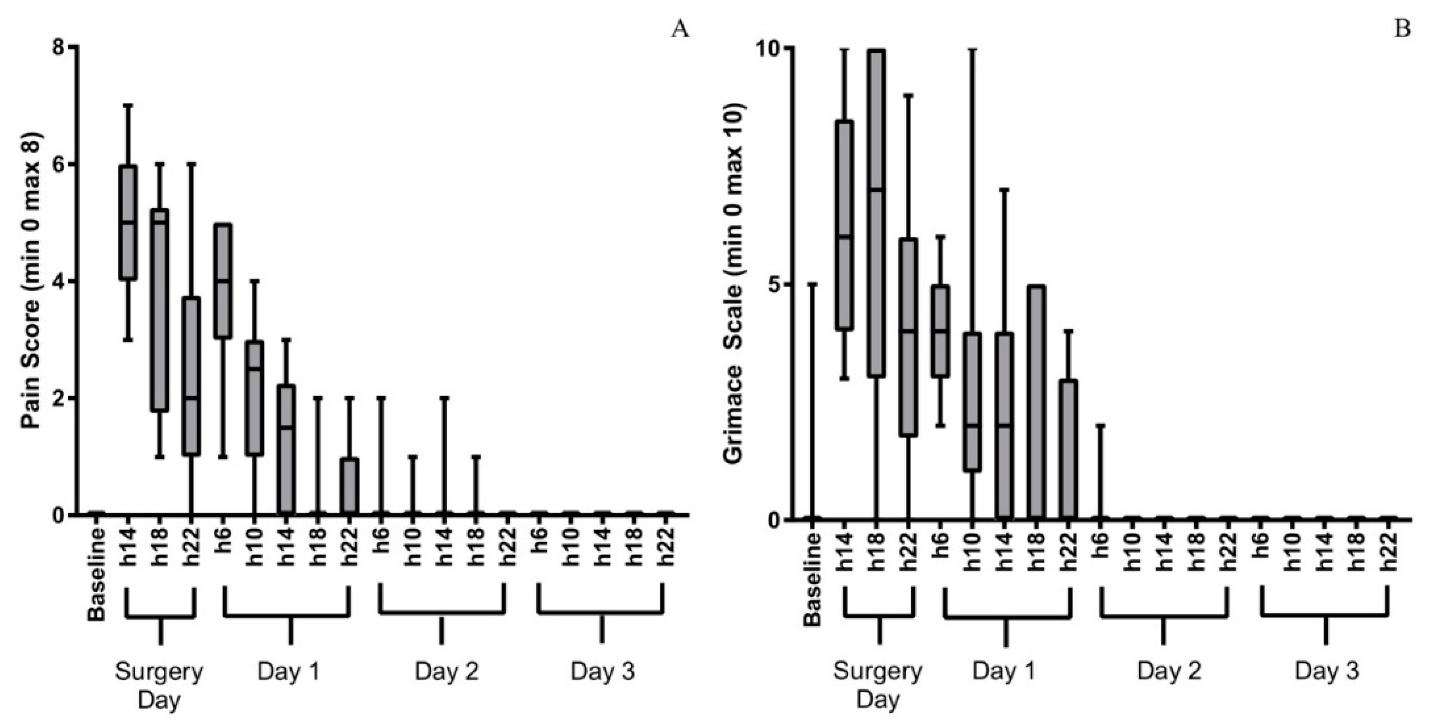
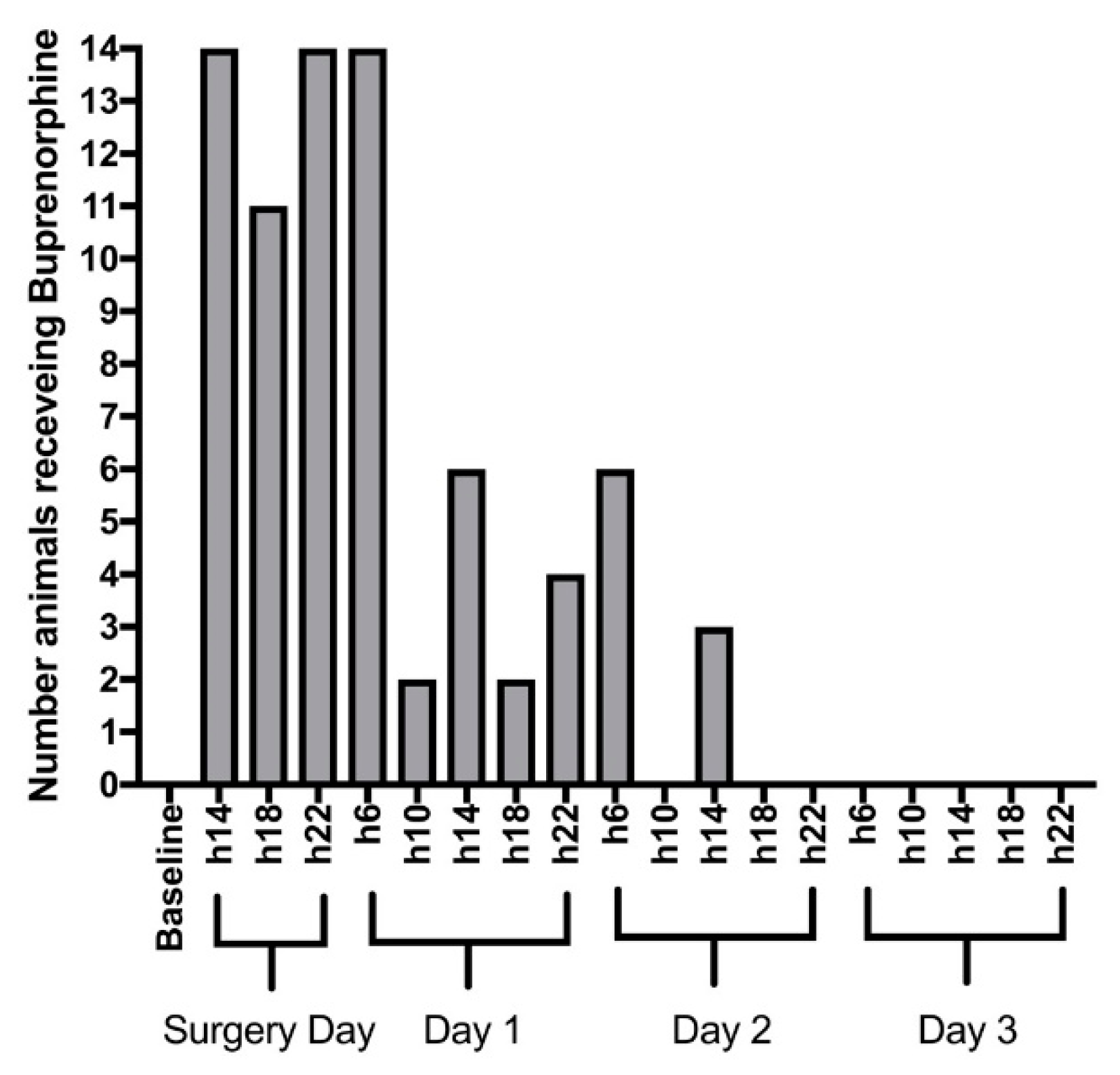
3. Results
4. Discussion
5. Conclusions
Author Contributions
Funding
Acknowledgments
Conflicts of Interest
References
- Rottgers, S.A.; Cray, J.J. Bone morphogenetic protein 2–mediated mandible reconstruction successfully heals bony defects but inhibits concurrent inferior alveolar nerve grafting. J. Craniofac. Surg. 2014, 25, 2241–2245. [Google Scholar] [CrossRef] [PubMed]
- Fujioka-Kobayashi, M.; Kobayashi, E. Effect of recombinant human bone morphogenic protein 9 (rhBMP9) loaded onto bone grafts versus barrier membranes on new bone formation in a rabbit calvarial defect model. J. Biomed. Mater. Res. 2017, 105, 2655–2661. [Google Scholar] [CrossRef] [PubMed]
- Turkseven, A.; Ozçelik, D. Does periosteal graft combined with platelet-rich plasma enhance the healing of bone defect? J. Craniofac. Surg. 2018, 29, 1072–1080. [Google Scholar] [CrossRef] [PubMed]
- Brooker, J.E.; Camison, L.B. Reconstruction of a calvarial wound complicated by infection. J. Craniofac. Surg. 2019, 30, 260–264. [Google Scholar] [CrossRef] [PubMed]
- Jeon, Y.R.; Kim, M.J. Scaffold free bone regeneration using platelet-rich fibrin in calvarial defect model. J. Craniofac. Surg. 2018, 29, 251–254. [Google Scholar] [CrossRef]
- Gohringer, I.; Muller, C.L.S. Would be prophylactic administrations of low concentration of alendronate an alternative for improving the craniofacial bone repair? A preliminary study focused in the period of cellular differentiation and tissue organization. J. Craniofac. Surg. 2017, 28, 1869–1873. [Google Scholar] [CrossRef]
- Seok, H.; Kim, M.K. Comparison of silkworm-cocoon–derived silk membranes of two different thicknesses for guided bone regeneration. J. Craniofac. Surg. 2014, 25, 2066–2069. [Google Scholar] [CrossRef]
- Schallenberger, M.A.; Rossmeier, K. Comparison of the osteogenic potential of OsteoSelect demineralized bone matrix putty to Novabone calcium-phosphosilicate synthetic putty in a cranial defect model. J. Craniofac. Surg. 2014, 25, 657–661. [Google Scholar] [CrossRef]
- Alberius, P.; Klinge, B. Management of craniotomy in young rabbits. Lab. Anim. 1989, 23, 70–72. [Google Scholar] [CrossRef]
- Smeets, R.; Knabe, C. Novel silk protein barrier membranes for guided bone regeneration. J. Biomed. Mater. Res. 2016, 11, 2603–2611. [Google Scholar] [CrossRef]
- Cai, Y.; Guo, J. Silk fibroin membrane used for guided bone tissue regeneration. Mat. Sci. Eng. 2017, 70, 148–154. [Google Scholar] [CrossRef] [PubMed]
- Jeong, W.H.; Roh, T.S. Acceleration of osteogenesis by platelet-rich plasma with acellular dermal matrix in a calvarial defect model. Childs Nerv. Sys. 2016, 32, 1653–1659. [Google Scholar] [CrossRef] [PubMed]
- Lappalainen, O.P.; Karhula, S.S. Micro-CT Analysis of Bone Healing in Rabbit Calvarial Critical-Sized Defects with Solid Bioactive Glass, Tricalcium Phosphate Granules or Autogenous Bone. J. Oral Maxillofac. Res. 2016, 7, 1–8. [Google Scholar] [CrossRef] [PubMed]
- Lappalainen, O.P.; Haapea, M. Iron-labeled adipose stem cells and neovascularization in rabbit calvarial critical-sized defects. Oral Surg. Oral Med. Oral Pathol. 2016, 121, e104–e110. [Google Scholar] [CrossRef] [PubMed]
- Wang, Z.; Hu, H. Sheet of osteoblastic cells combined with platelet-rich fibrin improves the formation of bone in critical-size calvarial defects in rabbits. Brit. J. Oral Max. Surg. 2016, 54, 316–321. [Google Scholar] [CrossRef] [PubMed]
- Lappalainen, O.P.; Karhula, S. Bone healing in rabbit calvarial critical-sized defects filled with stem cells and growth factors combined with granular or solid scaffolds. Childs Nerv. Syst. 2016, 32, 681–688. [Google Scholar] [CrossRef] [PubMed]
- Yue, X.; Niu, M. In vivo evaluation of a simvastatin-loaded nanostructured lipid carrier for bone tissue regeneration. Nanotechnology 2016, 27, 115708. [Google Scholar] [CrossRef] [PubMed]
- Tseng, C.L.; Chang, G.W. Cranioplasty using a novel osteoconductive scaffold and platelet gel. Ann. Plast. Surg. 2016, 76, S125–S129. [Google Scholar] [CrossRef]
- Semyari, H.; Rajipour, M. Evaluating the bone regeneration in calvarial defect using osteoblasts differentiated from adipose-derived mesenchymal stem cells on three different scaffolds: An animal study. Cell Tissue Bank. 2016, 17, 69–83. [Google Scholar] [CrossRef]
- Jeon, Y.R.; Jung, B.K. Comparing the effect of nonactivated platelet-rich plasma, activated platelet-rich plasma, and bone morphogenetic protein-2 on calvarial bone regeneration. J. Craniofac. Surg. 2016, 27, 317–321. [Google Scholar] [CrossRef]
- Kim, S.G.; Kim, M.K. Comparison of unprocessed silk cocoon and silk cocoon middle layer membranes for guided bone regeneration. Maxillofac. Plast. Reconstr. Surg. 2016, 38, 1–8. [Google Scholar] [CrossRef] [PubMed]
- You, H.; Yoon, S.R. Bone regenerative efficacy of limited-dose Escherichia coli–derived rhbmp-2 with biphasic calcium phosphate carrier in rabbit calvarial defect model. Implant Dent. 2016, 25, 16–23. [Google Scholar] [CrossRef] [PubMed]
- Acar, A.H.; Yolcu, U. Bone regeneration by low-level laser therapy and low-intensity pulsed ultrasound therapy in the rabbit calvarium. Arch. Oral Biol. 2016, 61, 60–65. [Google Scholar] [CrossRef] [PubMed]
- Kim, S.; Hwang, Y. Evaluation of bone regeneration on polyhydroxyethyl-polymethyl methacrylate membrane in a rabbit calvarial defect model. In Vivo 2016, 30, 587–591. [Google Scholar] [PubMed]
- Kilkenny, C.; Browne, W.J. Improving bioscience research reporting: The ARRIVE guidelines for reporting animal research. PLoS Biol. 2010, 8, e1000412. [Google Scholar] [CrossRef] [PubMed]
- Coulter, C.A.; Flecknell, P.A. Reported analgesic administration to rabbits undergoing experimental surgical procedures. BMC Vet. Res. 2011, 7, 12. [Google Scholar] [CrossRef]
- Raekallio, M.; Ansah, O.B. Some factors influencing the level of clinical sedation induced by medetomidine in rabbits. J. Vet. Pharmacol. Ther. 2002, 25, 39–42. [Google Scholar] [CrossRef]
- Grint, N.J.; Murison, P.J. A comparison of ketamine–midazolam and ketamine–medetomidine combinations for induction of anaesthesia in rabbits. Vet. Anaesth. Analg. 2008, 35, 113–121. [Google Scholar] [CrossRef]
- Association of Veterinary Anaesthetists (AVA). Available online: https://ava.eu.com/wp-content/uploads/2017/10/AVA-ASA-Low-Electronic-UK.pdf (accessed on 5 September 2017).
- Keating, S.C.J.; Thomas, A.A. Evaluation of EMLA Cream for Preventing Pain during Tattooing of Rabbits: Changes in Physiological, Behavioural and Facial Expression Responses. PLoS ONE 2012, 7, e44437. [Google Scholar] [CrossRef]
- Hampshire, V.; Robertson, S. Using the facial grimace scale to evaluate rabbit wellness in post-procedural monitoring. Lab. Anim. 2015, 44, 259–260. [Google Scholar] [CrossRef]
- Flecknell, P.A. Laboratory Animal Anaesthesia, 3rd ed.; Academic Press: Burlington, MA, USA, 2009. [Google Scholar]
- McDonell, W.N.; Kerr, C.L. Physiology, pathophysiology, and anesthetic management of patients with respiratory disease. In Veterinary Anesthesia and Analgesia, Lumb and Jones, 5th ed.; Grimm, K.A., Lamont, L.A., Tranquilli, W.J., Eds.; Wiley Blackwell: Hoboken, NJ, USA, 2015; pp. 513–555. [Google Scholar]
- Crotaz, I.R. Initial feasibility investigation of the v-gel airway: An anatomically designed supraglottic airway device for use in companion animal veterinary anaesthesia. Vet. Anaesth. Analg. 2010, 37, 579–580. [Google Scholar] [CrossRef] [PubMed]
- Wenger, S.; Mullhaupt, D. Experimental evaluation of four airway devices in anaesthetized New Zealand White rabbits. Vet. Anaesth. Analg. 2017, 44, 529–537. [Google Scholar] [CrossRef] [PubMed]
- Engbers, S.; Larkin, A. Comparison of a Supraglottic Airway Device (v-gel®) with Blind Orotracheal Intubation in Rabbits. Front. Vet. Sci. 2017, 4, 365–368. [Google Scholar] [CrossRef] [PubMed]
- Hedenqvist, P.; Orr, H.E. Anaesthesia with ketamine/medetomidine in the rabbit: Influence of route of administration and the effect of combination with butorphanol. Vet. Anaesth. Analg. 2002, 29, 14–19. [Google Scholar] [CrossRef] [PubMed]
- Eatwell, K.; Mancinelli, E. Use of arterial blood gas analysis as a superior method for evaluating respiratory function in pet rabbits (Oryctolagus cuniculus). Vet. Rec. 2013, 173, 166. [Google Scholar] [CrossRef] [PubMed]
- Orr, H.E.; Roughan, J.V. Assessment of ketamine and medetomidine anaesthesia in the domestic rabbit. Vet. Anaesth. Analg. 2005, 32, 271–279. [Google Scholar] [CrossRef]
- Murphy, K.L.; Roughan, J.V. Anaesthesia with a combination of ketamine and medetomidine in the rabbit: Effect of premedication with buprenorphine. Vet. Anaesth. Analg. 2010, 37, 222–229. [Google Scholar] [CrossRef]
- Gonzalez-Gil, A.; Villa, A. Effects of Dexmedetomidine and Ketamine-Dexmedetomidine with and without Buprenorphine on Corticoadrenal Function in Rabbits. J. Am. Assoc. Lab. Anim. Sci. 2010, 54, 299–303. [Google Scholar]
- Hedenqvist, P.; Roughan, J.V. Assessment of ketamine/medetomidine anaesthesia in the New Zealand White rabbit. Vet. Anaesth. Analg. 2001, 28, 18–25. [Google Scholar] [CrossRef]
- Brodbelt, D.C.; Blissitt, K.J. The risk of death: The Confidential Enquiry into Perioperative Small Animal Fatalities. Vet. Anaesth. Analg. 2008, 35, 365–373. [Google Scholar] [CrossRef]
- Grint, N.J.; Murison, P.J. Peri-Operative body temperatures in isoflurane-anaesthetized rabbits following ketamine-midazolam or ketamine-medetomidine. Vet. Anaesth. Analg. 2007, 34, 181–189. [Google Scholar] [CrossRef] [PubMed]
- Leach, M.C.; Allweiler, S. Behavioural effects of ovariohysterectomy and oral administration of meloxicam in laboratory housed rabbits. Res. Vet. Sci. 2009, 87, 336–347. [Google Scholar] [CrossRef] [PubMed]
- Weaver, L.A.; Blaze, C.A. A Model for Clinical Evaluation of Perioperative Analgesia in Rabbits (Oryctolagus cuniculus). J. Am. Assoc. Lab. Anim. Sci. 2010, 49, 845–851. [Google Scholar] [PubMed]
- Barter, L.S. Rabbit Analgesia. Vet. Clin. N. Am. 2011, 14, 93–104. [Google Scholar] [CrossRef] [PubMed]
- Sorge, R.E.; Totsch, S.K. Sex Differences in Pain. J. Neurosci. Res. 2017, 95, 1271–1281. [Google Scholar] [CrossRef] [PubMed]


| Sedation Scale in Rabbits [27] | ||
|---|---|---|
| Variable | Behaviour of the rabbit | Score |
| Spontaneous posture | Normal | 0 |
| Sedated but standing/sitting with head up | 1 | |
| Lying sternally head up | 2 | |
| Lying sternally head down | 3 | |
| Lying laterally, responding to stimuli | 4 | |
| Lying laterally, not moving when stimulated | 5 | |
| Resistance to being placed dorsally recumbent | Strong/normal resistance | 0 |
| Moderate resistance | 1 | |
| Slight resistance | 2 | |
| No resistance | 3 | |
| Jaw relaxation | Normal tonus | 0 |
| No resistance to mouth opening | 1 | |
| Palpebral reflex | Normal | 0 |
| Decreased | 1 | |
| Absent | 2 | |
| Limb withdrawal in response to pinching | Normal | 0 |
| Decreased | 1 | |
| No reaction | 2 | |
| Total sedation score | ||
| Quality of Induction and Condition for V-gel® Insertion Modified from Grint and Murison [28] | ||
| Description | Score | |
| Excitement, and some struggling, requires additional intravenous ketamine to permit V-gel® insertion | 1 | |
| Some excitement, vigorous swallowing, additional ketamine needed to permit V-gel® insertion | 2 | |
| Smooth induction but some swallowing and resistance to V-gel® insertion | 3 | |
| Very smooth induction of anaesthesia, no swallowing during V-gel® insertion | 4 | |
| Composite Behavioural Pain Scale | ||
|---|---|---|
| Variable | Behaviour of the Rabbit | Score |
| Attitude | Active, interested | 0 |
| Calm, move when touched | 1 | |
| Immobile, consolidated, uninterested | 2 | |
| Appetite | Normal, eating and drinking | 0 |
| Only drinking or only eating | 1 | |
| Not eating or drinking | 2 | |
| Mobility | Mobility is normal | 0 |
| Pain during action | 1 | |
| Inactive | 2 | |
| Appearance | Straight, bright pelt | 0 |
| Dull, slightly ruffled fur | 1 | |
| Ruffled fur | 2 | |
| Total score | (Best = 0, Worst = 8) | |
| Rabbit Grimace Scale [29,30] | ||
| Orbital Tightening | 0 | |
| 1 | ||
| 2 | ||
| Cheek Flattening | 0 | |
| 1 | ||
| 2 | ||
| Nose Shape | 0 | |
| 1 | ||
| 2 | ||
| Whisker Position | 0 | |
| 1 | ||
| 2 | ||
| Ear Position | 0 | |
| 1 | ||
| 2 | ||
| Total Pain score | (Best = 0, Worst = 10) | |
| Rabbit | 1 | 2 | 3 | 4 | 5 | 6 | 7 * | 8 | 9 | 10 | 11 * | 12 | 13 | 14 |
|---|---|---|---|---|---|---|---|---|---|---|---|---|---|---|
| Weight | 3.7 | 3.2 | 3.8 | 3.3 | 3.5 | 3.4 | 3.4 | 3.3 | 3.5 | 3.6 | 3.5 | 3.3 | 3.5 | 3.3 |
| Sedation score | 10 | 13 | 13 | 13 | 13 | 12 | 13 | 13 | 13 | 13 | 10 | 13 | 13 | 13 |
| Quality score | 2 | 4 | 4 | 4 | 4 | 4 | 3 | 4 | 4 | 4 | 2 | 4 | 4 | 4 |
| V-gel® | R4 | R4 | R4 | R4 | R4 | R4 | FM | R4 | R4 | R4 | R3 | R4 | R4 | R4 |
| % iso | 1–2.5 | 0.5-1.5 | 0–0.5 | 0.2–1 | 0.5–1.0 | 0.2–1.2 | 0 | 0 | 0.2–1 | 0–0.5 | 0–1 | 0 | 0 | 0 |
| PR (minx–max) beats minute−1 | 200–250 | 204–221 | 195–214 | 151–182 | 128–157 | 155–216 | 174–198 | 130–184 | 134–175 | 170–183 | 154–208 | 169–187 | 132–165 | 170–185 |
| MAP (min–max)(mmHg) | 55–65 | 72–77 | 57–74 | 64–74 | 69–77 | 61–79 | 64–77 | 59–76 | 64–82 | 58–67 | 56–82 | 69–83 | 67–75 | 61–68 |
| PE’CO2 (min–max)(kPa) | 5.3–7.5 | 4.7–7.2 | 5.2–8.3 | 5.3–6.1 | 4.3–6.5 | 5.5–8.3 | 8.4–10.8 | 6.7–9.7 | 6.1–8.7 | 5.0–7.0 | 3.3–NA | 5.6–6.8 | 4.8–6.4 | 5.0–6.7 |
| PE’CO2 (min–max)(mmHg) | 40–56 | 35–54 | 39–60 | 40–46 | 32–49 | 41–62 | 63–81 | 50–73 | 46–65 | 38–53 | 25–NA | 42–51 | 36–48 | 38–50 |
| Anaesth. Duration (min) | 80 | 65 | 75 | 60 | 65 | 60 | 65 | 70 | 70 | 70 | 85 | 65 | 55 | 60 |
| Surgery duration (min) | 45 | 30 | 30 | 35 | 35 | 30 | 35 | 40 | 40 | 30 | 40 | 30 | 30 | 30 |
| Return righting reflex (min) | 20 | 45 | 35 | 35 | 35 | 25 | 25 | 45 | 25 | 25 | 20 | 40 | 35 | 55 |
| Rabbit | 1 | 2 | 3 | 4 | 5 | 6 | 7 * | 8 | 9 | 10 | 11/1 | 11/2 * | 12/1 | 12/2 | 13/1 | 13/2 | 14/1 | 14/2 |
|---|---|---|---|---|---|---|---|---|---|---|---|---|---|---|---|---|---|---|
| pH | 7.29 | NA | 7.36 | 7.39 | 7.40 | 7.41 | 7.30 | 7.27 | 7.35 | 7.35 | 7.46 | 7.35 | 7.23 | 7.24 | 7.34 | 7.29 | 7.31 | 7.36 |
| PaO2 (kPa) | 13.2 | NA | 31.1 | 37.3 | 49.7 | 48.3 | 10.3 | 39.1 | 43.4 | 47.3 | 54.3 | 45.3 | 25.3 | 43.1 | 9.1 | 50 | 23.3 | 43.6 |
| PaO2 (mmHg) | 99 | NA | 233 | 280 | 373 | 362 | 77 | 293 | 306 | 355 | 407 | 340 | 190 | 323 | 68 | 375 | 175 | 327 |
| PaCO2 (kPa) | 4.8 | NA | 9.3 | 8.8 | 9.9 | 8.8 | 8.8 | 10.5 | 10.0 | 8.3 | 6.0 | 9.1 | 8.9 | 10.5 | 7.9 | 9.9 | 7.9 | 9.2 |
| PaCO2 (mmHg) | 36 | NA | 70 | 66 | 74 | 66 | 66 | 79 | 75 | 62 | 45 | 68 | 67 | 79 | 59 | 74 | 59 | 69 |
| BE | 16 | NA | 13 | 14 | 21 | 18 | 7 | 10 | 16 | 8 | 9 | 12 | 0 | 6 | 6 | 9 | 4 | 14 |
| Glucose (mmol L−1) | 20 | NA | 21 | 18 | 17 | 18 | 15 | 15 | 16 | 17 | 13 | 24 | 10 | 15 | 13 | 20 | 10 | 19 |
| Lactates (mmol L−1) | 1.4 | NA | 1.0 | 1.0 | 0.9 | 1.1 | 0.5 | 1.1 | 1.3 | 0.7 | 1.9 | 1.2 | 0.9 | 0.5 | 1.9 | 1.0 | 2.5 | 0.6 |
© 2019 by the authors. Licensee MDPI, Basel, Switzerland. This article is an open access article distributed under the terms and conditions of the Creative Commons Attribution (CC BY) license (http://creativecommons.org/licenses/by/4.0/).
Share and Cite
Raillard, M.; Detotto, C.; Grepper, S.; Beslac, O.; Fujioka-Kobayashi, M.; Schaller, B.; Saulacic, N. Anaesthetic and Perioperative Management of 14 Male New Zealand White Rabbits for Calvarial Bone Surgery. Animals 2019, 9, 896. https://doi.org/10.3390/ani9110896
Raillard M, Detotto C, Grepper S, Beslac O, Fujioka-Kobayashi M, Schaller B, Saulacic N. Anaesthetic and Perioperative Management of 14 Male New Zealand White Rabbits for Calvarial Bone Surgery. Animals. 2019; 9(11):896. https://doi.org/10.3390/ani9110896
Chicago/Turabian StyleRaillard, Mathieu, Carlotta Detotto, Sandro Grepper, Olgica Beslac, Masako Fujioka-Kobayashi, Benoit Schaller, and Nikola Saulacic. 2019. "Anaesthetic and Perioperative Management of 14 Male New Zealand White Rabbits for Calvarial Bone Surgery" Animals 9, no. 11: 896. https://doi.org/10.3390/ani9110896
APA StyleRaillard, M., Detotto, C., Grepper, S., Beslac, O., Fujioka-Kobayashi, M., Schaller, B., & Saulacic, N. (2019). Anaesthetic and Perioperative Management of 14 Male New Zealand White Rabbits for Calvarial Bone Surgery. Animals, 9(11), 896. https://doi.org/10.3390/ani9110896