1. Introduction
Given the increasingly severe concerns over antibiotic resistance and biosafety risks, natural polysaccharide bioactive molecules have attracted widespread attention in biofunctional molecule research and physiological regulation due to their excellent biocompatibility and multiple health-benefiting potentials [
1,
2]. Polysaccharides are widely present in algae, plants, fungi, and microbial metabolites, exhibiting multiple activities such as antioxidant, immunomodulatory, and antibacterial properties. However, their stability in the gastrointestinal environment, bioavailability, and interaction mechanisms with the gut microbiota, particularly how structural specificity influences their functional expression, remain key directions in current research [
3].
Microbial polysaccharides refer to a class of functional high-molecular-weight polymers synthesized and secreted by microorganisms such as bacteria and fungi during their metabolic processes, typically characterized by high structural complexity and branching degrees. Compared to polysaccharides derived from plants or animals, microbial polysaccharides offer advantages such as diverse sources, high production efficiency, ease of large-scale fermentation, and resistance to seasonal or environmental fluctuations, making them a recent focus of research on dietary functional components [
4]. β-glucans, as representative non-starch polysaccharides, are widely distributed in the cell walls of various biological sources such as yeast, bacteria, fungi, and grains. Their biological functions are closely related to their structural characteristics, including monosaccharide composition, glycosidic bond types, and branching patterns. Due to its unique β-1,3, β-1,4, and β-1,6 glycosidic bond structure, β-glucan has demonstrated immune-enhancing, anti-inflammatory, and intestinal barrier protective activities in various animal models [
5,
6,
7]. The physiological activity of polysaccharides is highly correlated with their structure. Specifically, monosaccharide composition, glycosidic bond types, degree of branching, molecular weight distribution, and spatial conformation collectively determine their resistance to digestion in the upper gastrointestinal tract and the degree to which they are utilized by specific microbial fermentation pathways in the intestine, thereby influencing the profile of short-chain fatty acids and the structure of microbial communities [
8].
Notably, galactoglucan, as a structurally distinct subclass of microbial polysaccharides (or more specifically, β-glucans), is characterized by the presence of galactose side chains attached to its backbone. This structural feature endows it with distinct physicochemical properties and physiological functions compared to typical linear or branched β-glucans. Given that these structural modifications are likely to influence its interactions with and modulation of the gut microbiota—a key mechanism for many polysaccharide bioactivities—investigating its specific effects is of particular interest. The core polysaccharide, L-β-galactoglucan targeted herein, has been fully structurally elucidated with established preparation protocols in previous research [
9]. Although the strain fermentation batches differ, the consistent fermentation and purification workflow yields a polysaccharide sharing the characteristic backbone structure of reported L-β-galactoglucan. Therefore, this study selected L-β-galactoglucan derived from the fermentation products of
Agrobacterium sp. FN01 isolated from highland soil. The molecular weight stability of this compound before and after in vitro digestion was analyzed using high-performance gel permeation chromatography–refractive index (HPSEC-RI). The L-β-galactoglucan-dominated crude fermentation product used for the in vivo feeding trial was obtained from a single production batch. Three independent production batches of purified L-β-galactoglucan were additionally prepared and subjected to HPSEC-RI analysis to assess batch-to-batch variation in molecular weight distribution. For the batch used in the pig feeding trial, the polysaccharide content was determined as 75.10% on a dry-matter basis, with residual components consisting of 1.72% moisture, 13.30% crude protein, and 9.5% ash. Notably, the purified L-β-galactoglucan for in vivo structural and enzymatic hydrolysis characterization was obtained by laboratory-scale isolation, and its yield was insufficient for large-scale pig feeding. Therefore, the industrial crude fermentation product described above was adopted for the in vivo trial. Additionally, animal experiments with growing pigs, metagenomic sequencing, and short-chain fatty acid detection were combined to systematically evaluate the effects of this crude fermentation product on intestinal microbiota and digestive metabolism. This study aimed to probe the potential mode of action of its major component, L-β-galactoglucan, and lay a theoretical foundation for its basic biological research and swine feed application.
2. Materials and Methods
2.1. Animal Ethics
The experimental design and procedures used in this study were approved by the Animal Care and Use Committee of the Institute of Subtropical Agriculture, Chinese Academy of Sciences (No. ISA-2024-00-16, approval date: 23 May 2024). All animal experiments were performed in accordance with the ARRIVE guidelines and relevant national laboratory animal welfare regulations.
2.2. Preparation and Basic Characterization of L-β-Galactoglucan
L-β-galactoglucan was produced by fermentation of
Agrobacterium sp. FN01 following a patented procedure. Briefly, the strain was cultivated in a liquid fermentation medium under controlled conditions, after which bacterial cells were removed by centrifugation, and the extracellular polysaccharide was recovered from the supernatant. The crude polysaccharide was then concentrated and subjected to ethanol precipitation, followed by deproteinization using a modified Sevag method. The resulting polysaccharide fraction was desalted, spray-dried, and supplied as a powder. The polysaccharide was a high-molecular-weight, branched galactoglucan mainly composed of glucose and galactose residues [
9].
2.3. HPSEC-RI for Molecular Weight Analysis
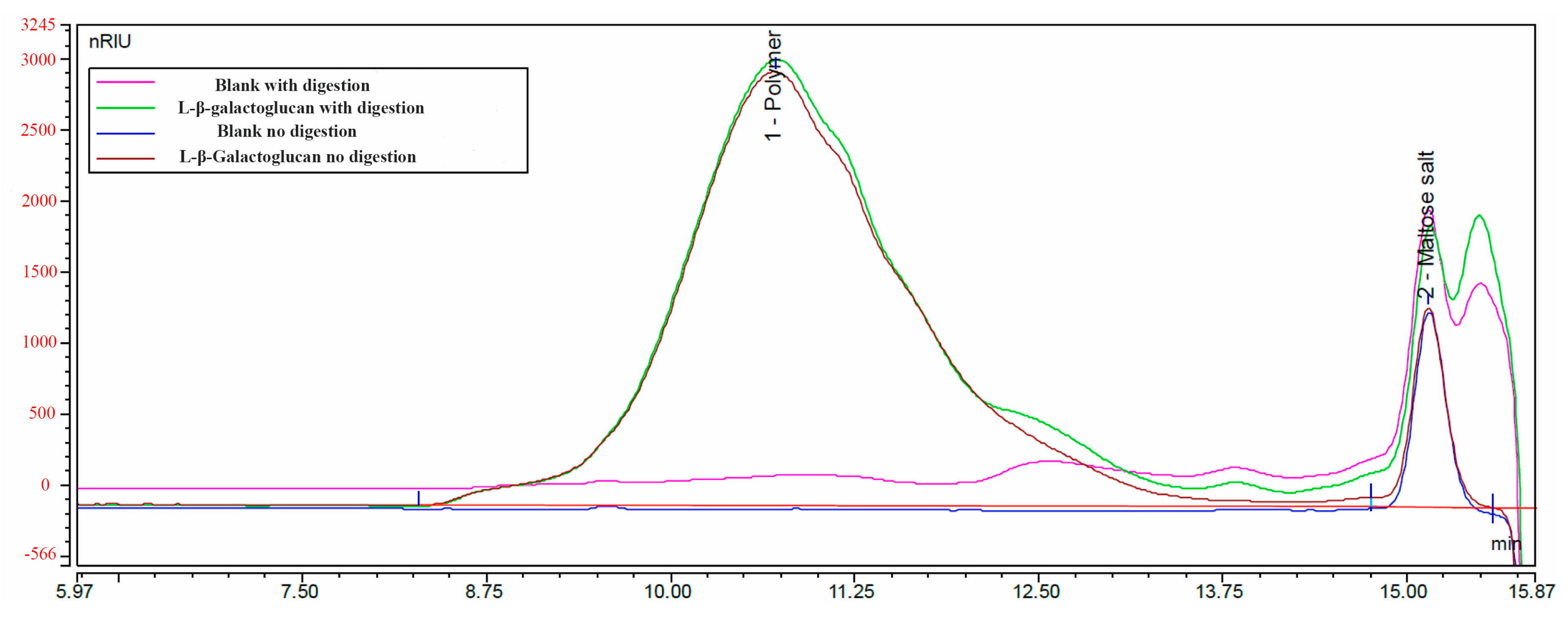
The molecular weight distribution of L-β-galactoglucan and its enzymatic hydrolysis products were analyzed using high-performance gel permeation chromatography–refractive index detection (HPSEC-RI). Digestion was performed according to AOAC 2017.16, involving sequential enzymatic hydrolysis using pancreatic α-amylase followed by amyloglucosidase to simulate the enzymatic environment of the small intestine. The digestion solution concentration was approximately 2.5 g/L (20 mg dissolved in 8 mL solution). After digestion, the samples were heated at 100 °C for 10 min to inactivate enzymes and microorganisms, diluted threefold, and filtered through a membrane filter for later use. Chromatographic separation was performed using three series-connected columns (including a guard column) of the Tosoh Bioscience TSKgel Super AW series (Tosoh Bioscience, Tokyo, Japan), with 0.4 M NaNO3 (pH 2.5, adjusted with nitric acid) as the mobile phase, and elution was carried out at 55 °C. The detector was a differential refractive index detector. Molecular weight calibration was performed using Pullulan series standards with known molecular weights and disaccharides (342 Da).
2.4. Animals and Experimental Design
The doses of crude L-β-galactoglucan used in the formal experiment were selected based on a preliminary 42 d feeding trial (
Supplementary Table S1), in which 28-day-old weaned piglets (Duroc × Landrace × Large White; 7.47 ± 0.23 kg) were fed diets supplemented with 0, 200, 400, or 600 mg/kg of crude L-β-galactoglucan. Based on the results of this preliminary study, 0, 200, and 400 mg/kg were selected for the subsequent experiment.
This experiment used 624 piglets (Duroc × Berkshire × Ningxiang pigs), weaned at 27 days of age with an initial body weight (BW) of 6.89 ± 1.38 kg. Approximately half of the pigs were castrated males, and the remaining were intact females. At 37 days of age, stratified randomization was conducted on pens rather than individual pigs. All pens were stratified based on the average initial body weight and sex ratio within each pen, and each whole pen was randomly assigned to one of the three treatments: 0 mg/kg (control group), 200 mg/kg, and 400 mg/kg crude L-β-galactoglucan in the diet. Each treatment included 16 replicate pens, and each pen housed 13 piglets with a balanced sex ratio at the beginning of the trial.
The feeding program included two phases: a nursery phase (37–65 days of age) and a finishing phase thereafter (66–196 days of age). The nursery and finishing diets used in this experiment were commercial feeds supplied by Anyou Biotechnology Group Co., Ltd. (Jiangyin, China), and their ingredient composition and nutrient levels are presented in
Table 1. All diets were formulated to meet the nutrient requirements recommended by NRC (2012) [
10]. Pigs were weighed under fasting conditions on the morning of D37, D66, D96, D153, and D197. Feeding phases were defined as D37–65 (29 d), D66–96 (31 d), D97–153 (57 d), and D154–196 (43 d), with a total feeding duration of 160 d, which corresponded to the start of the trial, nursery phase, early finishing phase, mid-finishing phase, and late finishing phase. The pen was the experimental unit for BW, ADG, ADFI, and F/G because feed intake was recorded per pen. The calculation formulas for growth performance indicators were as follows: ADG = (final phase average BW − initial phase average BW)/phase days; ADFI = total phase feed intake/total animal-days; and F/G = ADFI/ADG. Dead or removed animals were excluded when computing pen-level average body weights, while the pen was still kept as an experimental replicate. Animal-day adjustment was only applied for ADFI calculation to account for mortality or animal removal. Total feed consumption included only feed intake corresponding to the survival period of animals that died or were removed during the experimental period. No animal-day adjustment was performed for ADG calculation.
During the entire experimental period, the indoor temperature was maintained at 24–28 °C and relative humidity was controlled at 55–70%, and the pigs had ad libitum access to feed and water. Continuous mechanical ventilation was operated to ensure air quality inside the facility. Environmental management, including regular cleaning, disinfection, and control of temperature and humidity, was performed in accordance with standard farm protocols to maintain a dry and hygienic environment. No therapeutic antibiotics were administered during the feeding period. Other feeding management procedures followed routine protocols. Daily health monitoring was carried out throughout the trial. At day 66, pigs with body weight below 15 kg were removed from their pens according to the pre-defined trial criteria, as excessively low body weight would confound the evaluation of finishing-phase performance. These animals were included in the growth-performance calculation for the D37–66 period, but excluded from all finishing-phase indices (D66–196). Several pigs died or suffered severe illness during the experiment. These individual animals were excluded from growth-performance calculations, but no entire pen was excluded from statistical analysis. Mortality and animal-removal information for each pen is summarized in
Supplementary Table S2. At the end of the experiment, selected pigs were rendered unconscious via electrical stunning followed by exsanguination in accordance with standard commercial humane slaughter procedures.
2.5. Sample Collection
For digestibility, SCFA and metagenomic analyses, 10 pigs per treatment were selected from 10 separate pens, with one pig sampled from each pen. Three days before slaughter, fresh feces were collected every morning for three consecutive days by gently stimulating the rectum with sterile cotton swabs to induce defecation, avoiding contamination by urine and feed residues. All fresh fecal samples were placed into self-sealing bags and immediately stored at −20 °C after collection. Before nutrient determination, fecal samples collected over three days from the same pig were fully mixed for subsequent analysis. Meanwhile, approximately 200 g of diet samples per group were collected by the quartering method and stored at 4 °C. At the end of the trial, another 10 pigs per group were selected to collect colonic digesta. The samples were placed into sterile centrifuge tubes and preserved at −80 °C for subsequent metagenomic sequencing. All samples collected from each group were used for the determination of apparent digestibility. Eight samples per treatment were randomly selected for metagenomic sequencing and functional analysis. From these eight sequenced samples, six were further randomly subsampled for SCFA. Complete sample mapping information is provided in
Supplementary Table S4.
2.6. Apparent Digestibility
Diets and fecal samples were ground to pass through a 40-mesh screen. Fecal samples were dried at 65 °C and stored for subsequent analysis. Acid-insoluble ash (AIA) was used as an indicator to determine the apparent digestibility of dry matter (DM), gross energy (GE), crude protein (CP), and crude fat (EE) in the diet and feces. Dry matter was determined according to GB/T 6435-2014 [
11], and AIA was analyzed following GB/T 23742-2009 [
12]. Crude protein was measured using the Dumas combustion method, and nitrogen content was converted to CP using a factor of 6.25. Ether extract was determined by Soxhlet extraction using petroleum ether as the solvent. Gross energy was measured using an adiabatic bomb calorimeter. All analytical procedures were conducted in accordance with Chinese national standard methods. The formula for calculating the apparent digestibility of nutrients was: Apparent digestibility (%) = [1 − (A1 × F2)/(A2 × F1)] × 100, where A1 and A2 represent the AIA content (%) in the diet and fecal samples, respectively; F1 and F2 represent the nutrient content (%) in the diet and fecal samples, respectively.
2.7. DNA Extraction and Sequencing
Metagenomic DNA was extracted from colonic digesta samples using the DNeasy® PowerSoil® kit (Qiagen, Hilden, Germany). Approximately 1 μg of genomic DNA was fragmented to ~350 bp by Covaris sonication. Libraries were constructed following the NEBNext® Ultra™ DNA Library Preparation Kit protocol, including end repair, A-tailing, adapter ligation, purification, and PCR amplification. Library quality and insert size were verified by AATI, and effective library concentration was quantified via qPCR (only libraries >3 nM were qualified). Qualified libraries were pooled and sequenced on the Illumina NovaSeq platform with paired-end 150 bp (PE150) at Novogene Co., Ltd. (Beijing, China).
2.8. Metagenomic Data Analysis
Raw reads were preprocessed with fastp (v0.23.1) to obtain clean reads. Reads containing adapters, excessive low-quality bases (Q ≤ 5 over 50% of the length), or >10% ambiguous N bases were discarded. Bowtie2 (v2.5.4), was used to filter host-origin reads against the pig reference genome Sus scrofa11.1 (GCA_000003025.6) with parameters --end-to-end, --sensitive, -I 200, -X 400. Clean non-host reads were assembled using MEGAHIT (--presets meta-large, v1.2.9). Scaffolds were split at N gaps to generate scaftigs. ORF prediction on scaftigs (≥500 bp) was performed by MetaGeneMark.hmm (v2.1); sequences shorter than 100 nt were removed. Predicted genes were clustered by CD-HIT (v4.5.8) (-c 0.95, -aS 0.9, -G 0, -g 1, -d 0) to build a non-redundant gene catalog. Bowtie2 was applied to map clean reads to the gene catalogue, and genes with ≤2 mapped reads were filtered out. Gene abundance was calculated based on mapped read counts and gene length. For annotation, unigenes were aligned using DIAMOND (v2.1.9) with the parameters “blastp, -e 1 × 10
−5”. Taxonomic classification was conducted against Micro NR (sequences extracted from NCBI NR database,
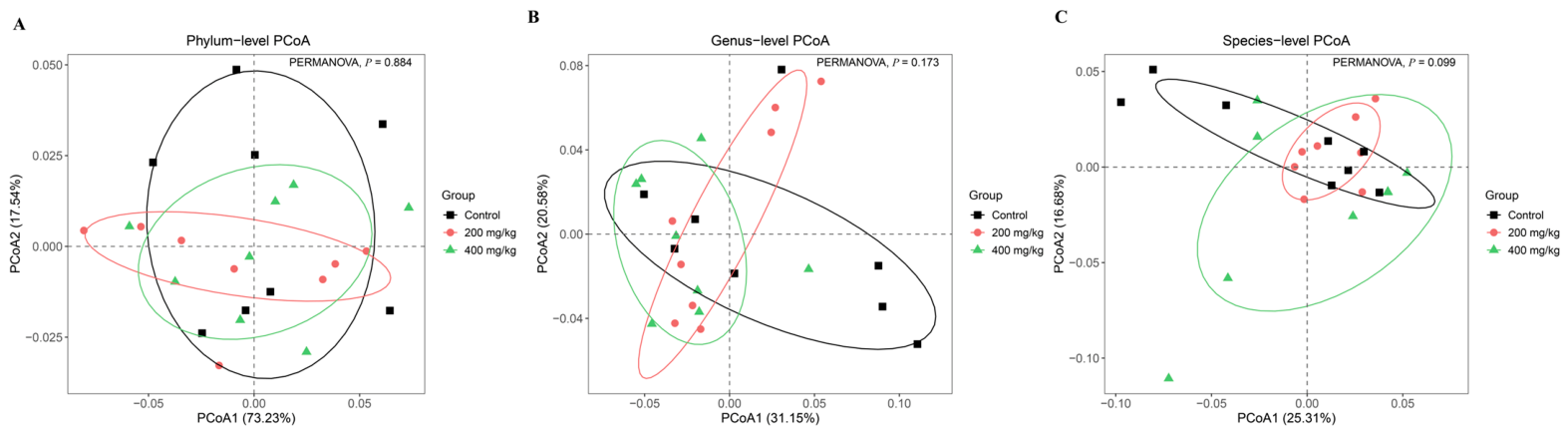
https://www.ncbi.nlm.nih.gov/, accessed 5 January 2025) using the LCA algorithm. Functional annotation was performed against KEGG, eggNOG v5.0, CAZy, VFDB and PHI databases. Database information: NCBI NR (downloaded: 10 January 2025), KEGG database (downloaded: 10 January 2025), and eggNOG v5.0 (downloaded: 10 January 2025). In community structure analysis, α-diversity indices (e.g., Shannon index, Chao 1 index) and β-diversity metrics (e.g., Bray–Curtis distance) were calculated at the phylum, genus, and species levels. The Bray–Curtis distance matrix was generated for principal coordinate analysis (PCoA), PERMANOVA, and PERMDISP, with 999 permutations applied for both permutation-based tests to evaluate intergroup differences in bacterial community structure. Differential taxa and functional genes (KO and eggNOG entries) were identified using the MetagenomeSeq (
https://bioconductor.org/packages/metagenomeSeq/, accessed 10 January 2025) approach. Raw abundance data were normalized by cumulative sum scaling (CSS). Prior to differential abundance testing, taxonomic features (phylum, genus, and species level) and functional profiles (KO, eggNOG) were pre-filtered by prevalence. Only features present in at least 15% of all samples were retained for subsequent differential analysis. Hypothesis testing was performed to generate raw
p-values, which were further adjusted via FDR correction to calculate
Q values. All statistical outputs, including raw
p-values,
Q-values, effect-size estimates, and prevalence information, are summarized in
Supplementary Table S6. Taxa and functional genes with
Q < 0.05 were selected for visualization.
2.9. Short-Chain Fatty Acid Analysis
Short-chain fatty acids (SCFAs) content in colonic digesta was determined using solid-phase extraction (SPE) combined with gas chromatography–flame ionization detection (GC-FID, Agilent Technologies, Santa Clara, CA, USA), following the method described by Zheng et al. [
13]. SCFAs included acetate, propionate, isobutyrate, butyrate, isovalerate, valerate, and caproate.
The specific method is as follows: Weigh 50 mg of colonic digesta sample and place it in a 2 mL centrifuge tube. Add 400 µL of acetone, homogenize using a handheld tissue grinder, then add 600 µL of acetone, and vortex-mix thoroughly for 3 min. Centrifuge the sample at 6000× g at 4 °C for 10 min, and collect the supernatant for SPE. Activate the SPE column (Bond Elut Plexa, Agilent) with 1 mL of acetone, remove any residual solvent, and load the entire supernatant onto the column. Allow the sample to elute by gravity and collect the eluent in a clean centrifuge tube. The collected solution is directly used for GC-FID analysis. GC-FID analysis conditions: Chromatography column: DB-FFAP (30 m × 0.25 mm × 0.25 µm, Agilent); carrier gas: nitrogen; flow rate: 1 mL/min; injection port temperature: 280 °C; detector temperature: 250 °C; and hydrogen and air flow rates: 40 mL/min and 300 mL/min, respectively. The column temperature program was set as follows: initial 50 °C, held for 1 min, then increased at 15 °C/min to 120 °C, followed by an increase at 6 °C/min to 200 °C, and finally increased to 235 °C and held for 3 min for washing. The injection needle was cleaned with high-purity water and acetone before each injection. The concentrations of SCFAs were calculated using the external standard method based on the standard curve.
2.10. Statistical Analysis
For correlation analysis, both Spearman’s rank-order correlation and partial correlation (treatment group as a confounder) were performed using the psych package in R software (Version 4.5.1; R Foundation for Statistical Computing, Vienna, Austria). Raw
p-values from Spearman and partial correlation were adjusted separately with the Benjamini–Hochberg FDR procedure to control multiple-testing risk, and the significance threshold was set at FDR-adjusted
Q < 0.05. Correlation network diagrams were plotted using the online platform CNSknowall (available at:
https://www.cnsknowall.com/, accessed on 20 August 2025).
Experimental data were analyzed using SPSS statistical software (Version 26.0; SPSS Inc., Chicago, IL, USA). The overall effects of Treatment, Time, and Treatment × Time interaction on repeatedly measured body weight were evaluated using a linear mixed model, with pen regarded as the random experimental unit. Other indicators were compared using one-way analysis of variance (ANOVA) and Tukey’s post hoc test. For datasets that failed to meet the homogeneity-of-variance and normality assumptions for parametric ANOVA, the Kruskal–Wallis H-test was applied. Differences were considered significant at p ≤ 0.05. A tendency toward difference was recognized when 0.05 < p ≤ 0.10, while no significant difference was observed at p > 0.05. The data are expressed as means and standard error of the mean (SEM).
4. Discussion
In recent years, growing attention has been paid to host gut microecological balance and safe natural bioactive molecules. Gut homeostasis regulation has become a core research hotspot for natural biological modulators. Prebiotic polysaccharides, due to their unique structural stability and digestive tolerance, can reach the colon intact and be utilized by the microbiota, thereby playing a significant role in regulating the gut environment and improving host nutrient metabolism. Previous studies have shown that β-glucan, as a typical dietary fiber, is largely indigestible in the small intestine and is primarily fermented by the colonic microbiota to produce short-chain fatty acids, thereby improving intestinal barrier function and immune status [
8,
14].
In this study, HPSEC-RI analysis showed that L-β-galactoglucan was resistant to digestive enzymes under in vitro conditions. Although polysaccharides generally exhibit digestive resistance, their physicochemical properties (e.g., molecular weight, polymerization degree, glycosidic bond type, and branched structure) have been reported to influence fermentation rates, microbial selectivity, and metabolic product composition [
9,
15]. These differences may lead to distinct effects on the gut microbiota. Therefore, crude L-β-galactoglucans may regulate microbial community composition and metabolic activity through their structural characteristics, which could contribute to improvements in intestinal health and growth performance.
Previous studies have shown that β-glucans derived from microorganisms (including yeast, bacteria, and algae) can reduce pathogenic bacteria, enhance immune responses, and improve intestinal health and growth performance in weaned piglets [
7,
16]. However, compared with yeast- and algae-derived β-glucans, studies on bacterial-derived β-glucans remain limited. β-glucan derived from
Agrobacterium sp. ZX09 has been reported to improve growth performance and intestinal function in weaned piglets and finishing pigs [
17,
18,
19]. In the present study, a long-term feed trial demonstrated that supplementation with 400 mg/kg improved growth performance, as indicated by increased daily gain and final body weight, along with a lower F/G ratio compared to diets without supplementation. Additionally, apparent digestibility of DM, CP, EE, and GE was increased in the groups supplemented with 200 mg/kg and 400 mg/kg, suggesting that the observed growth-promoting effects were associated with enhanced nutrient utilization. These findings are consistent with previous studies showing that β-glucans can improve the growth performance and nutrient digestibility in pigs [
20]. Consistent with the significant Treatment × Day interaction for body weight observed in linear mixed model analysis, supplementation with crude L-β-galactoglucan at 200 mg/kg and 400 mg/kg modulated the growth performance of pigs via distinct pathways, and such responses exhibited obvious stage-dependent effects. During the late finishing period (d 154–196), the ADG of the 200 mg/kg group was numerically higher than that of the 400 mg/kg group, although no significant difference was detected between treatments. Notably, the ADG of pigs receiving 200 mg/kg increased markedly from the mid-finishing stage to the late finishing stage (from 589.01 to 781.38 g/d). The 400 mg/kg supplementation level exerted growth-promoting effects through improving feed efficiency and nutrient digestibility. In the nursery and early-to-middle growing-finishing stages, ADFI showed minor differences between the two treatments. Nutrient utilization efficiency served as the core limiting factor for growth, and superior performance was observed in the 400 mg/kg group during these periods. In the late finishing stage, divergent growth trends emerged between doses, resulting in numerically greater ADG in the 200 mg/kg group. Collectively, these findings suggest that low and high supplemental doses of crude L-β-galactoglucan differ in their regulatory mechanisms for growth, and there is no single optimal dose applicable to the whole growth cycle of pigs.
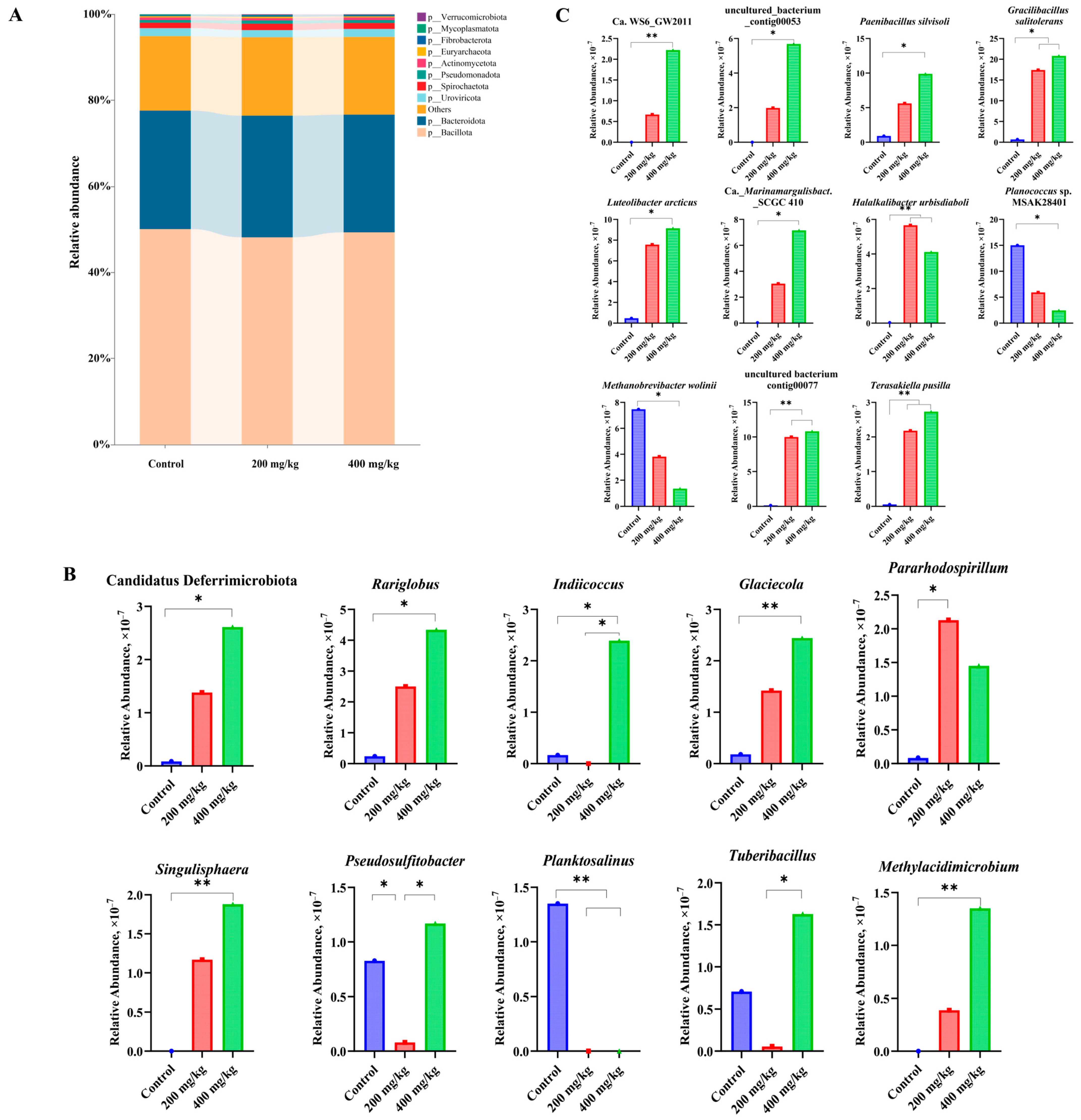
Metagenomic analysis revealed that crude L-β-galactoglucan did not significantly alter the dominant bacterial phyla, such as Bacillota and Bacteroidetes, across different supplementation levels, suggesting relative stability in the overall community structure. However, differential effects were observed at the level of low-abundance taxa. MetaGenomeSeq analysis indicated that the 400 mg/kg supplementation group increased the abundance of Candidatus Deferri
microbiota and several rare genera (e.g.,
Singulisphaera,
Glaciecola,
Methylacidimicrobium), while reducing the relative abundance of
Planktosalinus. At the species level, the 400 mg/kg treatment group enriched
Paenibacillus silvisoli,
Gracilibacillus salitolerans, and
Luteolibacter arcticus, and significantly inhibited the methanogenic bacterium
Methanobrevibacter wolinii. Additionally, the 200 mg/kg treatment group exhibited enrichment of several functionally related taxa, including
Halalkalibacter urbisdiaboli and
Terasakiella pusilla. These results suggest that crude L-β-galactoglucan may modulate the intestinal microbiota primarily through selective regulation of low-abundance taxa rather than directly dominant bacterial groups. Although the functional roles of these taxa in the pig intestine remain largely unclear, previous studies have reported that some of these genera possess capacities related to polysaccharide degradation and fermentation metabolism. For example,
Singulisphaera has been reported to degrade complex polysaccharides [
21,
22], and a polysaccharide utilization locus associated with Ulvan degradation has been identified in
Glaciecola, indicating its potential for utilizing sulfated polysaccharides [
23]. In addition,
Methylacidimicrobium has been characterized as a methanotrophic bacterium with methane and hydrogen metabolic pathways [
24]. Taken together, the structural characteristics of L-β-galactoglucan may provide selective ecological advantages for specific microbial taxa, thereby indirectly influencing intestinal metabolic processes and host nutrient utilization. Future studies are needed to verify these functional roles through metabolic prediction, functional annotation, or microbial isolation.
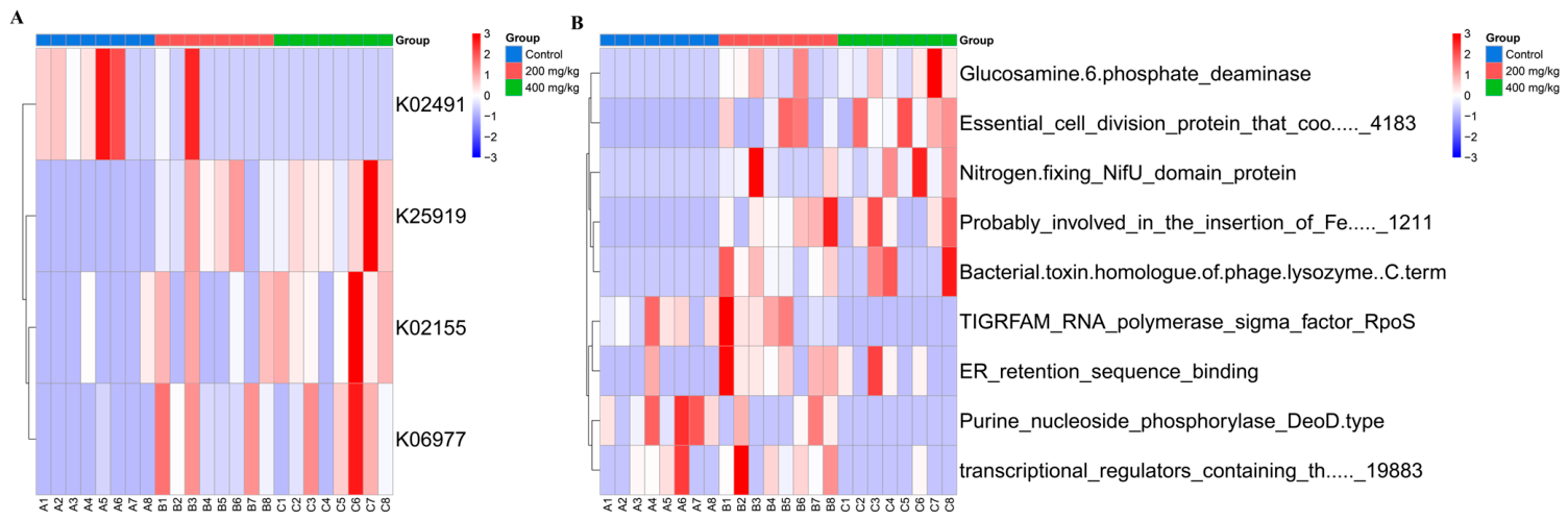
Functional annotation analysis indicated that the 400 mg/kg supplementation group increased the predicted relative abundance of selected functional genes. Specifically, genes related to Solabiose phosphorylase, V-type H-transporting ATPase, and the two-component system were enriched. V-type H-transporting ATPase is an important proton pump system involved in maintaining intracellular pH and regulating proton transmembrane transport. Although most studies have focused on eukaryotic systems, its role in microbial pH homeostasis has also been recognized [
25,
26]. Two-component systems, such as KdpD/KdpE, have been reported to sense environmental signals including potassium concentration, osmotic pressure, and cellular energy status, thereby regulating ion transport and stress adaptation [
27,
28]. At the eggNOG functional annotation level, the 400 mg/kg group increased the abundance of several metabolism-related functions, including essential cell division protein, NifU protein, and glucosamine-6-phosphate deaminase. These changes may indicate enhanced microbial growth potential, Fe–S cluster biosynthesis, and amino sugar metabolism [
29,
30]. Additionally, decreased abundance of RpoS and Purine nucleoside phosphorylase (DeoD-type) may reflect reduced stress response and a more stable metabolic homeostasis and state of the microbial community [
31]. In contrast, although the 200 mg/kg group also showed enrichment of certain metabolic functions, the magnitude of these changes was lower than that observed in the 400 mg/kg group. This suggests that higher supplementation levels have a greater impact on microbial functional modulation.
GC-FID analysis showed that acetate and propionate were the primary short-chain fatty acids in colonic digesta, followed by butyrate. Supplementation with 400 mg/kg increased the concentration of isobutyrate and isovalerate. Previous studies have shown that acetate serves as a substrate for multiple metabolic pathways, propionate can be utilized by the liver for gluconeogenesis, and butyrate acts as a primary energy source for colonic epithelial cells, supporting epithelial cell integrity and barrier function [
32]. In addition, inulin-type fructans have been reported to enhance short-chain fatty acid production, particularly butyrate [
33]. Isobutyrate and isovalerate are branched-chain fatty acids primarily derived from microbial fermentation of amino acids and are typically present at very low concentrations in the blood [
34]. Their production is influenced by dietary composition, particularly protein intake, and by metabolic characteristics of the gut microbiota [
35]. The increased levels of these metabolites in the present study may indicate that crude L-β-galactoglucans influenced not only the regulation of carbohydrate fermentation but also amino acid metabolism within the microbial community. These findings suggest that crude L-β-galactoglucans may modulate the metabolic profile of the gut microbiota through mechanisms that differ from those of conventional prebiotics such as β-glucans, inulin, and fructooligosaccharides.
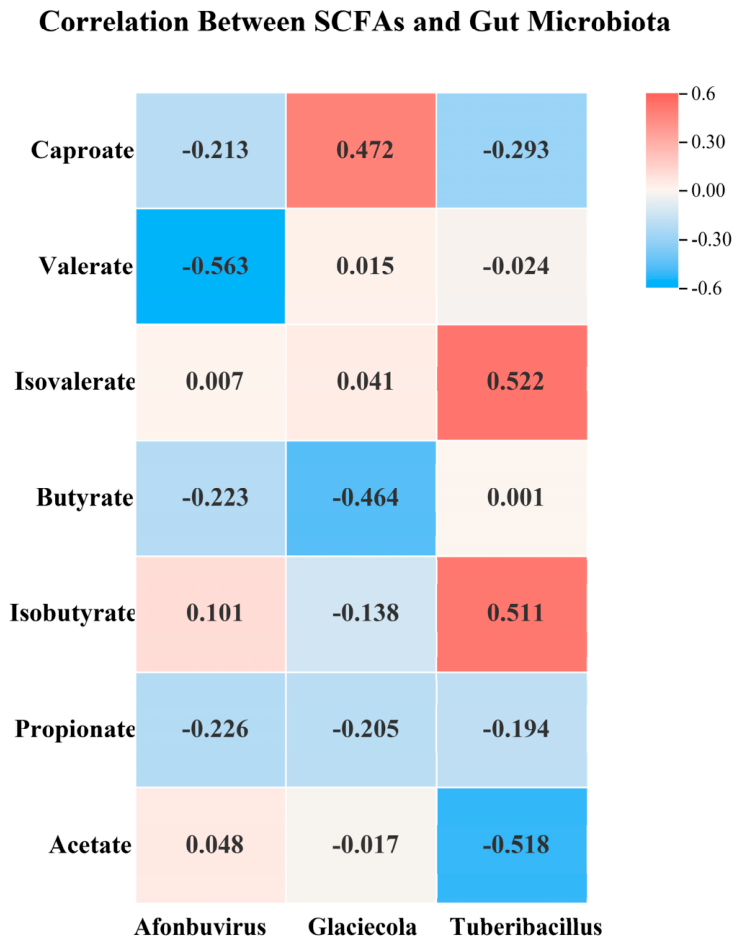
Analysis of the association between short-chain fatty acids and the microbiota showed that Tuberibacillus exhibited nominal positive associations with BCFAs at the raw p-value level, although these associations did not remain significant after Benjamini–Hochberg FDR correction. This observation hints that this genus might be potentially involved in protein degradation-related metabolism. Previous studies have demonstrated that some proteolytic microbes can produce isobutyrate and isovalerate via amino acid catabolism. It is speculated that crude L-β-galactoglucan might favor the proliferation or metabolic activity of such taxa, though this inference requires further experimental validation. In addition, Glaciecola showed nominal raw-p-level correlation with caproate, which may tentatively indicate altered functional niches of gut microbiota. Collectively, these correlative observations imply that crude L-β-galactoglucan could drive potential metabolic rearrangement toward multi-substrate and multi-pathway patterns in the intestinal microbiota, which remains to be verified by functional assays.




 ,
,