1. Introduction
Sow reproductive efficiency is pivotal for herd productivity and is closely linked to maternal skeletal health, a key determinant of sow longevity [
1]. During late gestation and lactation, sows must transfer large amounts of calcium to support rapid fetal growth and milk production, which inevitably increases mobilization of Ca from the maternal skeleton and predisposes sows to bone loss [
2,
3]. Importantly, evidence suggests that Ca supplementation alone is often insufficient to prevent this excessive skeletal mobilization in late gestation and lactation [
3,
4,
5], indicating that additional nutritional or endocrine regulators of Ca homeostasis are required.
Vitamin D plays an important role in maintaining calcium homeostasis for bone mineralization and skeletal muscle development [
6,
7]. Dietary vitamin D
3 undergoes sequential hydroxylation in the liver and kidneys to generate 1,25-(OH)
2-D
3, which regulates calcium balance by enhancing intestinal absorption and renal reabsorption and, when necessary, mobilizing skeletal calcium [
8,
9,
10]. By contrast, 25-hydroxyvitamin D
3 (25-OH-D
3) bypasses hepatic hydroxylation and is more readily available for subsequent activation [
11,
12]. In pigs, maternal 25-OH-D
3 supplementation has been reported to influence sow reproductive performance and colostrum composition and to support intestinal calcium absorption and bone properties in sow–piglet pairs [
13,
14]. Studies involving direct dietary supplementation of weaned piglets have associated 25-OH-D
3 with bone quality, antioxidant and immune indices, intestinal morphology, and tight-junction-related gene expression [
15,
16]. Thus, the reproductive and colostrum evidence derives from maternal sow studies, whereas much of the intestinal and immune evidence derives from direct supplementation studies in weaned piglets. Direct head-to-head comparisons among 25-OH-D
3 preparations produced by different chemical or microbial routes remain scarce, which provides the rationale and novelty for the present study.
Accordingly, we hypothesized that 25-OH-D3 preparations manufactured through different routes could produce source-dependent responses because differences in formulation, purity, stereochemical composition, stability, or recovery in feed may alter effective bioavailability even when the nominal supplemental dose is identical. The comparison was intended to evaluate biological responses among the available preparations rather than to attribute any observed difference solely to chemical or microbial origin. QV1 was commercially available, and chemically synthesized 25-OH-D3 was used as the reference material. QV2 and QV3 were biosynthesized 25-OH-D3 preparations produced using Saccharomyces cerevisiae and Bacillus subtilis as production strains, respectively. These strains were selected because they were the production hosts of the available candidate preparations and represented yeast- and bacterial-based biosynthetic platforms. Their inclusion was exploratory and was not based on prior evidence that one production strain would be biologically superior to the other in sows. The objective was to compare the three preparations during late gestation and lactation with respect to reproductive performance, maternal and offspring vitamin D-related indices, oxidative and inflammatory markers, piglet growth, femoral mineral concentrations, intestinal morphology, and barrier-related gene expression.
2. Materials and Methods
2.1. Animals and Experimental Design
This experiment was conducted at Saneng Pig Farm (Shanxi, China) and approved by the Animal Care and Use Committee of China Agricultural University (AW01705202-1-3, approval date: 10 July 2025). A total of 100 Large White × Landrace sows (parity: 4.25 ± 0.88; P2 backfat thickness: 17.50 ± 1.54 mm) were blocked by parity and P2 backfat thickness and then randomly assigned within each block to one of five dietary treatments (
n = 20 per treatment): CON (basal diet containing 25 µg/kg vitamin D
3), VD
3 (CON plus 50 µg/kg vitamin D
3), and QV1–QV3 (CON plus 50 µg/kg 25-hydroxyvitamin D
3 from three sources). Diets were formulated to meet NRC [
17] requirements for late gestation and lactation. The feeding period extended from day 90 of gestation to weaning (day 28 of lactation). Sows were individually housed with ad libitum access to water and received feed three times daily (08:00, 14:00, and 20:00).
2.2. Dietary Composition
Gestation and lactation diets were corn–soybean meal-based and supplemented with a vitamin–mineral premix (1%) and phytase. The premix supplied 25 µg/kg (1000 IU/kg) vitamin D
3, with additional vitamin D
3 or 25-OH-D
3 provided according to treatment. Because mineral metabolism and skeletal outcomes were central to the study, the basal gestation and lactation diets contained 0.67% and 0.88% calcium, 0.68% and 0.59% total phosphorus, and 0.26% and 0.38% available phosphorus, respectively (
Table 1).
Supplement Sources and Diet Verification
QV1 was a chemically synthesized 25-OH-D
3 used as the reference material. QV2 and QV3 were biosynthesized 25-OH-D
3 preparations produced using Saccharomyces cerevisiae and Bacillus subtilis as production strains, respectively. QV1, QV2, and QV3 were formulated premixes with a nominal 25-OH-D
3 content of 0.05% (
w/
w), with modified starch used as the carrier. The analyzed 25-OH-D
3 contents of the QV1, QV2, and QV3 premixes were 0.055%, 0.054%, and 0.056% (
w/
w), respectively, as determined according to the Chinese agricultural industry standard NY/T 3879-2021 [
18], Determination of 25-Hydroxyvitamin D
3 in Feeds. The premixes were added to the diets at 100 mg/kg, providing 50 μg/kg 25-OH-D
3. The CON and VD
3 diets received the same amount of modified starch carrier. Because the final dietary concentration was below the detection limit of the available method, dietary 25-OH-D
3 concentrations are reported as formulated values.
2.3. Sample Collection
Reproductive traits (total born, born alive, stillborns, mummified fetuses, piglets weaned, and farrowing duration) were recorded. Sow body weight and P2 backfat were measured at farrowing and weaning to calculate backfat loss; feed allowance during gestation and daily intake during lactation were recorded. Piglets were weighed at birth, day 14, and weaning, and litter weight gain was calculated. Six sows per treatment were randomly selected for blood and colostrum sampling. Sow blood samples were collected on gestation day 90 before the initiation of the experimental diets, at farrowing, and at weaning, and colostrum samples were collected at farrowing (n = 6 per treatment). At weaning, one clinically healthy piglet whose body weight was closest to the mean body weight of its litter was selected from each of six sampled litters per treatment and euthanized for collection of serum, liver, duodenum, jejunum, ileum, femur, and tibia. Piglet sex was not controlled during selection. The subset size was specified before sampling based on assay and terminal-tissue capacity while retaining six independent sows or litters per treatment. When cross-fostering was required, piglets were transferred only among sows in the same treatment, and litter performance outcomes were calculated using the actual post-fostering litter composition.
2.4. Vitamin D Metabolites, Biochemical Indices, Antioxidants, Cytokines, and Immunoglobulins
Serum and colostrum 25-OH-D3 were measured using a competitive ELISA kit (HB402-SH), and ALP using an assay kit (L-726-SH) (Shanghai Hengyuan Biotechnology Co., Shanghai, China). Concentrations of 1,25-dihydroxyvitamin D3 [1,25-(OH)2-D3], 24,25-dihydroxyvitamin D3 [24,25-(OH)2-D3], and vitamin D-binding protein (DBP) were quantified using commercial ELISA kits (HY028-NC, HY094-SH, and HY350-Pg, respectively). Serum calcium was measured by a GBHA-based micro-colorimetric method (520 nm), and serum phosphorus by a phosphomolybdate microplate method (660 nm). Antioxidant indices were determined using porcine ELISA kits for SOD (HB326X-Pg), GSH-Px (HB463X-Pg), CAT (HB251X-Pg), and MDA (HB443-Pg). Cytokines (IL-1β, HB354-Pg; IL-6, HB347-Pg; IL-10, HB359-Pg; TNF-α, HB015-Pg; IFN-γ, HB363-Pg) and colostrum IgG (HS173-Pg) and IgM (HS170-Pg) were quantified by ELISA. All ELISA kits were purchased from Shanghai Hengyuan Biotechnology Co., Ltd. (Shanghai, China) and were used according to the manufacturer’s instructions. The assay ranges and analytical sensitivities were 0.3–20 μg/L and 0.1 μg/L for 25-OH-D3, 3.2–200 pg/mL and 3 pg/mL for 1,25-(OH)2-D3, 0.32–20 ng/mL and 0.17 ng/mL for 24,25-(OH)2-D3, and 15.6–1000 μg/mL and 9.8 μg/mL for DBP, 1–45 ng/L and 0.25 ng/L for IL-1β, 50–1000 ng/L and 12.5 ng/L for IL-6, 8–180 ng/L and 2 ng/L for IL-10, 0.3–8 nmol/L and 0.075 nmol/L for MDA, 40–2000 U/L and 10 U/L for SOD, 5–165 IU/L and 1.25 IU/L for GSH-Px, 35–1500 U/L and 8.75 U/L for CAT, 10–400 pg/mL and 2.5 pg/mL for TNF-α, 100–2400 pg/mL and 25 pg/mL for IFN-γ, 12–400 μg/mL and 3 μg/mL for IgG and 1.2–40 μg/mL and 0.3 μg/mL for IgM, respectively. Absorbance was measured at 450 nm using a microplate reader. All samples were analyzed in duplicate, and samples with concentrations outside the lot-specific standard curve range were re-assayed after appropriate dilution. According to the manufacturer’s quality control specifications, the intra-assay and inter-assay coefficients of variation for the ELISA kits were less than 10% and 15%, respectively. The detection ranges and analytical sensitivities of individual kits followed the corresponding lot-specific instructions, and all reported concentrations fell within the respective standard curve ranges.
2.5. Quantitative Real-Time PCR
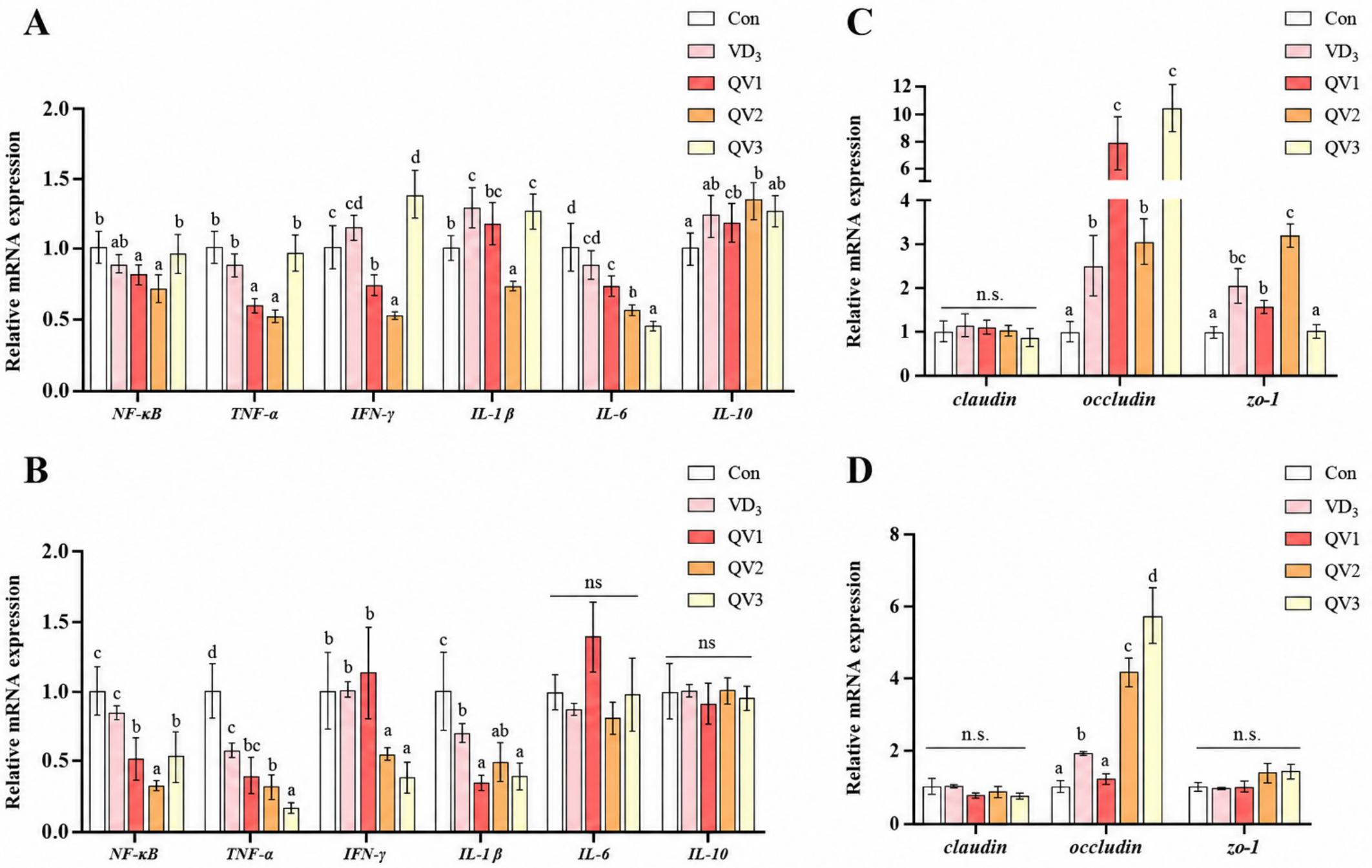
Jejunal and ileal mRNA expression of tight junction and inflammation-related genes was quantified by qRT-PCR (
Table 2). RNA was extracted with TRIzol (Invitrogen, Beijing, China), quantified by NanoDrop One (Thermo, Newark, DE, USA), reverse-transcribed (PrimeScript RT, Takara RR047A), and amplified on an ABI 7500 Fast system using TB Green (Takara RR820A). Expression was normalized to
GAPDH and calculated by 2^−ΔΔCt.
2.6. Intestinal Histology and Morphometric Analysis
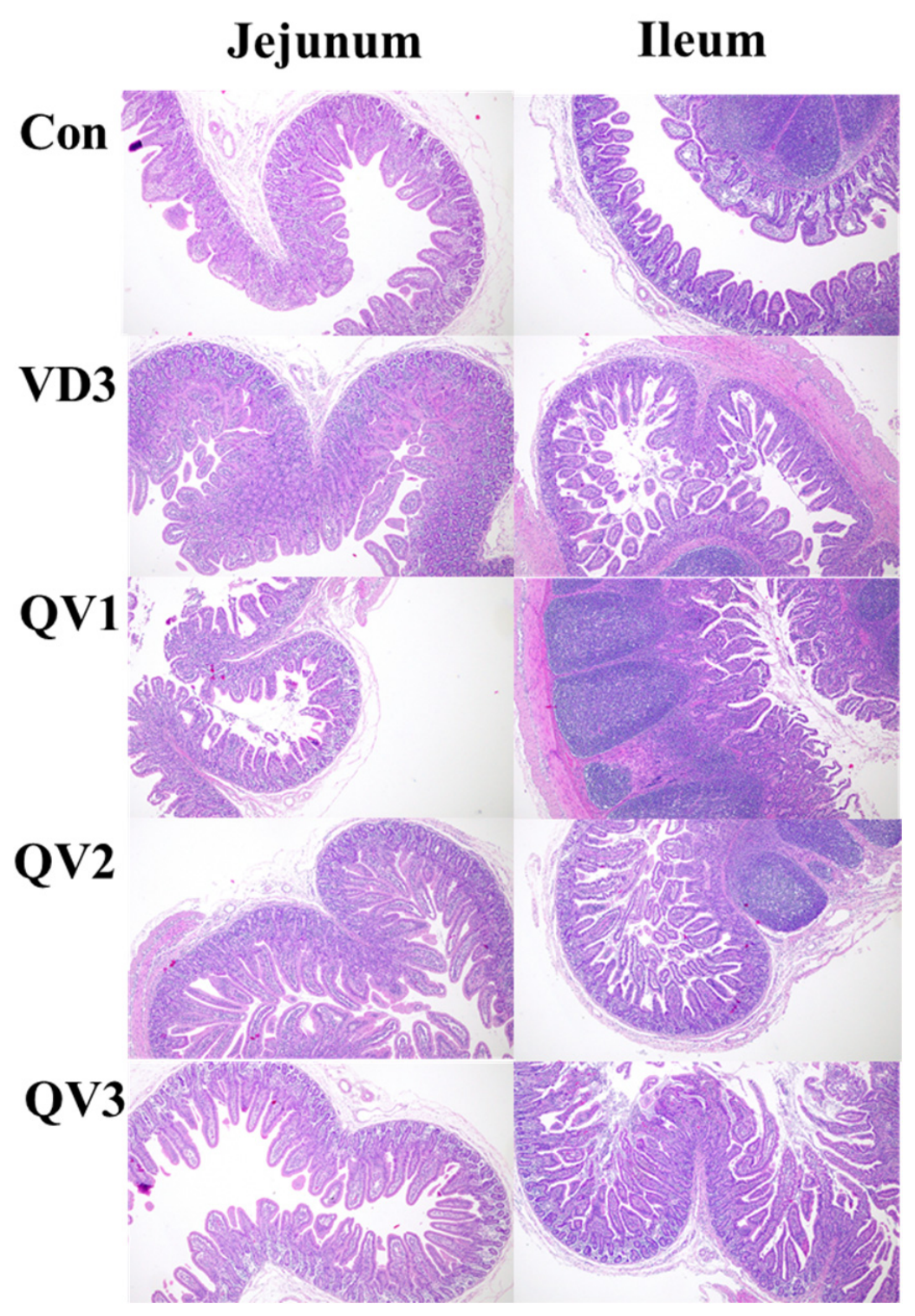
Jejunal and ileal tissues were fixed (4% paraformaldehyde), paraffin-embedded, sectioned (5 μm), and H&E-stained. Slides were scanned (Pannoramic MIDI, 3DHISTECH Ltd., Budapest, Hungary) and analyzed (CaseViewer, 3DHISTECH Ltd., Budapest, Hungary; Image-Pro Plus 6.0, Media Cybernetics, Inc., Rockville, MD, USA) to measure villus height, crypt depth, and villus-to-crypt ratio.
2.7. Bone Length and Mineral Determination
Femur and tibia lengths were measured with a digital caliper. Femur Ca, P, and ash were determined following GB/T 6438 [
19]: Ca by flame photometry after hot H
2SO
4 digestion, P by molybdenum–antimony colorimetry (700 nm) after H
2SO
4–H
2O
2 digestion, and ash by dry ashing to constant weight.
2.8. Colostrum Routine Composition Analysis
Colostrum was stored at −80 °C, thawed at 4 °C, warmed (~40 °C), and mixed. Fat, protein, lactose, and total solids were measured using an FTIR milk analyzer (MilkoScan FT series, FOSS Analytical A/S, Hillerød, Denmark) in duplicate.
2.9. Statistical Analysis
Statistical analyses were performed using SAS 9.4. For each response variable, data were analyzed by one-way ANOVA using the GLM procedure according to Yij = μ + Ti + εij, where μ is the overall mean, Ti is the fixed effect of dietary treatment, and εij is the residual error. The sow was the experimental unit for sow and litter outcomes, and the litter was the experimental unit for piglet serum and tissue outcomes (one piglet per litter). Farrowing and weaning endpoints were analyzed separately; therefore, no repeated-measures term was fitted. Residual normality and homogeneity of variance were evaluated before inference, and no material violations were detected. For litter performance, the actual litter size and composition after within-treatment cross-fostering were used in calculating outcomes; litter size and piglet sex were not fitted as separate covariates. Means were compared using Tukey’s HSD test. Results are presented as mean ± SEM; p < 0.05 was considered significant and 0.05 ≤ p < 0.10 was considered a tendency.
4. Discussion
Maternal 25-OH-D3 supplementation increased serum 25-OH-D3 concentrations in sows at farrowing and weaning and in piglets at weaning relative to the CON and VD3 groups; QV2 also yielded the highest colostrum concentration. However, treatment rankings were not uniform across endpoints: QV2 produced the strongest responses for selected outcomes, whereas QV1 or QV3 was similar or superior for others. The data therefore indicate product-specific responses among the three preparations tested and do not establish that biosynthesized 25-OH-D3 is generally superior to chemically synthesized 25-OH-D3.
Additional profiling of downstream vitamin D metabolites supported the treatment-related changes in vitamin D status. The absence of differences in 1,25-(OH)
2-D
3, 24,25-(OH)
2-D
3, and DBP on gestation day 90 confirmed comparable baseline conditions among treatments. At farrowing, maternal 25-OH-D
3 supplementation increased serum and colostrum concentrations of both 1,25-(OH)
2-D
3 and 24,25-(OH)
2-D
3, with QV2 generally showing the strongest response. These findings are consistent with the capacity of porcine tissues to convert 25-OH-D
3 through both 1α-hydroxylation and 24-hydroxylation pathways [
20,
21]. Similarly, Thayer et al. reported increased serum 24,25-(OH)
2-D
3 in progeny from 25-OH-D
3-supplemented sows, although no significant effect was detected in colostrum or milk [
22]. Differences in product formulation, sampling conditions, and analytical methods may account for the different colostrum responses. In weaned piglets, the increase in serum and duodenal 24,25-(OH)
2-D
3 without a corresponding increase in 1,25-(OH)
2-D
3 may reflect increased vitamin D turnover under homeostatic regulation. DBP remained unchanged across maternal and offspring samples, indicating that the changes in total vitamin D metabolites were not accompanied by detectable alterations in carrier abundance [
23].
Despite the increase in 25-OH-D
3 status, serum and colostrum calcium and phosphorus were unchanged, consistent with tight homeostatic regulation [
24]. Piglet femoral calcium concentration increased by 6–14% with the 25-OH-D
3 treatments, with the largest value in QV2, but bone length, phosphorus concentration, and ash were unaffected. Because total ALP rather than a bone-specific ALP isoenzyme was measured, the higher ALP activity should be interpreted only as supportive evidence of altered mineral or bone metabolic activity, not as direct proof of enhanced osteoblast activity, mineral deposition, skeletal development, or bone strength [
25]. These observations are broadly consistent with reports that maternal 25-OH-D
3 affects vitamin D status and selected indices of skeletal mineralization [
11,
26,
27].
Maternal 25-OH-D
3 supplementation did not affect litter size or birth weight. The significant growth response was confined to the first 14 days of life: QV2 increased litter weight on day 14 and litter weight gain from birth to day 14 by 9.8% relative to CON, whereas average piglet weight on day 14 showed only a tendency (
p = 0.09). One potential contributing factor is that, at approximately 21 days of lactation, some clinically smaller piglets were cross-fostered by farm personnel to other sows within the same dietary treatment. Although restricting transfers to the same treatment prevented direct cross-treatment contamination, late-lactation cross-fostering altered the composition of individual litters and may have increased variation in litter and piglet. weights at weaning, thereby attenuating the apparent persistence of the day-14 response. Litter weight, average piglet weight, and litter weight gain to weaning did not differ among treatments. Previous studies have reported variable growth responses to maternal 25-OH-D
3 supplementation [
14,
28,
29]. The source-dependent responses observed here may relate to product-specific composition or formulation. Regulatory assessments likewise treat manufacturing process, composition, and stability as product-specific attributes [
30]. Because epimer/isomer profiles, carrier composition, feed recovery, stability, and pharmacokinetics were not measured, the present study cannot identify the mechanism responsible for differences among QV1, QV2, and QV3.
Maternal 25-OH-D
3 supplementation altered the overall redox profile in sows, colostrum, and piglets, as reflected by coordinated changes in MDA, SOD, GSH-Px, and CAT rather than by any single antioxidant enzyme; however, the magnitude and direction of individual responses were not uniform across sources or sampling points. This combined pattern of lipid peroxidation and antioxidant defense markers is compatible with improved redox balance in selected matrices. Oxidative stress is common during reproduction and has been associated with placental dysfunction and impaired offspring development [
31,
32,
33]. In weaned piglets, 118 µg/kg 25-OH-D
3 and doses of 50 or 75 µg/kg have also been associated with combined changes in antioxidant indices [
34,
35]. Direct ROS measurements and tissue oxidative damage endpoints were not assessed in the present study. Because the intervention began on day 90 of gestation and the basal diet met NRC [
17] requirements, unchanged litter size and birth weight were not unexpected. These biochemical changes should therefore be interpreted as marker responses rather than evidence of improved reproductive performance, and the safety and long-term effects of supranutritional supplementation require further evaluation.
Maternal 25-OH-D
3 supplementation produced source-dependent changes in systemic immune markers. In sow serum, the three 25-OH-D
3 preparations generally reduced IL-1β, IL-6, TNF-α, and IFN-γ at farrowing and/or weaning, whereas increases in IL-10 were mainly observed at farrowing. In weaned piglets, QV2 and QV3 produced the most consistent reductions in circulating pro-inflammatory cytokines. These changes may be physiologically relevant because weaning is accompanied by increased intestinal expression of IL-1β, IL-6, and TNF-α in piglets [
36]. Madsen et al. similarly showed that improving 25-OH-D
3 status in piglets was associated with altered immune responses and potentially greater robustness during an Escherichia coli challenge, although their study did not demonstrate the same cytokine pattern observed here [
37]. Mechanistically, 1,25-(OH)
2-D
3–VDR signaling can attenuate Toll-like receptor-mediated inflammation by limiting NF-κB-associated signaling and strengthening negative feedback regulation [
38]. Intestinal epithelial VDR signaling has also been shown to reduce mucosal inflammation and epithelial injury in experimental models [
39,
40]. Collectively, the circulating and intestinal results support source-dependent immunomodulation by maternal 25-OH-D
3 supplementation, although they should not be interpreted as direct evidence of reduced histological inflammation.
Maternal 25-OH-D
3 supplementation also altered selected circulating and intestinal inflammatory markers. In piglet intestines, QV2 was associated with lower mRNA abundance of several inflammation-related genes, whereas QV1 and QV3 produced more selective transcriptional changes. These patterns are consistent with altered mucosal immune signaling [
41,
42,
43] but do not directly demonstrate inhibition of intestinal inflammation because protein abundance, pathway activation, immune cell infiltration, and histopathological inflammation were not measured. The immune marker changes may have accompanied the early litter growth response, but the present design does not establish a causal relationship, and the absence of significant effects on weaning weight or total litter gain to weaning warrants cautious interpretation. Mechanistic studies are needed to determine whether differences in absorption, metabolism, or tissue signaling explain the source-dependent responses.
QV2 and QV3 increased jejunal and ileal villus height, and selected 25-OH-D
3 treatments upregulated occludin and ZO-1 mRNA in a segment- and source-dependent manner. Villus architecture and tight-junction-related transcripts are relevant to absorptive and barrier biology [
44,
45], but intestinal permeability, nutrient absorption, and tight-junction protein abundance were not measured. Accordingly, these outcomes support changes in intestinal morphology and barrier-related gene expression but do not constitute direct evidence of enhanced intestinal barrier integrity or more efficient nutrient absorption.
Study Limitations
This study was conducted on one farm using one genotype and one supplemental dose and compared only three commercial preparations. The 25-OH-D3 content of each formulated premix was verified by HPLC; however, the chemical purity of the isolated active ingredients, epimer/isomer profiles, and pharmacokinetic characteristics were not independently determined. Therefore, the observed differences should be interpreted as product-specific responses among the preparations tested rather than as general differences between chemical and biosynthetic production routes. Vitamin D pharmacokinetics were not characterized by serial sampling. The biochemical and tissue subset comprised six independent sows or litters per treatment, cross-fostering occurred within treatment, and piglet sex was not controlled at terminal sampling. Direct ROS and tissue oxidative damage endpoints, inflammatory protein abundance, pathway activation, immune cell infiltration, histopathological inflammation, intestinal permeability, nutrient absorption, tight-junction proteins, bone strength, long-term reproductive outcomes, and economic return were not evaluated. These limitations restrict mechanistic interpretation and generalization of source-dependent effects.



 ,
,