1. Introduction
Nile tilapia (
Oreochromis niloticus) is among the most widely farmed freshwater fish species globally, with production reaching 5.3 million tonnes in 2022, owing to its rapid growth, omnivorous feeding habits, and tolerance to a wide range of environmental conditions [
1,
2,
3]. Across tropical and subtropical regions, tilapia aquaculture contributes to food security, affordable protein supply, and the economic stability of rural communities [
4].
However, as aquaculture systems intensify, the frequency and severity of infectious diseases have also increased. Among these,
Streptococcus agalactiae has emerged as one of the most destructive bacterial pathogens in tilapia culture worldwide [
5,
6,
7,
8]. This Gram-positive, β-hemolytic bacterium causes streptococcosis, a systemic infection characterized by septicemia, exophthalmia, erratic swimming, and high mortality, sometimes exceeding 50% during severe outbreaks [
9,
10,
11]. Such losses impose major economic and social burdens on small-scale and commercial farmers alike.
Antibiotics and chemotherapeutic agents remain the most common control measures against
S. agalactiae [
12]. While effective in the short term, their repeated and indiscriminate use has led to the emergence of antimicrobial-resistant strains [
13,
14], residue accumulation in fish tissues [
15,
16], and environmental contamination [
17,
18]. These consequences raise concerns about fish safety, human health, and the long-term sustainability of intensive aquaculture systems.
Vaccination provides a promising alternative, and several commercial vaccines have shown protective effects against
S. agalactiae. Nevertheless, vaccine efficacy often varies depending on strain diversity, fish age, environmental stress, and the need for booster doses [
19,
20]. Moreover, vaccines may not offer rapid protection during acute outbreaks, underscoring the need for reliable, sustainable, and cost-effective antimicrobial strategies for tilapia health management [
21,
22].
Plant-derived essential oils have increasingly attracted attention as environmentally friendly antimicrobial agents in aquaculture [
23,
24]. Oregano (
Origanum vulgare L.) essential oil (OE), rich in carvacrol and thymol, exhibits broad-spectrum antibacterial, antioxidant, and immunostimulatory activities against both Gram-positive and Gram-negative bacteria, including
Streptococcus spp. [
25,
26,
27,
28]. However, its high volatility, low aqueous solubility, and susceptibility to oxidation significantly limit its stability and bioavailability in aquatic environments [
29,
30,
31].
Lauric acid, a medium-chain fatty acid with potent antibacterial activity, particularly against Gram-positive pathogens, disrupts bacterial cell membranes through lipid bilayer destabilization and interference with membrane-associated functions [
32,
33]. Importantly, lauric acid can confer a cationic surface charge when incorporated into lipid-based nanocarriers, enhancing electrostatic interaction with negatively charged bacterial membranes and improving antimicrobial performance [
34,
35]. When co-formulated with essential oils, lauric acid may also facilitate synergistic membrane permeabilization, allowing lower effective doses of volatile compounds while mitigating cytotoxicity through controlled release [
36,
37].
Nanotechnology offers a promising approach to address these challenges by improving the encapsulation, protection, and controlled release of lipophilic compounds [
38]. Nanostructured lipid carriers (NLCs), which consist of solid and liquid lipids stabilized by surfactants, can protect bioactive compounds from oxidation, enhance solubility, and enable sustained release over time [
39]. In particular, cationic NLCs show stronger adhesion to negatively charged bacterial membranes and mucosal surfaces, which can improve antimicrobial activity and influence host–pathogen interactions [
8,
40,
41,
42].
However, despite the well-documented antimicrobial properties of oregano oil, lauric acid, and lipid-based nanocarrier systems individually, studies investigating their combined incorporation within cationic nanostructured lipid carriers for aquaculture applications remain limited. Previous studies have mainly focused on free oregano oil supplementation, nanoemulsions, or non-cationic delivery systems, which may exhibit limited stability, rapid release, or reduced interaction with negatively charged bacterial membranes and mucosal surfaces [
43,
44,
45,
46]. In addition, the synergistic antibacterial and safety effects of combining oregano oil and lauric acid within a cationic lipid nanocarrier against
S. agalactiae in Nile tilapia have not been comprehensively evaluated. Therefore, we hypothesized that this cationic nanoformulation could enhance antibacterial activity, improve physicochemical stability, prolong the release of bioactive compounds, and reduce acute toxicity compared with non-encapsulated and non-cationic formulations.
Building on these advantages, the present study aimed to develop and characterize OE-L+NLCs and to evaluate their in vitro antibacterial performance against S. agalactiae. The study systematically assessed the physicochemical properties, carvacrol release behavior, and antibacterial activity of the formulations, together with their stability under different storage temperatures and simulated gastrointestinal digestion conditions. In addition, in vivo toxicity and safety were evaluated through LC50 determination, survival analysis (Kaplan–Meier and Cox regression), and histopathological examination of major organs in Nile tilapia. Collectively, this study presents an integrated approach for the stabilization and delivery of plant-derived antimicrobials using cationic nanocarrier systems, highlighting their potential as feed-compatible alternatives to antibiotics for sustainable aquaculture.
2. Materials and Methods
2.1. Experimental Design
This study was conducted in sequential phases to evaluate the development, antibacterial efficacy, stability, and safety of OE-L+NLCs. First, the nanoparticles were prepared and characterized for their physicochemical properties, release behavior, and in vitro antibacterial activity against S. agalactiae. Next, their stability and retained antibacterial activity were assessed under different storage temperatures and simulated gastrointestinal conditions. Finally, in vivo safety was evaluated in Nile tilapia through acute toxicity, survival analysis, and histopathological examination.
2.2. Chemicals
Oregano oil (OE), derived from
Origanum vulgare L. (CAS No. 84012-24-8) and containing approximately 85% carvacrol and 3% thymol, was obtained from Katyani Exports (Delhi, India). Lauric acid cationic (L
+; CAS No. 60372-77-2), originally patented in Spain [
47] and now produced by Vedeqsa Lamirsa Group (Barcelona, Spain), was used as the cationic lipid. Medium-chain triglycerides (MCT) were purchased from Tulip Chemplast Co., Ltd. (Samut Sakhon, Thailand). Sorbitol monooleate (Span 20) and polysorbate 80 (Tween 80) were obtained from Merck Pte. Ltd. (Singapore). Montanov 82, polyethylene glycol, and glycerol monohydrate were sourced from Waruto Sama Co., Ltd. (Bangkok, Thailand). All reagents were of analytical grade and used without further purification.
2.3. Preparation of Oregano Oil–Lauric Acid Cationic Nanostructured Lipid Carriers
Nanostructured lipid carriers were prepared using a modified hot homogenization–ultrasonication method (
Figure S1). Briefly, the oil phase consisted of OE (10 g) and MCT (10–15 g) mixed with surfactants (span 20 and montanov 82, 1–3 g each) and maintained at 50–60 °C with gentle stirring (150 rpm). The aqueous phase consisted of purified water, Tween 80, glycerol monohydrate, polyethylene glycol (each 1–3 g), and L
+ (2 g), preheated to the same temperature. The aqueous phase was slowly added to the oil phase with continuous stirring, homogenized, and subsequently sonicated for 5 min using a Qsonica sonicator (35-amp pulse, 10 s on/5 s off cycles) following Yostawonkul et al. [
48]. The resulting emulsion was cooled to room temperature to obtain the final OE-L
+NLCs formulation. Control formulations included: (i) OE-NLCs (without L
+ in the aqueous phase), (ii) L
+NLCs (without OE in the oil phase), and (iii) blank NLCs (without both OE and L
+).
For comparative purposes, an OE solution (dissolved in 0.5% DMSO) and an L
+ solution (dissolved in sterile distilled water) were prepared. The composition of all formulations and solutions is summarized in
Table S1. The prepared formulations were subsequently subjected to physicochemical characterization, release profiling, and antibacterial evaluation, as described in the following sections.
2.4. Bacterial Strains and Culture Conditions
Six S. agalactiae strains, PC08 (Ia), SM81 (Ia), CT59 (III), SM86 (III), SM24 (Ia), and AY19 (Ia), were isolated from the brain tissue of diseased Nile tilapia collected from farms in Phetchaburi, Samut Sakhon, Chanthaburi, and Ayutthaya provinces, Thailand. Isolates were subcultured on Brain Heart Infusion (BHI) agar (pH 7.2) and incubated at 30 °C for 24 h. Bacterial suspensions were prepared in BHI broth under shaking conditions (150 rpm, 24 h). Cell density was standardized spectrophotometrically (OD600 = 0.2), corresponding to approximately 1 × 10⁸ CFU/mL. Culture purity was confirmed by Gram staining and microscopic examination prior to use.
2.5. Physicochemical Characterization of Nanoparticles
The physical appearance (color, homogeneity, and phase separation) of all formulations was visually assessed after 24 h of equilibration at room temperature. Particle size, zeta potential, and polydispersity index (PDI) were measured using dynamic light scattering (Zetasizer Ultra, Malvern Panalytical Ltd., Malvern, UK) at 25 °C. Measurements were performed in triplicate.
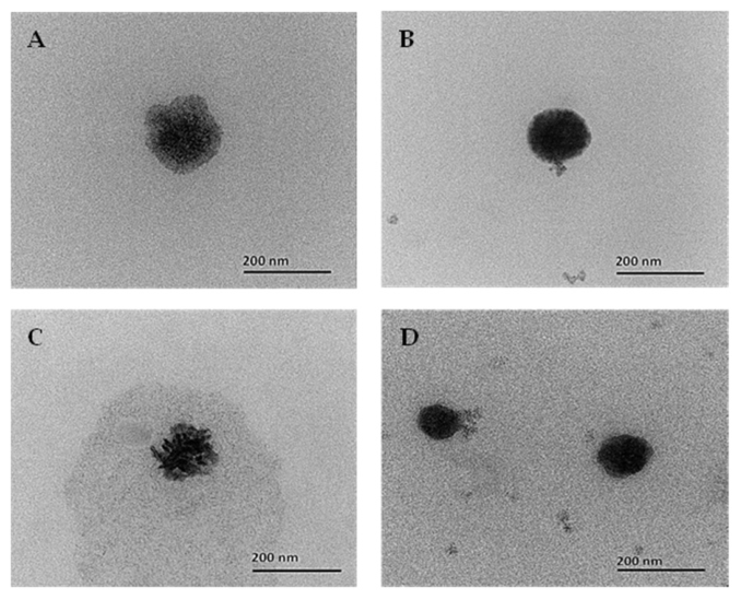
Nanoparticle morphology was examined using a transmission electron microscope (TEM; Talos F 200X, Thermo Fisher Scientific, Waltham, MA, USA). Samples were diluted (1:50,
v/
v) with deionized (DI) water, stained with 1% uranyl acetate, air-dried on carbon-coated grids, and imaged at an accelerating voltage of 200 kV [
49].
2.6. Encapsulation Efficiency, Drug Loading and In Vitro Release of Oregano Oil
The encapsulation efficiency (EE) and drug loading (DL) of carvacrol, the major bioactive compound of oregano oil, in OE-L
+NLCs and OE-NLCs were determined using an ultrafiltration–centrifugation method. Briefly, nanoparticle suspensions were transferred into Amicon Ultra-15 centrifugal filter units (Merck Millipore Ltd., Darmstadt, Germany) and centrifuged at 8000×
g for 1 h to separate free carvacrol from encapsulated fractions. The filtrate containing non-encapsulated carvacrol was passed through a 0.22 μm nylon membrane filter and quantified using gas chromatography coupled with flame ionization detection (GC-FID; Agilent Technologies 7890A, Agilent Technologies Inc., Santa Clara, CA, USA) following previously reported methods [
48,
50]. Encapsulation efficiency and drug loading were calculated according to the following equations:
where C
i represents the initial concentration of carvacrol added to the formulation, C
f represents the concentration of non-encapsulated carvacrol in the filtrate, W
i represents the initial weight of carvacrol added, W
f represents the weight of non-encapsulated carvacrol, and W
NLC represents the total weight of the nanostructured lipid carrier formulation.
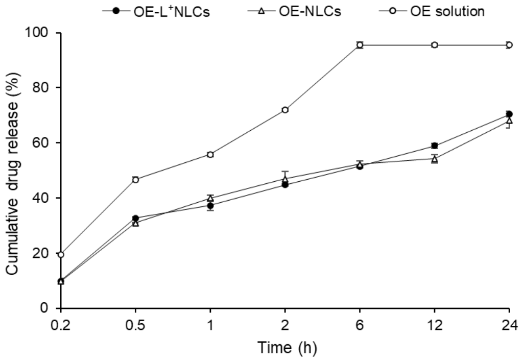
The in vitro release profile of carvacrol from OE-L
+NLCs, OE-NLCs and OE solution was evaluated using the dialysis bag diffusion method described by Yostawonkul et al. [
51]. Aliquots containing an amount equivalent to 10% (
w/
v) of carvacrol solubility were placed in dialysis membranes (MWCO 10 kDa) and immersed in phosphate-buffered saline (PBS, pH 7.4) supplemented with 0.5% Tween 80. Samples were incubated at 37 °C with constant stirring (40 rpm). At predetermined time points (0.2, 0.5, 1, 2, 6, 12 and 24 h), aliquots were withdrawn, filtered (0.22 µm), and analyzed for carvacrol content using gas chromatography–flame ionization (GC-FID) according to Lee et al. [
52]. The cumulative release (%) was calculated as:
2.7. Antibacterial Activity Assays
Antibacterial activity was evaluated under three conditions: baseline, post-storage, and post-digestion.
2.7.1. Baseline Antibacterial Activity Assays
Baseline antibacterial activity of OE-L
+NLCs, OE-NLCs, L
+NLCs, blank NLCs, OE solution, and L
+ solution was evaluated against six
S. agalactiae isolates using the broth microdilution method in accordance with NCCLS guidelines [
53,
54]. Stock solutions (125 mg/mL) were serially two-fold diluted in Muller–Hinton broth (MHB) to obtain concentrations ranging from 62.50 to 0.13 mg/mL. Each well of a 96-well microplate contained 100 µL of the diluted formulation and 100 µL of bacterial suspension adjusted to approximately 1 × 10
6 CFU/mL, resulting in a final bacterial concentration of approximately 5.0 × 10
5 CFU/mL/well, and a final concentration of sample solutions ranging from 31.25 to 0.06 mg/mL. Enrofloxacin (5 µg/mL) served as a positive control, while sterile water or 0.5% DMSO served as negative controls. Plates were incubated at 30 °C for 24 h. The minimum inhibitory concentration (MIC) was defined as the lowest concentration showing no visible growth. Minimum bactericidal concentration (MBC) was determined by subculturing aliquots from clear wells onto BHI agar and identifying the lowest concentration preventing colony formation after 24 h (
Figure S2).
2.7.2. Antibacterial Activity After Thermal Storage
To assess antibacterial activity following storage, the MIC and MBC values of all formulations were re-evaluated against
S. agalactiae strain AY19 after storage at different temperatures and for varying durations, as described in
Section 2.8. Strain AY19 was selected as a representative isolate based on its consistent growth characteristics and reproducible response in preliminary assays.
2.7.3. Antibacterial Activity After Simulated Digestion
Antibacterial activity of digested formulations obtained after simulated gastric and intestinal phases (see
Section 2.9) was evaluated against
S. agalactiae strain AY19 using the same broth microdilution protocol as described.
2.8. Thermal Stability Study
OE-L
+NLCs and control formulations were stored at 4 °C (refrigeration), 25 °C (room temperature), and 37 °C (physiological temperature) for 90 days [
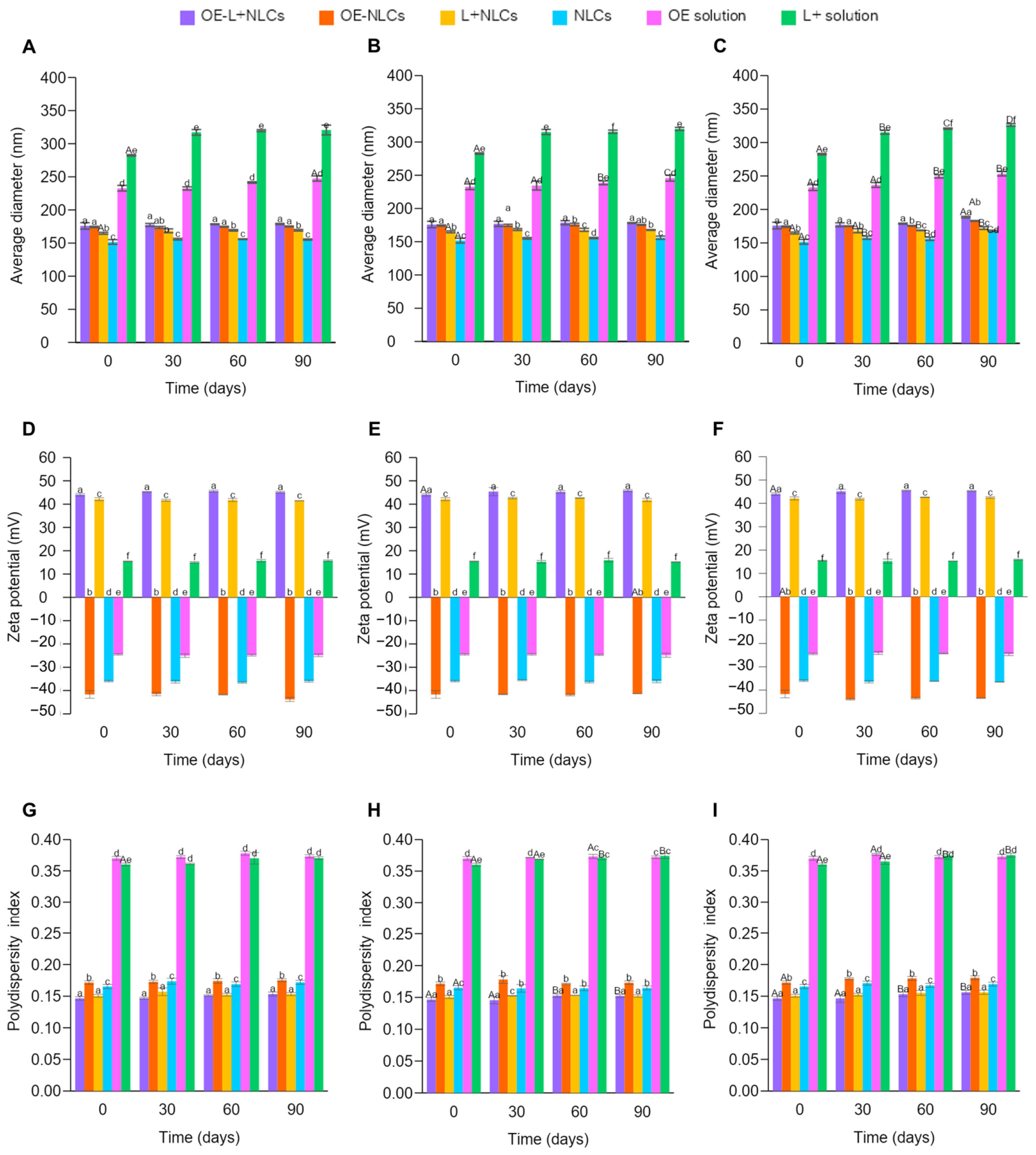
55]. To minimize contamination and environmental exposure during long-term storage, all formulations were prepared from a single batch and pre-aliquoted into sterile tubes according to each storage condition and sampling time point. Each aliquot was opened only once at the designated time point using aseptic conditions. Samples were analyzed at 0, 30, 60, and 90 days for physical appearance, particle size, zeta potential, and PDI. At each sampling interval, antibacterial activity against
S. agalactiae strain AY19 was evaluated as described in
Section 2.7.2 using freshly prepared culture media under standardized assay conditions. This analysis enabled evaluation of the relationship between physicochemical stability and retained antibacterial activity.
2.9. In Vitro Simulated Digestion
Nanoparticle stability under gastrointestinal conditions was evaluated using simulated gastric fluid (SGF) and simulated intestinal fluid (SIF). SGF (2 g/L NaCl, pH 2.0) and SIF (6.8 g/L KH
2PO
4, pH 7.0) were freshly prepared. Pepsin (10 mg/mL) and pancreatin (10 mg/mL) were added to SGF and SIF, respectively [
56]. For the gastric phase, 10 mL of each formulation was mixed with 9 mL SGF and incubated at 37 °C and 100 rpm for 2 h. For the intestinal phase, 10 mL of the gastric digest was transferred into 9 mL SIF containing 160 mg bile salts and incubated for an additional 2 h under identical conditions (
Figure S3). Following digestion, samples were centrifuged (1000×
g) to remove insoluble material. The supernatant was analyzed for particle size, zeta potential, and PDI. Antibacterial activity against
S. agalactiae strain AY19 was evaluated to assess bioactivity retention (
Figure S3). This simulated digestion model was used to assess whether nanoparticle integrity and antibacterial efficacy were maintained under gastrointestinal conditions.
Although the digestion experiments were conducted at 37 °C, which is higher than the typical physiological temperature range of Nile tilapia (approximately 28–30 °C), this condition was selected to maintain consistency with standardized in vitro digestion protocols frequently used in nanoencapsulation and lipid-based nanocarrier studies. This approach facilitates comparison with previously published studies and ensures reproducibility of enzymatic digestion conditions.
2.10. Experimental Design for Acute Toxicity Assessment
Healthy juvenile monosex (male) Nile tilapia (mean weight 10.0 ± 2.0 g; mean length 8.6 ± 1.2 cm) were acclimated for 14 days in 250 L fiberglass tanks with continuous aeration. Water quality parameters were maintained at 28–30 °C, pH 7.0–7.5, dissolved oxygen ≥ 5 mg/L, with a daily 20% water exchange.
During acclimation, fish were fed a commercial pellet diet at 3% of body weight per day. Following acclimation, fish were randomly assigned to six formulation groups (OE-L
+NLCs, OE-NLCs, L
+NLCs, OE solution, L
+ solution, and blank NLCs), with each group receiving nine concentrations of their assigned formulation (100.0, 50.0, 20.0, 10.0, 5.0, 3.3, 2.5, 2.0, and 0 mg/mL [control]). Each concentration was evaluated in triplicate, with 10 fish used per replicate (30 fish per concentration) [
57]. This resulted in 270 fish per formulation and a total of 1620 fish used in the acute toxicity experiment.
Each formulation was mixed with feed at approximately 30% moisture, air-dried on trays, and coated with 1% squid oil to minimize leaching of active compounds during feeding. Prepared diets were stored at 4 °C and used within 7 days. Control groups received feed treated with distilled water and squid oil to account for potential coating effects.
Fish were hand-fed the experimental diets at 5% of body weight per day, given as three equal feedings. Initial body weight was recorded to adjust feeding rates. Fish were monitored for mortality and clinical signs over a 96 h exposure period [
58]. Mortality was defined as the absence of opercular movement and no response to gentle stimulation. The LC
50 (median lethal concentration) was calculated using Probit analysis based on cumulative mortality across treatments.
2.11. Histopathological Evaluation of Dietary Toxicity in Nile Tilapia
At 96 h post-feeding, tissue samples (
n = 6 per group) were collected from the intestine, liver, kidney, spleen, brain, and gills of randomly selected fish. Prior to sampling, fish were anesthetized with clove oil (20 mg/L) [
59]. Collected tissues were fixed in 10% neutral buffered formalin for 24 h, followed by dehydration through a graded ethanol series (70% to absolute ethanol). Samples were then cleared in xylene and embedded in paraffin wax. Tissue sections (4–5 µm thick) were prepared and stained with hematoxylin and eosin for histopathological evaluation, following the method described by Moustafa et al. [
60].
2.12. Statistical Analysis
All data were expressed as mean ± standard deviation (n = 3) and analyzed using SPSS version 29 (IBM Corp., Armonk, NY, USA). Normality and homogeneity of variances were verified prior to analysis. Independent samples t-tests were used to compare EE and DL between OE-L+NLCs and OE-NLCs formulations. One-way ANOVA followed by Duncan’s multiple range test was used to compare physicochemical characteristics and antibacterial activity among formulations. Two-way ANOVA was used to assess the effects of storage temperature and duration on nanoparticle stability, while repeated-measures ANOVA evaluated differences in carvacrol release profiles over time. Acute toxicity was assessed using probit analysis to estimate median LC50 values together with slope parameters and confidence intervals, based on dose–response relationships. Survival data were analyzed using the Kaplan–Meier method to estimate survival probabilities over time, and differences among treatment groups were evaluated using the log-rank test. Additionally, Cox proportional hazards regression analysis was performed to determine the relative mortality risk among formulations and to quantify hazard ratios. A p-value < 0.05 was considered statistically significant.
4. Discussion
The physicochemical results indicate that the formulated NLCs have properties well suited for delivering bioactive compounds in aquaculture. Their nanoscale particle size is a key factor influencing biological interactions, mucosal contact, and the functional efficiency of feed additives in aquatic species [
61]. Similar particle sizes have been reported for other essential oil-loaded lipid nanocarriers, including clove oil-based NLCs, supporting the suitability of the present formulation approach [
62]. Surface charge was another important distinguishing feature. Incorporation of cationic lauric acid produced a strong positive surface charge, which improves colloidal stability through electrostatic repulsion and facilitates interaction with negatively charged bacterial membranes and fish mucosal surfaces [
63,
64]. This cationic behavior has been linked with improved antimicrobial activity and mucosal adhesion in lipid-based delivery systems [
65,
66]. By contrast, non-cationic essential oil-loaded NLCs usually carry an anionic surface charge, which may reduce membrane interaction [
62,
67]. The uniform spherical morphology observed further indicates optimized lipid–surfactant assembly, which is important for maintaining encapsulation efficiency and limiting premature release of volatile compounds [
68]. Collectively, these findings show that nanoencapsulation improves the physicochemical stability of oregano oil, while the inclusion of lauric acid further enhances properties associated with antimicrobial performance, consistent with previous reports on the synergistic benefits of lipid-based nanocarriers for essential oils [
69,
70,
71].
The sustained release behavior of carvacrol from OE-L
+NLCs and OE-NLCs, compared with free OE reflects effective entrapment of the volatile compound within the lipid matrix. Consistent with this observation, OE-L
+NLCs exhibited higher encapsulation efficiency and drug loading than OE-NLCs, suggesting that incorporation of lauric acid improved the retention capacity of the lipid system for hydrophobic bioactive compounds. The enhanced encapsulation performance may also have contributed to the prolonged release behavior by reducing premature leakage of oregano oil constituents during nanoparticle formation and storage. The observed release profile is characteristic of diffusion-controlled systems, where an initial release of surface-associated carvacrol is followed by gradual diffusion from the lipid core over time [
72,
73,
74]. Similar release kinetics and encapsulation improvements have been reported for essential oil-loaded NLCs and lipid-based nanocarriers containing hydrophobic phytochemicals, where nanoencapsulation delayed release and improved physicochemical stability relative to free compounds [
62,
75,
76,
77,
78]. For aquaculture feed applications, this controlled release is especially beneficial because it prolongs the availability of bioactive compounds, supports formulation stability, and helps explain the sustained antibacterial activity observed.
The strong antibacterial activity exhibited by OE-L
+NLCs and L
+NLCs highlights the central role of cationic lauric acid in inhibiting
S. agalactiae. Their performance, comparable to free L
+, is consistent with previous studies identifying L
+ as a potent antimicrobial agent against Gram-positive fish pathogens [
79]. The strong antibacterial activity exhibited by OE-L
+NLCs was comparable to that of L
+NLCs and free L
+ solution, indicating that the direct antibacterial effect against
S. agalactiae was primarily associated with the lauric acid component under the present in vitro conditions, while oregano oil may contribute to formulation stability, sustained release behavior, and lipid phase interactions within the nanocarrier system despite no additional measurable reduction in MIC/MBC values. Mechanistically, the cationic surface charge promotes electrostatic interaction with negatively charged bacterial membranes, facilitating membrane destabilization, while L
+ disrupts membrane integrity, interferes with electron transport, and inhibits membrane-associated enzymes, ultimately leading to bacterial cell lysis [
70,
80]. Nanoencapsulation may further contribute by stabilizing both L
+ and OE and prolonging the bioavailability of hydrophobic compounds relative to non-encapsulated forms [
81,
82]. These findings align with earlier reports demonstrating strong antibacterial activity of L
+ against
S. agalactiae [
79] and broad-spectrum antimicrobial effects of OE rich in carvacrol and thymol against
Streptococcus species [
83,
84]. The lack of activity in blank NLCs confirms that the antibacterial effects were due to the active compounds rather than the carrier matrix. Based on this initial screening, strain AY19 was chosen as the representative isolate for the thermal stability and digestion studies because its susceptibility pattern was similar to those of the other isolates, enabling a focused assessment of formulation stability and bioactivity without unnecessary duplication.
The NLC formulations demonstrated excellent thermal stability over prolonged storage at refrigeration, room, and physiological temperatures, as evidenced by their maintained nanoscale particle size, low PDI, and stable surface charge. In contrast, crystallization of the unencapsulated L
+ solution under cold storage highlights the greater temperature sensitivity of free compounds. The strong positive surface charge retained by OE-L
+NLCs and L
+NLCs further supports their robust colloidal stability, promoting electrostatic repulsion [
73] and interaction with negatively charged bacterial membranes [
85,
86]. Although particle size increased slightly at higher temperatures, formulation integrity was not compromised. These findings suggest that the NLC matrix protects OE and L
+ from thermal stress [
87,
88,
89], thereby reducing aggregation, volatilization, and degradation commonly seen in unencapsulated systems [
90,
91]. Similar stabilization has been reported for other lipid-based nanocarriers, including curcumin- and essential oil-loaded NLCs, where the lipid matrix provided long-term physicochemical protection during storage [
74,
81]. Importantly, the preserved physicochemical stability translated directly into sustained antibacterial efficacy. OE-L
+NLCs and L
+NLCs maintained consistent antibacterial activity across all storage conditions, whereas free OE lost activity rapidly at elevated temperatures. Comparable preservation of antimicrobial function has been reported for nanoencapsulated OE and other essential oils under environmental stress conditions [
84]. Collectively, these findings support the use of NLCs as effective carriers for thermolabile antimicrobial agents, ensuring long-term stability and functional reliability in aquaculture feed applications [
82].
The NLC formulations exhibited high stability under simulated gastric conditions, maintaining homogeneous appearance, nanoscale particle size, and low PDI, thereby preserving both nanoparticle integrity and bioactivity in an acidic environment [
92]. This gastric stability is essential for oral delivery in fish, as it ensures protection of encapsulated OE and L
+ during stomach passage [
90,
93]. In contrast, unencapsulated formulations showed pronounced instability, highlighting the protective role of the NLC matrix. Upon exposure to simulated intestinal conditions, all NLC formulations showed partial destabilization, characterized by increased particle size, elevated PDI, and visible structural changes. This behavior is likely driven by the combined effects of elevated pH, bile salts, and digestive enzymes, which disrupt lipid–surfactant interfaces and promote nanoparticle restructuring or aggregation [
94,
95,
96]. Comparable digestion-dependent behavior has been observed in other lipid-based and essential oil-loaded nanocarriers, which are generally stable under gastric conditions but undergo structural rearrangements in the intestinal environment [
97,
98]. Consistent with these physicochemical changes, OE-L
+NLCs and L
+NLCs retained strong antibacterial activity after gastric digestion, confirming that nanoencapsulation effectively protects active compounds from acidic degradation [
92,
99]. However, antibacterial activity was markedly reduced under simulated intestinal conditions, likely due to carrier destabilization [
97,
100], rapid release of the bioactives [
101], and the complex intestinal environment (including bile salts, digestive enzymes, ionic strength, and pH changes), which may alter the nanoparticle integrity, weaken electrostatic interactions, and reduce the sustained antibacterial functionality of the intact nanocarrier system [
100]. Similar digestion-related reductions in antimicrobial efficacy have been reported for essential oil-loaded nanoparticles [
31], where sustained activity throughout the intestinal phase often requires further formulation optimization [
102,
103].
Building on these in vitro findings, the in vivo toxicity assessment provides important insight into the biological safety of the formulations. LC
50 values (5.89–12.48 mg/mL) revealed marked formulation-dependent differences, with NLC-based formulations showing lower toxicity than non-encapsulated forms. The OE solution had the lowest LC
50 (5.89 mg/mL), whereas L
+NLCs showed the highest (12.48 mg/mL), demonstrating that encapsulation substantially improved safety [
104]. This improved safety profile is consistent with the controlled release behavior and physicochemical stability observed in vitro, as nanoencapsulation reduces sudden peak exposure to active compounds and enables more gradual and controlled release, thereby minimizing acute toxic effects [
105]. The biocompatible lipid composition of NLCs may further enhance safety compared with other nanoparticle systems [
93], while also improving the stability and delivery efficiency of hydrophobic phytochemicals such as essential oils [
105,
106,
107]. Similar benefits have been reported for encapsulation approaches that improve solubility, reduce leaching, and enhance compound retention in aquatic environments [
108].
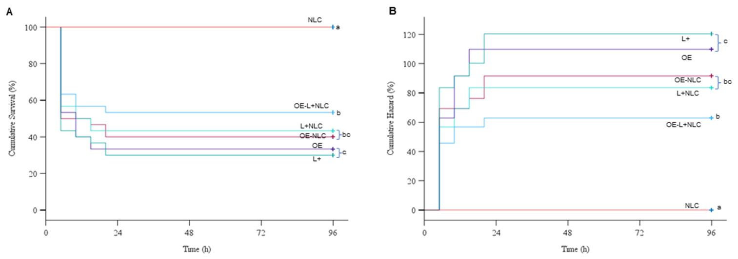
The Kaplan–Meier survival analysis and Cox regression results further supported these findings, demonstrating that NLC formulations maintained higher survival probabilities compared with non-encapsulated treatments. Significant differences among treatment groups, confirmed by the log-rank test and Cox model, indicate that formulation significantly influenced mortality risk. The consistency between LC
50 values and survival outcomes reinforces the reliability of the toxicity assessment, as higher LC
50 values correspond to lower acute toxicity and reduced mortality risk [
104]. This agreement highlights the importance of formulation design in determining not only efficacy but also safety in aquaculture applications.
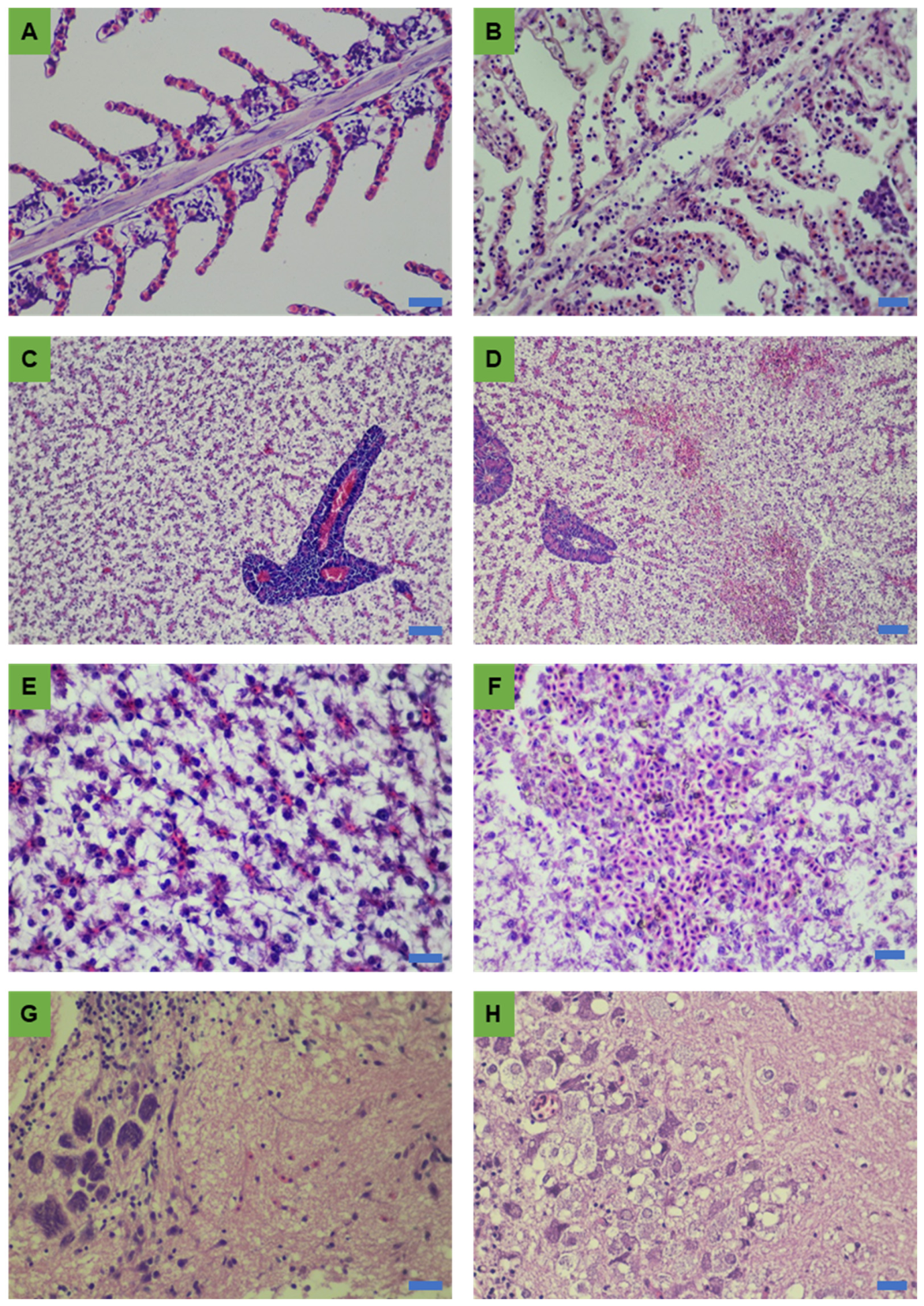
Histopathological observations provided additional evidence supporting the improved safety of the NLC formulations. Gill lesions, including edema, vascular congestion, and pillar capillary dissociation, were more pronounced in non-encapsulated treatments and are well-recognized indicators of toxicant exposure [
109,
110]. The liver exhibited multifocal hemorrhages, hepatocellular swelling, and disruption of hepatic cord organization, consistent with tissue injury reported in fish exposed to high doses of essential oils [
111,
112]. In contrast, reduced hepatic damage in NLC-treated fish suggests that encapsulation moderated tissue distribution and accumulation, as previously observed for lipid-based nanocarriers [
38,
113]. The most severe changes were observed in the brain, where neuronal edema, cytoplasmic vacuolation, and neuropil degeneration were evident. Given the limited regenerative capacity of neural tissue, these findings are of particular toxicological concern. However, the reduced severity of brain lesions in NLC-treated groups suggests a potential neuroprotective effect, possibly due to controlled release and reduced systemic exposure to toxic metabolites [
113,
114,
115]. The overall agreement among LC
50 values, survival data, and histopathological findings represents a major strength of the study and supports the conclusion that nanoencapsulation improves the therapeutic index of bioactive compounds in fish [
38,
104,
106,
113].
These findings collectively indicate that nanoencapsulation enhances the stability, antibacterial efficacy, and safety profile of OE and L+, primarily through controlled release behavior and improved physicochemical protection. While OE-L+NLCs demonstrated strong performance under gastric conditions, the reduced antibacterial activity observed in simulated intestinal environments highlights a key limitation that requires further formulation optimization. In addition, as the present study was limited to acute toxicity assessment, future studies should focus on chronic exposure, tissue distribution, elimination kinetics, and long-term safety under practical aquaculture conditions.





 ,
,