Article

Species Identification and Antimicrobial Resistance of Streptococcus spp. Isolated from Nasal Swabs of Wild Boars in Avellino Province, Southern Italy


Department of Veterinary Medicine and Animal Production, University of Naples Federico II, Via F. Delpino, n. 1, 80137 Naples, Italy
*
Authors to whom correspondence should be addressed.
These authors contributed equally to this work.
Animals 2026, 16(11), 1619; https://doi.org/10.3390/ani16111619
Submission received: 23 April 2026 / Revised: 16 May 2026 / Accepted: 23 May 2026 / Published: 26 May 2026
(This article belongs to the Section Wildlife)

Simple Summary

Wild boars may serve as reservoirs for various bacteria that can affect both livestock and humans. This study focused on the bacterial flora of the nasal cavities of wild boars, especially Streptococcus species, which were found to be the most prevalent, to understand their diversity and levels of antimicrobial resistance. Our findings revealed the presence of several important streptococcal species, many of which showed high resistance to commonly used antibiotics like tetracycline. These results highlight the potential role of wild boars in the environmental circulation of resistant bacteria, emphasizing the need for continued monitoring to protect animal and public health.

Abstract

From an ecological epidemiology perspective, wildlife nasal cavities may serve as critical ecological niches for the maintenance and spread of bacterial pathogens relevant to both animal and human health. This study investigated Streptococcus spp. as the most prevalent genus in the nasal cavities of 82 wild boars (Sus scrofa), sampled across six hunting districts in Avellino Province (Southern Italy), assessing the diversity of the colonizing streptococcal populations and their antimicrobial resistance profiles. The sampled population consisted of 43 males (52%) and 39 females (48%). A total of 173 Gram-positive isolates were recovered, with 74 identified as Streptococcus spp. by MALDI-TOF MS. These isolates originated from 59 positive swabs (72%; 59/82). Streptococcus gallolyticus was the most prevalent species (41%; 30/74), followed by Streptococcus porcinus (27%; 20/74) and Streptococcus suis (19%; 14/74). No statistically significant geographic association was found across sampling districts (p = 0.491), whereas a significant association was found between bacterial species and antimicrobial resistance profiles (χ2 = 16.70, p < 0.001). Specifically, all Streptococcus porcinus isolates (100%; 20/20) exhibited resistance to sulfamethoxazole–trimethoprim and tetracycline, while high tetracycline resistance was also noted in Streptococcus suis (93%; 13/14) and Streptococcus gallolyticus (83%; 25/30). Overall, 69% (51/74) of isolates were classified as MDR and 5% (4/74) as XDR. These findings suggest the potential role of wild boars as reservoirs for antimicrobial-resistant streptococci, emphasizing the need for continuous surveillance under a One Health framework.

1. Introduction

While the role of livestock in the dissemination of antimicrobial resistance is well-documented [1,2], the epidemiological contribution of wildlife, particularly wild boars (Sus scrofa), remains insufficiently characterized and represents a critical knowledge gap [3,4]. Wild boars act as mobile sentinels and potential reservoirs for antimicrobial resistance genes in the environment [3]. Within these hosts, the nasal cavity represents a distinct clinical sampling site, mediating the balance between commensalism and infection. It serves as a primary niche for the colonization and persistence of both commensal and pathogenic species, influencing local immune responses and host susceptibility to respiratory infections [5]. Furthermore, the nasal cavity hosts a complex microbiota, typically dominated by major, stable genera such as Staphylococcus, that is essential for defending against infections [5]. However, the nasal microbial composition may exhibit significant diversity; indeed, the genus Streptococcus also represents a dominant and highly diverse taxon within this niche [6].
The genus Streptococcus currently includes over 100 species, ranging from essential commensals to highly virulent pathogens responsible for significant public health burdens worldwide [7,8,9]. In veterinary medicine, several Streptococcus species are of major clinical importance, including Streptococcus canis in dogs and cats, Streptococcus equi subsp. equi, the causative agent of strangles in horses, and Streptococcus agalactiae, a major cause of bovine mastitis. In addition, Streptococcus suis is a major zoonotic swine pathogen associated with severe systemic conditions such as meningitis, septicemia, arthritis, and endocarditis [10]. Other streptococcal species have also been identified in a wide range of hosts, including birds, aquatic animals, and livestock [11].
Research on Streptococcus species in wild boars remains limited compared with the extensive literature available for domestic pigs [12,13]. Nevertheless, several species, including Streptococcus porcorum and Streptococcus suis, have been isolated from wild boar populations, often in the absence of evident clinical disease. This supports the hypothesis that wild boars may act as asymptomatic reservoirs of potentially zoonotic strains [14]. Molecular surveys detecting bacterial DNA in clinically healthy animals further suggest that streptococcal colonization may occur without associated lesions, contributing to environmental persistence and interspecies transmission [12,15].
Prevalence studies have reported substantial carriage rates in some regions. In northwestern Europe, Streptococcus suis was isolated from up to 92% of tonsil samples collected from wild boars, with a proportion of isolates carrying virulence-associated genes [16]. These findings highlight the potential zoonotic relevance of wild boar populations, particularly for individuals with occupational or recreational exposure, such as butchers and hunters. Although wild boars are often asymptomatic carriers, clinical disease has occasionally been reported, including fatal cases of septicemia and meningitis [17]. Population-level investigations have also revealed considerable genetic diversity among wild boar isolates, which frequently differ from the dominant clonal complexes identified in domestic pigs and humans [18]. Consequently, wild boars may represent a localized interface at the wildlife–livestock–human boundary, although the actual spillover risk likely depends on the extent of overlap with agricultural environments [19].
Beyond Streptococcus suis, other Streptococcus species have occasionally been reported in wild boars, suggesting that this host harbors a broader streptococcal community than currently recognized [18,20]. However, the occurrence of specific species such as Streptococcus gallolyticus remains sporadically documented, and their ecological and epidemiological significance at the wildlife–livestock–human interface is still poorly understood [21].
Therefore, this study aimed to characterize the species distribution and antimicrobial resistance profiles of the Streptococcus species most frequently isolated from the nasal cavities of healthy wild boars hunted in the Avellino Province (Southern Italy), to expand the currently limited epidemiological data on nasal microbiota and antimicrobial resistance in wildlife.

2. Materials and Methods

2.1. Ethics Statement

Ethical approval was not required for this study because the animals were not killed specifically for research purposes. Wild boars were legally harvested in their natural environment by licensed hunters in accordance with the 2025–2026 annual hunting plan authorized by the Province of Avellino (Campania Region, Italy). Samples were collected post-mortem during the official hunting season in compliance with national wildlife management regulations. Informed consent to participate in this research study was obtained by licensed and specialized hunters, who provided wild boar nasal swabs.

2.2. Study Area and Sample Collection

2.2.1. Study Area

The study was conducted in the Province of Avellino, located in the inland sector of the Campania region, Southern Italy (Figure 1). The Campania region occupies the south-western portion of the Italian Peninsula, covering an area of 13,590 km2 and extending along approximately 350 km of coastline on the Tyrrhenian Sea. The Province of Avellino is centered approximately at 40°55′00″ N and 14°47′00″ E and is characterized by predominantly mountainous and hilly terrain, with elevations ranging from about 200 to over 1800 m above sea level, including part of the Apennine chain. The area exhibits high environmental heterogeneity, with extensive forested habitats interspersed with agricultural land, creating a complex mosaic of wildlife habitats. Climatic conditions are typical of inland Mediterranean environments, with significant seasonal thermal variation. This heterogeneous topography, together with the strong connectivity between forested and semi-natural landscapes, provides favorable conditions for the expansion and high population density of wild boar (Sus scrofa), promoting frequent interactions with agricultural environments and human settlements.
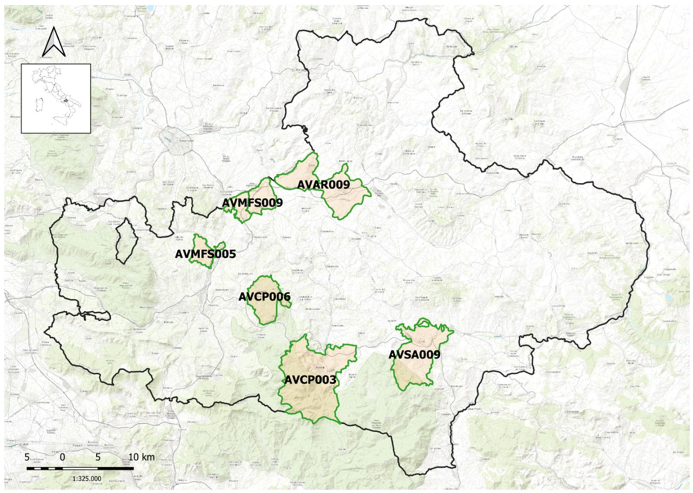
Specifically, the study focused on wild boar hunting activities across six different hunting districts within the Avellino Province as reported in Figure 2.

2.2.2. Study Design and Sample Collection

This cross-sectional study employed convenience sampling of wild boars harvested by authorized hunting teams during the 2025–2026 hunting season (from 1 October 2025 to 1 January 2026). After hunting, the animals were transported to a central processing site for carcass dressing and sample collection. Nasal swabs were collected from clinically healthy male and female wild boars, weighing between 15 and 160 kg, showing no gross pathological lesions at post-mortem inspection. The age of the hunted animals was estimated based on tooth eruption patterns, and individuals were classified into three age groups: juveniles (0–12 months), subadults (13–36 months), and adults (>36 months). The sampling was performed by inserting and rotating a single sterile swab in both nostrils of each animal. Each swab was placed in Stuart W/O CH transport medium (Aptaca Spa, Asti, Italy) to maintain bacterial viability and prevent desiccation. The specimens were then labeled and transported within 48 h to the laboratory in an insulated icebox containing refrigerant packs, ensuring that the temperature did not exceed 10 °C. Bacteriological analyses were conducted at the Bacteriology Diagnostic Laboratory of the Department of Veterinary Medicine and Animal Production, University of Naples Federico II.

2.3. Species Identification and Antimicrobial Susceptibility Testing

Nasal swabs were cultured in parallel on different solid agar media for the isolation of both Gram-positive bacteria (Columbia CNA blood agar and Mannitol Salt Agar) and Gram-negative bacteria (Mac Conkey agar), as well as in Brain Heart Infusion (BHI) broth for enrichment, and incubated aerobically at 37 °C for 24 h. The following day, turbid BHI cultures were subcultured onto the same agar media. All media were purchased from Liofilchem S.r.l. (Teramo, Italy).
Specifically, the isolation of Streptococcus spp. was performed using Columbia CNA agar (CNA) supplemented with 5% sheep blood (Liofilchem S.r.l., Teramo, Italy), a selective medium for Gram-positive bacterial isolation. Once bacterial growth was observed on CNA plates, colonies suspected to belong to Streptococcus spp. (small, translucent colonies surrounded by alpha- or beta-hemolysis) were preliminarily screened using standard rapid identification techniques, including colony morphology assessment, Gram staining, and catalase testing. Subsequently, isolates were identified to the species level using Matrix-Assisted Laser Desorption/Ionization–Time of Flight Mass Spectrometry (MALDI–TOF MS) using the MALDI Biotyper Sirius System (Bruker Daltonics Inc., Bremen, Germany). For each isolate, analyses were performed in triplicate to ensure reproducibility of the spectral profiles. Spectra were acquired in linear positive ion mode using FlexControl 3.4 software (Bruker Daltonics, Bremen, Germany). Identification was achieved by comparing the acquired spectra with the Bruker MSP database using the Bruker MBT Compass HT software (MBT Compass HT RUO version 5.1.400.9024) with default parameters. Identification accuracy was determined in accordance with the manufacturer’s scoring thresholds. Specifically, scores ≥ 2.00 were considered indicative of high-confidence species identification, while values ranging between 1.70 and 1.99 were used to establish a confident identification at the genus level. Any scores falling below 1.70 were treated as providing no reliable identification. To ensure analytical precision and maintain quality control throughout the process, a bacterial test standard (BTS) (Bruker Daltonics, Bremen, Germany) was employed as the system calibrator. Streptococcus equi subsp zooepidemicus ATCC® 53698TM and Staphylococcus aureus ATCC® 33591TM were included as quality control strains.
Susceptibility testing was performed by agar disk diffusion method on Mueller–Hinton agar plates (Liofilchem S.r.l., Teramo, Italy). The collected isolates were evaluated for their susceptibility to eleven antimicrobial agents belonging to eight different antimicrobial classes. These agents, along with their respective classes, are listed in Table 1. All tested antimicrobial disks were purchased from Liofilchem S.r.l. (Teramo, Italy).
Specifically, isolates were classified as susceptible, intermediate, or resistant to amoxicillin/clavulanic acid (AMC), ampicillin (AMP), penicillin (P), cefalexin (CL), enrofloxacin (ENR), gentamicin (CN), and erythromycin (E) according to the Clinical and Laboratory Standards Institute (CLSI) guidelines [22]. Susceptibility to imipenem (IMI), meropenem (MRP), tetracycline (TE), and sulfamethoxazole–trimethoprim (SXT) was interpreted according to the European Committee on Antimicrobial Susceptibility Testing (EUCAST) guidelines [23]. In addition, the recovered Streptococcus species were categorized as multidrug-resistant (MDR), extensively drug-resistant (XDR) and pandrug-resistant (PDR) following the criteria proposed by Magiorakos et al. [24]. For classification purposes, isolates displaying resistance to at least one antimicrobial agent per category were considered. MDR isolates were defined as those resistant to at least one agent in three or more antimicrobial categories, XDR isolates as those resistant to at least one agent in most antimicrobial categories, and PDR isolates as those resistant to all antimicrobial categories tested.
Streptococcus spp. isolates were then preserved in 16% v/v glycerol broth and in Microbank tubes (Pro-Lab Diagnostics, Round Rock, TX, USA) at −80 °C for further investigation.

2.4. Data Handling and Descriptive Statistical Analysis

All microbiological and antimicrobial susceptibility results generated by the Microbiological Diagnostic Laboratory were recorded in a Microsoft Excel™ spreadsheet (Microsoft 365, Microsoft Corp., Redmond, WA, USA). For each isolate, the following variables were recorded: sample identification code, sampling district, host sex and body weight, Streptococcus species, and antimicrobial resistance profile for each tested antimicrobial agent. Descriptive statistical analyses were performed by calculating absolute frequencies, percentages, and 95% confidence intervals (95% CI) to determine the occurrence of the different Streptococcus species and the distribution of antimicrobial resistance phenotypes among recovered isolates. Graphical representations were generated using Microsoft Excel™. The association between categorical variables was assessed using Pearson’s Chi-square test (R × C contingency tables), specifically for the distribution of Streptococcus species across sampling districts, the distribution of multidrug resistance phenotypes (MDR/XDR) among bacterial species, and the influence of host age categories (0–12, 13–36, >36 months) on MDR/XDR phenotypes. Cramer’s V coefficient was calculated to estimate the strength of the association where applicable. To analyze the association between Streptococcus species and antimicrobial phenotypes, resistance profiles were dichotomized into an ‘MDR/XDR’ group versus a ‘non-MDR’ group to ensure statistical robustness. Standardized residuals (SR) were then evaluated to identify specific isolates contributing significantly to the results. For the analysis of host sex (male vs. female) in relation to the occurrence of MDR/XDR profiles, Fisher’s Exact Test (2 × 2 table) was employed. Statistical analysis was performed using JASP (Version 0.96.0; JASP Team 2026, Amsterdam, The Netherlands). A p-value < 0.05 was considered statistically significant. Since multiple bacterial isolates could originate from the same animal (co-colonization), statistical analyses were performed at the isolate level. Consequently, the assumption of independence required for standard inferential tests was only partially satisfied due to potential clustering effects within individual hosts. This limitation was carefully considered when interpreting the significance of the associations, which should be interpreted as exploratory.

3. Results

3.1. Sampling Area

A total of 82 nasal swabs were collected from six hunting districts within the Avellino Province (Figure 2). Specifically, 16 nasal samples were obtained from the AVMFS009 Medio Fiume Sabato District (Venticano/Pietradefusi); 19 from the AVAR009 Arianese District (Bonito-Grottaminarda); 14 from the AVCP006 Picentini District (Chiusano); 18 from AVMFS005 Medio Fiume Sabato District (Prata di Principato Ultra); 8 from AVCP003 Picentini District (Montella); and 7 from AVSA009 Sant’Angelo District (Lioni). The sampled population consisted of 43 males (52%) and 39 females (48%). Regarding age distribution, juveniles (0–12 months) were the most represented class with 38 animals (46.3%), followed by subadults (13–36 months) with 24 animals (29.3%), and adults (>36 months) with 20 animals (24.4%).

3.2. Isolation and Identification of Species of the Genus Streptococcus

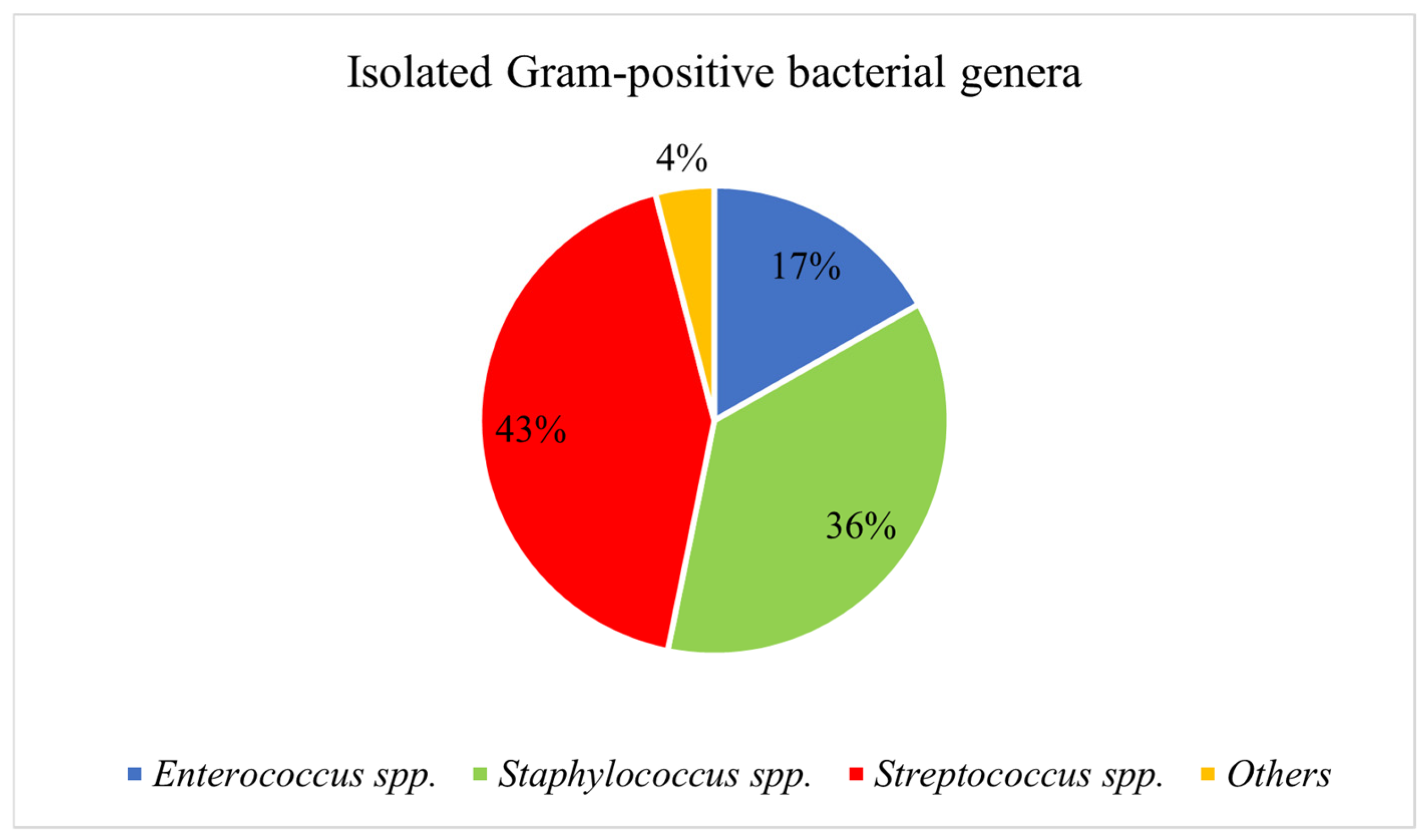
A total of 173 Gram-positive bacterial isolates were recovered from 82 nasal swabs. Among these, 74 isolates were identified as Streptococcus spp., originating from 59 positive swabs (72%; 59/82; 95% CI: 60.9–81.3%). Regarding the Gram-positive nasal microbiota, Streptococcus spp. was the most frequent genus identified, accounting for 43% (74/173; 95% CI: 35.3–50.5%) of the isolates, as illustrated in Figure 3. The second most frequent genus was Staphylococcus spp., representing 36% (63/173; 95% CI: 29.3–44.0%) of the isolates, followed by Enterococcus spp. at 17% (30/173; 95% CI: 12.0–24.0%). The remaining 4% (6/173; 95% CI: 1.3–7.4%) consisted of other Gram-positive bacterial genera detected at lower frequencies.
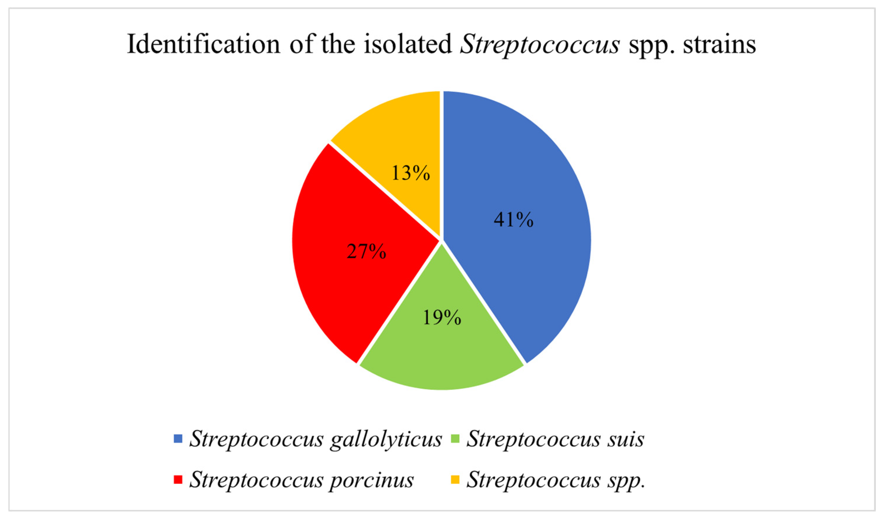
As shown in Figure 4, among Streptococcus spp. isolated from wild boar nasal cavities, Streptococcus gallolyticus was the most frequently detected species (41%; 30/74; 95% CI: 29.3–52.5%), followed by Streptococcus porcinus (27%; 20/74; 95% CI: 17.6–38.6%) and Streptococcus suis (19%; 14/74; 95% CI: 10.8–29.7%). Additional species identified in smaller numbers included Streptococcus pluranimalium (7%; 5/74; 95% CI: 2.2–15.1%), Streptococcus infantarius (3%; 2/74; 95% CI: 0.3–9.4%), Streptococcus dysgalactiae (3%; 2/74; 95% CI: 0.3–9.4%), and Streptococcus lutetiensis (1%; 1/74; 95% CI: 0.0–7.3%). MALDI-TOF MS log(score) values for all Streptococcus species isolates ranged between 2.2 and 2.3, confirming highly probable species-level identification.
Co-colonization by different Streptococcus species within the same animal was observed in 15 out of 59 positive swabs (25%; 95% CI: 15.3–38.3%). The most frequent combination was the association of Streptococcus porcinus and Streptococcus gallolyticus (47%; 7/15; 95% CI: 21.3% –73.4%), followed by the association of Streptococcus suis and Streptococcus gallolyticus (27%; 4/15; 95% CI: 7.8–55.1%).
Regarding spatial distribution, no statistically significant association was found between sampling districts and species occurrence (χ2 = 14.46, df = 15, p = 0.491), with a Cramer’s V of 0.255. Descriptively, Streptococcus gallolyticus was most frequently detected in Bonito-Grottaminarda (55%; 11/20; 95% CI: 31.5–76.9%) and Venticano/Pietradefusi (50%; 6/12; 95% CI: 21.1–78.9%), whereas Streptococcus suis showed its highest occurrence in Lioni (67%; 2/3; 95% CI: 9.4–99.2%). Streptococcus porcinus predominated in Chiusano (37%; 7/19; 95% CI: 16.3–61.6%), while other identified Streptococcus species were detected at lower frequencies across districts. The isolation frequency of Streptococcus species across the six sampling districts is detailed in Table 2.

3.3. Antimicrobial Resistance Profiles of Identified Streptococcus spp.

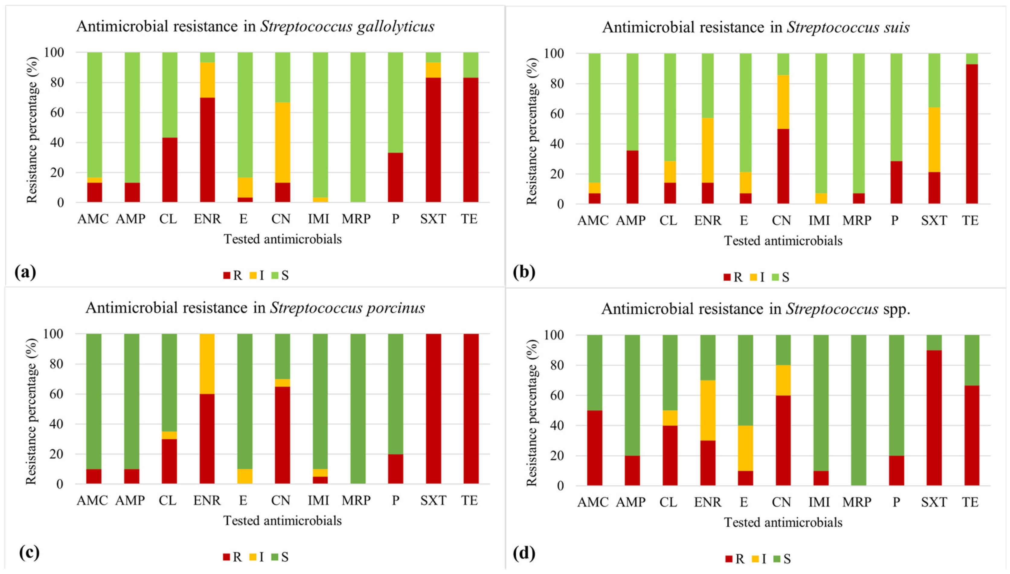
The antimicrobial resistance profiles of the recovered Streptococcus species showed marked variability across the tested antimicrobials (Figure 5). All Streptococcus porcinus isolates (100%; 20/20; 95% CI: 83.2–100%) were resistant to both sulfamethoxazole–trimethoprim (SXT) and tetracycline (TE). High resistance to TE was also observed in Streptococcus suis (93%; 13/14; 95% CI: 66.1–99.8%) and Streptococcus gallolyticus (83%; 25/30; 95% CI: 65.3–94.4%). Similarly, elevated SXT resistance frequencies were recorded for Streptococcus gallolyticus (83%; 25/30; 95% CI: 65.3–94.4%) and the other identified Streptococcus spp. group (90%; 9/10; 95% CI: 55.5–99.7%). Furthermore, high levels of intermediate susceptibility were noted in Streptococcus suis for enrofloxacin (ENR) and SXT (43%; 6/14; 95% CI: 17.7–71.1%), as well as gentamicin (CN) (36%; 5/14; 95% CI: 12.8–64.9%). Intermediate susceptibility was also identified in Streptococcus gallolyticus for CN (53%; 16/30; 95% CI: 34.3–71.7%) and in Streptococcus porcinus for ENR (40%; 8/20; 95% CI: 19.1–63.9%).

3.4. Occurrence of Multidrug- and Extensively Drug-Resistant Streptococcus Isolates

Based on their antimicrobial susceptibility profiles, the recovered isolates were further classified according to multidrug resistance phenotype. Overall, 69% (51/74; 95% CI: 57.1–79.2%) of the isolates were classified as MDR, whereas 5% (4/74; 95% CI: 1.5–13.3%) were identified as XDR. No PDR isolates were detected. The highest MDR isolation frequency was observed in Streptococcus porcinus (85%; 17/20; 95% CI: 62.1–96.8%), followed by Streptococcus gallolyticus (73%; 22/30; 95% CI: 54.1–87.7%), and the other identified Streptococcus spp. group (70%; 7/10; 95% CI: 34.8–93.3%). Conversely, MDR phenotypes were less frequent in Streptococcus suis, accounting for 36% (5/14; 95% CI: 12.8–64.9%) of the isolates. XDR strains were sporadically identified, with one isolate each detected in Streptococcus gallolyticus (3%; 1/30; 95% CI: 0.1–17.2%) and Streptococcus porcinus (5%; 1/20; 95% CI: 0.1–24.9%), and two in the other identified Streptococcus spp. group (20%; 2/10; 95% CI: 2.5–55.6%). No XDR isolates were recovered from Streptococcus suis.
Chi-squared analysis revealed a significant association between bacterial species and the combined MDR/XDR resistance profile (χ2 = 16.70, df = 3, p < 0.001), with a Cramer’s V effect size of 0.472. Analysis of standardized residuals (SR) identified Streptococcus suis as the species with the lowest occurrence of MDR/XDR phenotypes within the studied isolates (SR = −3.92). In contrast, Streptococcus porcinus contributed positively to the distribution of multidrug resistance (SR = 1.97), while Streptococcus gallolyticus and the other identified Streptococcus spp. group showed lower positive deviations toward multidrug resistance (SR = 0.53 and SR = 1.28, respectively).
Furthermore, host-related factors did not appear to influence the distribution of resistance. No significant association was observed between host sex and the occurrence of MDR/XDR strains (p = 0.325), with MDR/XDR phenotypes detected in 76% (26/34) of males and 88% (22/25) of females. Similarly, age categories did not significantly affect the distribution of resistance profiles (χ2 = 2.315, df = 2, p = 0.314), with MDR/XDR observed at 83% (25/30) in the 0–12 months group, 67% (8/12) in the 13–36 months group, and 88% (15/17) in the >36 months group.

4. Discussion

To the best of our knowledge, this is the first study in Italy specifically aimed at characterizing Streptococcus species and their antimicrobial resistance profiles from the nasal cavities of wild boars hunted in the Avellino Province, Southern Italy. Streptococcus spp. were prioritized in this investigation due to their high prevalence in the nasal microbiota of the sampled animals. Therefore, the present study addresses a current knowledge gap by providing specific data on the presence and relative abundance of Streptococcus spp. in wild boar nasal samples.
Specifically, in our study, the detection of Streptococcus gallolyticus, Streptococcus porcinus, and Streptococcus suis suggests that the upper respiratory tract of wild suids represents an important ecological niche for diverse streptococcal species, in addition to Staphylococcus spp., Enterococcus spp. and Escherichia coli, as recently reported [25,26]. Although these three species are all Gram-positive cocci belonging to the genus Streptococcus, they occupy distinct ecological and clinical niches.
Streptococcus suis is a major zoonotic swine pathogen; in domestic pigs, the nasal cavity and tonsils are recognized as primary colonization sites where it can be carried asymptomatically while maintaining the potential to cause systemic disease under favorable conditions [27]. Consequently, wild boars may represent a noteworthy component in the ecology and potential transmission dynamics of this agent. Its detection in free-ranging populations is of significant epidemiological importance, as these animals could facilitate environmental persistence and potentially act as natural reservoirs within transmission cycles [15,17]. Notably, the bacterium has been identified even in the absence of associated lesions, further highlighting the role of wild boars in the silent maintenance of the pathogen [15]. From a One Health perspective, the identification of Streptococcus gallolyticus in nasal samples is also noteworthy. Although typically a gastrointestinal commensal in mammals, including swine, this Lancefield group D Streptococcus (Streptococcus bovis complex) can act as an opportunistic pathogen under specific conditions [28]. Its detection outside the intestinal tract may indicate transient colonization, environmental contamination, or broader mucosal distribution in wildlife hosts than currently recognized. Finally, Streptococcus porcinus, a β-hemolytic Streptococcus species (not yet classically grouped), although less studied than Streptococcus suis, has been reported among atypical streptococci capable of causing systemic infections in pigs, including septicemia and meningitis [29]. Our findings of Streptococcus porcinus in nasal cavities of wild boars may suggest either commensal colonization or early-stage carriage, although epidemiological data in wildlife remain extremely limited.
Beyond the microbiological characterization, the spatial distribution of these species was also evaluated. In the present study, no statistically significant association was found between sampling districts and species occurrence (χ2 = 14.46, p = 0.491). Although this result suggests the absence of marked spatial variation within the analyzed sample, it should not be interpreted as conclusive evidence of a homogeneous epidemiological distribution across the province. Rather, these non-significant findings indicate only the absence of statistically detectable geographic clustering within the constraints of the present study design. Some limitations should be acknowledged. The relatively small sample sizes in specific locations, such as Montella (AVCP003 Picentini District) and Lioni (AVSA009 Sant’Angelo District), limited the statistical power to detect subtle geographic differences. Furthermore, this study utilized an opportunistic sampling strategy, as samples were collected exclusively from animals harvested during the official hunting season. This may have introduced selection bias, meaning the sampled animals may not fully represent the broader wild boar population. Second, because the statistical analysis was conducted at the isolate level rather than animal level, potential clustering effects from multiple isolates per animal must be considered when interpreting inferential estimates and the significant associations should be interpreted as exploratory.
Despite these limitations, the simultaneous detection of multiple Streptococcus species, such as Streptococcus gallolyticus, Streptococcus porcinus and Streptococcus suis, highlights the complexity of the upper respiratory microbiota in wildlife. These findings may further highlight the potential function of wild boars as an interface species between wildlife ecosystems, domestic pigs, and humans. Given the zoonotic potential of several swine-associated streptococci, nasal carriage in wild boars could represent a potential source of occupational exposure for hunters, veterinarians, and individuals involved in carcass handling.
The antimicrobial resistance levels observed in this study may indicate a potential reduced susceptibility to several antimicrobial agents among the recovered isolates. However, as data on direct antimicrobial exposure history were not available, the specific drivers of these resistance phenotypes cannot be definitively determined. The high resistance levels to tetracycline and sulfamethoxazole-trimethoprim, notably the resistance in all Streptococcus porcinus (100%), the high frequency in Streptococcus suis (93%) and Streptococcus gallolyticus (83%), may reflect multiple, non-mutually exclusive factors. These include hypothesized environmental exposure to residues, indirect contact with livestock-associated bacterial populations, species-specific susceptibility profiles, or intrinsic reduced susceptibility to certain antimicrobial classes.
In Streptococcus species, tetracycline resistance is frequently linked to transferable determinants such as tetM, tetO, and tetS, which are widely disseminated across the genus [30]. While tetM gene has been reported in up to 93% of tetracycline-resistant isolates in literature [31], no molecular characterization was performed in the present study; therefore, the specific genetic basis of the observed phenotypes remains to be elucidated. From an epidemiological perspective, these levels appear consistent with historical reports of antimicrobial use in animal production [32], which could have contributed to the long-term maintenance of resistance genes in bacterial reservoirs even after regulatory restrictions [31]. Beyond individual resistance profiles, the frequency of multidrug resistance observed in this study (69%) deserves attention. This finding suggests that a substantial proportion of Streptococcus isolates circulating in the wild boar population exhibited resistance to at least three different antimicrobial classes. The detection of XDR profiles (5%), although at a relatively low frequency, further highlights the presence of isolates with limited therapeutic options. While no PDR strains were identified, these findings remain relevant from both clinical and epidemiological perspectives. Our results are consistent with previous studies reporting the widespread occurrence of MDR bacteria in wild boar populations and other wildlife species, supporting the hypothesis of free-ranging animals as potential environmental sentinels for antimicrobial resistance dissemination [3,33]. In particular, comparable MDR prevalence has been described in bacterial isolates from wild boars in Southern Italy, further suggesting a possible influence of anthropogenic pressures at the wildlife–livestock interface [25,26].
In the present study, Chi-squared analysis and standardized residuals confirmed that the occurrence of resistance profiles is significantly associated with the bacterial species (p < 0.001). Overall, these findings suggest that antimicrobial resistance in streptococci isolated from wild boars is associated with the specific bacterial species, with specific zoonotic pathogens displaying divergent resistance risks within the limits of this exploratory study. Specifically, Streptococcus suis was identified as the species with the lowest occurrence of MDR/XDR phenotypes (SR = −3.92), whereas Streptococcus porcinus contributed positively to the distribution of multidrug resistance (SR = 1.97). The occurrence of elevated levels of multidrug resistance in wildlife, which are generally not directly exposed to antimicrobial treatments, could hypothetically reflect environmental selective pressures or the horizontal transfer of resistance genes within the nasal microbiota [26]. These findings suggest a potential spill-over effect, whereby wildlife might mirror the environmental resistome shaped by anthropogenic activities and by ecological proximity to livestock production systems, highlighting the potential role of free-ranging animals as indicators of antimicrobial resistance circulation across the One Health interface [4,34], regardless of host demographics.
Resistance levels to enrofloxacin and gentamicin (approximately 60%), while lower than those for tetracycline, could also merit attention as these antimicrobials are often considered alternative therapeutic options when first-line drugs fail. These findings suggest a reduced phenotypic susceptibility, though the ecological factors contributing to these patterns, including potential co-selection phenomena, remain unclear. Resistance genes on mobile genetic elements often cluster, potentially facilitating the persistence of multidrug-resistant phenotypes even under selective pressure from a single antimicrobial class [33]. Importantly, the species-specific differences observed in resistance and intermediate susceptibility profiles might reflect the potential influence of species ecology, genetic plasticity, and hypothesized antimicrobial exposure history in shaping resistance evolution [33]. For instance, in Streptococcus suis, tetracycline resistance determinants such as tetO and tetW are widely documented, with a large proportion of isolates carrying at least one tetracycline resistance determinant, highlighting the extensive dissemination of these genes in animal-associated streptococci as reported in previous studies [35].
Another relevant finding is the high proportion of isolates showing intermediate susceptibility, such as in Streptococcus suis for enrofloxacin (43%) and in Streptococcus gallolyticus for gentamicin (53%). While these findings appear to align with global observations of antimicrobial resistance in various Streptococcus species [36], the descriptive nature of this study warrants a cautious interpretation of such trends, underlining the need for continued species-specific surveillance and antimicrobial stewardship.
Taken together, these results suggest that resistance phenotypes might be stabilized within the investigated bacterial populations, highlighting the potential importance of wild boars as potential sentinels for environmental antimicrobial resistance. Future investigations incorporating molecular characterization and longitudinal surveillance are necessary to better understand these dynamics and inform targeted stewardship interventions. Furthermore, the high resistance levels detected for multiple antimicrobial classes may compromise therapeutic options and facilitate the persistence and spread of resistant Streptococcus spp. strains within wildlife populations. This dissemination, potentially extending along the food chain, may pose significant veterinary and public health concerns.

5. Conclusions

In conclusion, this descriptive study highlights the presence of a variety of Streptococcus species in the nasal cavities of wild boars from Avellino Province in Southern Italy. The observed frequency of isolates with multidrug-resistant phenotypes, which showed a significant species-dependent distribution, suggests that these bacterial populations could potentially harbor diverse resistance determinants. However, given the exploratory nature of this study and the convenience sampling approach, these findings should be interpreted strictly as a preliminary snapshot of the local ecological context. While wild boars could potentially contribute to the environmental maintenance of antimicrobial resistance, further longitudinal studies incorporating molecular data are required to better understand the possible dynamics at the wildlife–livestock–human interface. Within a One Health framework, these results underline the importance of including wildlife in surveillance programs to better elucidate their potential contribution to the global burden of antimicrobial resistance.

Author Contributions

Conceptualization, L.D.M., and R.C.; methodology, F.P.N., C.L., and S.D.; software, A.R., R.S., and F.P.N.; formal analysis, F.P.N., C.L., A.R., and R.S.; investigation, F.P.N., C.L., A.R., R.S., C.R., V.I., N.P., and R.C.; data curation, F.P.N., C.L., A.R., R.S., L.D.M., S.D., and R.C.; writing—original draft preparation, F.P.N., and L.D.M.; writing—review and editing, F.P.N., L.D.M., S.D., and R.C.; visualization, F.P.N., C.L., A.R., R.S., N.P., C.R., V.I., L.D.M., S.D., and R.C. supervision, L.D.M., and R.C.; All authors have read and agreed to the published version of the manuscript.

Funding

This research received no external funding.

Institutional Review Board Statement

Not applicable. This study did not involve the use of living wild boars, thus ethical approval was not required.

Informed Consent Statement

Informed consent to participate in this research study was obtained by licensed and specialized hunters, who provided wild boar nasal swabs.

Data Availability Statement

The original contributions presented in this study are included in the article. Further inquiries can be directed to the corresponding authors.

Acknowledgments

The authors are grateful to Giuseppe Marzatico for his assistance with the cartographic representation. Raffaele Antonio, the Territorial Hunting Area (ATC) of the province of Avellino, and the authorized hunters who enabled the collection of samples are gratefully acknowledged.

Conflicts of Interest

The authors declare no conflicts of interest.

Abbreviations

The following abbreviations are used in this manuscript:
AMCamoxicillin/clavulanic acid
AMPampicillin
BHIBrain Heart Infusion broth
BTSbacterial test standard
CLcephalexin
CNAColumbia CNA blood agar with 5% sheep blood
ENRenrofloxacin
Eerythromycin
CN gentamicin
IMIimipenem
MDRmultidrug-resistant
MRPmeropenem
Ppenicillin
PDRpandrug-resistant
SXTsulfamethoxazole-trimethoprim
TEtetracycline
XDRextensively drug-resistant

References

  1. Marshall, B.M.; Levy, S.B. Food animals and antimicrobials: Impacts on human health. Clin. Microbiol. Rev. 2011, 24, 718–733. [Google Scholar] [CrossRef]
  2. Farabi, A.A.; Hossain, H.; Brishty, K.A.; Rahman, M.H.; Rahman, M.; Siddiqui, M.S.I.; Atikuzzaman, M.; Saleh, A.; Binsuwaidan, R.; Selim, H.M.R.M.; et al. Prevalence, Risk Factors, and Antimicrobial Resistance of Staphylococcus and Streptococcus Species Isolated from Subclinical Bovine Mastitis. Foodborne Pathog. Dis. 2025, 22, 467–476. [Google Scholar] [CrossRef]
  3. Torres, R.T.; Fernandes, J.; Carvalho, J.; Cunha, M.V.; Caetano, T.; Mendo, S.; Serrano, E.; Fonseca, C. Wild boar as a reservoir of antimicrobial resistance. Sci. Total Environ. 2020, 717, 135001. [Google Scholar] [CrossRef]
  4. Smoglica, C.; Carcagnì, A.; Angelucci, S.; Di Tana, F.; Marsilio, F.; López-Olvera, J.R.; Di Francesco, C.E. Systematic review and meta-analysis of antimicrobial resistant bacteria in free-ranging wild mammals. BMC Vet. Res. 2025, 21, 150. [Google Scholar] [CrossRef]
  5. Pirolo, M.; Espinosa-Gongora, C.; Bogaert, D.; Guardabassi, L. The porcine respiratory microbiome: Recent insights and future challenges. Anim. Microbiome 2021, 3, 9. [Google Scholar] [CrossRef]
  6. Correa-Fiz, F.; Fraile, L.; Aragon, V. Piglet nasal microbiota at weaning may influence the development of Glässer’s disease during the rearing period. BMC Genom. 2016, 17, 404. [Google Scholar] [CrossRef]
  7. Gao, X.Y.; Zhi, X.Y.; Li, H.W.; Klenk, H.P.; Li, W.J. Comparative genomics of the bacterial genus Streptococcus illuminates evolutionary implications of species groups. PLoS ONE 2014, 9, e101229. [Google Scholar] [CrossRef]
  8. Kumar, S.; Bansal, K.; Sethi, S.K. Comparative genomics reconciliations of genus Streptococcus resolve its taxonomy and elucidates biotechnological importance of their constituent species. Ecol. Genet. Genom. 2023, 29, 100205. [Google Scholar] [CrossRef]
  9. Kosecka-Strojek, M.; Sabat, A.J.; Akkerboom, V.; Kooistra-Smid, A.M.D.M.; Miedzobrodzki, J.; Friedrich, A.W. Development of a reference data set for assigning Streptococcus and Enterococcus species based on next generation sequencing of the 16S–23S rRNA region. Antimicrob. Resist. Infect. Control 2019, 8, 178. [Google Scholar] [CrossRef]
  10. Obradovic, M.R.; Segura, M.; Segalés, J.; Gottschalk, M. Review of the speculative role of co-infections in Streptococcus suis-associated diseases in pigs. Vet. Res. 2021, 52, 49. [Google Scholar] [CrossRef] [PubMed]
  11. Hyeon, J.Y.; Kim, J.; Chung, D.H.; Helal, Z.H.; Polkowski, R.; Lee, D.H.; Risatti, G.R. Genome analysis of Streptococcus spp. isolates from animals in pre-antibiotic era with respect to antibiotic susceptibility and virulence gene profiles. Vet. Res. 2024, 55, 51. [Google Scholar] [CrossRef]
  12. Vela, A.I.; Sánchez, V.; Mentaberre, G.; Lavín, S.; Domínguez, L.; Fernández-Garayzábal, J.F. Streptococcus porcorum sp. nov., isolated from domestic and wild pigs. Int. J. Syst. Evol. Microbiol. 2011, 61, 1585–1589. [Google Scholar] [CrossRef][Green Version]
  13. Vötsch, D.; Willenborg, M.; Weldearegay, Y.B.; Valentin-Weigand, P. Streptococcus suis—The “Two Faces” of a Pathobiont in the Porcine Respiratory Tract. Front. Microbiol. 2018, 9, 480. [Google Scholar] [CrossRef]
  14. Dresen, M.; Murray, G.G.R.; Valentin-Weigand, P.; Fulde, M.; Weinert, L.A. Streptococcus suis avian expansion suggests shared antibiotic use drives host jumps. BMC Biol. 2025, 23, 358. [Google Scholar] [CrossRef]
  15. Lopes, C.E.; Lopes, B.C.; Cerva, C.; Bertagnolli, A.C.; de Castro, L.T.; Lima, R.P.; Menegatt, J.C.O.; Loiko, M.R.; Driemeier, D.; Mayer, F.Q. Streptococcus suis in free-ranging wild boars from Southern Brazil: A preliminary survey. Vet. Res. Commun. 2025, 50, 7. [Google Scholar] [CrossRef]
  16. Baums, C.G.; Verkühlen, G.J.; Rehm, T.; Silva, L.M.; Beyerbach, M.; Pohlmeyer, K.; Valentin-Weigand, P. Prevalence of Streptococcus suis genotypes in wild boars of Northwestern Germany. Appl. Environ. Microbiol. 2007, 73, 711–717. [Google Scholar] [CrossRef]
  17. Risco, D.; Fernández-Llario, P.; Cuesta, J.M.; García-Jiménez, W.L.; Gonçalves, P.; Martínez, R.; García, A.; Rosales, R.; Gómez, L.; de Mendoza, J.H. Fatal case of Streptococcus suis infection in a young wild boar (Sus. scrofa) from Southwestern Spain. J. Zoo Wildl. Med. 2015, 46, 370–373. [Google Scholar] [CrossRef]
  18. Sánchez del Rey, V.; Fernández-Garayzábal, J.F.; Mentaberre, G.; Briones, V.; Lavín, S.; Domínguez, L.; Gottschalk, M.; Vela, A.I. Characterisation of Streptococcus suis isolates from wild boars (Sus scrofa). Vet. J. 2014, 200, 464–467. [Google Scholar] [CrossRef]
  19. Fernández-Aguilar, X.; Gottschalk, M.; Aragon, V.; Càmara, J.; Ardanuy, C.; Velarde, R.; Galofré-Milà, N.; Castillo-Contreras, R.; López-Olvera, J.R.; Mentaberre, G.; et al. Urban Wild Boars and Risk for Zoonotic Streptococcus suis, Spain. Emerg. Infect. Dis. 2018, 24, 1083–1086. [Google Scholar] [CrossRef]
  20. Yang, G.; Shi, C.; Zhang, S.; Liu, Y.; Li, Z.; Gao, F.; Cui, Y.; Yan, Y.; Li, M. Characterization of the bacterial microbiota composition and evolution at different intestinal tract in wild pigs (Sus. scrofa ussuricus). PeerJ 2020, 8, e9124. [Google Scholar] [CrossRef]
  21. Vedel, G.; Triadó-Margarit, X.; Linares, O.; Moreno-Rojas, J.M.; la Peña, E.; García-Bocanegra, I.; Jiménez-Martín, D.; Carranza, J.; Casamayor, E.O. Exploring the potential links between gut microbiota composition and natural populations management in wild boar (Sus. scrofa). Microbiol. Res. 2023, 274, 127444. [Google Scholar] [CrossRef]
  22. CLSI. Performance Standards for Antimicrobial Disk and Dilution Susceptibility Tests for Bacteria Isolated from Animals, 4th ed.; CLSI Supplement VET08; Clinical and Laboratory Standards Institute: Wayne, PA, USA, 2018. [Google Scholar]
  23. EUCAST. The European Committee on Antimicrobial Susceptibility Testing. Breakpoint Tables for Interpretation of MICs and Zone Diameters. Version 15.0. 2025. Available online: http://www.eucast.org (accessed on 7 January 2025).
  24. Magiorakos, A.P.; Srinivasan, A.; Carey, R.B.; Carmeli, Y.; Falagas, M.E.; Giske, C.G.; Harbarth, S.; Hindler, J.F.; Kahlmeter, G.; Olsson-Liljequist, B.; et al. Multidrug-resistant, extensively drug-resistant and pandrug-resistant bacteria: An international expert proposal for interim standard definitions for acquired resistance. Clin. Microbiol. Infect. 2012, 18, 268–281. [Google Scholar] [CrossRef]
  25. Nocera, F.P.; Ferrara, G.; Scandura, E.; Ambrosio, M.; Fiorito, F.; De Martino, L. A preliminary study on antimicrobial susceptibility of Staphylococcus spp. and Enterococcus spp. grown on Mannitol Salt Agar in European wild boar (Sus. scrofa) hunted in Campania Region-Italy. Animals 2022, 12, 85. [Google Scholar] [CrossRef]
  26. Arslan, S.; Schena, R.; Romano, A.; Hohenegger, M.; De Martino, L.; Nocera, F.P. Characterization of antimicrobial resistance and biofilm formation in Escherichia coli isolated from wild boars. Res. Vet. Sci. 2026, 204, 106148. [Google Scholar] [CrossRef]
  27. Dutkiewicz, J.; Zając, V.; Sroka, J.; Wasiński, B.; Cisak, E.; Sawczyn, A.; Kloc, A.; Wójcik-Fatla, A. Streptococcus suis: A re-emerging pathogen associated with occupational exposure to pigs or pork products. Part II—Pathogenesis. Ann. Agric. Environ. Med. 2018, 25, 186–203. [Google Scholar] [CrossRef]
  28. Sitthicharoenchai, P.; Burrough, E.R.; Arruda, B.L.; Sahin, O.; Dos Santos, J.G.; Magstadt, D.R.; Piñeyro, P.E.; Schwartz, K.J.; Rahe, M.C. Streptococcus gallolyticus and Bacterial Endocarditis in Swine, United States, 2015–2020. Emerg. Infect. Dis. 2022, 28, 192–195. [Google Scholar] [CrossRef]
  29. Costinar, L.; Badea, C.; Marcu, A.; Pascu, C.; Herman, V. Multiple Drug Resistant Streptococcus Strains-An Actual Problem in Pig Farms in Western Romania. Antibiotics 2024, 13, 277. [Google Scholar] [CrossRef]
  30. Chopra, I.; Roberts, M. Tetracycline antibiotics: Mode of action, applications, molecular biology, and epidemiology of bacterial resistance. Microbiol. Mol. Biol. Rev. 2001, 65, 232–260. [Google Scholar] [CrossRef]
  31. Glajzner, P.; Szewczyk, E.M.; Szemraj, M. Phenotypic and Genotypic Characterization of Antimicrobial Resistance in Streptococci Isolated from Human and Animal Clinical Specimens. Curr. Microbiol. 2023, 80, 228. [Google Scholar] [CrossRef]
  32. Van Boeckel, T.P.; Brower, C.; Gilbert, M.; Grenfell, B.T.; Levin, S.A.; Robinson, T.P.; Teillant, A.; Laxminarayan, R. Global trends in antimicrobial use in food animals. Proc. Natl. Acad. Sci. USA 2015, 112, 5649–5654. [Google Scholar] [CrossRef]
  33. Hadjirin, N.F.; Miller, E.L.; Murray, G.G.R.; Yen, P.L.K.; Phuc, H.D.; Wileman, T.M.; Hernandez-Garcia, J.; Williamson, S.M.; Parkhill, J.; Maskell, D.J.; et al. Large-scale genomic analysis of antimicrobial resistance in the zoonotic pathogen Streptococcus suis. BMC Biol. 2021, 19, 191. [Google Scholar] [CrossRef] [PubMed]
  34. Vezeau, N.; Kahn, L. Spread and mitigation of antimicrobial resistance at the wildlife-urban and wildlife-livestock interfaces. J. Am. Vet. Med. Assoc. 2024, 262, 741–747. [Google Scholar] [CrossRef] [PubMed]
  35. Li, K.; Lacouture, S.; Lewandowski, E.; Thibault, E.; Gantelet, H.; Gottschalk, M.; Fittipaldi, N. Molecular characterization of Streptococcus suis isolates recovered from diseased pigs in Europe. Vet. Res. 2024, 55, 117. [Google Scholar] [CrossRef] [PubMed]
  36. Shivaee, A.; Kazemi, F.; Navidifar, T.; Golab, N.; Parvizi, E.; Mofid, M.; Sholeh, M.; Beig, M. Global trends in macrolide and lincosamide resistance in Streptococcus species: A comprehensive systematic review and meta-analysis. BMC Infect. Dis. 2025, 25, 1577. [Google Scholar] [CrossRef]
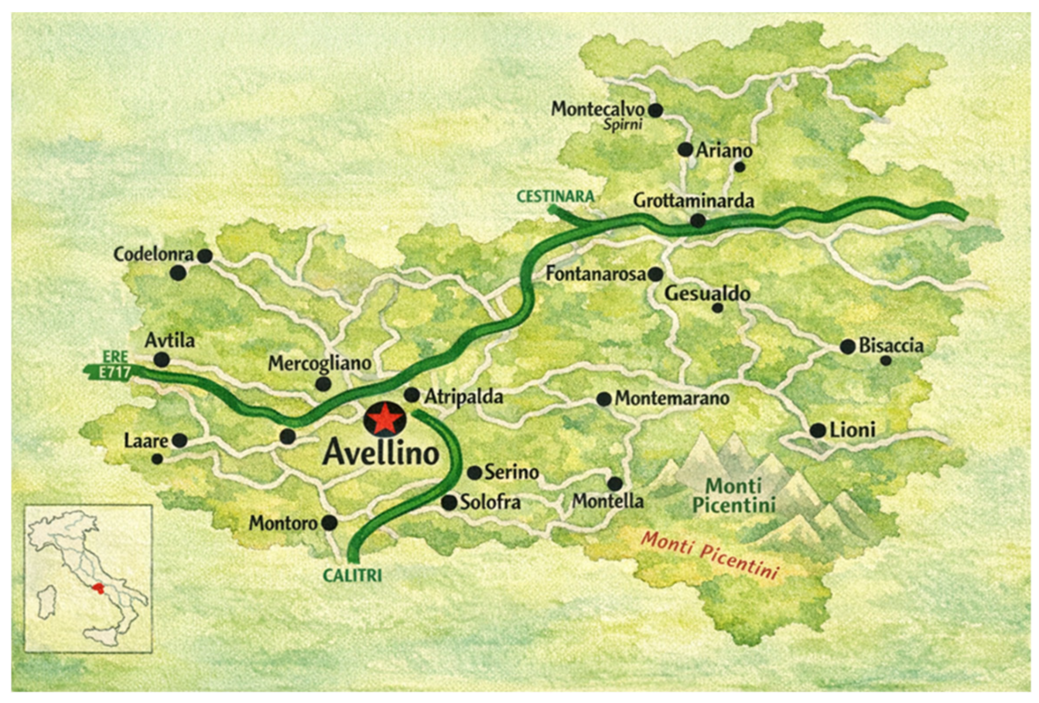
Figure 1. Geographic location of the study area: detailed map of the Avellino Province (Irpinia) located in Campania Region, Southern Italy.
Animals 16 01619 g001
Figure 2. Spatial distribution of the sampling districts within the Avellino Province. Highlighted areas represent the specific hunting districts where wild boar specimens were collected: AVMFS009 Medio Fiume Sabato District (Venticano/Pietradefusi); AVAR009 Arianese District (Bonito-Grotta-minarda); AVCP006 Picentini District (Chiusano); AVMFS005 Medio Fiume Sabato District (Prata di Principato Ultra); AVCP003 Picentini District (Montella); AVSA009 Sant’Angelo District (Lioni).
Animals 16 01619 g002
Figure 3. Identification frequency of Gram-positive bacterial genera.
Animals 16 01619 g003
Figure 4. Identification frequency of Streptococcus species isolated from nasal cavities of wild boars.
Animals 16 01619 g004
Figure 5. Antimicrobial resistance profiles of the identified Streptococcus spp. isolates. (a) Streptococcus gallolyticus (n = 30); (b) Streptococcus suis (n = 14); (c) Streptococcus porcinus (n = 20); (d) other identified Streptococcus spp. (n = 10). Tested antimicrobials: AMC: amoxicillin/clavulanic acid; AMP: ampicillin; CL: cephalexin; ENR: enrofloxacin; E: erythromycin; CN: gentamicin; IMI: imipenem; MRP: meropenem; P: penicillin; SXT: sulfamethoxazole-trimethoprim; TE: tetracycline.
Animals 16 01619 g005
Table 1. Antimicrobial classes and agents tested for susceptibility profiling.
AntimicrobialsDisk ContentAntimicrobial ClassReferences for
Breakpoints
Amoxicillin/Clavulanic Acid (AMC)20/10 µgPenicillins[22]
Ampicillin (AMP)10 µg[22]
Penicillin (P)10 IU[22]
Cefalexin (CL)30 µgFirst-generation cephalosporins[22]
Enrofloxacin (ENR)5 µgFluoroquinolones[22]
Gentamicin (CN)10 µgAminoglycosides[22]
Imipenem (IMI)10 µgCarbapenems[23]
Meropenem (MRP)10 µg[23]
Erythromycin (E)15 µgMacrolides[22]
Tetracycline (TE)30 µgTetracyclines[23]
Sulfamethoxazole-trimethoprim (SXT)23.75/1.25 µgSulphonamides[23]
Table 2. Distribution of Streptococcus species by sampling district.
Sampling District n. Nasal SwabsStreptococcus gallolyticus
n (%)
Streptococcus suis
n (%)
Streptococcus porcinus
n (%)
Other Streptococcus spp.
n (%)
Total Isolates (N) *
Bonito-Grottaminarda1911 (55%)4 (20%)4 (20%)1 (5%)20
Prata Principato Ultra 185 (42%)1 (8%)4 (33%)2 (17%)12
Venticano/Pietradefusi166 (50%)1 (8%)3 (25%)2 (17%)12
Chiusano145 (26%)3 (16%)7 (37%)4 (21%)19
Montella83 (38%)3 (38%)1 (13%)1 (13%)8
Lioni70 (0%)2 (67%)1 (33%)0 (0%)3
Total 8230 1420 1074
* Percentages are calculated based on the total number of isolates recovered per location (N).
Disclaimer/Publisher’s Note: The statements, opinions and data contained in all publications are solely those of the individual author(s) and contributor(s) and not of MDPI and/or the editor(s). MDPI and/or the editor(s) disclaim responsibility for any injury to people or property resulting from any ideas, methods, instructions or products referred to in the content.